Supporting Online Material for - sciencemag.org · was centrifuged at 8,000 g for 20 min, and the...
17
www.sciencemag.org/cgi/content/full/1156970/DC1 Supporting Online Material for S-Nitrosylation and Thioredoxins Regulate Conformational Changes of NPR1 in Establishing Plant Immunity Yasuomi Tada, Steven H. Spoel, Karolina Pajerowska-Mukhtar, Zhonglin Mou, Junqi Song, Xinnian Dong* *To whom correspondence should be addressed. E-mail: [email protected] Published 17 July 2008 on Science Express DOI: 10.1126/science.1156970 This PDF file includes: Materials and Methods Figs. S1 to S10 References
Transcript of Supporting Online Material for - sciencemag.org · was centrifuged at 8,000 g for 20 min, and the...

www.sciencemag.org/cgi/content/full/1156970/DC1
Supporting Online Material for
S-Nitrosylation and Thioredoxins Regulate Conformational Changes of NPR1 in Establishing Plant Immunity
Yasuomi Tada, Steven H. Spoel, Karolina Pajerowska-Mukhtar, Zhonglin Mou, Junqi Song, Xinnian Dong*
*To whom correspondence should be addressed. E-mail: [email protected]
Published 17 July 2008 on Science Express
DOI: 10.1126/science.1156970
This PDF file includes:
Materials and Methods Figs. S1 to S10 References

Supporting Online Material Methods Plant transformation and growth conditions Site-directed mutagenesis of cysteine 156 in NPR1 was performed in pCB302-35S::NPR1-GFP (S1) with a QuickChange site-directed mutagenesis kit (Stratagene, La Jolla, CA). 35S::NPR1 tagged with tandem affinity purification-tag (TAP), which consists of a calmodulin binding peptide, a tobacco etch virus (TEV) protease cleavage site, and IgG-binding units of protein A (S2) was introduced into the plant transformation vector pBI1.4T. The resulting plasmids were electroporated into Agrobacterium tumefaciens strain GV3101 (pMP90), and the resulting bacteria were used to transform the npr1-1 mutant. The herbicide Basta (glufosinate ammonium, dilution 1:2000) was sprayed to select 35S::NPR1C156A-GFP plants. 35S::NPR1-TAP plants were selected by SA tolerance test as described (S3). Arabidopsis thaliana (ecotype Columbia) plants and the transgenic plants were grown on soil (Metro Mix 200; Grace-Sierra, Milpitas, CA) at 22°C under a 12/12 hr light/dark cycle. For induction, 3- to 4-week-old plants were sprayed with SA at the indicated concentration. T-DNA insertion knockout mutants were obtained from Salk Institute (SALK 111160 for At5g42980; SALK 144259 for At1g45145; SALK 039152 for At2g17420). Microscopy Leaf tissues were mounted in 10% glycerol and viewed with a Leica (Wetzlar, Germany) DMRB fluorescence microscope. GFP signal was viewed with an excitation wavelength of 488 nm and a bandpass 510- to 525-nm emission filter. Real-time RT-PCR RNA samples were prepared with a previously reported protocol (S4) and reverse transcribed into cDNA with SuperScript Reverse Transcriptase (Invitrogen, Carlsbad, CA). The cDNA was quantified with gene specific primers and the QuantiTect reagent (Qiagen, Valencia, CA) in a LightCycler (Roche Diagnostics, Mannheim, Germany). The experiments were done three times. Bacterial pathogen infection Three-week-old Arabidopsis plants were inoculated with 10 mM MgCl2 (- SAR) or the avirulent bacterial pathogen Pseudomonas syringae pv. maculicola (Psm) ES4326/avrRpt2 (+ SAR) at the concentration of OD600 = 0.02. The elicited plants were challenge inoculated three days later with the virulent Psm ES4326 at OD600 = 0.001. The bacterial growth and symptom were monitored as described (S4). Recombinant TRX and TRX-immobilized resin Cloning of the Arabidopsis thioredoxin (TRX-h) genes, protein production and immobilization of TRX on a resin were carried out as described (S5,S6). The wild-type TRX-h3 and TRX-h5 and the corresponding cysteine mutants were cloned into the NdeI and XhoI sites of pET-24c (Novagen, Madison, WI) and were transformed into E. coli
1

BL21 (DE3). A 250 ml Escherichia coli culture overexpressing each recombinant protein was centrifuged at 8,000 g for 20 min, and the pellet was suspended in 3 ml BugBuster (Novagen, Madison, WI). The cell extract was centrifuged twice at 15,000 g for 20 min, and the supernatant was desalted with PD-10 column (GE Healthcare, Piscataway, NJ) with buffer A, 50 mM potassium phosphate buffer, pH 8.0, 300 mM NaCl. The protein mixture was incubated with 500 μl Ni-NTA (Qiagen) resin at 4°C overnight. Beads were washed with 5 ml buffer A containing 10 to 40 mM imidazole, followed by elution with elution buffer, 50 mM potassium phosphate buffer, pH 8.0, 300 mM NaCl, 350 mM imidazole. The samples were treated with 20 mM dithiothreitol (DTT) at RT for 15 min to reduce TRX and dialyzed against 10 mM potassium phosphate buffer, pH 8.0, and stored at -20°C until use. TRX (2.5 mg) in reaction buffer (0.1 M sodium carbonate buffer and 0.5 M NaCl) was incubated with CNBr-activated Sepharose 4B resin (GE Healthcare) at 4°C overnight. After washing beads with 10 ml reaction buffer, the remaining reactive side chains were blocked with inactivation buffer, 0.1 M Tris-HCl, pH8.0, 0.5 M NaCl, at 4°C overnight. The inactivation buffer was replaced with reaction buffer after centrifugation. Recombinant wild-type and mutant NPR1 The N terminus of NPR1 (NH; nucleotides 1 to 246) was cloned into pET-28a (Novagen, Madison, WI) with NdeI and BamHI. The plasmid pCB302-35S::NPR1-GFP (described above) was used as template to clone the mutant form of NH into pET-28a. The NH proteins were purified from E. coli by Ni-NTA resin as described above. Purified proteins were dialyzed against 25 mM Hepes buffer, pH7.7, and stored at -20°C until use. In vitro re-oligomerization assay Cytosolic extracts were obtained from 1 g of 3- to 4-week-old leaves by grinding in 1 ml extraction buffer (25 mM potassium phosphate buffer, pH 7.4, 2 mM EDTA, MG115, and protease inhibitor cocktail) and were centrifuged twice at 14,000 g for 15 min. The supernatant of extracts was treated with 50 mM DTT at RT for 20 min, followed by dialysis against 25 mM potassium phosphate buffer (pH 7.4) to remove DTT. Total proteins were mixed with 40 μM MG115 and incubated at RT for the indicated time. To test the effect of the oxidants on the oligomerization of NPR1, the samples were treated with the NO donors, GSNO and sodium nitroprusside (SNP, Fluka, Buchs, Switzerland), diethylamine-NO (DEA/NO, Sigma, St. Louis, MO) and H2O2 at the indicated concentrations for 2 hr. The samples were separated on 8% nonreducing (-DTT) and reducing (+DTT) sodium dodecyl sulfate polyacrylamide gel electrophoresis (SDS-PAGE) gels and immunoblotted for NPR1-GFP. The recombinant NH solution (in 25 mM Hepes, pH 7.7) was mixed with 1 mM EDTA and 0.1 mM neocuproine, and then treated with 250 μM GSNO for 30 min in the dark. The NO donor was removed by a MicroBioSpin6 column pre-equilibrated in 25 mM Hepes, pH 7.7, 1 mM EDTA, 0.1 mM neocuproine. The samples were incubated with or without 50 mM sodium ascorbate, which specifically reduce S-nitrosylated thiols, at RT for 15 min, followed by dialysis against 25 mM Hepes buffer, pH7.7. Proteins were incubated at RT for the indicated time points in the dark to see if S-nitrosylation facilitates oligomerization of NH. Biotin switch assay
2
The biotin switch assay was performed as described (S7,S8). Leaf tissues were homogenized in HEN buffer (250 mM Hepes, pH 7.7, 1 mM EDTA, 0.1 mM neocuproine) containing protease inhibitor cocktail and 40 μM MG115. The extract was centrifuged twice at 14,000 g for 15 min, and the protein concentration of the supernatant was determined by Bio-Rad protein assay (Bio-Rad, Hercules, CA). Samples were diluted to 1 mg protein/ml with HEN buffer plus 2.5% SDS and 25 mM S-methylmethane thiosulfonate (MMTS), and incubated at 50°C for 20 min with frequent vortex mixing. Proteins were precipitated by adding one volume of acetone at -20°C for 20 min and centrifuged at 5,000 g for 5 min. The pellet was washed 3 times with 50% acetone and dissolved in 850 μl HEN buffer containing 1% SDS, followed by addition of 50 μl of 1 M sodium ascorbate and 100 μl of 2.5 mg/ml biotin-HPDP. After labeling for 90 min, proteins were precipitated with 50% acetone, and briefly washed with 70% acetone. Samples were then dissolved in suspension buffer, 25 mM Hepes, pH 7.7, 1 mM EDTA, 1% SDS, and subjected to precipitation with 50% acetone. The pellet was resuspended in 250 μl suspension buffer and 750 μl neutralization buffer, 25 mM Hepes, pH 7.7, 100 mM NaCl, 1 mM EDTA, 0.5% Triton X-100. After centrifugation at 10,000 g for 5 min, the supernatant was added to 40 μl streptavidin beads and incubated at 4°C overnight. Beads were washed 5 times with washing buffer, 25 mM Hepes, pH 7.7, 600 mM NaCl, 1 mM EDTA, 0.5% Triton X-100. After washing with neutralization buffer once, proteins were eluted with 40 μl elution buffer, 25 mM Hepes, pH 7.7, 100 mM NaCl, 1 mM EDTA, 100 mM DTT, at 50°C for 30 min. The samples were separated on 8% SDS-PAGE gels and immunoblotted for NPR1 or NPR1-GFP. In vitro SNO test Cytosolic extracts were obtained from 3- to 4-week-old leaves by grinding in extraction buffer, 25 mM Hepes, pH 7.7, 1 mM EDTA, 40 μM MG115, and protease inhibitor cocktail. The supernatant of extracts was treated with 50 mM DTT at RT for 20 min, followed by dialysis against 25 mM Hepes buffer, pH 7.7, to remove DTT. The samples were mixed with 1 mM EDTA and 0.1 mM neocuproine, followed by incubation with 250 μM GSNO or 250 μM SNP at RT for 30 min in the dark. The biotin switch assay was then performed by immunoprecipitation of NPR1-GPF protein. SNO-NPR1-GFP was detected using a streptavidin-horseradish peroxidase (streptavidin-HRP) conjugate. In vitro pull-down assay Total protein was obtained from 2 g of 3- to 4-week-old Col-0 leaves by grinding in 2 ml extraction buffer, 50 mM potassium phosphate buffer, pH 7.4, 300 mM NaCl, 1 mM EDTA, 40 μM MG115, and protease inhibitor cocktail. The supernatant of the extract was recovered after centrifugation twice at 14,000 g for 15 min. Purified 200 μg of NH oligomers were mixed with 2 ml of extract and 0.5 ml Ni-NTA resin, followed by incubation at RT for 90 min. Beads were washed 5 times with 1 ml extraction buffer containing 20 mM imidazole. Protein was eluted with extraction buffer with 350 mM imidazole and separated on 15% SDS-PAGE gels and immunoblotted for TRX-h3 and TRX-h5 (S9). To pull-down NPR1 with TRX, the TRX-h3M- or TRX-h5M-immobilized resin was mixed with total protein obtained from 2 g of 3- to 4-week-old Col-0 tissue as described above. After incubation at RT for 90 min, precipitated resin was washed 6 times with 50 mM potassium phosphate buffer, pH 7.4, 1 M NaCl, 1 mM EDTA, and
3
bound protein was eluted with the same buffer containing 150 mM DTT at 70°C for 20 min. The eluate was separated on 12% SDS-PAGE gels and immunoblotted for NPR1. Detection of in vivo NPR1-TRX interaction using co-immunoprecipitation Five grams of leaf tissue from 3- to 4-week-old 35S::NPR1-TAP plants treated with 1 mM SA for 24 hr were ground under liquid nitrogen, and 5 ml “fixation buffer” (25 mM Hepes, pH 7.7, 2.5% SDS,1 mM EDTA, 0.1 mM neocuproine, 25 mM MMTS, 40 μl MG115 and protease inhibitor cocktail) was added and further ground by a mortar and pestle on ice. The supernatant was recovered by centrifugation at 14,000 g for 20 min, and the protein was precipitated by incubation with 50% acetone at -20°C for 20 min. The pellet was washed once with 50% acetone, and dissolved in 3 ml of 25 mM Hepes, pH 7.7, 1% SDS, 1 mM EDTA, 0.1 mM neocuproine, followed by addition of 9 ml neutralization buffer, 25 mM Hepes, pH 7.7, 0.5% Triton X-100, 1 mM EDTA, 0.1 mM neocuproine. The protein extract was then incubated with 100 μl of IgG sepharose (GE Healthcare) to pull down NPR-TAP at 4°C overnight. Beads were washed 6 times with 25 mM Hepes, pH 7.7, 150 mM NaCl, 1 mM EDTA, 0.1 mM neocuproine, and bound protein was eluted with 400 μl of the washing buffer containing 2.5% SDS by heating at 70°C for 20 min. The eluate was precipitated by 70% acetone at -20°C for 20 min, and the pellet was dissolved in 40 μl of 1x loading buffer (62.5 mM Tris-HCl, pH 6.8, 2% SDS, 10% glycerol, 0.01% bromophenol blue). Immunoblot was performed for TRX-h5 on a 15% SDSPAGE gel. The leaf tissue from 35S::NPR1-GFP plants (5 g) treated with 1 mM SA for 24 hr was used as a control. Reduction of NPR1 oligomer to monomer by TRX-h5 Two grams of leaf tissue from uninduced 35S::NPR1-GFP were homogenized with 1.5 ml of 10 mM Hepes, pH 7.7, 40 μM MG132, and protease inhibitor cocktail. The supernatant was recovered by centrifugation at 14,000 g for 20 min, and dialyzed against 10 mM Hepes, pH 7.7. After addition of 40 μM MG132 and protease inhibitor cocktail, 90 μl of plant extracts were incubated with or without 50 μM TRX-h5 and 0.33 mM DTT for the indicated time. Immunoblot was performed on a 7.5% SDS-PAGE gel for NPR1-GFP. Measurement of TRX-h activity The TRX-h activity was evaluated as described (S5). The reaction mixture contained 100 mM potassium phosphate buffer, pH 7.0, 2 mM EDTA, and 130 μM bovine insulin with or without 2 μM TRX. The enzymatic reaction was initiated by adding 0.33 mM DTT. The turbidity caused by TRX was monitored at 650 nm. Yeast two-hybrid analysis Yeast two-hybrid analysis was performed with the Matchmaker Gal4 system following the manufacturer’s instructions (Clontech, Palo Alto, CA). The NPR1 and npr1-C156A cDNA was cloned into the pGBKT7 bait vector and transformed into yeast strain Y187 (MATα). Seven TGA cDNAs were cloned into the pGADT7 prey vector and transformed into yeast strain AH109 of opposite mating type (MATa). Protein-protein interaction was determined by growth of mating zygotes on SD-Leu-Trp-His plates containing 3 mM 3-AT. The picture was taken 6 days after incubation at 28ºC.
4

Computational modeling of NPR1-BTB PHYRE (http://www.sbg.bio.ic.ac.uk/phyre/) was used to search for structural homologues of the NPR1-BTB domain. This yielded the crystal structures of the BTB domain of human BCL-6 (PDB model 1R29) and the C-terminal domain of the NF-κB-like protein CSL (PDB model 1TTU). NPR1-BTB was threaded over the structures of 1R29 and 1TTU at the SwissModel server (http://swissmodel.expasy.org/) and optimized with DeepView (S10) The topology of the NPR1-BTB oligomer was predicted according to the structure of the crystallized PLZF homo-oligomer (S11).
5

Fig. S1. Induction of NPR1 monomer release and oligomer formation in wild-type Columbia (Col-0) and atgsnor1-3 plants. (A) Four-week-old Col-0 plants were treated with 0.5 mM SA for the indicated time. Total protein was subjected to SDS-PAGE with (+) or without (-) DTT (100 mM) and analyzed by immunoblot using a polyclonal anti-NPR1 antibody. (O) oligomer (M) monomer (T) total protein. (B) Cytosolic proteins were extracted from Col-0 and atgsnor1-3 plants treated with 0.5 mM SA for the indicated time. Samples were subjected to non-reducing (-DTT) and reducing (+DTT) SDS-PAGE followed by immunoblotting with an NPR1 antibody. (O) Oligomer; (M) monomer; (T) total.
6

Fig. S2. The NPR1-TAP protein is biologically active. Col-0, npr1-1, and 35S::NPR1-TAP (in npr1-1) plants carrying the SA-responsive BGL2::GUS transgene were sprayed with 0.5 mM SA for 24 hours. Subsequently, plants were analyzed for the expression of GUS as described previously (S4).
7
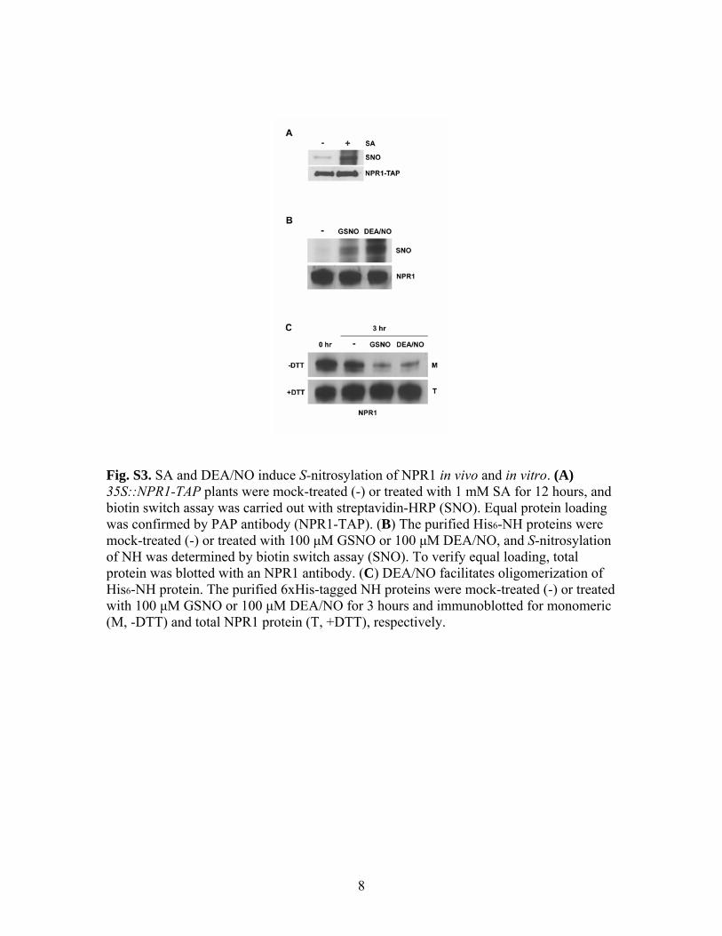
Fig. S3. SA and DEA/NO induce S-nitrosylation of NPR1 in vivo and in vitro. (A) 35S::NPR1-TAP plants were mock-treated (-) or treated with 1 mM SA for 12 hours, and biotin switch assay was carried out with streptavidin-HRP (SNO). Equal protein loading was confirmed by PAP antibody (NPR1-TAP). (B) The purified His6-NH proteins were mock-treated (-) or treated with 100 μM GSNO or 100 μM DEA/NO, and S-nitrosylation of NH was determined by biotin switch assay (SNO). To verify equal loading, total protein was blotted with an NPR1 antibody. (C) DEA/NO facilitates oligomerization of His6-NH protein. The purified 6xHis-tagged NH proteins were mock-treated (-) or treated with 100 μM GSNO or 100 μM DEA/NO for 3 hours and immunoblotted for monomeric (M, -DTT) and total NPR1 protein (T, +DTT), respectively.
8
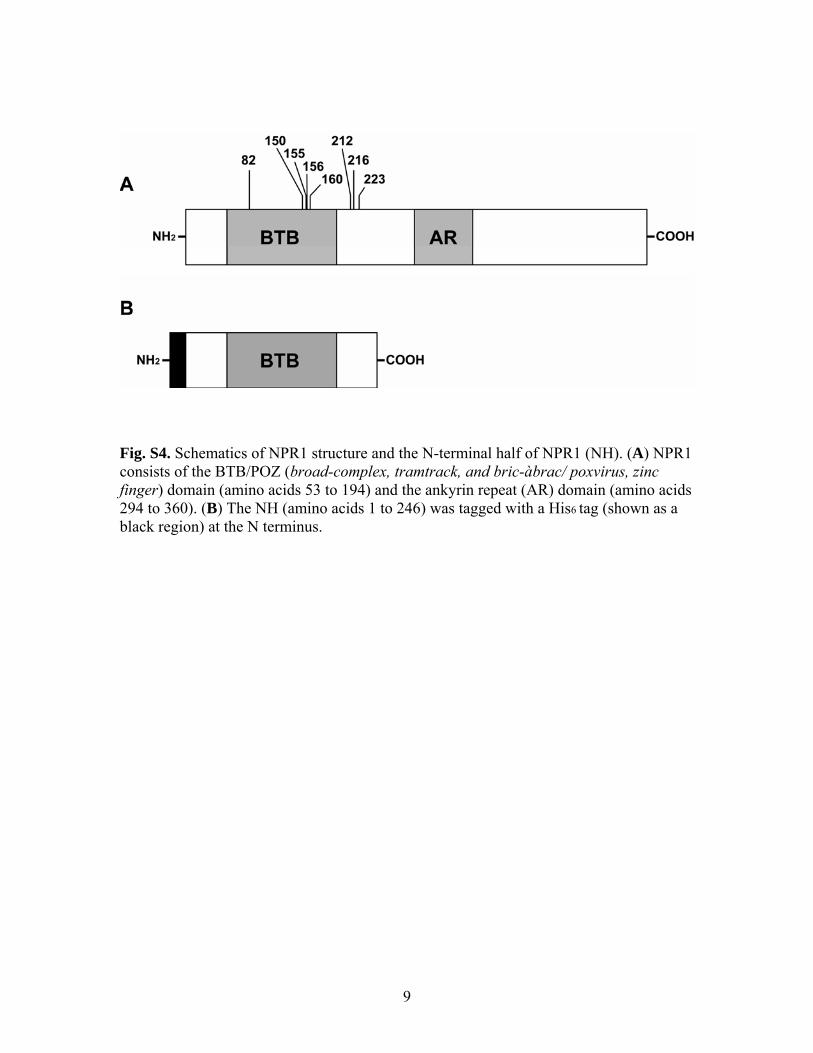
ig. S4. Schematics of NPR1 structure and the N-terminal half of NPR1 (NH). (A) NPR1
cids
Fconsists of the BTB/POZ (broad-complex, tramtrack, and bric-àbrac/ poxvirus, zinc finger) domain (amino acids 53 to 194) and the ankyrin repeat (AR) domain (amino a294 to 360). (B) The NH (amino acids 1 to 246) was tagged with a His6 tag (shown as a black region) at the N terminus.
9

Fig. S5. Computational model of the topology of the NPR1 oligomer. (A) Ribbon representation of the predicted NPR1-BTB dimer. Cysteine residues important for oligomerization and stability of NPR1 are indicated. The model indicates that the NPR1-BTB dimer may be stabilized by a disulfide bond between two C82 residues. (B) Topology prediction of higher-order NPR1-BTB oligomer formation. NPR1-BTB homodimers may interact through the “bottom” side of each dimer. This brings residues C150, C155, C156, and C160 (CCCC) of one dimer in close proximity to the equivalent residues (CCCC’’) of another dimer. Thus, one or more disulfide bonds may form between these equivalent residues. This results in a continuous string-like oligomer in which the dimers are approximately 90 degrees offset relative to each other.
10

Fig. S6. NPR1C156A interacts with TGAs in yeast. Yeast- two-hybrid analysis revealed interaction of NPR1C156 with seven TGA transcription factor in a similar manner to NPR1. pGBKT7 and pGADT7 are empty bait and prey vectors, respectively. The picture was taken 6 days after incubation at 28ºC.
11

Fig. S7. Expression profiles of Arabidopsis TRX-hs in response to SAR induction. Half leaves of 4-week-old plants were inoculated with Psm ES4326/avrRpt2 (OD600 = 0.02) and the other halves were collected at indicated time points and gene expression was profiled using Affymetrix ATH1 GeneChip (24,000 genes) for Arabidopsis. The expression of the eight TRX-h genes was plotted individually according to data from two biological replicates.
12

Fig. S8. Enzymatic interaction of TRXs with NPR1 oligomer. (A) An antibody against NPR1 demonstrated binding with TRX-h3M and TRX-h5M. (B) The enzymatic activities of TRX-h5 and TRX-h5M were measured with insulin as a substrate. The turbidity of the reaction mixture was monitored at 650 nm at RT. (C) Purified TRX-h5 or TRX-h5M protein was immobilized on a resin and incubated with the cell lysate obtained from 35S:NPR1-GFP. The amount of trapped NPR1-GFP protein was assessed by immunoblot with a GFP antibody.
13

Fig. S9. Proposed mechanism of SNO and TRX regulating the NPR1 oligomer/monomer exchange reactions in SA-mediated defense response. See text for details.
14

Fig. S10. SA induces NPR1 oligomerization and monomerization sequentially. 35S::NPR1-GFP (in npr1-1) plants were treated with a combination of 0.5 mM SA, 50 μM of the protein synthesis inhibitor cycloheximide (CHX), and 100 μM of the proteasome inhibitor MG115. At the indicated times plants were harvested and subjected to non-reducing (-DTT) and reducing (+DTT) SDS-PAGE followed by immunoblotting with an anti-GFP antibody. (O) Oligomer; (M) monomer; (T) total.
15

Supplemental References and Notes S1. Z. Mou, W. Fan, X. Dong, Cell 113, 935 (2003). S2. G. Rigaut et al., Nat Biotechnol 17, 1030 (1999). S3. M. Kinkema, W. Fan, X. Dong, Plant Cell 12, 2339 (2000). S4. H. Cao, S. A. Bowling, S. Gordon, X. Dong, Plant Cell 6, 1583 (1994). S5. D. Yamazaki, K. Motohashi, T. Kasama, Y. Hara, T. Hisabori, Plant Cell Physiol
45, 18 (2004). S6. K. Motohashi, A. Kondoh, M. T. Stumpp, T. Hisabori, PNAS 98, 11224 (2001). S7. S. R. Jaffrey, H. Erdjument-Bromage, C. D. Ferris, P. Tempst, S. H. Snyder, Nat
Cell Biol 3, 193 (2001). S8. E. J. Whalen et al., Cell 129, 511 (2007). S9. N. Mouaheb, D. Thomas, L. Verdoucq, P. Monfort, Y. Meyer, Proc Natl Acad Sci
U S A 95, 3312 (1998). S10. N. Guex, M. C. Peitsch, Electrophoresis 18, 2714 (1997). S11. X. Li et al., Cancer Res 59, 5275 (1999).
16