SUPPLEMENTARY NOTES ON THE ANATOMY OF Title …
18
RIGHT: URL: CITATION: AUTHOR(S): ISSUE DATE: TITLE: SUPPLEMENTARY NOTES ON THE ANATOMY OF METARUNCINA SETOENSIS (BABA, 1954), (N. G.) (OPISTHOBRANCHIA- CEPHALASPIDEA) Baba, Kikutaro Baba, Kikutaro. SUPPLEMENTARY NOTES ON THE ANATOMY OF METARUNCINA SETOENSIS (BABA, 1954), (N. G.) (OPISTHOBRANCHIA-CEPHALASPIDEA). PUBLICATIONS OF THE SETO MARINE BIOLOGICAL LABORATORY 1967, 15(3): 185-197 1967-10-30 http://hdl.handle.net/2433/175467
Transcript of SUPPLEMENTARY NOTES ON THE ANATOMY OF Title …

RIGHT:
URL:
CITATION:
AUTHOR(S):
ISSUE DATE:
TITLE:
SUPPLEMENTARY NOTES ON THEANATOMY OF METARUNCINASETOENSIS (BABA, 1954), (N. G.)(OPISTHOBRANCHIA-CEPHALASPIDEA)
Baba, Kikutaro
Baba, Kikutaro. SUPPLEMENTARY NOTES ON THE ANATOMY OF METARUNCINASETOENSIS (BABA, 1954), (N. G.) (OPISTHOBRANCHIA-CEPHALASPIDEA). PUBLICATIONSOF THE SETO MARINE BIOLOGICAL LABORATORY 1967, 15(3): 185-197
1967-10-30
http://hdl.handle.net/2433/175467

SUPPLEMENTARY NOTES ON THE ANATOMY OF MET ARUNCINA SETOENSIS (BABA, 1954), (N.G.)
(OPISTHOBRANCHIA-CEPHALASPIDEA)')
KIKUTAR6 BABA
Biological Institute, Osaka Kyoiku University2 )
With Plates II- V
The species which forms the subject of this paper was originally reported by me
as Runcina setoensis BABA, 1954. After that paper was published, there was found in the years 1955,-.....,1959 a great increase in the population of Runcina around the type
locality, and part of them were used as the material for the studies of their spawn and developing embryos in the laboratory (BABA and HAMATANI, 1959). With this
advantage of having abundant material at my disposal, I decided to go further to make more detailed accounts of the species on the basis of a microscopic study of the
animals. In the beginning of October, 1963, I had fortunately chances to discuss with
Dr. M.T. GHISELIN, who was at Seto at that time, about the phylogeny of the Run
cinidae. His ingenious ways of research on Runcina brought him successful results (GHISELIN, 1963) and stimulated me as valuable suggestions for my subsequent works.
Before going to summarize my studies, I have to mention that I was affected partly by Mr. R. BuRN (1963): he showed his talent in the systematic classification of Runcina
and its allies.
Metaruncina BABA, n. g.
Most closely allied to Ildica BERGH, 1889 (type: I. nana BERGH, 1889.-Mau
ritius) in the possession of a single cephalaspidean gill on the right posterior side of body, but differentiated from it in having a greatly degraded radula which remains in a triseriate type ( 1.1.1.) in Ildica. Shell posterior, terminal, internal in Metaruncina,
and external ( ?) in Ildica. Genitalia without a spermatheca (bursa copulatrix).
Type: Runcina setoensis BABA, 1954.
I) Contributions from the Seto Marine Biological Laboratory, No. 470. 2) Former Osaka Gakugei University.
Publ. Seto Mar. Bioi. Lab., XV (3), 185-197, 1967. (Article 11)

186 K. BABA
Metaruncina setoensis (BABA, 1954)
Runcina setoensis BABA, 1954, pp. 373-374, text-fig. 1.-Seto, Kii; BABA & HAMATANI, 1959, pp. 281-290, pl. 22, figs. 1-19; pl. 23, figs. 1-10; pl. 24, figs. 1-7 (direct development).-Seto, Kii.
Dissection work under a binocular microscope was made usually on live specimens
taken from the sea. For histological examination eighteen specimens listed below
were used in serial preparations.
Sp. Nos. 1-4. Oct. 12, 1958. (H.S.) Sp. Nos. 5-7. Oct. 5, 1959. (H.S.) Sp. No. 8. Oct. 12, 1958. (L.S.)
Sp. Nos. 9-12. Sp. Nos. 13-14. Sp. Nos. 15-18.
Oct. 4, 1959. (L.S.) Oct. 12, 1958. (T.S.) Oct. 4, 1959. (T.S.)
They were all in a matured state each. The fixative in general use was that of BourN; only the specimens Nos. 5 to 7 were fixed with NAVASHIN's fluid. The dyes
for many of the preparations were DELAFIELD's haematoxylin and eosin; a selective
mucus staining by toluidine blue was applied to a single case of the specimen No. 4. Externals: The general body-form and colours were approximately as shown
previously (BABA, 1954; BABA & HAMATANI, 1959). The gill in this species was
rightly discussed by GmsELIN (1963, pp. 391-392). It is single, attached to the right posterior side of body, and freely projecting behind. The pinnae, 4-6 in number on each side of the rachis, are arranged alternately. The first rudiment of the gill is to be seen on a young individual immediately after hatching (BABA &
HAMATANI, 1959, text-fig. lA). It is notable that such a cephalaspidean gill is
possessed in common only by two of the different genera, Ildica and Metaruncina ( cf.
BuRN, 1963, p. 19). In Runcina (in a restricted sense) the gill is right posterior in position, but it consists of 2-3, paucipinnate plumes ( cf. BuRN, 1963, pp. 20, 21). In the genera Runcinida and Runcinella the gill is formed of 4-5, paucipinnate plumes
which are set in a semi-circle round the anus ( cf. BuRN, 1963, p. 21). The anus is
posterior and median as usual. In Metaruncina the opening of the opaline gland lies just to the left of the anus (in Runcinida this opening is low down the level of anus, on the left of the median line). From the opening of the opaline gland there occurs
an occasional jet of colourless, hyaline mucous matter which encloses whitish grains as the products of the opaline gland proper ( cf. GmsELIN, 1963, p. 390).
In Metaruncina the common genital orifice lies moderately apart from the gill
(in Runcinida and Runcinella it is found rather closely in front of the gill). The copulatory behavior (including the act of transmission of spermatophores) stated by GHISELIN (1963, pp. 394-395) has repeatedly been confirmed by me. The male orifice
opens on the right side of the mouth (in Runcinida it comes more closely to the mouth). The external seminal groove and the foldings of the HANCOCK's organs are as shown
by GHISELIN. The shell of Metaruncina varies greatly in form according to specimens. Typically
it is of an elongated haliotiform, highly flattened, more or less widened in front but narrowed to a produced rostrum behind, and marked on the surface with fine growth-

Anatomy of Metaruncina setoensis 187
lines; in consistency it is calcareous, opaque and whitish. The total length of the
shell ranges from 0.2 mm to 0.4 mm. As was reported previously (BABA & HAMATANI,
1959, p. 284), the shell of Metaruncina begins to develop within the mantle-tissue at
an early stage of intra-capsular veliger, and is kept as a complete internal shell through
out the life of the animal. Hence there is no formation of a protoconch on the rostrum
of the shell in question. The external (?) shell of Ildica needs to be re-examined on
fresh material. Ildica divae of MARCus & MARcus, 1963 has a true nautiloid shell.
The whole outer surface of the body including the mantle, sides, gill and sole is
covered with fine cilia.
Internals: The jaw-plates and radula are nearly as given before (BABA, 1954, p. 373; see also GHISELIN, 1963, p. 391). The paired salivary glands are long, band
like and colourless. The alimentary canal (oesophagus and intestine) is yellow
pigmented, its lumen having a strong ciliary current in a fresh state. The oesophageal
diverticulum lies a short distance in front of the gizzard. It appears to be a simple
blind sac without a secretory structure. It is only slightly ciliated internally. The
gizzard plates, colourless when fresh, show each a series of 11-12, acutely pointed
laminae in side view. The stomach does not form a well defined chamber. It is
accompanied by paired liver-lobes which are symmetrical in the intra-capsular veliger
stage (BABA & HAMATANI, 1959, p. 284), but later these structures acquire an asym
metry. Thus in the adult the right liver which is the smaller of the two takes an
antero-ventral position; the left liver is decidedly larger than the right one and lies
postero-dorsal to the stomach. The oesophagus passes into the stomach at its antero
ventral corner on the left side; the intestine leaves the antero-dorsal corner of the
stomach, and runs down as far as to the postero-median anus. The intestine has an
internal longitudinal fold at its origin from the stomach. The liver-mass in fresh is
generally of an ashy yellowish brown. Under the microscope the liver cells appear
to take different facies as follows: ( 1) some are filled with clear and colourless granules
which tend to be easily destroyed in the sectioned material; (2) some are packed with
mostly fine granules of a dull colour, and (3) others contain coaser, yellow-tinted
granules. The contents of the cell-types (2) and (3) may remain as they were before
on mounted sections.
The opaline gland consists of a moderate number of compound glandules inter
mingled with smaller mucous cells. In a fresh state each of the compound glandules
is formed of cells full of whitish grains (these latter, however, may easily be destroyed
by a fixing fluid). Externally it is covered by a muscular layer which apparently
controlls the contraction of the body of the glandule. The whole organ of the opaline
gland has a common lumen which opens to the exterior through a small pore. An
actual case of secretion from the opaline gland was already noted before. Lying
just below the shell and located above the rectum there is a small compound gland of
unknown function. Its duct opens at a point immediately above the posterior
insertion of the gill, and so this gland is named tentatively as a supra-branchial

188 K. BABA
gland.
The central nervous system of Metaruncina was studied and discussed with some reservations by GHISELIN ( 1963, pp. 392-393). Unfortunately I have not been able
to prepare much informations of this system on the basis of my own research material. The primitively cephalaspidean character ofthe central nervous system of the runcinid
is shown by the possession of strongly ganglionated nerves (labial and olfactory nerves) supplied from the cerebro-pleural (here identified as such by the presence of the pleural nerves) ganglia to the HANCOCK's organ on either side of the head (cf. GmART, 1901, fig. 55.-Scaphander). A tendency of great shortening of the visceral
loop occurs in some small-sized forms (Philinoglossa, etc.) of the Opisthobranchia
(GHISELIN, 1963, p. 395). It seems likely that the ganglionic mass (RP+ of
GHISELIN, 1963) just behind the right cerebro-pleural ganglion consists mainly of the supra-intestinal elements. It takes its position on the dorsal side of the oesophagus.
The ganglionic mass (LP+ of GHISELIN) immediately behind the left cerebro-pleural ganglion may be presumed as having been formed by the union of the infra-intestinal
and the visceral ganglia. This mass is as large as the right-sided partner, and lies below the oesophagus. Only it must be remarked that the fine nerves from the
visceral loop remained undetermined on the microscopic preparations. The statocysts contain each a single statolith. There is no formation of an osphradium.
The heart occupies its position in the right posterior part of the haemocoele (cf.
Pluscula of MARcus, 1953, and Philinoglossa). The auricle receives blood from the
lacunar system of the gill. On the serial preparations the aorta appears to pass forward to open directly into the haemocoele. The kidney is a spacious sac lying to the left side of the vascular system. It is combined with the pericardium by a simple
reno-pericardia! canal. The nephroproct is found just below the anterior insertion
of the gill. There occur abundant plasma cells wandering about in the lacunose
subepithelial connective tissue and in the haemocoele. Amoebocytes were found
gathered especially within the lacunae of the gill.
The gonad is seen at about the middle of the body and located to the right of the median line. It consists of a single elongated testis accompanied on the periphery
by a moderate number of ovarian follicles. These follicles may assume each a rusty
brown tint owing to the eggs matured within. Posteriorly the gonad passes to form a slightly bulged sac (ampulla) for keeping endogenous sperms. Then follows the
pallial part of gonoduct with an accessory female gland mass filling the hindmost lumen of the haemocoele. The details of the pallial gonoduct, however, could not satisfactorily be analized from the viewpoint of functional anatomy ( cf. GHISELIN,
1963, p. 394). Distally the seemingly monaulic canal of the pallial gonoduct in
Metaruncina has no formation of a spermatheca (bursa copulatrix) for the reception of introduced spermatophores (a stalked spermatheca is clearly present in Runcinida).
The male copulatory organ consists of three parts: the penis sac, the prostate, and the spermatic bulb (GHISELIN, 1963, p. 394). In a live animal the whole organ is

Anatomy of Metaruncina setoensis 189
greatly extensible or contractile owing to the muscular covering contammg circular
and longitudinal fibres. The everted penis sac acts as a temporary penis. The
prostate is yellow-pigmented, and the spermatic bulb has a melanin-black epithelium. It is to be added that a spermatic bulb is present also in the copulatory apparatus of
Runcinida.
Systematical Notes
When making a general survey of the genera and species of the Runcinoidea, it
was felt necessary to pay a special attention to the nautiloid shell possessed by Ildica
divae MARCUS & MARCus, 1963 from Curac;ao. The external ( ?) shell of the digest
ed animal of Ildica nana BERGH, 1889 cannot rightly be accepted. It is very unfortunate that the type of Runcina FoRBES, 1851 has long been left without being·
anatomized exactly by later investigators. The sole external feature to be found in this type concerns the gill. So I am going to prepare below a re-arrangement of all the genera of the Runcinoidea on the basis mainly of the comparison of their gill characters. This does not mean, however, to neglect the systematic classification of
the Runcinoidea advanced by BuRN (1963, pp. 19-22).
Superfamily Runcinoidea
Family Runcinidae. Shell reduced to a non-convoluted internal shell, or it may be missing altogether. A. A single cephalaspidean gill on the right posterior body-side.
I. Itdica Bergh, 1889. Type: Ildica nana BERGH, 1889.-Mauritius. Radula I.!. I. Shell posterior, terminal, and external (?).
2. Metaruncina BABA, n.g. Type: Runcina setoensis BABA, 1954.-Seto, Kii. Radula greatly reduced, not in distinct rows. Shell posterior, terminal, and completely internal in formation.
B. Gill formed of 2-3 separate plumes, these being right posterior in position. I. Runcina FoRBEs, 1851. Type: Runcina hancocki FoRBEs, 1851 =Petta corona fa
QuATREFAGEs, 1844.-England. Radula and shell unknown. Several species have hitherto been referred provisionally to this genus.
C. Gill formed of 4-5 separate plumes set in a semi-circle round the posterior median anus. A shell absent.
I. Runcinella 0DHNER, 1924. Type: Runcinella zetandica 0DHNER, 1924.-New Zealand. Radula 2.1.2.
2. Runcinida BURN, 1963. Type: Runcina etioti BABA, 1937.-Amakusa. Radula 1.1.1.
D. Gill missing. I. Ilbia BuRN, 1963. Type: Ilbia ilbi BuRN, 1963.-Victoria. No shell. Radula
I. I.!.
REFERENCES
ALDER, J. & HANCOCK, A. 1846. Notices of some new and rare British species of naked Mollusca. Ann. Mag. Nat. Hist., vol. 18. (Petta)
BABA, K. 1937. Opisthobranchia of Japan. I. Journ. Dept. Agric. Kyushu Imp. Univ., vol. 5, no. 4. (Runcina etioti)

190 K. BABA
BABA, K. 1954. Runcina setoensis, a new and rare species from the coast of Kii, Middle Japan (Opisthobranchia). Pub!. Seta Mar. Bioi. Lab., val. 3, no. 3.
BABA, K. & HAMATANI, I. 1959. The direct development in Runcina setoensis BABA (OpisthobranchiaCephalaspidea). Pub!. Seta Mar. Bioi. Lab., val. 7, no. 2.
BERGH, R. 1889. Malakologische Untersuchungen. Heft 16, pt. 2. (Ildica nana)
BoETTGER, C.R. 1955. Die Systematik der euthyneuren Schnecken. Zoo!. Anz., Suppl., Bd. 18. (Runcinidae)
BuRN, R. 1963. Australian Runcinacea (Mollusca: Gastropoda). Australian Zoologist, val. 13, pt. I.
1966. The opisthobranchs of a caulerpan microfauna from Fiji. Proc. malac. Soc. London, val. 37, pt. I. (Runcina marshae)
CoLOSI, G. 1915. Osservazioni anatomo-istologiche sulla Runcina calaritana n. sp. Mem. Accad. Sci. Torino, ser. 2, tom. 66.
CRAWFORD, T. 1965. Some molluscs associated with Posidonia weed at Flinders, Victoria. Australian
Newsletter, val. 13, no. 50. (Ilbia ilbi)
EALES, N.B. 1952. The littoral fauna of Great Britain. Cambridge Univ. Press. (Pelta coronata)
FoRBES, E. 1851. In: A history of British molluscs and their shells. val. 3. (Runcina hancocki, not directly referred to)
GANTES, H. 1956. Complement a !'etude des opisthobranches des cotes du Maroc. Bull. Soc. nat. Phys. Maroc, tom. 36. (Runcina africana)
GHISELIN, M.T. 1963. On the functional and comparative anatomy of Runcina setoensis BABA, an opisthobranch gastropod. Pub!. Seta Mar. Bioi. Lab., val. II, no. 2.
----- 1966. Reproductive function and the phylogeny of opisthobranch gastropods. Malacologia, val. 3, no. 3.
GurART,j. 1901. Contribution a !'etude des gasteropodes opisthobranches et en particulier des cephalaspides. Mem. Soc. Zoo!. France, tom. 14.
HAEFELFINGER, H.R. 1959. Catalogue des opisthobranches de la rade de Villefranche-sur-Mer et ses environs (Alpes Maritimes). Rev. suisse de Zoo!., tom. 67, no. 27. (Runcina coronata)
HoFFMANN, H. 1932-40. BRONNS Klassen und Ordnungen des Tier-Reichs. Bd. 3, Mollusca, Abt. 2, Gastropoda, Buch 3, Opisthobranchia, Teil I-II.
KAWAGUTI, S. & YAMAsu, T. 1961. Self-fertilization in the bivalved gastropod with special references to the reproductive organs. Bioi. Journ. Okayama Univ., val. 7, nos. 3-4.
LEMCHE, H. 1948. Northern and Arctic tectibranch gastropods. I. The larval shells. II. A revision of the cephalaspid species. Kgl. Dansk Vid. Selsk. Bioi. Skr., Bd. 5, no. 3. (Runcina coronata)
1956. The anatomy and histology ofC',ylichna (Gastropoda Tectibranchia). Skrifter Zoo!.
Mus., 16. 1965. Pelta QuATREFAGEs, 1844 or Runcina FoRBEs, 1851 (Gastropoda): two competing
names for a place on the Official List. Z.N. (S.) 580. Bull. zoo!. Nomencl., val. 22, pt. I.
MARcus, Er. 1953. Three Brazilian sand-Opisthobranchia. Bal. Fac. Cien., Letr. Univ. Sao Paulo, Zoo!., no. 18. (Pluscula cuica)
MARcus, Ev. & MARCus, Er. 1954. tiber Philinoglossacea und Acochlidiacea. Kieler Meeresf., Bd. 10, Heft 2. (Philinoglossa)
1963. Opisthobranchs from the Lesser Antilles. Studies on the fauna of Cura<;ao and other Caribbean islands, val. 19. (Ildica divae)
MAzzARELLI, G. 1893. Ricerche sulle Peltidae del Golfo di Napoli. Atti Rend. Accad. Sci. Fis. mat. Napoli, val. 6, ser. 2, no. 4. (Pelta capreensis, not directly referred to)
Mi:iRCH, M.O.A.L. 1863. Contributions a Ia faune malacologique des Antilles danoises. Journ. Conchylio., val. II. (Pelta prasina)
0DHNER, N. 1924. Papers from Dr. Th. MoRTENSEN's Pacific Expedition 1914-16. 19. New Zealand Mollusca. Vid. Medd. Dansk naturh. Foren, Bd. 77. (Runcinella zelandica)
PELSENEER, P. 1894. Recherches sur divers opisthobranches. Mem. cour. Acad. Roy. Belgique, tom. 53. (Pelta)

Anatomy of Metaruncina setoensis 191
PILSBRY, H.A. 1896. Manual of Conchology. Vol. 16. (Runcinidae) PRUVOT-FOL, A. 1953. Etude de quelques opisthobranches de Ia cote Atlantique du Maroc et du
Senegal. Trav. l'Inst. Sci. Cherifien, no. 5. (Runcina africana)
----- 1954. Faune de France. 58. Mollusques opisthobranches. (Runcinidae) 1960. Les organes genitaux des opisthobranches. Arch. Zoo!. Exp. et Gen., tom. 99,
fasc. 2. Risso-DoMINGUEz, C.J. 1963. Measuring nudibranchs: a standardization for descriptive purposes.
Proc. malac. Soc. London, vol. 35, pt. 5. RussELL, L. 1929. The comparative morphology of the elysioid and aeolidioid types of the molluscan
nervous system, and its bearing on the relationships of the ascoglossan nudibranchs. Proc. Zoo!. Soc. London, pt. 2.
TAYLOR, D.H. & SoHL, N.F. 1962. An outline of gastropod classification. Malacologia, vol. I, no. I. (Runcinidae)
THIELE, J. 1931. Handbuch der systematischen Weichtierkunde. Teil 2. (Runcinidae) VAYSSIERE, A. 1883. Recherches anatomiques sur les genres Petta et Tytodina. Ann. Sci. nat. Zoo!.,
ser. 6, tom. 15. (Petta coronata, not directly referred to) ----- 1885. Recherches zoologiques et anatorniques sur les mollusques opisthobranches du
Golfe de Marseille. Tectibranches. Ann. Mus. d'Hist. nat. Marseille, Zoo!., tom. 2. (Petta coronata)
----- 1900. Note sur un nouveau cas de condensation embryogenique observe chez le Petta
coronata, type de tectibranche. Zoo!. Anz., Bd. 23. ----- 1929. Faune et flore de Ia Mediterranee. 12. Opisthobranchiata. (Petta coronata)
VERRILL, A.E. 1901. Additions to the fauna of the Bermudas. Trans. Conn. Acad. Arts Sci., vol. II, pt. I. (Runcina inconspicua, not directly referred to)
Opinion 811. Runcina Forbes, 1851 (Gastropoda): validated under the plenary powers. Bull. zoo!. Nomencl., vol. 24, pt. 2.
Postscript
1. Additional specimens of Runcinida elioti (BABA, 1937) have been collected from several stations of our seas. Anatomical notes of this species will be prepared
in a separate paper. 2. In my report of Volvatella of July, 1966, I could not refer to V. ficula BuRN,
1966, a species which had been recorded from Fiji. Some of the southern Pacific
species of Volvatella will tentatively be taken up as below: (i) V. fragilis PEASE, 1860 (Hawaii; live animal white); (ii) V. evansi (KAY, 1961) (Hawaii; live animal
orange); (iii) V.ficula BuRN, 1966 (Fiji; live animal creamy white); (iv) V. vigourouxi
(MoNTROUZIER, 1861) (New Caledonia; shell only); and (v) V. kawamurai HABE, 1946 (Okinawa and Amami Groups; colour of live animal unknown).
Literature: BuRN, R. 1966. The opisthobranchs of a caulerpan ficuta); KAY, A. 1961. A new opisthobranch mollusc from Hawaii. evansi)
microfauna from Fiji. (Volvatella
Pac. Sci., vol. 15, no. I. (Arthessa
3. Two of the latest works by BuRN (1965 and 1966) have given us solid grounds
for establishing advanced taxonomy of the recent taxa of the bivalved sacoglossans. Privately I am going to agree with him, and Edenttellina, Tamanovalva and Midorigai
are accepted as distinct from one another either concho1ogically or anatomically.
Now the specific name T. limax KAWAGUTI & BABA, 1959 stands as it was first pub-

192 K. BABA
lished. Tamanovalva comprises also a number of exotic species such as T. corallensis
(HEDLEY, 1920) (Australia), T. babai BuRN, 1965 (=Berthelinia typica BuRN, 1960) (Australia), T. jijiensis BuRN, 1966 (Fiji), T. chloris (DALL, 1918) (Baja California), and some others. Berthelinia, ( ?) Anomalomya and Cossmannella ( =Ludovicia) are fossil genera, the first of these being based on an undifferentiated larval shell.
Literature: BuRN, R. 1965. Rediscovery and taxonomy of Edenttellina typica GATLIFF and GABRIEL. Nature, vol. 206, no. 4985;---1966. The opisthobranchs of a caulerpan microfauna from Fiji. ( Tamanovalva jijiensis)
4. In response to the suggestion by BuRN, 1966, it was decided to create a new genus as follows: Embletoniella BABA, n. g. Type: Embletonia paucipapillata BABA &
HAMATANI, 1963. This new genus is distinguished from Embletonia ALDER & HANcocK, 1851 by the branchial papillae which are marked each with four apical twigs,
and by having a prostatic and unarmed penis. Embletonia gracilis of RxsBEC, 1928 belongs to this new genus, but it was not satisfactorily anatomized. Now the two species of Embletoniella are shown thus: (i) E. paucipapillata (BABA & HAMATANI,
1963) (Japan) and (ii) E. gracilis (RISBEC, 1928) (New Caledonia, Australia, and
Japan). Literature: BABA, K. 1959. A new record of an interesting species, Embletonia gracile RISBEC, from Japan (Nudibranchia-Eolidacea). Pub!. Seto Mar. Bioi. Lab., vol. 7, no. 3; BABA, K. & HAMATANI, I. 1963. Anatomy of Embletonia gracilis paucipapillata n. ssp. from Osaka Bay, Japan (NudibranchiaEolidoidea). Pub!. Seto Mar. Bioi. Lab., vol. II, no. 2; BuRN, R. 1966. Descriptions of Australian Eolidacea (Mollusca: Opisthobranchia). 3. Journ. malac. Soc. Australia, no. 9. (Embletonia gracilis);
LEMCHE, H. 1964. Embletonia ALDER & HANCOCK, 1851 (Gastropoda): proposed validation under the plenary powers. Z.N. (S.) 1100. Bull. zoo!. Nomencl., vol. 21, pt. 2.
5. Some of the species of the Aeolidiidae from our seas will provisionally be
designated as below: (i) Berghia amakusana (BABA, 1937) =Baeolidia major amakusana BABA, 1937. This name change follows MARCus', 1958. Baeolidia major ELIOT, 1903, from the sea
round its type locality Zanzibar, is hoped to be revised. Recently the species ama
kusana was found from the Gulf of California (FARMER, 1966). (ii) Berghia japonica
(BABA, 1937) =Baeolidiajaponica BABA, 1937. This name change follows MARcus', 1958. I have a plan to make a complete anatomy of this species at any chance in
future. (iii) Limenandrafusiformis (BABA, 1949) =Baeolidiajusiformis BABA, 1949. This name change was suggested by HAEFELFINGER & STAMM, 1959. Specimens of this
species will also be revised by me in the future.
Literature: FARMER, W. 1966. Range extension of Berghia amakusana (BABA) to the east Pacific. Veliger, vol. 9, no. 2; HAEFELFINGER, H.R. & STAMM, R.A. 1959. Limenandra nodosa gen. et sp. nov., un opisthobranche nouveau de Ia Mediterranee. Vie et Milieu, tom. 9, fasc. 4; MARCus, Er. 1958. On western Atlantic opisthobranchiate gastropods. Amer. Mus. Novitates, no. 1906. (Baeolidia)

Anatomy of Metaruncina setoensis 193
EXPLANATION OF PLATES 11-V
Metaruncina setoensis. All the specimens for this study were collected from the shores around the Seto Marine Biological Laboratory, Seto, Kii, Middle Japan, during
the years 1951-65.
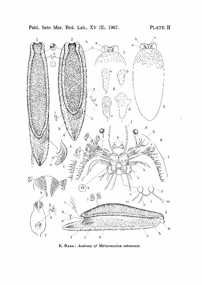
PLATE II
Fig. 1. Matured specimen in an actively crawling position (Aug. 1, 1963). Measurements (after the Codes in Dorids): total body-length from head to tail-tip (Code A) 6 mm; mantle-length (Code Am) 4 mm; mantle-width (Code M) I mm; mantle-height (Code H) 1 mm; tail-length (Code C) 2 mm; sole-width (Code B) 1 mm; gill-length 0.8 mm. Mantle truncated or shallowly sinuated
in front, obtuse behind, lateral margins parallel, tail projecting. General groundcolour above ashy yellow, wholly sprinkled with chocolate-brown spots, the
melanin-black pigment prevailing at the centre of mantle, on mantle-margin,
on sides, on foot-margin, and on median line of tail. Paired eyes shining through, the internal shell usually not visible from outside. Gill deep black. a. cluster
of chocolate-brown pigment cells, b. rachis of gill, c. lateral pinnae of gill, d. anus. Fig. 2. The same animal in a resting position. b. male genital orifice (actually
this is more close to mouth), c. external seminal groove, d. common genital
orifice, e. spermatophores ( 1-2) erroneously planted near the genital orifice,
f. gill, g. anus, h. opening of opaline gland, i. position of internal shell. The
spermatophores an; papilliform, and opaque white in appearance.
Fig. 3. A resting animal from right side (Oct. 6, 1963). a. everted penis sac forming
a whitish cone of temporary penis, b. feeble foldings of HANCOCK's organ visible on under side of mantle-margin, c. external seminal groove, d. common genital
orifice, e. nephroproct (secured by serial sections), f. gill, g. anus, i. secretion from the opening (h) of opaline gland, j. isolated epithelial cells with melanin pigment.
Fig. 4. The same animal from ventral side. Sole ashy yellow covered with chocolate
brown spots as above. a. external seminal groove, b. male genital orifice, c. mouth-slit. There is no formation of oral tentacles.
Fig. 5. The same animal as above. a. appearance of a temporary penis.
Fig. 6. Some of the differently shaped shells (X 45). Fresh material. a-b (Aug. 1,
1963); c-d (Oct. 6, 1963).
Fig. 7. Pharyngeal bulb from above. Fresh material (Aug. 1, 1963). a. paired
jaw-plates covered with spiny denticles (X 130), b. group of rudimentary radula
teeth (X 180).
Fig. 8. General view of the central nervous system (X 50). Fresh material (Oct.
6, 1963). Details supplemented by observation of serial preparations (Sp. No.
1 and others). a. buccal ganglion, b. cerebro-pleural ganglion, c. pedal ganglion,
194 K. BABA
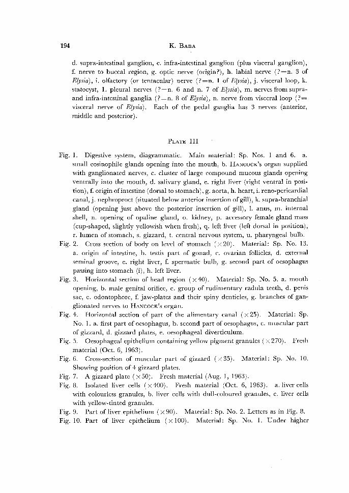
d. supra-intestinal ganglion, e. infra-intestinal ganglion (plus visceral ganglion),
f. nerve to buccal region, g. optic nerve (origin?), h. labial nerve (?=n. 3 of
Elysia), i. olfactory (or tentacular) nerve (?=n. 1 of E[vsia), j. visceral loop, k. statocyst, 1. pleural nerves (?=n. 6 and n. 7 of E£vsia), m. nerves from supraand infra-intestinal ganglia (?=n. 8 of Elysia), n. nerve from visceral loop (?= visceral nerve of Elysia). Each of the pedal ganglia has 3 nerves (anterior, middle and posterior).
PLATE III
Fig. 1. Digestive system, diagrammatic. Main material: Sp. Nos. 1 and 6. a. small eosinophile glands opening into the mouth, b. HANCOCK's organ supplied
with ganglionated nerves, c. cluster of large compound mucous glands opening ventrally into the mouth, d. salivary gland, e. right liver (right ventral in posi
tion),£ origin of intestine (dorsal to stomach), g. aorta, h. heart, i. reno-pericardia!
canal, j. nephroproct (situated below anterior insertion of gill), k. supra-branchial gland (opening just above the posterior insertion of gill), 1. anus, m. internal
shell, n. opening of opaline gland, o. kidney, p. accessory female gland mass
(cup-shaped, slightly yellowish when fresh), q. left liver (left dorsal in position), r. lumen of stomach, s. gizzard, t. central nervous system, u. pharyngeal bulb.
Fig. 2. Cross section of body on level of stomach (X 20). Material: Sp. No. 13.
a. origin of intestine, b. testis part of gonad, c. ovarian follicles, d. external seminal groove, e. right liver, f. spermatic bulb, g. second part of oesophagus
passing into stomach (i), h. left liver. Fig. 3. Horizontal section of head region (X 40). Material: Sp. No. 5. a. mouth
opening, b. male genital orifice, c. group of rudimentary radula teeth, d. penis sac, e. odontophore, £ jaw-plates and their spiny denticles, g. branches of gan
glionated nerves to HANcocK's organ. Fig. 4. Horizontal section of part of the alimentary canal (X 25). Material: Sp.
No. 1. a. first part of oesophagus, b. second part of oesophagus, c. muscular part
of gizzard, d. gizzard plates, e. oesophageal diverticulum. Fig. 5. Oesophageal epithelium containing yellow pigment granules (X 270). Fresh
material (Oct. 6, 1963). Fig. 6. Cross-section of muscular part of gizzard (X 35). Material: Sp. No. 10.
Showing position of 4 gizzard plates. Fig. 7. A gizzard plate (X 50). Fresh material (Aug. 1, 1963).
Fig. 8. Isolated liver cells (X 400). Fresh material (Oct. 6, 1963). with colourless granules, b. liver cells with dull-coloured granules,
with yellow-tinted granules.
a. liver cells c. liver cells
Fig. 9. Part of liver epithelium (X 90). Material: Sp. No. 2. Letters as in Fig. 8. Fig. 10. Part of liver epithelium (X 100). Material: Sp. No. 1. Under higher

Anatomy of Metaruncina setoensis 195
magnification the yellow-tinted granules may be seen included each within a
vacuole. Letters as in Fig. 8. Fig. 11. Cross section of intestine (a) and rectum (b) (x90). Material: Sp. No.
14. The excrement consists of fine granules of unknown nature. Fig. 12. Intestinal epithelium containing yellow pigment granules (X 270). Fresh
material (Oct. 6, 1963). Fig. 13. Horizontal section of opaline gland (X 100). Material: Sp. No. 6. a.
compound glandules, b. opening of opaline gland, c. common lumen of opaline
gland, d. mucous cells, e. invaginated epithelium. Fig. 14. Part of opaline gland (xl50). Material: Sp. No.1. a. external muscular
layer, b. individual gland cells filled with eosinophile grains, c. lumen of the
individual glandule, d. mucous cells. Fig. 15. Individual glandule in the isolated state (X 150). Fresh material (Oct. 6,
1963). b. individual gland cells filled with whitish grains. These gland cells
may sometimes contain traces of yellow pigment. Fig. 16. Cross section of opaline gland (X 80). Material: Sp. No. 13. a. showing
destruction of secretory grains into finer granulations, b. lumen of an individual glandule.
PLATE IV
Fig. 1. Horizontal section of the right posterior part of body on level of the heart
(X 35). Material: Sp. No. 6. a. intestine, b. aorta, c. ventricle, d. auricle, e. reno-pericardia! canal, f. nephroproct (situated below anterior insertion of
gill), g. gill, h. anus, i. opening of opaline gland (j), k. rectum, I. lumen ofkidney, m. left liver, n. testis part of gonad.
Fig. 2. Median longitudinal section of head region (X 35). Material: Sp. No. 8. a. ovarian follicles, b. supra-intestinal ganglion, c. cerebro-pleural ganglion,
d. oesophagus, e. buccal ganglion, f. branches of ganglionated nerves to
HANCOCK's organ, g. pharynx, h. spiny denticles of jaw-plate, i. cuticular lining, j. mouth opening, k. pedal ganglion, 1. cluster oflarge compound mucous glands,
m. prostatic part of male copulatory organ, n.; right liver, o. left liver. Fig. 3. Submedian longitudinal section ofposterior part ofbody (x35). Material:
Sp. No. 8. a. left liver, b. gland C (?=mucous gland) of the accessory female gland mass, c. gland A (?=albumen gland) of the same, d. opening of opaline gland.
Fig. 4. Median longitudinal section of posterior part of body (X 35). Material:
Sp. No. 10. a. shell within a shell sac, b. supra-branchial gland, c. anus guarded by small eosinophile glands.
Fig. 5. Longitudinal section of posterior part of body, passing through heart (X 35). Material: Sp. No. 11. a. testis, b. ovarian follicles, c. common gonoduct, d. lumen
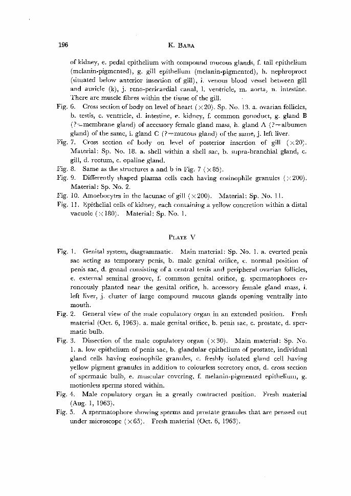
196 K. BABA
of kidney, e. pedal epithelium with compound mucous glands, f. tail epithelium
(melanin-pigmented), g. gill epithelium (melanin-pigmented), h. nephroproct
(situated below anterior insertion of gill), i. venous blood vessel between gill and auricle (k), j. reno-pericardia! canal, 1. ventricle, m. aorta, n. intestine. There are muscle fibres within the tissue of the gill.
Fig. 6. Cross section of body on level of heart (X 20). Sp. No. 13. a. ovarian follicles,
b. testis, c. ventricle, d. intestine, e. kidney, f. common gonoduct, g. gland B (?=membrane gland) of accessory female gland mass, h. gland A (?=albumen
gland) of the same, i. gland C (?=mucous gland) of the same, j. left liver. Fig. 7. Cross section of body on level of posterior insertion of gill (X 20).
Material: Sp. No. 18. a. shell within a shell sac, b. supra-branchial gland, c. gill, d. rectum, e. opaline gland.
Fig. 8. Same as the structures a and bin Fig. 7 (X85). Fig. 9. Differently shaped plasma cells each having eosinophile granules (X 200).
Material: Sp. No. 2.
Fig. 10. Amoebocytes in the lacunae of gill (x200). Material: Sp. No. 11. Fig. 11. Epithelial cells of kidney, each containing a yellow concretion within a distal
vacuole (X 180). Material: Sp. No. 1.
PLATE v
Fig. 1. Genital system, diagrammatic. Main material: Sp. No. 1. a. everted penis sac acting as temporary penis, b. male genital orifice, c. normal position of
penis sac, d. gonad consisting of a central testis and peripheral ovarian follicles, e. external seminal groove, f. common genital orifice, g. spermatophores er
roneously planted near the genital orifice, h. accessory female gland mass, i.
left liver, j. cluster of large compound mucous glands opening ventrally into mouth.
Fig. 2. General view of the male copulatory organ in an extended position. Fresh
material (Oct. 6, 1963). a. male genital orifice, b. penis sac, c. prostate, d. spermatic bulb.
Fig. 3. Dissection of the male copulatory organ (X 30). Main material: Sp. No. 1. a. low epithelium of penis sac, b. glandular epithelium of prostate, individual
gland cells having eosinophile granules, c. freshly isolated gland cell having yellow pigment granules in addition to colourless secretory ones, d. cross section
of spermatic bulb, e. muscular covering, f. melanin-pigmented epithelium, g.
motionless sperms stored within.
Fig. 4. Male copulatory organ in a greatly contracted position. Fresh material
(Aug. 1, 1963). Fig. 5. A spermatophore showing sperms and prostate granules that are pressed out
under microscope (X 65). Fresh material (Oct. 6, 1963).

Anatomy of Metaruncina setoensis 197
Fig. 6. Longitudinal section of body passing through the common genital opening
(X 35). Seen from right side. Material: Sp. No. 8. a. three spermatophores
about to pass simultaneously into the common gonoduct (b). Fig. 7. Horizontal section along the length of the gonad (X 15). Sp. No. I. a. testis
(accumulation of melanin pigment granules occurs in the outer wall of testis),
b. hermaphrodite duct ( =preampullar portion of coelomic gonoduct), c. ampulla, d. ovarian follicles.
Fig. 8. Enlarged figure of ovarian follicle (a) and part of testis (b) ( X 60). Material:
Sp. No. 18. Fig. 9. Reconstructed genital organs from above, diagrammatic (X 35). Main
material: Sp. No. I. a. spermatophore passing into common genital orifice (b), c. common gonoduct (=distal canal of pallial gonoduct), d. testis part of gonad, e. hermaphrodite duct, f. a small ganglion present near the genital complex, g. area of gland A (?=albumen gland), h. area of gland C (?=mucous gland),
i. area of gland B (?=membrane gland). Fig. 10. Analysis ofvarious parts of genital organs shown in Fig. 9. a. hermaphrodite
duct, b. ampulla, c. straight canal with strong cilia, d. ciliated chamber, e. connecting canal to gland C (f), g. connecting canal to gland A (h), i. communication centre between 3 different canals, j. gland B. The secretory granules in gland A are coarse and eosinophile. This gland was
identified as an albumen gland. From special staining reaction (from e to f)
to toluidine blue solution the gland C was determined as a mucous gland. The
secretory granules of gland B are finer than in gland A, and eosinophile. This
gland was referred to a membrane gland with many questions. No further histo-chemical examination of these glands was possible for me. The above analysis was made certain on some other series of sections (Sp. Nos. 2-7, 13, 16, and 18), but actual ways of transporting sexual elements (eggs, endogenous
sperms, and exogenous ones) to their respective destination could not be under
stood by this study. Moreover, the positional arrangement of these glandular areas from A, C to B seems to be different from the order of substances attached
to the periphery of each of the oviposited eggs.
Fig. 11. Egg-cell in a newly laid egg-band (X 40). Fresh material (Oct. 6, 1963).
a. opaque, whitish fluid (? albumen), b. soft, colourless substance (mucus), c. colourless membrane of egg, d. animal pole. The egg-cell itself is of a deep
orange-yellow, being somewhat paler towards the animal pole.
Seto Mar. Bioi. Lab., Publ. XV (3), 1967.
K.
2
5
•• 1 ~ : : \ :. ·.: #' •• , .....
\l '
.
'
I _d 6 - I
0 I
~ ~( ~~, (( ~,
' e )d c
'f f
I
I
f e .
1 Metaruncina setoensis BABA : Anatomy o
PLATE II
4

Publ. Seto Mar. Biol. Lab., XV (3)., 1967.
1
r~
13
K. BABA: Anatomy of Metaruncina setoensis
PLATE Ill
e
b ~~c . 0."~ . .,) ou oo
I 8
2
~ ~
a

Publ. Seto Mar. Bioi. Lab., XV (3), 1967.
10~. I@ H.'>\0@ ~@0
@@
.I 1
I m
2
K. BABA: Anatomy of Metaruncina setoensis
n I
PLATE IV
5
7
~b
'C
3
f
~g
- -h
- -i
'j

Publ. Seto Mar. Biol. Lab., XV (3), 1967. PLATE V
a
-C
4
K. BABA: Anatomy of Metaruncina setoensis