Supplementary Material for - Nizet Lab: Bacterial...
22
www.sciencemag.org/content/359/6370/eaao5610/suppl/DC1 Supplementary Material for Recurrent infection progressively disables host protection against intestinal inflammation Won Ho Yang, Douglas M. Heithoff, Peter V. Aziz, Markus Sperandio, Victor Nizet, Michael J. Mahan, Jamey D. Marth* *Corresponding author. Email: [email protected] Published 22 December 2017, Science 359, eaao5610 (2017) DOI: 10.1126/science.aao5610 This PDF file includes: Figs. S1 to S10 References
Transcript of Supplementary Material for - Nizet Lab: Bacterial...

www.sciencemag.org/content/359/6370/eaao5610/suppl/DC1
Supplementary Material for Recurrent infection progressively disables host protection against
intestinal inflammation
Won Ho Yang, Douglas M. Heithoff, Peter V. Aziz, Markus Sperandio, Victor Nizet, Michael J. Mahan, Jamey D. Marth*
*Corresponding author. Email: [email protected]
Published 22 December 2017, Science 359, eaao5610 (2017) DOI: 10.1126/science.aao5610
This PDF file includes:
Figs. S1 to S10 References
9
Supplementary Figure S1
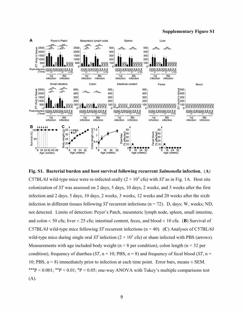
Fig. S1. Bacterial burden and host survival following recurrent Salmonella infection. (A)
C57BL/6J wild-type mice were re-infected orally (2 × 10³ cfu) with ST as in Fig. 1A. Host site
colonization of ST was assessed on 2 days, 5 days, 10 days, 2 weeks, and 3 weeks after the first
infection and 2 days, 5 days, 10 days, 2 weeks, 3 weeks, 12 weeks and 20 weeks after the sixth
infection in different tissues following ST recurrent infections (n = 72). D, days; W, weeks; ND,
not detected. Limits of detection: Peyer’s Patch, mesenteric lymph node, spleen, small intestine,
and colon < 50 cfu; liver < 25 cfu; intestinal content, feces, and blood < 10 cfu. (B) Survival of
C57BL/6J wild-type mice following ST recurrent infections (n = 40). (C) Analyses of C57BL/6J
wild-type mice during single oral ST infection (2 × 10³ cfu) or sham infected with PBS (arrows).
Measurements with age included body weight (n = 8 per condition), colon length (n = 32 per
condition), frequency of diarrhea (ST, n = 10; PBS, n = 8) and frequency of fecal blood (ST, n =
10; PBS, n = 8) immediately prior to infection at each time point. Error bars, means ± SEM.
***P < 0.001; **P < 0.01; *P < 0.05; one-way ANOVA with Tukey’s multiple comparisons test
(A).
10
Supplementary Figure S2
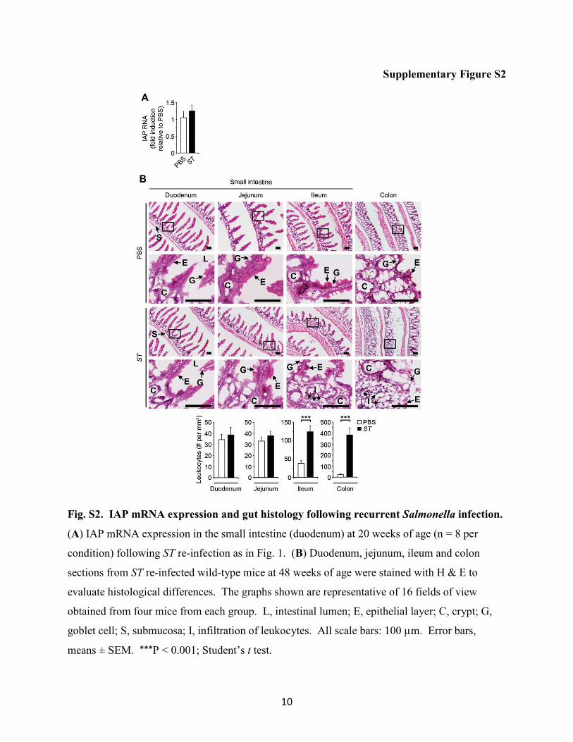
Fig. S2. IAP mRNA expression and gut histology following recurrent Salmonella infection.
(A) IAP mRNA expression in the small intestine (duodenum) at 20 weeks of age (n = 8 per
condition) following ST re-infection as in Fig. 1. (B) Duodenum, jejunum, ileum and colon
sections from ST re-infected wild-type mice at 48 weeks of age were stained with H & E to
evaluate histological differences. The graphs shown are representative of 16 fields of view
obtained from four mice from each group. L, intestinal lumen; E, epithelial layer; C, crypt; G,
goblet cell; S, submucosa; I, infiltration of leukocytes. All scale bars: 100 µm. Error bars,
means ± SEM. ***P < 0.001; Student’s t test.
11
Supplementary Figure S3
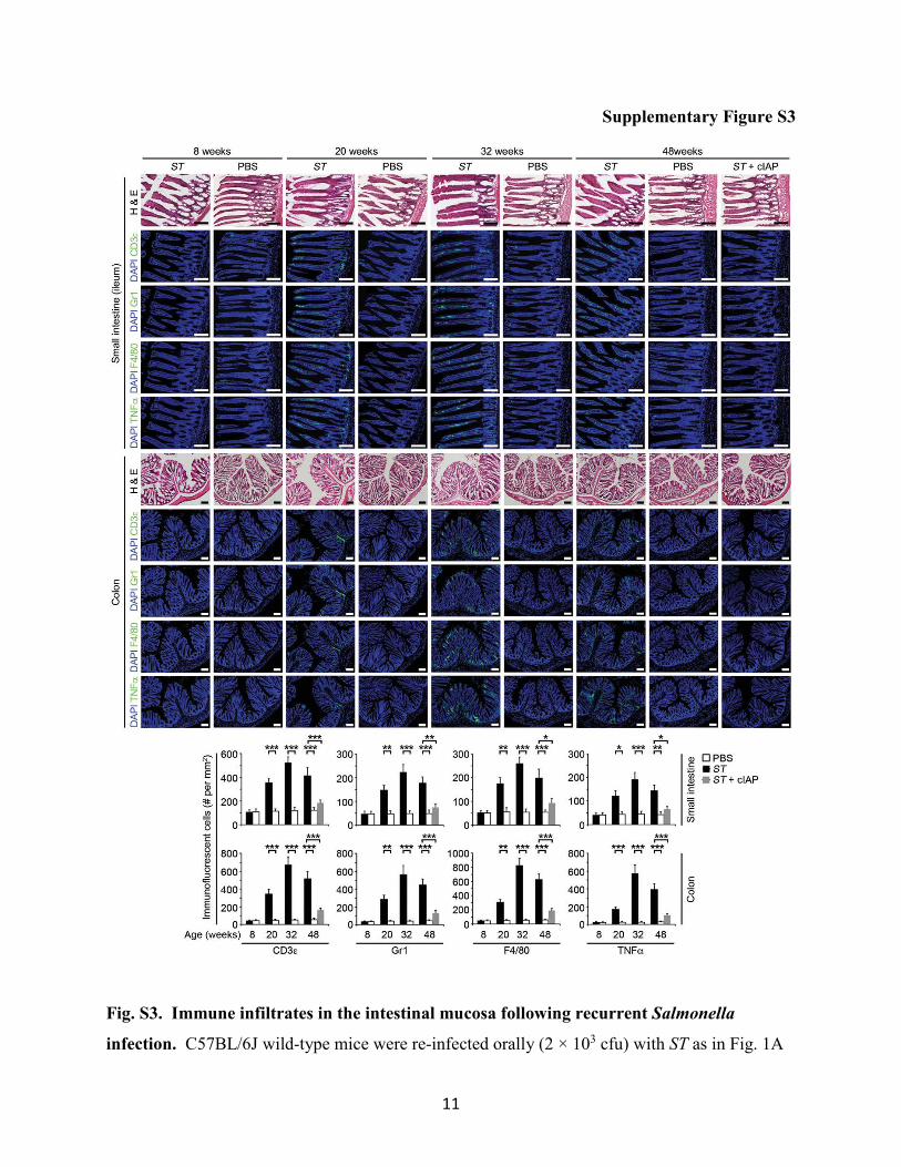
Fig. S3. Immune infiltrates in the intestinal mucosa following recurrent Salmonella
infection. C57BL/6J wild-type mice were re-infected orally (2 × 10³ cfu) with ST as in Fig. 1A

12
in the presence or absence of cIAP. Colon and small intestine serial sections were stained with
H&E or fluorescence using antibodies specific for CD3ε, Gr1, F4/80, or TNFα at 8 (prior to the
first infection), 20 (prior to 4th infection), 32 weeks (4 weeks after the sixth infection) and 48
weeks age. DNA is stained with DAPI. The graphs shown indicate the abundance of
immunofluorescent cells and are representative of ten fields of view obtained from four mice of
each condition and time point. Error bars, means ± SEM. ***P < 0.001; **P < 0.01; *P < 0.05;
one-way ANOVA with Tukey’s multiple comparisons test. All scale bars: 100 µm.

13
Supplementary Figure S4
Fig. S4. Endocytosis and altered glycosylation of enterocyte membrane proteins following
recurrent Salmonella infection. (A–C) In situ localization, abundance and lectin binding of
sucrase-isomaltase (SIM) at the cell surface among enterocytes of the small intestine (duodenum)
at 20 weeks of age (immediately before the fourth ST infection) (n = 8 per condition). (D–F) In
situ localization, abundance and lectin binding of dipeptidyl peptidase 4 (DPP4) at the cell
surface among enterocytes of the small intestine (duodenum) at 20 weeks of age (immediately
before the fourth ST infection) (n = 8 per condition). (G–I) In situ localization, abundance and
lectin binding of lactase (LCT) at the cell surface among enterocytes of the small intestine
(duodenum) at 20 weeks of age (immediately before the fourth ST infection) (n = 8 per
condition). Error bars, means ± SEM. **P < 0.01; *P < 0.05; Student’s t test.
14
Supplementary Figure S5
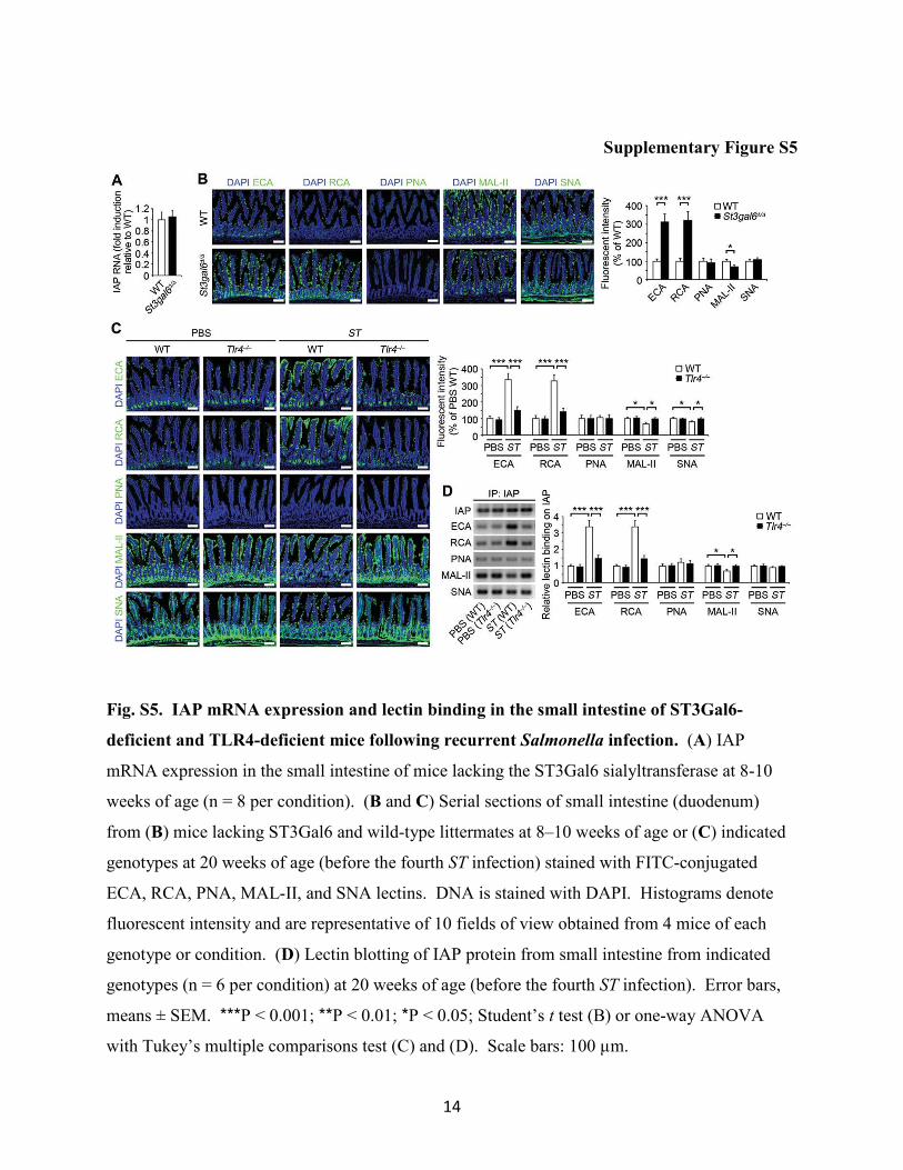
Fig. S5. IAP mRNA expression and lectin binding in the small intestine of ST3Gal6-
deficient and TLR4-deficient mice following recurrent Salmonella infection. (A) IAP
mRNA expression in the small intestine of mice lacking the ST3Gal6 sialyltransferase at 8-10
weeks of age (n = 8 per condition). (B and C) Serial sections of small intestine (duodenum)
from (B) mice lacking ST3Gal6 and wild-type littermates at 8–10 weeks of age or (C) indicated
genotypes at 20 weeks of age (before the fourth ST infection) stained with FITC-conjugated
ECA, RCA, PNA, MAL-II, and SNA lectins. DNA is stained with DAPI. Histograms denote
fluorescent intensity and are representative of 10 fields of view obtained from 4 mice of each
genotype or condition. (D) Lectin blotting of IAP protein from small intestine from indicated
genotypes (n = 6 per condition) at 20 weeks of age (before the fourth ST infection). Error bars,
means ± SEM. ***P < 0.001; **P < 0.01; *P < 0.05; Student’s t test (B) or one-way ANOVA
with Tukey’s multiple comparisons test (C) and (D). Scale bars: 100 µm.
15
Supplementary Figure S6
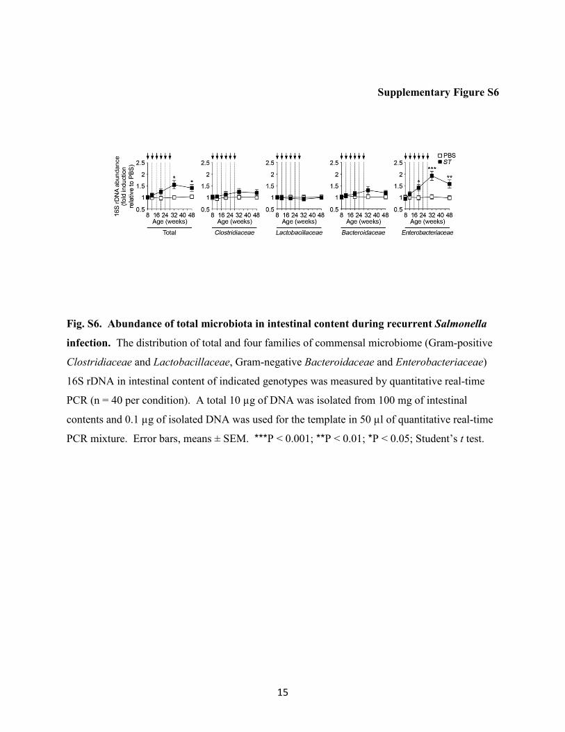
Fig. S6. Abundance of total microbiota in intestinal content during recurrent Salmonella
infection. The distribution of total and four families of commensal microbiome (Gram-positive
Clostridiaceae and Lactobacillaceae, Gram-negative Bacteroidaceae and Enterobacteriaceae)
16S rDNA in intestinal content of indicated genotypes was measured by quantitative real-time
PCR (n = 40 per condition). A total 10 µg of DNA was isolated from 100 mg of intestinal
contents and 0.1 µg of isolated DNA was used for the template in 50 µl of quantitative real-time
PCR mixture. Error bars, means ± SEM. ***P < 0.001; **P < 0.01; *P < 0.05; Student’s t test.

16
Supplementary Figure S7
Fig. S7. Altered IAP abundance and glycosylation in the gut following oral LPS
administration. (A) Total AP activity in feces following single oral LPS administration of
multiple doses (E. coli 0111:B4) (n = 8 per condition). (B) Total AP activity in feces following
repeated oral LPS administrations (24-h intervals for 10 days, arrows) of multiple doses (n = 8
per condition). (C) Lectin blot analyses of identical amounts of IAP isolated from the small
intestine of 8-week-old wild-type mice on day 6 following repeated LPS administrations (n = 8
per condition). (D) Serial tissue sections of small intestine (duodenum) of 8-week-old wild-type
mice on day 6 following repeated LPS administrations (100 mg/kg; E. coli 0111:B4, 24-h
intervals for 10 days) stained with fluorescence using FITC-conjugated ECA, RCA, PNA, MAL-
II, and SNA lectins. DNA is stained with DAPI. Plots indicate the fluorescent intensity and are
representative of ten fields of view obtained from four mice from each group. Scale bars, 100
µm. (E) In situ localization and co-localization (yellow) of IAP with various intracellular
compartments in the small intestine (duodenum) of 8-week-old wild-type mice on day 6
following LPS administrations. Small intestine (duodenum) serial sections were stained with H

17
& E or by fluorescence using antibodies as in Fig. 3D. DNA was stained with DAPI. The
graphs indicate the percentage of IAP co-localized (yellow) with the above intracellular
compartments and IAP abundance at the cell surface and are representative of ten fields of view
obtained from four mice of each condition. Scale bars: 10 µm. Error bars, means ± SEM. ***P
< 0.001; **P < 0.01; *P < 0.05; one-way ANOVA with Tukey’s multiple comparisons test (B) or
Student’s t test (C) to (E).
18
Supplementary Figure S8
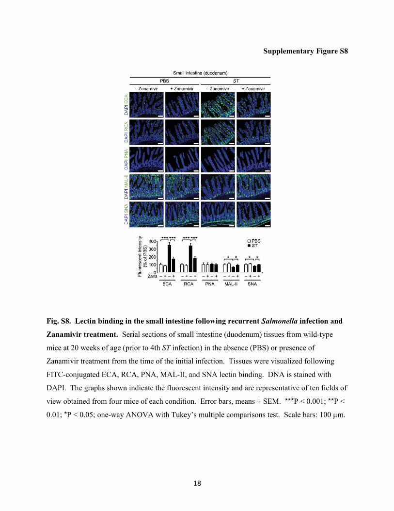
Fig. S8. Lectin binding in the small intestine following recurrent Salmonella infection and
Zanamivir treatment. Serial sections of small intestine (duodenum) tissues from wild-type
mice at 20 weeks of age (prior to 4th ST infection) in the absence (PBS) or presence of
Zanamivir treatment from the time of the initial infection. Tissues were visualized following
FITC-conjugated ECA, RCA, PNA, MAL-II, and SNA lectin binding. DNA is stained with
DAPI. The graphs shown indicate the fluorescent intensity and are representative of ten fields of
view obtained from four mice of each condition. Error bars, means ± SEM. ***P < 0.001; **P <
0.01; *P < 0.05; one-way ANOVA with Tukey’s multiple comparisons test. Scale bars: 100 µm.
19
Supplementary Figure S9
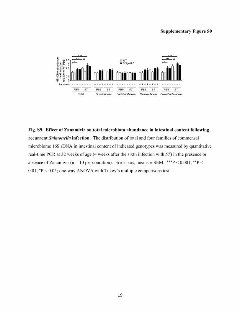
Fig. S9. Effect of Zanamivir on total microbiota abundance in intestinal content following
recurrent Salmonella infection. The distribution of total and four families of commensal
microbiome 16S rDNA in intestinal content of indicated genotypes was measured by quantitative
real-time PCR at 32 weeks of age (4 weeks after the sixth infection with ST) in the presence or
absence of Zanamivir (n = 10 per condition). Error bars, means ± SEM. ***P < 0.001; **P <
0.01; *P < 0.05; one-way ANOVA with Tukey’s multiple comparisons test.
20
Supplementary Figure S10
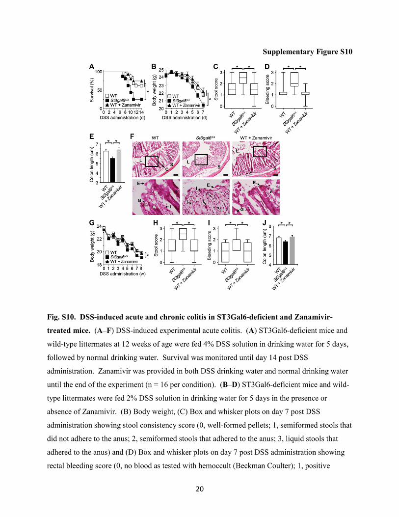
Fig. S10. DSS-induced acute and chronic colitis in ST3Gal6-deficient and Zanamivir-
treated mice. (A–F) DSS-induced experimental acute colitis. (A) ST3Gal6-deficient mice and
wild-type littermates at 12 weeks of age were fed 4% DSS solution in drinking water for 5 days,
followed by normal drinking water. Survival was monitored until day 14 post DSS
administration. Zanamivir was provided in both DSS drinking water and normal drinking water
until the end of the experiment (n = 16 per condition). (B–D) ST3Gal6-deficient mice and wild-
type littermates were fed 2% DSS solution in drinking water for 5 days in the presence or
absence of Zanamivir. (B) Body weight, (C) Box and whisker plots on day 7 post DSS
administration showing stool consistency score (0, well-formed pellets; 1, semiformed stools that
did not adhere to the anus; 2, semiformed stools that adhered to the anus; 3, liquid stools that
adhered to the anus) and (D) Box and whisker plots on day 7 post DSS administration showing
rectal bleeding score (0, no blood as tested with hemoccult (Beckman Coulter); 1, positive

21
hemoccult; 2, blood traces in stool visible; 3, gross rectal bleeding) were checked daily (n = 10
per condition). (E) Colon length was measured on day 7 post DSS administration (n = 8 per
condition). (F) Histopathological analysis in colon cross sections were examined by H&E
staining. Images shown are representative of ten fields of view obtained from four mice of each
condition. L, intestinal lumen; E, epithelial layer; C, crypt; G, goblet cell; S, submucosa; I,
infiltration of leukocytes. All scale bars: 100 µm. (G–J) DSS-induced experimental chronic
colitis. (G) Body weight, (H) Box and whisker plots on week 8 post DSS administration
showing stool consistency score, and (I) Box and whisker plots on week 8 post DSS
administration showing rectal bleeding score (n = 12 per condition). (J) Colon length was
measured on week 8 post DSS administration (n = 8 per condition). Error bars, means ± SEM.
*P < 0.05; log-rank test (A), one-way ANOVA with Tukey’s multiple comparisons test (B), (E),
(G), and (J), or Kruskal-Wallis test with Dunn’s multiple comparisons test (C), (D), (H), and (I).
References
1. B. Khor, A. Gardet, R. J. Xavier, Genetics and pathogenesis of inflammatory bowel disease. Nature
474, 307–317 (2011). doi: 10.1038/nature10209; Medline
2. A. Kaser, S. Zeissig, R. S. Blumberg, Inflammatory bowel disease. Annu. Rev. Immunol. 28, 573–
621 (2010). doi: 10.1146/annurev-immunol-030409-101225; Medline
3. P. J. Sansonetti, War and peace at mucosal surfaces. Nat. Rev. Immunol. 4 , 953–964 (2004). doi:
10.1038/nri1499; Medline
4. D. Knights, K. G. Lassen, R. J. Xavier, Advances in inflammatory bowel disease pathogenesis:
Linking host genetics and the microbiome. Gut 62, 1505–1510 (2013). doi: 10.1136/gutjnl-2012-
303954; Medline
5. J. Halfvarson, Genetics in twins with Crohn’s disease: Less pronounced than previously believed?
Inflamm. Bowel Dis. 17, 6–12 (2011). doi: 10.1002/ibd.21295; Medline
6. L. Eckmann, Animal models of inflammatory bowel disease: Lessons from enteric infections. Ann.
N. Y. Acad. Sci. 1072, 28–38 (2006). doi: 10.1196/annals.1326.008; Medline
7. A. Sonnenberg, Seasonal variation of enteric infections and inflammatory bowel disease. Inflamm.
Bowel Dis. 14, 955–959 (2008). doi: 10.1002/ibd.20408; Medline
8. Centers for Disease Control and Prevention (CDC), Vital signs: Incidence and trends of infection
with pathogens transmitted commonly through food—Foodborne Diseases Active Surveillance
Network, 10 U.S. sites, 1996–2010. MMWR Morb. Mortal. Wkly. Rep. 60, 749–755 (2011). Medline
9. E. Scallan, R. M. Hoekstra, F. J. Angulo, R. V. Tauxe, M.-A. Widdowson, S. L. Roy, J. L. Jones, P. M.
Griffin, Foodborne illness acquired in the United States—Major pathogens. Emerg. Infect. Dis. 17, 7–
15 (2011). doi: 10.3201/eid1701.P11101; Medline
10. S. E. Majowicz, J. Musto, E. Scallan, F. J. Angulo, M. Kirk, S. J. O’Brien, T. F. Jones, A. Fazil, R. M.
Hoekstra; International Collaboration on Enteric Disease 'Burden of Illness’ Studies, The global
burden of nontyphoidal Salmonella gastroenteritis. Clin. Infect. Dis. 50, 882–889 (2010). doi:
10.1086/650733; Medline
11. H. K. de Jong, C. M. Parry, T. van der Poll, W. J. Wiersinga, Host-pathogen interaction in invasive
Salmonellosis. PLOS Pathog. 10, e1002933 (2012). doi: 10.1371/journal.ppat.1002933; Medline
12. N. A. Feasey, G. Dougan, R. A. Kingsley, R. S. Heyderman, M. A. Gordon, Invasive non-
typhoidal salmonella disease: An emerging and neglected tropical disease in Africa. Lancet 379, 2489–
2499 (2012). doi: 10.1016/S0140-6736(11)61752-2; Medline
13. B. F. Hinnebusch, A. Siddique, J. W. Henderson, M. S. Malo, W. Zhang, C. P. Athaide, M. A.
Abedrapo, X. Chen, V. W. Yang, R. A. Hodin, Enterocyte differentiation marker intestinal alkaline
phosphatase is a target gene of the gut-enriched Krüppel-like factor. Am. J. Physiol. Gastrointest. Liver
Physiol. 286, G23–G30 (2004). doi: 10.1152/ajpgi.00203.2003; Medline
14. K. Poelstra, W. W. Bakker, P. A. Klok, M. J. Hardonk, D. K. Meijer, A physiologic function for
alkaline phosphatase: Endotoxin detoxification. Lab. Invest. 76, 319–327 (1997). Medline
15. I. Koyama, T. Matsunaga, T. Harada, S. Hokari, T. Komoda, Alkaline phosphatases reduce toxicity
of lipopolysaccharides in vivo and in vitro through dephosphorylation. Clin. Biochem. 35, 455–461
(2002). doi: 10.1016/S0009-9120(02)00330-2; Medline
16. J. M. Bates, J. Akerlund, E. Mittge, K. Guillemin, Intestinal alkaline phosphatase detoxifies
lipopolysaccharide and prevents inflammation in zebrafish in response to the gut microbiota. Cell Host
Microbe 2, 371–382 (2007). doi: 10.1016/j.chom.2007.10.010; Medline
17. N. L. Sussman, R. Eliakim, D. Rubin, D. H. Perlmutter, K. DeSchryver-Kecskemeti, D. H. Alpers,
Intestinal alkaline phosphatase is secreted bidirectionally from villous enterocytes. Am. J. Physiol. 257,
G14–G23 (1989). Medline
18. L. G. Ellies, M. Sperandio, G. H. Underhill, J. Yousif, M. Smith, J. J. Priatel, G. S. Kansas, K. Ley, J.
D. Marth, Sialyltransferase specificity in selectin ligand formation. Blood 100 , 3618–3625 (2002). doi:
10.1182/blood-2002-04-1007; Medline
19. W. H. Yang, C. Nussbaum, P. K. Grewal, J. D. Marth, M. Sperandio, Coordinated roles of ST3Gal-
VI and ST3Gal-IV sialyltransferases in the synthesis of selectin ligands. Blood 120, 1015–1026
(2012). doi: 10.1182/blood-2012-04-424366; Medline
20. N. Kamada, S. U. Seo, G. Y. Chen, G. Nunez, Role of the gut microbiota in immunity and
inflammatory disease. Nat. Rev. Immunol. 13 , 321–335 (2013). doi: 10.1038/nri3430; Medline
21. N. A. Nagalingam, S. V. Lynch, Role of the microbiota in inflammatory bowel diseases. Inflamm.
Bowel Dis. 18, 968–984 (2012). doi: 10.1002/ibd.21866; Medline
22. N. Figueroa-Bossi, S. Uzzau, D. Maloriol, L. Bossi, Variable assortment of prophages provides a
transferable repertoire of pathogenic determinants in Salmonella. Mol. Microbiol. 39, 260–271 (2001).
doi: 10.1046/j.1365-2958.2001.02234.x; Medline
23. E. Monti, E. Bonten, A. D’Azzo, R. Bresciani, B. Venerando, G. Borsani, R. Schauer, G.
Tettamanti, Sialidases in vertebrates: A family of enzymes tailored for several cell functions. Adv.
Carbohydr. Chem. Biochem. 64, 403–479 (2010). doi: 10.1016/S0065-2318(10)64007-3; Medline
24. W. H. Yang, P. V. Aziz, D. M. Heithoff, M. J. Mahan, J. W. Smith, J. D. Marth, An intrinsic
mechanism of secreted protein aging and turnover. Proc. Natl. Acad. Sci. U.S.A. 112, 13657–13662
(2015). doi: 10.1073/pnas.1515464112; Medline
25. A. Poltorak , X. He, I. Smirnova, M.-Y. Liu, C. Van Huffel, X. Du, D. Birdwell, E. Alejos, M. Silva, C.
Galanos, M. Freudenberg, P. Ricciardi-Castagnoli, B. Layton, B. Beutler, Defective LPS signaling in
C3H/HeJ and C57BL/10ScCr mice: Mutations in Tlr4 gene. Science 282, 2085–2088 (1998). doi:
10.1126/science.282.5396.2085; Medline
26. B. Beutler, Endotoxin, toll-like receptor 4, and the afferent limb of innate immunity. Curr. Opin.
Microbiol. 3 , 23–28 (2000). doi: 10.1016/S1369-5274(99)00046-6; Medline
27. M. T. Abreu, Toll-like receptor signalling in the intestinal epithelium: How bacterial recognition
shapes intestinal function. Nat. Rev. Immunol. 10 , 131–144 (2010). doi: 10.1038/nri2707; Medline
28. H. Huhta , O. Helminen, J. H. Kauppila, T. Salo, K. Porvari, J. Saarnio, P. P. Lehenkari, T. J.
Karttunen, The expression of Toll-like receptors in normal human and murine gastrointestinal organs
and the effect of microbiome and cancer. J. Histochem. Cytochem. 64 , 470–482 (2016). doi:
10.1369/0022155416656154; Medline
29. R. Dheer , R. Santaolalla, J. M. Davies, J. K. Lang, M. C. Phillips, C. Pastorini, M. T. Vazquez-
Pertejo, M. T. Abreu, Intestinal epithelial Toll-like receptor 4 signaling affects epithelial function and
colonic microbiota and promotes a risk for transmissible colitis. Infect. Immun. 84 , 798–810 (2016).
doi: 10.1128/IAI.01374-15; Medline
30. C. L. Leaphart , J. Cavallo, S. C. Gribar, S. Cetin, J. Li, M. F. Branca, T. D. Dubowski, C. P. Sodhi, D.
J. Hackam, A critical role for TLR4 in the pathogenesis of necrotizing enterocolitis by modulating
intestinal injury and repair. J. Immunol. 179 , 4808–4820 (2007). doi: 10.4049/jimmunol.179.7.4808;
Medline
31. A. Moscona, Neuraminidase inhibitors for influenza. N. Engl. J. Med. 353, 1363–1373 (2005). doi:
10.1056/NEJMra050740; Medline
32. K. Hata , K. Koseki, K. Yamaguchi, S. Moriya, Y. Suzuki, S. Yingsakmongkon, G. Hirai, M.
Sodeoka, M. von Itzstein, T. Miyagi, Limited inhibitory effects of oseltamivir and zanamivir on human
sialidases. Antimicrob. Agents Chemother. 52, 3484–3491 (2008). doi: 10.1128/AAC.00344-08;
Medline
33. N. M. Stamatos , I. Carubelli, D. van de Vlekkert, E. J. Bonten, N. Papini, C. Feng, B. Venerando,
A. d’Azzo, A. S. Cross, L. X. Wang, P. J. Gomatos, LPS-induced cytokine production in human dendritic
cells is regulated by sialidase activity. J. Leukoc. Biol. 88, 1227–1239 (2010). doi:
10.1189/jlb.1209776; Medline
34. Y.-L. Huang, C. Chassard, M. Hausmann, M. von Itzstein, T. Hennet, Sialic acid catabolism drives
intestinal inflammation and microbial dysbiosis in mice. Nat. Commun. 6, 8141 (2015). doi:
10.1038/ncomms9141; Medline
35. S. Ramasamy , D. D. Nguyen, M. A. Eston, S. N. Alam, A. K. Moss, F. Ebrahimi, B. Biswas, G.
Mostafa, K. T. Chen, K. Kaliannan, H. Yammine, S. Narisawa, J. L. Millán, H. S. Warren, E. L.
Hohmann, E. Mizoguchi, H.-C. Reinecker, A. K. Bhan, S. B. Snapper, M. S. Malo, R. A. Hodin,
Intestinal alkaline phosphatase has beneficial effects in mouse models of chronic colitis. Inflamm.
Bowel Dis. 17 , 532–542 (2011). doi: 10.1002/ibd.21377; Medline
36. T. Dolowschiak , A. A. Mueller, L. J. Pisan, R. Feigelman, B. Felmy, M. E. Sellin, S. Namineni, B. D.
Nguyen, S. Y. Wotzka, M. Heikenwalder, C. von Mering, C. Mueller, W.-D. Hardt, IFN-γ hinders
recovery from mucosal inflammation during antibiotic therapy for Salmonella gut infection. Cell Host
Microbe 20 , 238–249 (2016). doi: 10.1016/j.chom.2016.06.008; Medline
37. S. L. Foster, R. Medzhitov, Gene-specific control of the TLR-induced inflammatory response.
Clin. Immunol. 130 , 7–15 (2009). doi: 10.1016/j.clim.2008.08.015; Medline
38. K. Kamdar, S. Khakpour, J. Chen, V. Leone, J. Brulc, T. Mangatu, D. A. Antonopoulos, E. B.
Chang, S. A. Kahn, B. S. Kirschner, G. Young, R. W. DePaolo, Genetic and metabolic signals during
acute enteric bacterial infection alter the microbiota and drive progression to chronic inflammatory
disease. Cell Host Microbe 19, 21–31 (2016). doi: 10.1016/j.chom.2015.12.006; Medline
39. S. Manco , F. Hernon, H. Yesilkaya, J. C. Paton, P. W. Andrew, A. Kadioglu, Pneumococcal
neuraminidases A and B both have essential roles during infection of the respiratory tract and sepsis.
Infect. Immun. 74, 4014–4020 (2006). doi: 10.1128/IAI.01237-05; Medline
40. S. M. Schwerdtfeger, M. F. Melzig, Sialidases in biological systems. Pharmazie 65, 551–561
(2010). Medline
41. A. Varki, P. Gagneux, Multifarious roles of sialic acids in immunity. Ann. N. Y. Acad. Sci. 1253,
16–36 (2012). doi: 10.1111/j.1749-6632.2012.06517.x; Medline
42. K. Yamaguchi , K. Koseki, M. Shiozaki, Y. Shimada, T. Wada, T. Miyagi, Regulation of plasma-
membrane-associated sialidase NEU3 gene by Sp1/Sp3 transcription factors. Biochem. J. 430, 107–117
(2010). doi: 10.1042/BJ20100350; Medline
43. W. Ma , W. Lim, K. Gee, S. Aucoin, D. Nandan, M. Kozlowski, F. Diaz-Mitoma, A. Kumar, The p38
mitogen-activated kinase pathway regulates the human interleukin-10 promoter via the activation of
Sp1 transcription factor in lipopolysaccharide-stimulated human macrophages. J. Biol. Chem. 276,
13664–13674 (2001). doi: 10.1074/jbc.M011157200; Medline
44. V. S. Carl, J. K. Gautam, L. D. Comeau, M. F. Smith Jr., Role of endogenous IL-10 in LPS-
induced STAT3 activation and IL-1 receptor antagonist gene expression. J. Leukoc. Biol. 76, 735–742
(2004). doi: 10.1189/jlb.1003526; Medline
45. Y. Kakugawa , T. Wada, K. Yamaguchi, H. Yamanami, K. Ouchi, I. Sato, T. Miyagi, Up-regulation
of plasma membrane-associated ganglioside sialidase (Neu3) in human colon cancer and its
involvement in apoptosis suppression. Proc. Natl. Acad. Sci. U.S.A. 99, 10718–10723 (2002). doi:
10.1073/pnas.152597199; Medline
46. S. Kawamura , I. Sato, T. Wada, K. Yamaguchi, Y. Li, D. Li, X. Zhao, S. Ueno, H. Aoki, T. Tochigi,
M. Kuwahara, T. Kitamura, K. Takahashi, S. Moriya, T. Miyagi, Plasma membrane-associated
sialidase (NEU3) regulates progression of prostate cancer to androgen-independent growth through
modulation of androgen receptor signaling. Cell Death Differ. 19, 170–179 (2012). doi:
10.1038/cdd.2011.83; Medline
47. K. Yamaguchi , K. Shiozaki, S. Moriya, K. Koseki, T. Wada, H. Tateno, I. Sato, M. Asano, Y.
Iwakura, T. Miyagi, Reduced susceptibility to colitis-associated colon carcinogenesis in mice lacking
plasma membrane-associated sialidase. PLOS ONE 7, e41132 (2012). doi:
10.1371/journal.pone.0041132; Medline
48. T. Wada, Y. Yoshikawa, S. Tokuyama, M. Kuwabara, H. Akita, T. Miyagi, Cloning, expression,
and chromosomal mapping of a human ganglioside sialidase. Biochem. Biophys. Res. Commun. 261,
21–27 (1999). doi: 1006/bbrc.1999.0973; Medline
49. A. Mozzi , M. Forcella, A. Riva, C. Difrancesco, F. Molinari, V. Martin, N. Papini, B. Bernasconi,
S. Nonnis, G. Tedeschi, L. Mazzucchelli, E. Monti, P. Fusi, M. Frattini, NEU3 activity enhances EGFR
activation without affecting EGFR expression and acts on its sialylation levels. Glycobiology 25 , 855–
868 (2015). doi: 10.1093/glycob/cwv026; Medline
50. K. Ohtsubo , S. Takamatsu, M. T. Minowa, A. Yoshida, M. Takeuchi, J. D. Marth, Dietary and
genetic control of glucose transporter 2 glycosylation promotes insulin secretion in suppressing
diabetes. Cell 123, 1307–1321 (2005). doi: 10.1016/j.cell.2005.09.041; Medline
51. B. Weinhold, R. Seidenfaden, I. Röckle, M. Mühlenhoff, F. Schertzinger, S. Conzelmann, J. D.
Marth, R. Gerardy-Schahn, H. Hildebrandt, Genetic ablation of polysialic acid causes severe
neurodevelopmental defects rescued by deletion of the neural cell adhesion molecule. J. Biol. Chem.
280 , 42971–42977 (2005). doi: 10.1074/jbc.M511097200; Medline
52. P. K. Grewal, M. Boton, K. Ramirez, B. E. Collins, A. Saito, R. S. Green, K. Ohtsubo, D. Chui, J.
D. Marth, ST6Gal-I restrains CD22-dependent antigen receptor endocytosis and Shp-1 recruitment in
normal and pathogenic immune signaling. Mol. Cell. Biol. 26, 4970–4981 (2006). doi:
10.1128/MCB.00308-06; Medline
53. K. Ohtsubo, J. D. Marth, Glycosylation in cellular mechanisms of health and disease. Cell 126,
855–867 (2006). doi: 10.1016/j.cell.2006.08.019; Medline
54. P. R. Crocker, J. C. Paulson, A. Varki, Siglecs and their roles in the immune system. Nat. Rev.
Immunol. 7, 255–266 (2007). doi: 10.1038/nri2056; Medline
55. F.-T. Liu, G. A. Rabinovich, Galectins: Regulators of acute and chronic inflammation. Ann. N. Y.
Acad. Sci. 1183, 158–182 (2010). doi: 10.1111/j.1749-6632.2009.05131.x; Medline
56. A. Tuin, K. Poelstra, A. de Jager-Krikken, L. Bok, W. Raaben, M. P. Velders, G. Dijkstra, Role of
alkaline phosphatase in colitis in man and rats. Gut 58, 379–387 (2009). doi:
10.1136/gut.2007.128868; Medline
57. M. Lukas, P. Drastich, M. Konecny, P. Gionchetti, O. Urban, F. Cantoni, M. Bortlik, D. Duricova,
M. Bulitta, Exogenous alkaline phosphatase for the treatment of patients with moderate to severe
ulcerative colitis. Inflamm. Bowel Dis. 16, 1180–1186 (2010). doi: 10.1002/ibd.21161; Medline
58. K. Molnár, Á. Vannay, B. Szebeni, N. F. Bánki, E. Sziksz, Á. Cseh, H. Győrffy, P. L. Lakatos, M.
Papp, A. Arató, G. Veres, Intestinal alkaline phosphatase in the colonic mucosa of children with
inflammatory bowel disease. World J. Gastroenterol. 18, 3254–3259 (2012). doi:
10.3748/wjg.v18.i25.3254; Medline
59. S. N. Alam, H. Yammine, O. Moaven, R. Ahmed, A. K. Moss, B. Biswas, N. Muhammad, R.
Biswas, A. Raychowdhury, K. Kaliannan, S. Ghosh, M. Ray, S. R. Hamarneh, S. Barua, N. S. Malo,

A. K. Bhan, M. S. Malo, R. A. Hodin, Intestinal alkaline phosphatase prevents antibiotic-induced
susceptibility to enteric pathogens. Ann. Surg. 259, 715–722 (2014). doi:
10.1097/SLA.0b013e31828fae14; Medline
60. J. J. Miklavcic, T. D. L. Hart, G. M. Lees, G. K. Shoemaker, K. L. Schnabl, B. M. K. Larsen, O. F.
Bathe, A. B. R. Thomson, V. C. Mazurak, M. T. Clandinin, Increased catabolism and decreased
unsaturation of ganglioside in patients with inflammatory bowel disease. World J. Gastroenterol. 21,
doi: 10080–10090 (2015). 10.3748/wjg.v21.i35.10080; Medline
61. R. F. Goldberg, W. G. Austen Jr., X. Zhang, G. Munene, G. Mostafa, S. Biswas, M. McCormack,
K. R. Eberlin, J. T. Nguyen, H. S. Tatlidede, H. S. Warren, S. Narisawa, J. L. Millán, R. A. Hodin,
Intestinal alkaline phosphatase is a gut mucosal defense factor maintained by enteral nutrition. Proc.
Natl. Acad. Sci. U.S.A.. 105, 3551–3556 (2008). doi: 10.1073/pnas.0712140105; Medline
62. D. M. Heithoff, W. R. Shimp, J. K. House, Y. Xie, B. C. Weimer, R. L. Sinsheimer, M. J. Mahan,
Intraspecies variation in the emergence of hyperinfectious bacterial strains in nature. PLOS Pathog. 8,
e1002647 (2012). doi: 10.1371/journal.ppat.1002647; Medline
63. J. Fu, B. Wei, T. Wen, M. E. V. Johansson, X. Liu, E. Bradford, K. A. Thomsson, S. McGee, L.
Mansour, M. Tong, J. M. McDaniel, T. J. Sferra, J. R. Turner, H. Chen, G. C. Hansson, J. Braun, L.
Xia, Loss of intestinal core 1-derived O-glycans causes spontaneous colitis in mice. J. Clin. Invest.
121, 1657–1666 (2011). doi: 10.1172/JCI45538; Medline
64. S. Narisawa, L. Huang, A. Iwasaki, H. Hasegawa, D. H. Alpers, J. L. Millán, Accelerated fat
absorption in intestinal alkaline phosphatase knockout mice. Mol. Cell. Biol. 23, 7525–7530 (2003).
doi: 10.1128/MCB.23.21.7525-7530.2003; Medline
65. K. Ohtsubo, M. Z. Chen, J. M. Olefsky, J. D. Marth, Pathway to diabetes through attenuation of
pancreatic beta cell glycosylation and glucose transport. Nat. Med. 17, 1067–1075 (2011). doi:
10.1038/nm.2414; Medline
66. L. G. Ellies, D. Ditto, G. G. Levy, M. Wahrenbrock, D. Ginsburg, A. Varki, D. T. Le, J. D. Marth,
Sialyltransferase ST3Gal-IV operates as a dominant modifier of hemostasis by concealing
asialoglycoprotein receptor ligands. Proc. Natl. Acad. Sci. U.S.A. 99, 10042–10047 (2002). doi:
10.1073/pnas.142005099; Medline
67. M. R. Davis Jr., J. B. Goldberg, Purification and visualization of lipopolysaccharide from Gram-
negative bacteria by hot aqueous-phenol extraction. J. Vis. Exp. 2012 , 3916 (2012). doi:
10.3791/3916; Medline
68. C.-H. Lee, C.-M. Tsai, Quantification of bacterial lipopolysaccharides by the purpald assay:
Measuring formaldehyde generated from 2-keto-3-deoxyoctonate and heptose at the inner core by
periodate oxidation. Anal. Biochem. 267, 161–168 (1999). doi: 10.1006/abio.1998.2961; Medline
69. N. J. Foot, Y. A. Leong, L. E. Dorstyn, H. E. Dalton, K. Ho, L. Zhao, M. D. Garrick, B. Yang, D.
Hiwase, S. Kumar, Ndfip1–deficient mice have impaired DMT1 regulation and iron homeostasis.
Blood 117 , 638–646 (2011). doi: 10.1182/blood-2010-07-295287; Medline
70. A. Fuhrer, N. Sprenger, E. Kurakevich, L. Borsig, C. Chassard, T. Hennet, Milk sialyllactose
influences colitis in mice through selective intestinal bacterial colonization. J. Exp. Med. 207, 2843–
2854 (2010). doi: 10.1084/jem.20101098; Medline
71. M. H. Zaki, K. L. Boyd, P. Vogel, M. B. Kastan, M. Lamkanfi, T.-D. Kanneganti, The NLRP3
inflammasome protects against loss of epithelial integrity and mortality during experimental colitis.
Immunity 32, 379–391 (2010). doi: 10.1016/j.immuni.2010.03.003; Medline