
Supplementary Material10.1007/s12012... · Web viewand the Apex of the heart at 1 and 0.2 Hz pacing...
15
Supplementary Material d,l-sotalol concentrations Base Intermediate Apex 1 Hz 0.2 Hz 1 Hz 0.2 Hz 1 Hz 0.2 Hz TRI (ms) 0 317 ± 319 ± 8 319 ± 319 ± 8 316 ± 7 322 ± 8 3 µM 314 ± 323 ± 319 ± 9 328 ± 314 ± 8 327 ± 9 10 µM 337 ± 8 370 ± 9 335 ± 8 366 ± 333 ± 7 366 ± 9 30 µM 370 ± 9 398 ± 9 366 ± 9 412 ± 7 366 ± 8 424 ± 7 100 µM 396 ± 9 459 ± 393 ± 9 453 ± 395 ± 9 491 ± Tp (ms) 0 55 ± 5 57 ± 5 78 ± 3 72 ± 4 65 ± 5 66 ± 5 3 µM 52 ± 6 63 ± 4 74 ± 3 68 ± 3 65 ± 5 62 ± 4 10 µM 64 ± 5 73 ± 5 79 ± 4 84 ± 3 73 ± 5 72 ± 5 30 µM 69 ± 5 74 ± 6 88 ± 4 105 ± 5 78 ± 6 88 ± 6 100 µM 70 ± 6 93 ± 4 98 ± 4 120 ± 4 84 ± 6 120 ± 7 AT (ms) 0 31 ± 2 31 ± 2 26 ± 1 26 ± 1 26 ± 1 25 ± 1 3 µM 31 ± 2 31 ± 2 26 ± 2 26 ± 1 26 ± 1 25 ± 1 10 µM 31 ± 2 31 ± 2 26 ± 2 25 ± 1 26 ± 1 25 ± 1 30 µM 32 ± 2 32 ± 2 26 ± 2 27 ± 1 26 ± 1 25 ± 1 100 µM 32 ± 2 32 ± 2 25 ± 1 26 ± 1 26 ± 1 25 ± 1 ARI (ms) 0 196 ± 213 ± 149 ± 172 ± 189 ± 233 ± 3 µM 185 ± 211 ± 158 ± 164 ± 187 ± 273 ± 10 µM 228 ± 8 233 ± 173 ± 187 ± 201 ± 225 ± 30 µM 218 ± 265 ± 190 ± 218 ±17 214 ±22 291 ± 100 µM 239 ± 302 ± 193 ± 239 ± 229 ± 240 ± Table 1: Quantitative effects of increasing concentrations of d,l-sotalol (3, 10, 30, 100 µM, n=6) on Activation Time (AT), 1
-
Upload
vuongthien -
Category
Documents
-
view
221 -
download
6
Transcript of Supplementary Material10.1007/s12012... · Web viewand the Apex of the heart at 1 and 0.2 Hz pacing...

Supplementary Material
d,l-sotalol concentrations
Base Intermediate Apex
1 Hz 0.2 Hz 1 Hz 0.2 Hz 1 Hz 0.2 Hz
TRI (ms)0 317 ± 11 319 ± 8 319 ± 10 319 ± 8 316 ± 7 322 ± 83 µM 314 ± 11 323 ± 10 319 ± 9 328 ± 11 314 ± 8 327 ± 910 µM 337 ± 8 370 ± 9 335 ± 8 366 ± 10 333 ± 7 366 ± 930 µM 370 ± 9 398 ± 9 366 ± 9 412 ± 7 366 ± 8 424 ± 7100 µM 396 ± 9 459 ± 16 393 ± 9 453 ± 13 395 ± 9 491 ± 21
Tp (ms)0 55 ± 5 57 ± 5 78 ± 3 72 ± 4 65 ± 5 66 ± 53 µM 52 ± 6 63 ± 4 74 ± 3 68 ± 3 65 ± 5 62 ± 410 µM 64 ± 5 73 ± 5 79 ± 4 84 ± 3 73 ± 5 72 ± 530 µM 69 ± 5 74 ± 6 88 ± 4 105 ± 5 78 ± 6 88 ± 6100 µM 70 ± 6 93 ± 4 98 ± 4 120 ± 4 84 ± 6 120 ± 7
AT (ms)0 31 ± 2 31 ± 2 26 ± 1 26 ± 1 26 ± 1 25 ± 13 µM 31 ± 2 31 ± 2 26 ± 2 26 ± 1 26 ± 1 25 ± 110 µM 31 ± 2 31 ± 2 26 ± 2 25 ± 1 26 ± 1 25 ± 130 µM 32 ± 2 32 ± 2 26 ± 2 27 ± 1 26 ± 1 25 ± 1100 µM 32 ± 2 32 ± 2 25 ± 1 26 ± 1 26 ± 1 25 ± 1
ARI (ms)0 196 ± 27 213 ± 27 149 ± 12 172 ± 14 189 ± 43 233 ± 31 3 µM 185 ± 28 211 ± 32 158 ± 11 164 ± 15 187 ± 26 273 ± 7010 µM 228 ± 8 233 ± 34 173 ± 11 187 ± 20 201 ± 28 225 ± 3430 µM 218 ± 32 265 ± 37 190 ± 10 218 ±17 214 ±22 291 ± 70100 µM 239 ± 25 302 ± 48 193 ± 11 239 ± 11 229 ± 23 240 ± 31
Table 1: Quantitative effects of increasing concentrations of d,l-sotalol (3, 10, 30, 100 µM, n=6) on Activation Time (AT), Action Recovery Interval (ARI), complete duration (TRI), Peak-to-end duration (Tp) of the unipolar electrograms recorded at the Base, the Intermediate
1

and the Apex of the heart at 1 and 0.2 Hz pacing rates. Data were averaged per electrode and are expressed in ms.
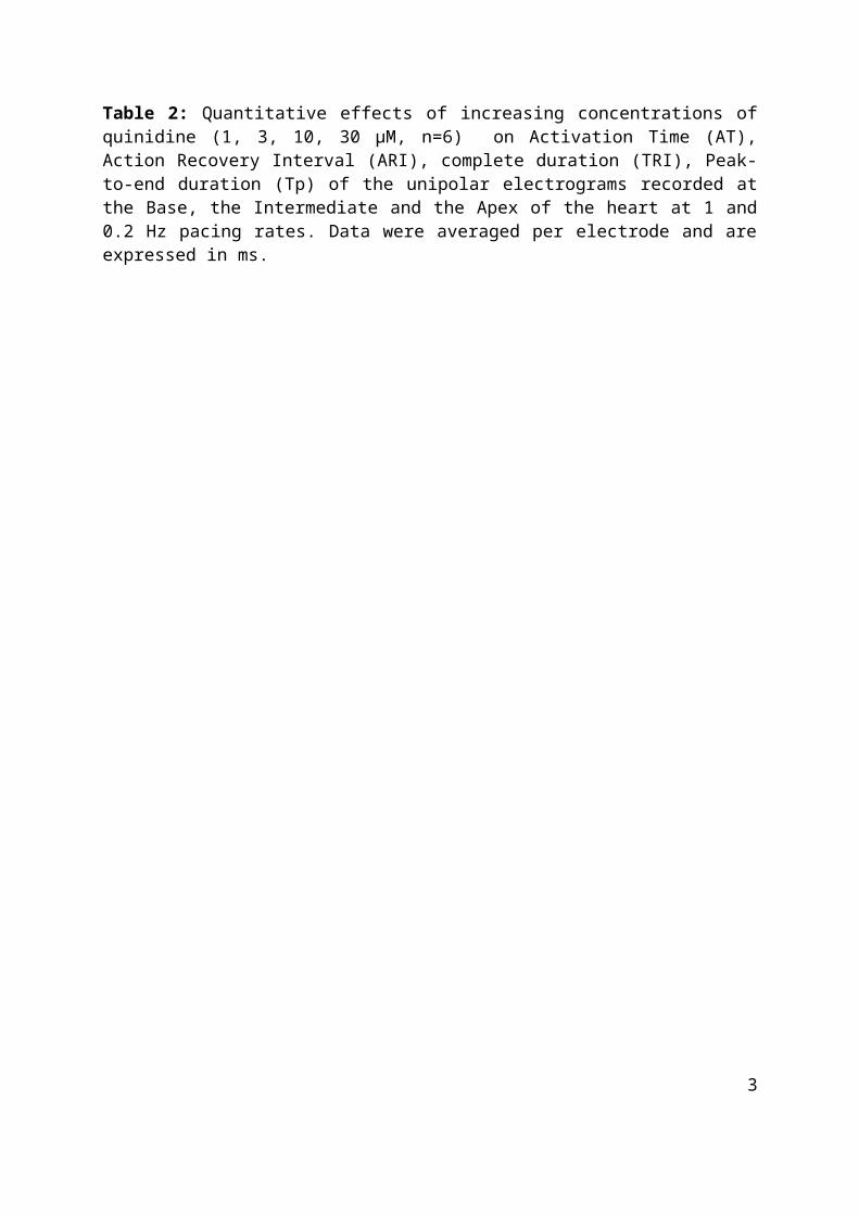
quinidine concentrations
Base Intermediate Apex
1 Hz 0.2 Hz 1 Hz 0.2 Hz 1 Hz 0.2 Hz
TRI (ms)0 317 ± 11 319 ± 8 319 ± 10 319 ± 8 316 ± 7 322 ± 81 µM 314 ± 11 323 ± 10 319 ± 9 328 ± 11 314 ± 8 327 ± 93 µM 337 ± 8 370 ± 9 335 ± 8 366 ± 10 333 ± 7 366 ± 910 µM 370 ± 9 398 ± 9 366 ± 9 412 ± 7 366 ± 8 424 ± 730 µM 396 ± 9 459 ± 16 393 ± 9 453 ± 13 395 ± 9 491 ± 21
Tp (ms)0 55 ± 5 57 ± 5 78 ± 3 72 ± 4 65 ± 5 66 ± 51 µM 52 ± 6 63 ± 4 74 ± 3 68 ± 3 65 ± 5 62 ± 43 µM 64 ± 5 73 ± 5 79 ± 4 84 ± 3 73 ± 5 72 ± 510 µM 69 ± 5 74 ± 6 88 ± 4 105 ± 5 78 ± 6 88 ± 630 µM 70 ± 6 93 ± 4 98 ± 4 120 ± 4 84 ± 6 120 ± 7
AT (ms)0 35 ± 0 35 ± 1 33 ± 2 31 ± 3 20 ± 3 21 ± 21 µM 36 ± 1 35 ± 1 32 ± 3 31 ± 3 22 ± 2 22 ± 23 µM 36 ± 1 35 ± 1 32 ± 3 32 ± 3 23 ± 2 22 ± 210 µM 44 ± 2 37 ± 1 40 ± 4 34 ± 3 27 ± 2 23 ± 230 µM 70 ± 7 45 ± 2 61 ± 8 41 ± 4 39 ± 3 28 ± 3
ARI (ms)0 229 ± 39 239 ± 8 196 ± 26 222 ± 8 161 ± 57 218 ± 171 µM 221 ± 22 267 ± 12 177 ± 26 232 ± 14 168 ± 29 176 ± 403 µM 246 ± 20 289 ± 12 191 ± 21 255 ± 26 193 ± 40 238 ± 3610 µM 271 ± 20 299 ± 8 204 ± 25 268 ± 19 215 ± 38 248 ± 2430 µM 276 ± 12 293 ± 6 218 ± 22 253 ± 14 203 ± 36 221 ± 23
Table 2: Quantitative effects of increasing concentrations of quinidine (1, 3, 10, 30 µM, n=6) on Activation Time (AT), Action Recovery Interval (ARI), complete duration (TRI), Peak-to-end duration (Tp) of the unipolar electrograms recorded at the Base, the Intermediate and the Apex of the heart at 1 and 0.2 Hz pacing rates. Data were averaged per electrode and are expressed in ms.
2
3
-10 0 10 20 30 40 50
3µM10µM
30µM100µM
% vs (C)
d,l-sotalol concentrations
-10 0 10 20 30 40 50
3 µM10
µM
30 µ
M10
0 µ
M
% vs (C)
d,l-sotalol concentrations
**
**
**
EndoM
idEpi
TRI
Tp
EndoM
idEpi
Base
IntA
pex
Base
IntA
pex
TRI
Tp
-10
10 30 50 70 90
110
130
150
1µM3µM
10µM30µM
% vs (C)
quinidine concentrations
-10
10 30 50 70 90
110
130
150
1µM3µM
10µM30µM
% vs (C)
quinidine concentrations
**
*
****
**
EndoM
idEpi
TRI
Tp
EndoM
idEpi
Base
IntA
pex
Base
IntA
pex
TRI
Tp
AB
CD

Figure 1: Effects of d,l-sotalol (3, 10, 30, 100 µM, n=6) (upper panel) and quinidine (1, 3, 10, 30 µM, n=6) (lower panel) on TRI and Tp in the Endocardium (Endo), the Midmyocardium (Mid) and the Epicardium (Epi) of the heart (A&C) and at the Base, Intermediate part (Int) and Apex of the heart (B&D). Data were grouped per needle and are expressed in % vs. Control. *: p<0.05, **: p<0.01.
4

A
d,l-sotalol concentrations(µM
)d,l-sotalol concentrations (µM
)d,l-sotalol concentrations (µM
)
quinidine concentrations (µM)
quinidine concentrations (µM)
quinidine concentrations (µM)
B
EN
DO
MID
EP
I
EN
DO
MID
EP
I
-10 10 30 50 70 90
110
130
150
13
1030
†
†
† **
-10 10 30 50 70 90
110
130
150
13
1030
Tp
Tp (+C
HR
O)
-10 10 30 50 70 90
110
130
150
13
1030
†
†
† **
-10 10 30 50 70 90
110
130
150
13
1030
Tp
Tp (+C
HR
O)
-10 10 30 50 70 90
110
130
150
13
1030
-10 10 30 50 70 90
110
130
150
13
1030
Tp
Tp (+C
HR
O)
**
†
†
†
-10 0 10 20 30 40 50 60 70
310
30100
**†
-10 10 30 50 70 90
110
130
150
13
1030
Tp
Tp (+C
HR
O)
-10 0 10 20 30 40 50 60 70
310
30100
**†
-10 10 30 50 70 90
110
130
150
13
1030
Tp
Tp (+C
HR
O)
-10 0 10 20 30 40 50 60 70
310
30100
**†
-10 10 30 50 70 90
110
130
150
13
1030
Tp
Tp (+C
HR
O)
% vs (C) % vs (C)
Figure 2: Quantitative effects on Tp in the Endocardium (Endo), the Midmyocardium (Mid) and the Epicardium (Epi) of the heart of increasing concentrations of (A) d,l-sotalol (3, 10, 30, 100 µM, n=6) (B) quinidine (1, 3, 10, 30 µM, n=6) at the stimulation rate of 1 Hz combined (+CHRO) or not with 20 µM chromanol 293B. Data were grouped per electrode and are expressed in % vs. Control. *: p<0.05, **: p<0.01, †: p<0.01.
5

-10 10 30 50 70 90
110
130
150
13
1030
% vs (C)
quinidine concentrations (µM)
-10 10 30 50 70 90
110
130
150
310
30100
% vs (C)
quinidine concentrations (µM)
-10 0
10
20
30
40
50
31
03
01
00
% vs (C )
d,l-so
talol co
ncen
tration
s (µM)
-10 0 10 20 30 40 50
310
30100
% vs (C)
d,l-sotalol concentrations (µM)
EndoM
idEpi
ATA
RI
EndoM
idEpi
EndoM
idEpi
ATA
RI
EndoM
idEpi
Base
IntA
pex
Base
IntA
pex
ATA
RI
Base
IntA
pex
Base
IntA
pex
ATA
RI
Figure 3: Quantitative effects on AT and TRI of (upper panel) d,l-sotalol (3, 10, 30, 100 µM, n=6) and (lower panel) quinidine (1, 3, 10, 30 µM, n=6) at the stimulation rate of 1 Hz. Data were grouped per electrode and are expressed in % vs. Control.
6

-20 0 20 40 60 80
100
120
310
30100
ARI (% vs C)
d,l-sotalol concentrations (µM)
END
O-C
HR
O
+CH
RO
-20 0 20 40 60 80
100
120
310
30100
ARI(% vs C)d,l-sotalol concentrations (µM
)
MID
-CH
RO
+CH
RO
-20 0 20 40 60 80
100
120
310
30100
ARI(% vs C)
d,l-sotalol concentrations (µM)
EPI-C
HR
O
+CH
RO
-20
-10 0 10 20 30 40 50 60
13
1030
ARI (% vs C)
Quinidine concentrations (µM
)
END
O-C
HR
O
+CH
RO
-20
-10 0 10 20 30 40 50 60
13
1030
ARI (% vs C)
Quinidine concentrations (µM
)
MID
-CH
RO
+CH
RO
-20
-10 0 10 20 30 40 50 60
13
1030
ARI (% vs C)Q
uinidine concentrations (µM)
EPI-C
HR
O
+CH
RO
Figure 4: Quantitative effects on ARI in the Endocardium (Endo), the Midmyocardium (Mid) and the Epicardium (Epi) of (upper panel) d,l-sotalol (3, 10, 30, 100 µM, n=6) and (lower panel) quinidine (1, 3, 10, 30 µM, n=6) at the stimulation rate of 1 Hz combined (+CHRO) or not with 20 µM chromanol 293B. Data were grouped per electrode and are expressed in % vs. Control.
7

-20 0 20 40 60 80
100
120
310
30100
ARI (% vs C)
d,l-sotalol concentrations (µM)
BASE
-CH
RO
+CH
RO
-20 0 20 40 60 80
100
120
310
30100
ARI (% vs C)d,l-sotalol concentrations (µM
)
INT
-CH
RO
+CH
RO
-20 0 20 40 60 80
100
120
310
30100
ARI (% vs C)
d,l-sotalol concentrations (µM)
APEX-C
HR
O
+CH
RO
-40
-20 0 20 40 60 80
13
1030
ARI(% vs C)
Quinidine concentrations (µM
)
BASE
-CH
RO
+CH
RO
-40
-20 0 20 40 60 80
13
1030
ARI(% vs C)
Quinidine concentrations (µM
)
INT
-CH
RO
+CH
RO
-40
-20 0 20 40 60 80
13
1030
ARI (% vs C)Q
uinidine concentrations (µM)
APEX-C
HR
O
+CH
RO
Figure 5: Quantitative effects on ARI in the Base, the Intermediate part (Int) and the Apex of the heart of (upper panel) d,l-sotalol (3, 10, 30, 100 µM, n=6) and (lower panel) quinidine (1, 3, 10, 30 µM, n=6) at the stimulation rate of 1 Hz combined or not with 20 µM chromanol 293B (CHRO). Data were grouped per electrode and are expressed in % vs. Control.
8

0 20 40 60 80
100
120
140
13
1030
Tp (% vs C)
Quinidine concentrations (µM
)
END
O-C
HR
O
+CH
RO
0 20 40 60 80
100
120
140
13
1030
Tp (% vs C)Q
uinidine concentrations (µM)
MID
-CH
RO
+CH
RO
0 20 40 60 80
100
120
140
13
1030
Tp (% vs C)
Quinidine concentrations (µM
)
EPI-C
HR
O
+CH
RO
0 20 40 60 80
100
120
140
160
13
1030
Tp (% vs C)
Quinidine concentrations (µM
)
BASE
-CH
RO
+CH
RO
0 20 40 60 80
100
120
140
160
13
1030
Tp (% vs C)
Quinidine concentrations (µM
)
INT
-CH
RO
+CH
RO
0 20 40 60 80
100
120
140
160
13
1030
Tp (% vs C)Q
uinidine concentrations (µM)
APEX-C
HR
O
+CH
RO
Figure 6: Quantitative effects on AT of quinidine (1, 3, 10, 30 µM, n=6) at the stimulation rate of 1 Hz combined or not with 20 µM chromanol 293B (CHRO, upper panel) in the Endocardium (Endo), the Midmyocardium (Mid) and the Epicardium (Epi) and (lower panel) in the Base, the Intermediate part (Int) and the Apex of the heart. Data were grouped per electrode and are expressed in % vs. Control.
9

AB
BA
SE
INT
AP
EX
BA
SE
INT
AP
EX
-20 0 20 40 60 80100120
140160180
13
1030
‡
‡‡ **
-10 10 30 50 70 90
110
130
150
13
1030
Tp
Tp
(+C
HR
O)
-20 0 20 40 60 80100120140160180
13
1030
††
**
-10 10 30 50 70 90
110
130
150
13
1030
Tp
Tp
(+CH
RO
)
-20 0 20 40 60 80100120
140160
180
13
1030
††
**
-10 10 30 50 70 90
110
130
150
13
1030
TpTp (+CH
RO
)
-20 0 20 40 60 80
100
120
310
30100
**
-10 10 30 50 70 90
110
130
150
13
1030
TpTp (+CH
RO
)
-20 0 20 40 60 80
100
120
310
30100
**
-10 10 30 50 70 90
110
130
150
13
1030
Tp
Tp
(+C
HR
O)
-20 0 20 40 60 80
100
120
310
30100
**†
-10 10 30 50 70 90
110
130
150
13
1030
Tp
Tp
(+C
HR
O)
d,l-sotalol concentrations(µM
)d,l-sotalol concentrations (µM
)d,l-sotalol concentrations (µM
)
quinidine concentrations (µM)
quinidine concentrations (µM)
quinidine concentrations (µM)
% vs (C) % vs (C)
Figure 7: Quantitative effects on Tp in the Base, the Intermediate part (Int) and the Apex (Epi) of the heart of increasing concentrations of (A) d,l-sotalol (3, 10, 30, 100 µM, n=6) and (B) quinidine (1, 3, 10, 30 µM, n=6) at the stimulation rate of 1 Hz combined or not with 20 µM chromanol 293B (CHRO, n=12). Data were grouped per needle and are expressed in % vs. Control. *, #: p<0.05, **, †: p<0.01, ‡: p<0.001.
10


