Supplementary Information - Royal Society of ChemistrySupplementary Information Target-selective...
15
1 Supplementary Information Target-selective photo-degradation of a sialyl Lewis A (sLe a ) conjugate and photo-cytotoxicity against sLe a positive cancer cells using an anthraquinone-antibody hybrid Daisuke Takahashi, Takashi Nagao, Shota Sotokawa, and Kazunobu Toshima* Department of Applied Chemistry, Faculty of Science and Technology, Keio University, 3-14-1 Hiyoshi, Kohoku-ku, Yokohama 223-8522, Japan Page Contents S-2 ・・・ General methods for chemical synthesis S-2 ・・・ Materials S-3 ・・・ Synthesis of AQ-mAb hybrids 6 and 7 S-8 ・・・ Synthesis of anti-sLe a mAb-Alexa fluor 555 conjugate 16 S-8 ・・・ Bradford assay S-9 ・・・ ELISA assay S-9 ・・・ Photo-degradation of HSA-sLe a 4 using hybrid 6 S-10 ・・・ Cell Culture S-10 ・・・ MTT assay S-10 ・・・ Fluorescence microscopy analysis S-10 ・・・ References S-11 ・・・ 1 H and 13 C NMR spectrum charts Electronic Supplementary Material (ESI) for MedChemComm. This journal is © The Royal Society of Chemistry 2016
Transcript of Supplementary Information - Royal Society of ChemistrySupplementary Information Target-selective...

1
Supplementary Information
Target-selective photo-degradation of a sialyl Lewis A (sLea)
conjugate and photo-cytotoxicity against sLea positive cancer
cells using an anthraquinone-antibody hybrid
Daisuke Takahashi, Takashi Nagao, Shota Sotokawa, and Kazunobu Toshima*
Department of Applied Chemistry, Faculty of Science and Technology, Keio University,
3-14-1 Hiyoshi, Kohoku-ku, Yokohama 223-8522, Japan
Page Contents
S-2 ・・・ General methods for chemical synthesis
S-2 ・・・ Materials
S-3 ・・・ Synthesis of AQ-mAb hybrids 6 and 7
S-8 ・・・ Synthesis of anti-sLea mAb-Alexa fluor 555 conjugate 16
S-8 ・・・ Bradford assay
S-9 ・・・ ELISA assay
S-9 ・・・ Photo-degradation of HSA-sLea 4 using hybrid 6
S-10 ・・・ Cell Culture
S-10 ・・・ MTT assay
S-10 ・・・ Fluorescence microscopy analysis
S-10 ・・・ References
S-11 ・・・ 1H and
13C NMR spectrum charts
Electronic Supplementary Material (ESI) for MedChemComm.This journal is © The Royal Society of Chemistry 2016
2
General methods for chemical synthesis
NMR spectra were recorded on a JEOL Lamda (300 MHz for 1H) or a JEOL ECA-500 (125
MHz for 13
C) spectrometer in the indicated solvent. 1H-NMR data are reported as follows;
chemical shift in parts par million (ppm) downfield or upfield from tetramethylsilane (TMS) (δ
0.00), CD3OD (δ 3.34), or CDCl3 (δ 7.26), integration, multiplicity (s = singlet, d = doublet, t =
triplet, q = quartet, and m = multiplet) and coupling constants (Hz). 13
C-NMR data are reported
as follows; chemical shift in parts par million (ppm) downfield or upfield from CD3OD (δ
49.70), CDCl3 (δ 77.16) or external standard acetone (δ 30.89). Melting points were determined
on a micro hot-stage (Yanako MP-S3). ESI-TOF mass spectra were measured on a Waters LCT
premier XE. Matrix-assisted laser desorption ionization time-of-flight mass spectrometry
(MALDI TOF MS) was conducted using a Bruker Ultraflex mass spectrometer with detection in
linear mode. Sinapinic acid was used as the matrix, with positive ionization mode. UV-vis
spectra were recorded on a JASCO V-650DS spectrophotometer. Silica gel TLC and column
chromatography were performed using Merck TLC 60F-254 (0.25 mm) and Silica gel 60 N
(spherical, neutral) (Kanto Chemical Co., Inc.), respectively. Reverse phase column
chromatography separations were performed using Wakosil 25C18 (Wako pure chemical
industries, Ltd.). Air- and/or moisture-sensitive reactions were carried out under an atmosphere
of argon using oven-dried glass ware. In general, organic solvents were purified and dried using
an appropriate procedure, and evaporation and concentration were carried out under reduced
pressure below 30 °C, unless otherwise noted.
Materials
Sulfo-SMCC (13) was purchased from Dojindo. Human serum albumin (HSA) was
purchased from Funakoshi. Anti-sLea mAb CA19-9-203 (12) was purchased from Abcam.
HSA-sLea conjugate 4 and HSA-Le
a conjugate 5 were purchased from GlycoTech. Anti-KLH
mAb 14 and anti-HSA mAb were purchased from R&D Systems. Anti-Lea mAb was purchased
from SPRING. The human epidermoid squamous carcinoma cell line, A431 (RCB0202), the
human cervix epidermoid carcinoma cell line, HeLa (RCB0007), and the human hepatocellular
carcinoma cell line, HuH-7 (RCB1942) were provided by the RIKEN BRC through the National
Bio-Resource Project of the MEXT, Japan. The human colon adenocarcinoma cell lines, WiDr
(JCRB0224) was purchased from the Japanese Collection of Research Bioresources (JCRB)
Cell Bank (Osaka, Japan).

3
Synthesis of AQ-mAb hybrids 6 and 7
Compound S1
To a solution of 8[1]
(1.07 g, 3.81 mmol) in dry DMF (20.0 mL) was dropwisely added
thioacetic acid potassium salt (KSAc) (522 mg, 4.57 mmol) at 0 °C. After being stirred for 1 h at
room temperature, the reaction mixture was added to water (50.0 mL). The aqueous layer was
extracted with three portions of EtOAc (50.0 mL). The combined extracts were washed with
brine (10.0 mL), dried over anhydrous Na2SO4, filtered, and concentrated in vacuo. The residue
was subjected to silica gel column chromatography (40.0 g, hexane/EtOAc = 1/2 to 0/1) to give
S1 (1.01 g, 3.66 mmol, 96% yield) as a white solid. Rf 0.48 (1/2 hexane/EtOAc); m.p.
103-105 °C; 1H-NMR (300 MHz, CDCl3, TMS): δ 3.55 (2H, s), 3.33 (2H, t, J = 5.7 Hz), 3.26
(2H, t, J = 5.6 Hz), 2.42 (3H, s), 1.45 (9H, s); 13
C-NMR (125 MHz, CDCl3, TMS): δ 195.4,
168.6, 156.6, 79.6, 40.9, 40.1, 32.9, 30.3, 28.4; HRMS (ESI-TOF) m/z 299.1033 (299.1041
calcd for C11H20N2O4SNa, [M+Na]+).
Compound 9
To a solution of S1 (579 mg, 2.09 mmol) in dry CH2Cl2 (17.0 mL) was dropwisely added
TFA (17.0 mL) at 0 °C. After being stirred for 1 h at room temperature, the reaction mixture was
concentrated in vacuo. The residue was subjected to reverse phase column chromatography
(H2O:MeOH = 1:0 to 0:1) to give 9 (339 mg, 1.17 mmol, 56% yield) as a light yellow oil. Rf
0.33 (3/1 CHCl3/MeOH); 1H-NMR (300 MHz, CD3OD, TMS): δ 3.56 (2H, s), 3.38 (2H, t, J =
6.0 Hz), 2.97 (2H, t, J = 5.9 Hz), 2.28 (3H, s); 13
C-NMR (125 MHz, CD3OD, TMS): δ 196.32
171.9, 40.6, 38.5, 33.6, 30.0; HRMS (ESI-TOF) m/z 177.0691 (177.0698 calcd for C6H13N2O2S,
[M+H]+).
Compound 10
To a solution of 3[2]
(140 mg, 0.421 mmol) in DMF (7.00 mL) were added TBTU (271 mg,
0.843 mmol), NEM (160 μL, 1.26 mmol) and 9 (89.0 mg, 0.505 mmol). After being stirred for
16 h at room temperature, the reaction mixture was concentrated in vacuo. The residue was
subjected to reverse phase column chromatography (H2O:MeOH = 1:0 to 0:1). The resulting

4
solid was dissolved in water (5.00 mL) and recrystallized with brine (5.00 mL) to give 2/6-
disubstituted 10 (80.5 mg, 0.164 mmol, 39% yield) as a brown solid. Rf 0.59 (3/2
CHCl3/MeOH); m.p.>300 C̊; 1
H-NMR (300 MHz, D2O): δ 7.78-8.12 (6H, m), 3.51 (2H, s),
3.37-3.39 (4H, m), 2.13 (3H, s); 13
C-NMR (125 MHz, D2O, acetone): δ 199.8, 182.8, 182.6,
172.1, 168.3, 149.1, 139.5, 134.8, 134.4, 133.7, 133.4, 133.0, 132.2, 128.8, 128.3, 126.3, 124.7,
40.0, 39.6, 33.5, 30.1; HRMS (ESI-TOF) m/z 489.0414 (489.0426 calcd for C21H17N2O8S2,
[M−H]−).
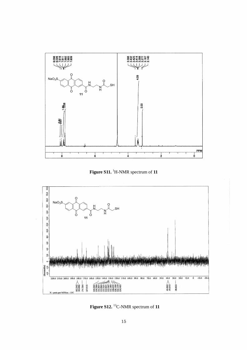
Compound 11
To a solution of 10 (16.4 mg, 33.4 μmol) in dry MeOH (1.60 mL) was added 28% NaOMe
in MeOH (7.03 μL, 40.1 μmol) at room temperature. After being stirred at room temperature for
20 min, the reaction mixture was filtered, and quenched with Amberlite. The residue was
filtered and concentrated in vacuo to give 11 (11.2 mg, 25.1 μmol, 75% yield) as a brown oil. Rf
0.72 (3/2 CHCl3/MeOH ); 1H-NMR (300 MHz, D2O): δ 7.81-8.10 (6H, m), 3.40 (4H, q, J = 7.8
Hz), 3.14 (2H, s); 13
C-NMR (125 MHz, D2O, acetone): δ 182.3, 182.0, 174.7, 167.9, 149.2,
139.3, 134.3, 133.9, 133.7, 133.0, 132.6, 132.2, 128.7, 128.2, 126.1, 124.6, 40.0, 39.6, 28.0;
HRMS (ESI-TOF) m/z 447.0308 (447.0321 calcd for C19H15N2O7S2, [M−H]−).
AQ-mAb hybrid 6
To the column (Ab-Rapid SPiN Ex, Protenova) was added anti-sLea mAb 12 (500 L, 0.2
mg/mL) at room temperature. After the suspension was gently shaken at room temperature for
90 min, the resulting gel was washed three times with 10 mM PBS (pH 7.4). And then, 12 was
eluted with elution buffer (400 L, 0.1 M Glycine-HCl, pH 2.8) and promptly neutralized with
neutralization buffer (9.0 L, 1 M Tris). To a solution of 12 (100 g685 pmol) in 10 mM PBS
(pH 7.4, 130 L) was added sulfo-SMCC (13) (29.9 g, 68.5 nmol) in DMF (6.85L) at room
temperature. After the reaction mixture was incubated for 1 h at 25 °C, Amicon 3K centrifugal
filter device (Millipore) was used to separate from excess 13 and to concentrate the sample
solution. And then, to a solution of anti-sLea mAb-SMCC S2 in 10 mM PBS (pH 7.4, 100 L)
was added 11 (30.7 g, 68.5 nmol) in 10 mM PBS (pH 7.4, 37.0 L) at room temperature. After
the reaction mixture was incubated for 16 h at 4 °C, Amicon 3K centrifugal filter device
(Millipore) was used to separate from excess 11 and to concentrate the sample solution to give
AQ-mAb hybrid 6 (0.27 mg/mL, 100 L, 27% yield in 2 steps). The chemical yield of 6 was
calculated based on the Bradford method.

5
AQ-mAb hybrid 7
To a solution of anti-KLH mAb 14 (100 g685 pmol) in 10 mM PBS (pH 7.4, 130 L)
was added sulfo-SMCC (13) (29.9 g, 68.5 nmol) in DMF (6.85L) at room temperature. After
the reaction mixture was incubated for 1 h at 25 °C, Amicon 3K centrifugal filter device
(Millipore) was used to separate from excess 13 and to concentrate the sample solution. And
then, to a solution of anti-KLH mAb-SMCC S3 in 10 mM PBS (pH 7.4, 100 L) was added 11
(30.7 g, 68.5 nmol) in 10 mM PBS (pH 7.4, 37.0 L) at room temperature. After the reaction
mixture was incubated for 16 h at 4 °C, Amicon 3K centrifugal filter device (Millipore) was
used to separate from excess 11 and to concentrate the sample solution to give AQ-mAb hybrid
7 (0.54 mg/mL, 100 L, 55% yield in 2 steps). The chemical yield of 7 was calculated based on
the Bradford method.
Figure S1. UV spectra of 6, 12, and 7. These compounds (1.2 M) were dissolved in PBS (10
mM, pH 7.4).
0
0.01
0.02
0.03
0.04
0.05
0.06
220 230 240 250 260 270 280 290
Abs [
-]
Wavelenth [nm]
AQ-mAb hybrid 6
Anti-sLea mAb 12
AQ-mAb hybrid 7
AQ-mAb hybrid 6
Anti-sLea mAb 12
AQ-mAb hybrid 7
6
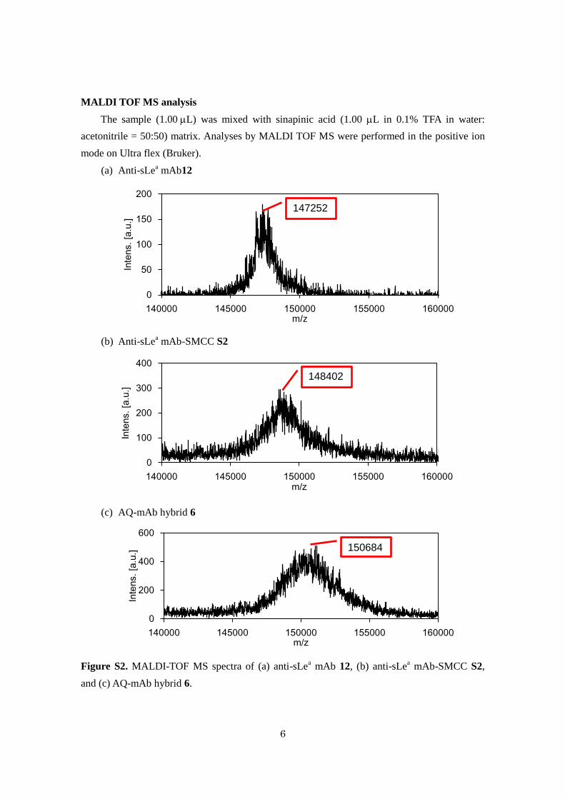
MALDI TOF MS analysis
The sample (1.00L) was mixed with sinapinic acid (1.00 L in 0.1% TFA in water:
acetonitrile = 50:50) matrix. Analyses by MALDI TOF MS were performed in the positive ion
mode on Ultra flex (Bruker).
(a) Anti-sLea mAb12
(b) Anti-sLea mAb-SMCC S2
(c) AQ-mAb hybrid 6
Figure S2. MALDI-TOF MS spectra of (a) anti-sLea mAb 12, (b) anti-sLe
a mAb-SMCC S2,
and (c) AQ-mAb hybrid 6.
0
50
100
150
200
140000 145000 150000 155000 160000
Inte
ns.
[a.u
.]
m/z
147252
0
100
200
300
400
140000 145000 150000 155000 160000
Inte
ns.
[a.u
.]
m/z
148402
0
200
400
600
140000 145000 150000 155000 160000
Inte
ns.
[a.u
.]
m/z
150684

7
(a) Anti-KLH mAb 14
(b) Anti-KLH mAb-SMCC S3
(c) AQ-mAb hybrid 7
Figure S3. MALDI-TOF MS spectra of (a) anti-KLH mAb 14, (b) anti-KLH mAb-SMCC S3,
and (c) AQ-mAb hybrid 7.
0
200
400
600
800
1000
140000 145000 150000 155000 160000
Inte
ns.
[a.u
.]
m/z
147549
0
200
400
600
800
1000
140000 145000 150000 155000 160000
Inte
ns.
[a.u
.]
m/z
148952
0
50
100
140000 145000 150000 155000 160000
Inte
ns.
[a.u
.]
m/z
150537

8
Synthesis of anti-sLea mAb-Alexa fluor 555 conjugate 16
Scheme S1. Synthetic scheme of anti-sLea mAb-AF555 16.
Preparation of anti-sLea mAb-AF555 16
To a solution of anti-sLea mAb 12 (100 g685 pmol) in 1 M sodium bicarbonate buffer
(pH 9.0) was added Alexa Fluor 555 2,3,5,6-tetrafluorophenyl ester (29.9 g, 34.3 nmol) in
DMF (6.85L) at room temperature. After the reaction mixture was incubated for 1 h at 25 °C,
Amicon 3K centrifugal filter device (Millipore) was used to separate from excess AF555 and to
concentrate the sample solution to give anti-sLea mAb-AF555 16 (0.54 mg/mL, 100 L, 54%
yield). The chemical yield of 16 was calculated based on the Bradford method.
Anti-sLea mAb-AF555 16
Figure S4. MALDI-TOF MS spectra of anti-sLea mAb-AF555 16.
Bradford assay
To a sample (10.0 L) in test tube, a volume of 300L Bradford assay reagent (Thermo
Scientific) was added, and then the resulting mixture was blended by gentle vortex mixing.
After 5 min, absorbance at 595 nm was measured in plastic 96-well microplate against a reagent
blank using SpectraMax i3 (Molecular Devices). The calibration curve was built using BSA
samples (0.100-0.500 g).
0
1000
2000
3000
4000
140000 145000 150000 155000 160000
Inte
ns.
[a.u
.]
m/z
148212

9
ELISA assay
HSA-sLea 4 (1.00 μg/mL) in 10 mM PBS (pH 7.4) was coated overnight at 4 °C on a
96-well ELISA plate (Sumitomo Bakelite Co., Ltd.). The solution was then removed, and 300
μL of the blocking buffer (10 mM PBS, 1% nonfat dry milk) was added to each well and
incubated at room temperature for 2 h. The wells were then washed with 300 μL of 10 mM
PBS-T (pH 7.4, 0.05% Tween-20). Hybrid 6 was prepared in 10 mM PBS (pH 7.4) at a range of
concentrations, and was added (100 μL) to each well. The plate was then incubated for 2 h at
room temperature. The sample solutions were then removed, and the wells washed three times
with PBS-T. To detect the sample, 100 μL of anti-mouse IgG-peroxidase conjugate (1:800
dilution) (GE healthcare) was added into each well and incubated for 1 h at room temperature.
The solutions were then removed and the plate was washed with PBS-T three times. OPD (3.7
mM, 100 μL/well) in citrate buffer (citric acid 26.5 mM, sodium hydrogen phosphate 57.8 mM
and H2O2 3.82 M) was then added into each well. After approximately 15 min, when the
yellow color was sufficiently visible, 50.0 μL of 4 N H2SO4 solution was added into each well
to stop the enzymatic reaction as indicated by the solution turning brown. The absorbance was
read using a microplate reader at 490 nm within 30 min.
Photo-degradation of HSA-sLea 4 using hybrid 6
To a solution of HSA-sLea
4 (5.00L, 1 pmol) in 10 mM PBS (pH 7.4) was added a
solution of hybrid 6 (5.00L, 0.3-10 pmol) in 10 mM PBS. After being incubated at room
temperature for 30 min, the mixture was incubated at 25 °C for 2 h with or without
photo-irradiation using a UV lamp (365 nm, 100 W, Blak-ray (B-100A), UVP. Inc.) placed 10
cm from the sample. And then, 5.00L of electrophoresis buffer consisted of SDS (5%, wt/vol),
glycerol (27%, vol/vol), DTT (0.5%, wt/vol) and bromophenol blue (0.007%, wt/vol) was added
to the photo-irradiated samples. The photo-degradation products were separated by SDS-PAGE
in 7% polyacrylamide gels and electrotransfered onto Amersham Hybond ECL Nitrocellulose
Membrane (GE Healthecare). The membrane was blocked with 10 mM Tris-buffered
saline-0.1% Tween 20 (TBS-T) containing 5% nonfat dry milk for 2 h at room temperature.
After a short wash in TBS-T, the membrane was incubated in a 1:1000 dilution of a primary
antibody (abcam) in TBS-T for 14 h at 4 °C followed by washing with TBS-T for 30 min. The
bound antibody was then detected with horseradish peroxidase-conjugated secondary antibody
anti-mouse IgG (GE healthcare) diluted at 1:3000 in TBS-T and 2% (wt/vol) nonfat dry milk by
incubation with it for 2 h at 4 °C. After having been washed for 30 min in TBS-T, the
immunocomplexes were detected by using ECL reagent, Immobilon Western (Millipore,
Billerica, MA). Exposure to RX-U films (Fuji Film, Kanagawa, Japan) was carried out for 1
min to 10 min.

10
Cell Culture
< A431, WiDr >
The A431 or WiDr cell line was routinely grown in Dulbecco’s modified Eagle’s medium
(DMEM) supplemented with 5% (v/v) Fetal bovine serum, 0.5% (v/v) penicillin and
kanamycine. The cells were maintained at 37 °C in a humidified atmosphere containing 5%
CO2.
<HuH-7>
The HuH-7 cell line was routinely grown in RPMI medium supplemented with 5% (v/v)
Fetal bovine serum, 0.5% (v/v) penicillin and kanamycine. The cells were maintained at 37 °C
in a humidified atmosphere containing 5% CO2.
<HeLa>
The HeLa cell line was routinely grown in Dulbecco’s modified Eagle’s medium (DMEM)
supplemented with 5% (v/v) Bovine Calf Serum, 0.5% (v/v) penicillin and kanamycine. The cells
were maintained at 37 °C in a humidified atmosphere containing 5% CO2.
MTT assay
1×10³ cells in 90.0L of medium were cultured in 96-well microplates, and then incubated
for 24 h at 37 °C prior to the addition of experimental sample. To each well was added 10.0 L of
hybrid 6 (10.0 pmol/well) in medium and the culture was incubated for 24 h. The plate was
irradiated with a UV lamp (368 nm, 30 W, FL15BLB-368, Sankyo Denki Co., Ltd.) from 20 cm
for 30 min, and then incubated for 72 h at 37 °C. Cell viability was evaluated using the MTT
assay. 10.0L of 5.00 mg/mL MTT dissolved in PBS was added to each well. After incubation
for 3 h at 37 °C, medium was aspirated and 50.0L of DMSO was added to each well, and
color intensity was measured using SpectraMax i3 (Molecular Devices) micro plate reader at
540 nm.
Fluorescence microscopy analysis
Cancer cells were plated in 96-well plate (1×10³ cells/well) and incubated overnight at
37 °C. Cells were then treated with anti-sLea mAb-AF555 16 for 1 h at 4 °C or at 37 °C. Cells
were washed with PBS (3 × 100 L) followed by PBS + DAPI (1 g/mL) and imaged using a
fluorescence microscopy EVOS AMF-4302.
References
1) H. Y. Kim, S. Rhim, J. J. Park and J. Kang, J. Incl. Phenom. Macrocycl. Chem., 2012, 74,
317.
2) Y. Imai, S. Hirono, H. Matsuba, T. Suzuki, Y. Kobayashi, H. Kawagishi, D. Takahashi and

11
K. Toshima, Chem. Asian J., 2012, 7, 97.
1H and
13C NMR
spectrum charts

12
Figure S5. 1H-NMR spectrum of S1
Figure S6. 13
C-NMR spectrum of S1

13
Figure S7. 1H-NMR spectrum of 9
Figure S8. 13
C-NMR spectrum of 9

14
Figure S9. 1H-NMR spectrum of 10
Figure S10. 13
C-NMR spectrum of 10
15
Figure S11. 1H-NMR spectrum of 11
Figure S12. 13
C-NMR spectrum of 11