Supplementary Information Chemical-induced cardiac …10.1038...2018-04-18Supplementary Information...
20
Supplementary Information Chemical-induced cardiac reprogramming in vivo Chenwen Huang 1 , Wanzhi Tu 2, 3 , Yanbin Fu 1 , Jinxi Wang 4 , Xin Xie 1, 2, 3, 5, * 1 Shanghai Key Laboratory of Signaling and Disease Research, Laboratory of Receptor-based Bio-medicine, School of Life Sciences and Technology, Tongji University, Shanghai 200092, China 2 CAS Key Laboratory of Receptor Research, the National Center for Drug Screening, Shanghai Institute of Materia Medica, Chinese Academy of Sciences, Shanghai 201203, China 3 School of Life Science and Technology, ShanghaiTech University, Shanghai 200031, China 4 Key Laboratory of Stem Cell Biology and Laboratory of Molecular Cardiology, Shanghai Institutes for Biological Sciences, Chinese Academy of Sciences, Shanghai Jiao Tong University School of Medicine, Shanghai, 200031, China 5 Stake Key Laboratory of Drug Research, the National Center for Drug Screening, Shanghai Institute of Materia Medica, Chinese Academy of Sciences, Shanghai, China *Address correspondence to: Dr. Xin Xie, 189 Guo Shou Jing Road, Shanghai 201203, China. Tel: 86-21-50801313 ex 156; E-mail: [email protected].
-
Upload
truongdieu -
Category
Documents
-
view
217 -
download
0
Transcript of Supplementary Information Chemical-induced cardiac …10.1038...2018-04-18Supplementary Information...
Supplementary Information
Chemical-induced cardiac reprogramming in vivo
Chenwen Huang1, Wanzhi Tu2, 3, Yanbin Fu1, Jinxi Wang4, Xin Xie1, 2, 3, 5, *
1Shanghai Key Laboratory of Signaling and Disease Research, Laboratory of
Receptor-based Bio-medicine, School of Life Sciences and Technology, Tongji
University, Shanghai 200092, China 2CAS Key Laboratory of Receptor Research, the National Center for Drug Screening,
Shanghai Institute of Materia Medica, Chinese Academy of Sciences, Shanghai
201203, China 3School of Life Science and Technology, ShanghaiTech University, Shanghai 200031,
China 4Key Laboratory of Stem Cell Biology and Laboratory of Molecular Cardiology,
Shanghai Institutes for Biological Sciences, Chinese Academy of Sciences, Shanghai
Jiao Tong University School of Medicine, Shanghai, 200031, China 5Stake Key Laboratory of Drug Research, the National Center for Drug Screening,
Shanghai Institute of Materia Medica, Chinese Academy of Sciences, Shanghai,
China
*Address correspondence to: Dr. Xin Xie, 189 Guo Shou Jing Road, Shanghai
201203, China. Tel: 86-21-50801313 ex 156; E-mail: [email protected].
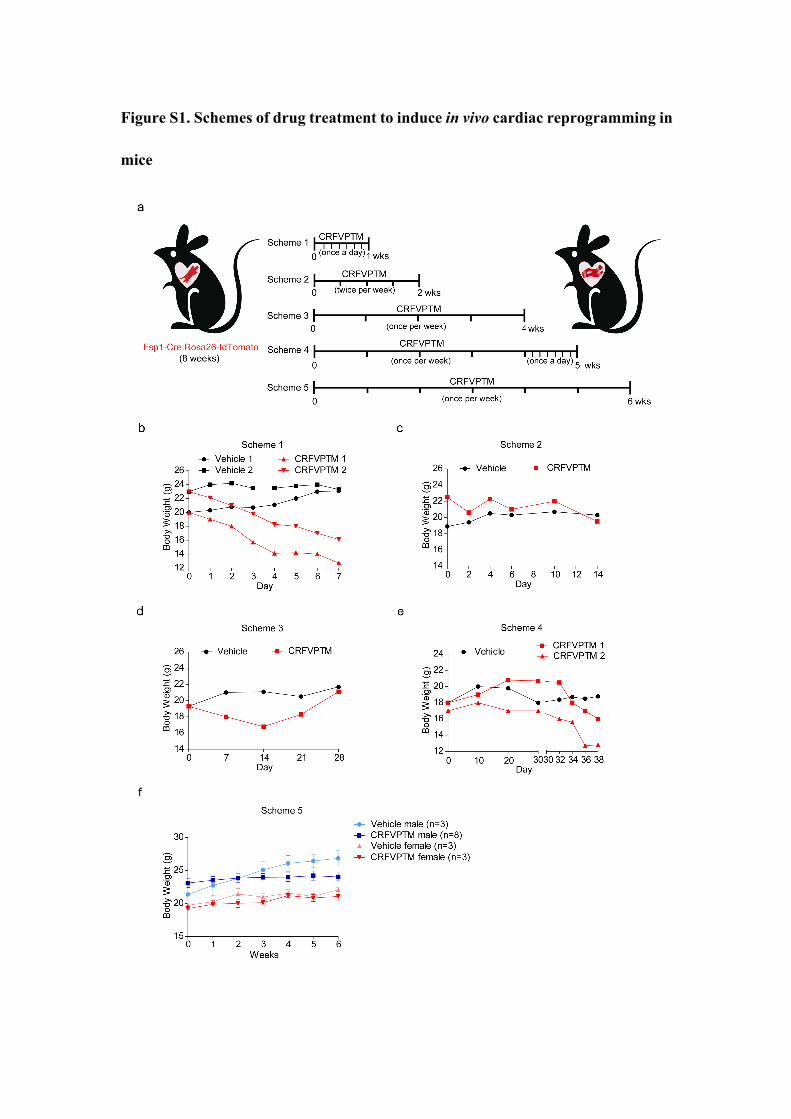
Figure S1. Schemes of drug treatment to induce in vivo cardiac reprogramming in
mice
(a) Schemes of drug treatment to induce in vivo cardiac reprogramming in Fsp1-
cre:R26RtdTomato mice. Scheme 1, mice were given CRFVPTM once a day for a week;
Scheme 2, mice were given CRFVPTM twice a week for 2 weeks; Scheme 3, mice
were given CRFVPTM once a week for 4 weeks; Scheme 4, mice were given
CRFVPTM once a week for 4 weeks, then once a day for another week; Scheme 5,
mice were given CRFVPTM once a week for 6 weeks. (b-f) Body weight of the Fsp1-
cre:R26RtdTomato mice treated with CRFVPTM or vehicle as illustrated in Scheme 1 to
5.
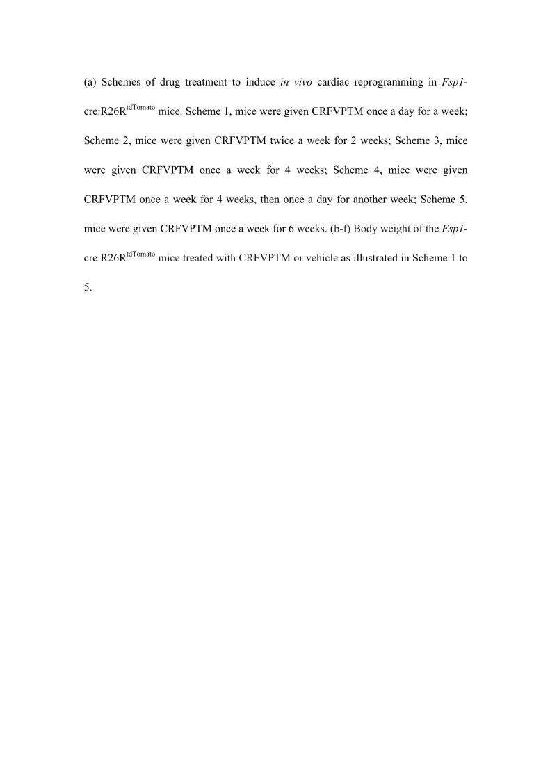
Figure S2. Characterization of tdTomato+ cells in the heart of Fsp1-
cre:R26RtdTomato mouse
Immunofluorescence staining of Fsp1, a-SMA, Vimentin, CD11b, cTnI and Gata4 in
the cryosections of the hearts from Fsp1-cre:R26RtdTomato mice before drug treatment.
Nuclei were stained with hoechst. Scale bar, 20 µm.
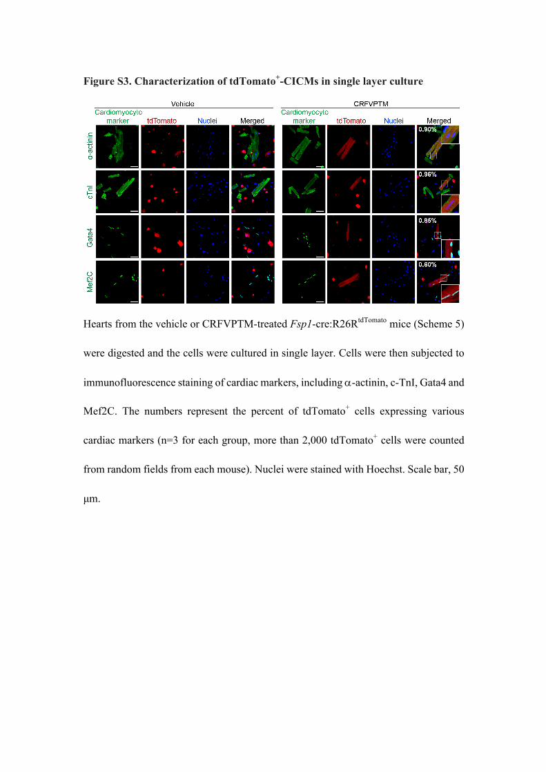
Figure S3. Characterization of tdTomato+-CICMs in single layer culture
Hearts from the vehicle or CRFVPTM-treated Fsp1-cre:R26RtdTomato mice (Scheme 5)
were digested and the cells were cultured in single layer. Cells were then subjected to
immunofluorescence staining of cardiac markers, including a-actinin, c-TnI, Gata4 and
Mef2C. The numbers represent the percent of tdTomato+ cells expressing various
cardiac markers (n=3 for each group, more than 2,000 tdTomato+ cells were counted
from random fields from each mouse). Nuclei were stained with Hoechst. Scale bar, 50
µm.
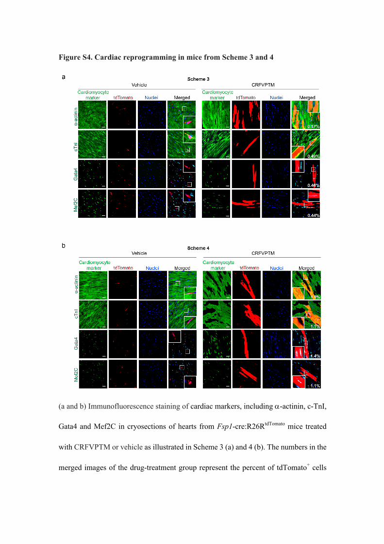
Figure S4. Cardiac reprogramming in mice from Scheme 3 and 4
(a and b) Immunofluorescence staining of cardiac markers, including a-actinin, c-TnI,
Gata4 and Mef2C in cryosections of hearts from Fsp1-cre:R26RtdTomato mice treated
with CRFVPTM or vehicle as illustrated in Scheme 3 (a) and 4 (b). The numbers in the
merged images of the drug-treatment group represent the percent of tdTomato+ cells

expressing various cardia markers (5 sections from each mouse were analyzed). Nuclei
were stained with Hoechst. Scale bar, 20 µm.
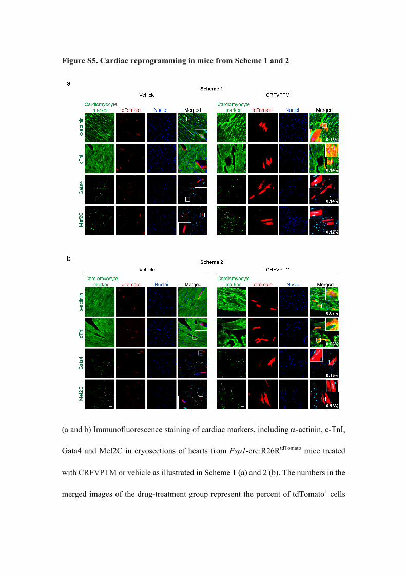
Figure S5. Cardiac reprogramming in mice from Scheme 1 and 2
(a and b) Immunofluorescence staining of cardiac markers, including a-actinin, c-TnI,
Gata4 and Mef2C in cryosections of hearts from Fsp1-cre:R26RtdTomato mice treated
with CRFVPTM or vehicle as illustrated in Scheme 1 (a) and 2 (b). The numbers in the
merged images of the drug-treatment group represent the percent of tdTomato+ cells

expressing various cardia markers (5 sections from each mouse were analyzed).
Nuclei were stained with Hoechst. Scale bar, 20 µm.
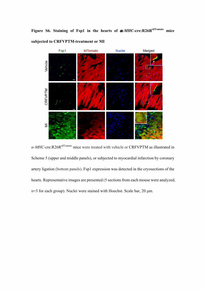
Figure S6. Staining of Fsp1 in the hearts of a-MHC-cre:R26RtdTomato mice
subjected to CRFVPTM-treatment or MI
α-MHC-cre:R26RtdTomato mice were treated with vehicle or CRFVPTM as illustrated in
Scheme 5 (upper and middle panels), or subjected to myocardial infarction by coronary
artery ligation (bottom panels). Fsp1 expression was detected in the cryosections of the
hearts. Representative images are presented (5 sections from each mouse were analyzed,
n=3 for each group). Nuclei were stained with Hoechst. Scale bar, 20 µm.

Figure S7. Staining of cardiac markers in the skeletal muscle, tail tip, lung and
liver of Fsp1-cre:R26RtdTomato mice from Scheme 5
(a) Immunofluorescence staining of Gata4, a-MHC, a-actinin and nebulin in the
cryosections of the tibialis anterior muscle isolated from Fsp1-cre:R26RtdTomato mice
treated with CRFVPTM or vehicle for 6 weeks (Scheme 5). Nuclei were stained with
Hoechst. Scale bar, 20 µm. (b-d) Immunofluorescence staining of cTnI, Gata4, or
MEF2C and in the cryosections of the tail tip (b), lung (c) and liver (d) isolated from
Fsp1-cre:R26RtdTomato mice treated with CRFVPTM or vehicle for 6 weeks (Scheme 5).
Nuclei were stained with Hoechst. Scale bar, 20 µm.
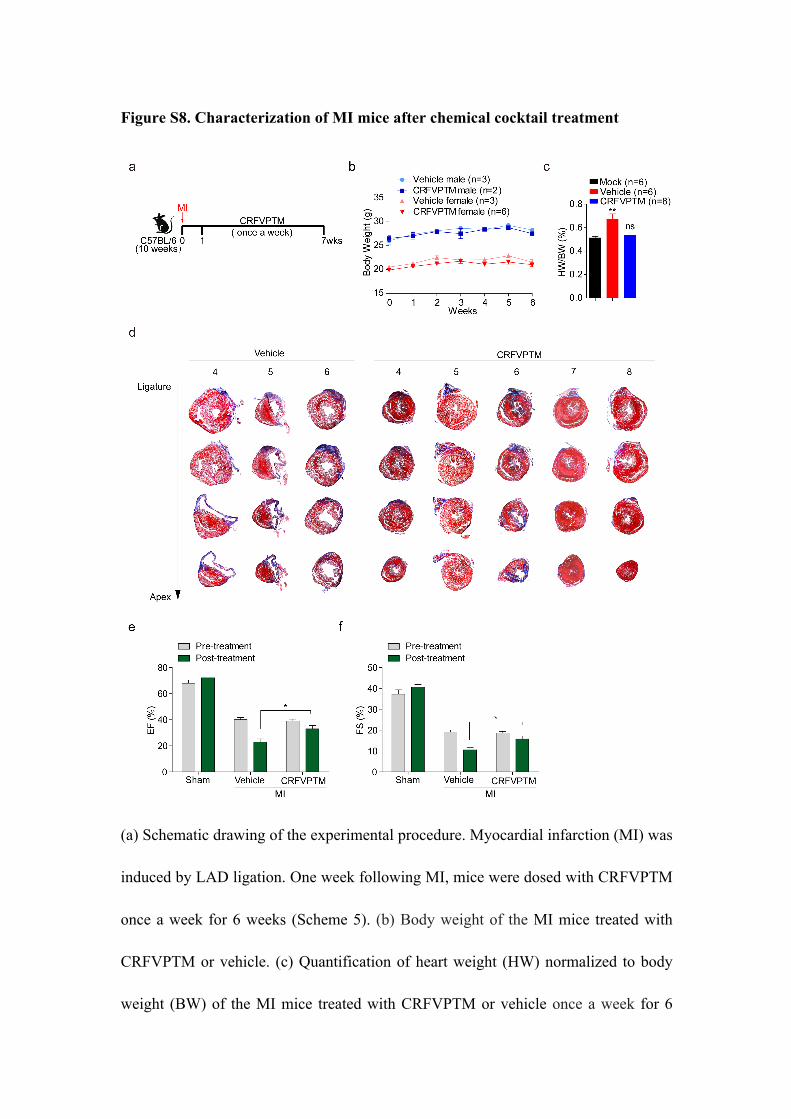
Figure S8. Characterization of MI mice after chemical cocktail treatment
(a) Schematic drawing of the experimental procedure. Myocardial infarction (MI) was
induced by LAD ligation. One week following MI, mice were dosed with CRFVPTM
once a week for 6 weeks (Scheme 5). (b) Body weight of the MI mice treated with
CRFVPTM or vehicle. (c) Quantification of heart weight (HW) normalized to body
weight (BW) of the MI mice treated with CRFVPTM or vehicle once a week for 6
weeks. Data are presented as means ± SEM. **P < 0.01 versus mock group. (d) Images
of Masson trichrome staining of the heart sections (as presented in Figure 1e) from the
MI mice treated with CRFVPTM (mice number 4-8) or vehicle (mice number 4-6) once
a week for 6 weeks. (e, f) Evaluation of cardiac functions with echocardiography. Mice
were subjected sham operation or MI by LAD ligation. Sham operated mice received
CRFVPTM once a week for 6 weeks. MI mice were treated with vehicle and
CRFVPTM once a week for 6 weeks. Ejection fraction (EF, e) and fractional shortening
(FS, f) of pre- and post-treated mice were presented. Pre-treatment refers to the 2 or 3
days after LAD ligation while post-treatment refers to 1 weeks after completion of the
drug treatment. Data are presented as means ± SEM (n=8 for sham groups, n=9 for MI
vehicle group, n=10 for MI drug-treated group). *P < 0.05 (Student’s t-test).
Materials and Methods
Mouse line
The Fsp1-Cre mice (The Jackson Laboratory) that express Cre recombinase under
the control of the fibroblast specific protein-1 (Fsp1 or S100A4) promoter were mated
with the R26RtdTomato mice (The Jackson Laboratory), in which the expression of
tdTomato is terminated by a loxP-flanked STOP cassette. The progeny mice (Fsp1-
Cre:R26RtdTomato) with specific expression of the red fluorescent protein tdTomato in
the fibroblasts were used for lineage tracing experiments1-3. The α-MHC-Cre mice (The
Jackson Laboratory) that express Cre recombinase under the control of the cardiac-
specific alpha myosin-heavy chain (α-MHC-Cre or Myh6) promoter were mated with
the R26RtdTomato mice too. The progeny mice (α-MHC-Cre:R26RtdTomato) with specific
expression of the red fluorescent protein tdTomato in the cardiomyocytes4,5. All
experiment procedures for the use and the care of the animals complied with
international guidelines for the care and use of laboratory animals and were approved
by the Animal Ethics Committee of Shanghai Institute of Materia Medica.
Drug treatment
CRFTM, including CHIR99021 (C, 14 mg/kg), Repsox (R, 8.6 mg/kg), Forskolin
(F, 61.6 mg/kg), TTNPB (T, 1 mg/kg) and Rolipram (M, 2.5 mg/kg), were mixed in
0.5% CMC-Na (sodium carboxymethyl cellulose)/saline. VP, including VPA (V, 250
mg/kg) and Parnate (P, 2.7 mg/kg), were dissolved in saline. Mice were treated with
C6FTR by oral gavage and VP by intraperitoneal injection with intervals illustrated in
Figure S1. The vehicle group only received 0.5% CMC-Na/saline by oral gavage and
saline by intraperitoneal injection.
Immunofluorescent staining
Hearts were removed and perfused via aortic cannulation with PBS with a constant
flow of 1 ml/min to ensure the removing of all blood cells. Hearts, skeletal muscles and
other tissue were fixed in 4% PFA at 4°C overnight and incubated in 20% sucrose at
4°C overnight. Tissue were embedded in OCT medium, frozen at -80 °C and sections
with 10-15 µm thickness were prepared. After permeabilization with 1% Triton X-100
for 30 minutes, cryosections were treated with 5% BSA for 1-2 h, and then incubated
with various primary antibodies at 4°C overnight. After thorough washing, secondary
antibodies conjugated with Alexa Fluor 555, Alexa Fluor 488 and Alexa Fluor 647 were
used. Nuclei were visualized with Hoechst 33342 (10 µg/ml). Images were captured
with an Olympus FV10i confocal microscope. Antibodies used in this study are as
following: Gata4 (SAB4501129, Sigma), c-TnI (ab47003, Abcam), α-Actinin (A7811,
Sigma), Mef2c (5030S, CST), Connexin 43 (610061, BD Biosciences), N-cadherin
(ab76057, Abcam), α-MHC (ab50967, Abcam), Nebulin (N9891, Sigma), Fsp1(ABF32,
Merckmillipore), α-SMA ( ab2547, Abcam) ,Vimentin (ab8978, Abcam) ,CD11b
(ab184308, Abcam) .
Isolation of adult cardiomyocytes
Adult mice were anaesthetized with 10% chloral hydrate. Hearts were removed and
perfused retrogradely via aortic cannulation with cardiomyocyte isolation buffer (CIB,
containing 137 mM NaCl, 2.7 mM KCl, 1.05 mM MgSO4·7H2O, 0.42 mM
NaH2PO4·2H2O, 11.9 mM NaHCO3, 5.6 mM glucose, 0.6 mg/mL Taurine and 1 mg/ml
2,3-Butanedione monoxime, pH=7.4) with a constant flow of 0.5 ml / min at 37℃ by
peristaltic pump to remove blood cells. Hearts were then perfused with perfusion buffer
(CIB supplemented with 0.4 mM EGTA). After 3 min, hearts were digested with
digestion buffer (CIB supplemented with 0.06 mg/ml Protease XIV, 0.36 mg/ml
Collagenase II, 0.48 mg/ml Collagenase IV and 300 nM CaCl2) with a constant flow of
1 ml/min at 37℃ till the flow out of the cardiomyocytes. Hearts were then washed with
growth medium (DMEM containing 10% FBS) to terminate the digestion, followed by
perfusion with CIB to collect cardiomyocytes by gently squeezing of the hearts.
Patch clamp recording
Patch clamp recording was performed in a temperature-controlled room at
approximately 25°C. The Giga-Ohm seal was achieved and the action potentials (APs)
were recorded under the current-clamp at zero applied current (Axopatch-200B
amplifier). The pipette solution contained 145 mM KCl, 1 mM MgCl2, 5 mM EGTA,
10 mM HEPES and 10 mM Na2ATP (pH=7.3 with KOH). Extracellular solution
contained 140 mM NaCl, 3 mM KCl, 2 mM CaCl2, 1.5 mM MgCl2, 10 mM HEPES
and 10 mM glucose (pH=7.4 with NaOH). Signals were filtered at 1 kHz, and digitized
using a DigiData 1440 with pClamp9.2 software (Molecular Devices).
Mouse MI model
Permanent ligation of the left anterior descending coronary artery (LAD) at the left
ventricle was performed in 10-wk-old C57BL/6 mice. The mice were anesthetized with
2.4% isoflurane/97.6% oxygen and placed in a supine position. Thoracotomy was
performed at the third intercostal space, and self-retaining micro-retractors were placed
to separate the third and fourth rib to visualize the LAD. The LAD was surgically
ligated at 1.5 mm distal to the left atrial appendage without tearing the pericardial sac.
After LAD ligation, the retractors were removed and the chest was closed.
Measurement of scar area
The fibrotic scar formation in the hearts with MI was visualized with trichrome
staining of the cryosections with a Masson’s trichrome staining kit (Sigma, HT15-1)
according to the manufacturer’s instruction. The scar size was measured with ImagePro
software (scar area (blue), healthy area (red)) on transections spanning four levels
running from the ligation site to the apex of the heart (about 0.5 mm between adjacent
levels, with the first level starting right below the ligation). From each level, 4 slides of
heart tissue were measured (a total of 16 sections from each heart).
Echocardiography
All mice were lightly anesthetized by 1.5-3% isoflurane inhalation. Heart function
was evaluated by transthoracic echocardiography using a Vevo 2100 instrument and
data was analyzed with Vevo 2100 software package. Echocardiography performed 2-
3 days post MI demonstrated a significantly reduction in ejection fraction before drug
treatment. Echocardiography was then performed 1 week after completion of the drug
treatment.
Statistical Analysis

Values are reported as the Means ± SEM. P values were calculated by Student’s t-
test, P < 0.05 was considered statistically significant. All graphs were plotted with
GraphPad Prism software.
References
1. Jayawardena, T. M. et al., MicroRNA induced cardiac reprogramming in vivo:
evidence for mature cardiac myocytes and improved cardiac function. CIRC RES
116 418 (2015).
2. Jayawardena, T. M. et al., MicroRNA-mediated in vitro and in vivo direct
reprogramming of cardiac fibroblasts to cardiomyocytes. CIRC RES 110 1465
(2012).
3. Qian, L. et al., In vivo reprogramming of murine cardiac fibroblasts into induced
cardiomyocytes. NATURE 485 593 (2012).
4. Palermo, J., Gulick, J., Colbert, M., Fewell, J. & Robbins, J., Transgenic
remodeling of the contractile apparatus in the mammalian heart. CIRC RES 78 504
(1996).
5. Agah, R. et al., Gene recombination in postmitotic cells. Targeted expression of
Cre recombinase provokes cardiac-restricted, site-specific rearrangement in adult
ventricular muscle in vivo. J CLIN INVEST 100 169 (1997).