Supplemental material JCB
2
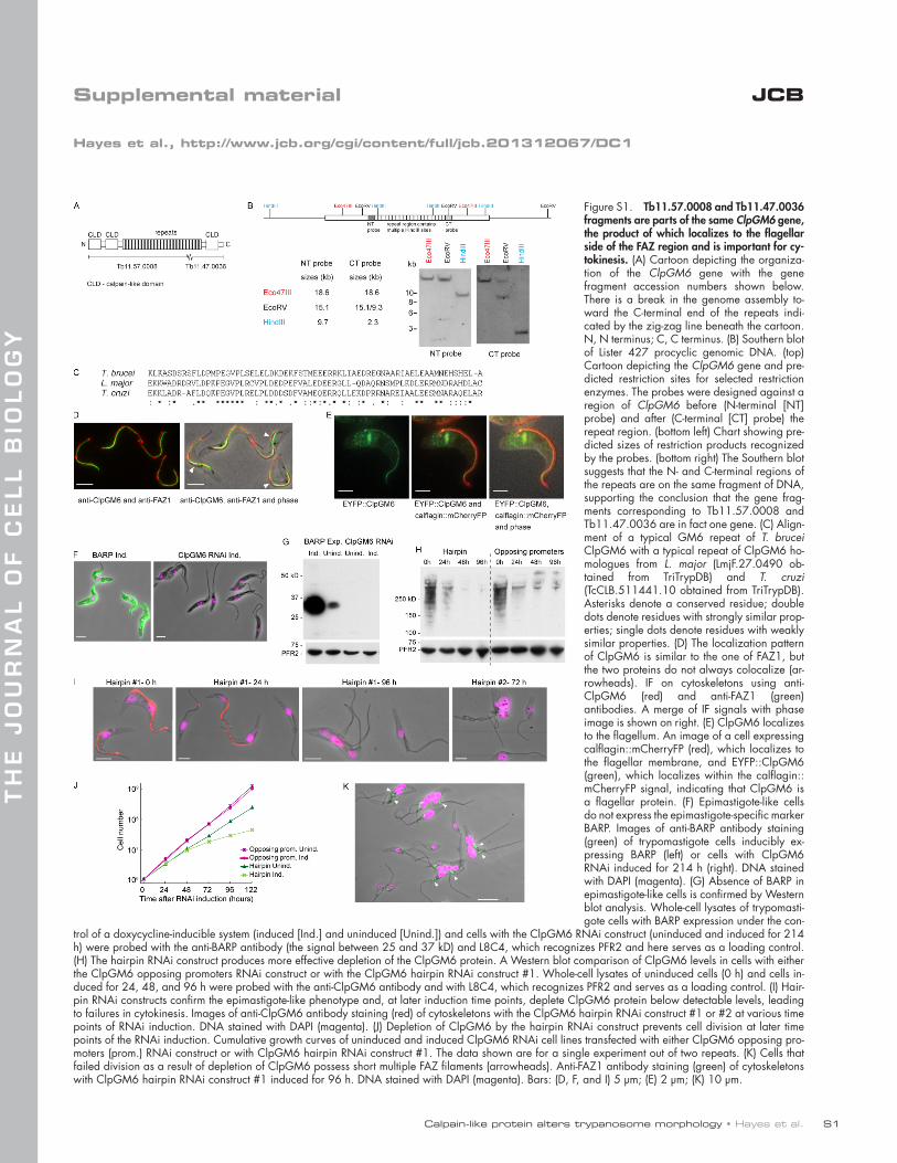
Calpain-like protein alters trypanosome morphology • Hayes et al. S1 JCB THE JOURNAL OF CELL BIOLOGY Supplemental material Hayes et al., http://www.jcb.org/cgi/content/full/jcb.201312067/DC1 Figure S1. Tb11.57.0008 and Tb11.47.0036 fragments are parts of the same ClpGM6 gene, the product of which localizes to the flagellar side of the FAZ region and is important for cy- tokinesis. (A) Cartoon depicting the organiza- tion of the ClpGM6 gene with the gene fragment accession numbers shown below. There is a break in the genome assembly to- ward the C-terminal end of the repeats indi- cated by the zig-zag line beneath the cartoon. N, N terminus; C, C terminus. (B) Southern blot of Lister 427 procyclic genomic DNA. (top) Cartoon depicting the ClpGM6 gene and pre- dicted restriction sites for selected restriction enzymes. The probes were designed against a region of ClpGM6 before (N-terminal [NT] probe) and after (C-terminal [CT] probe) the repeat region. (bottom left) Chart showing pre- dicted sizes of restriction products recognized by the probes. (bottom right) The Southern blot suggests that the N- and C-terminal regions of the repeats are on the same fragment of DNA, supporting the conclusion that the gene frag- ments corresponding to Tb11.57.0008 and Tb11.47.0036 are in fact one gene. (C) Align- ment of a typical GM6 repeat of T. brucei ClpGM6 with a typical repeat of ClpGM6 ho- mologues from L. major (LmjF.27.0490 ob- tained from TriTrypDB) and T. cruzi (TcCLB.511441.10 obtained from TriTrypDB). Asterisks denote a conserved residue; double dots denote residues with strongly similar prop- erties; single dots denote residues with weakly similar properties. (D) The localization pattern of ClpGM6 is similar to the one of FAZ1, but the two proteins do not always colocalize (ar- rowheads). IF on cytoskeletons using anti- ClpGM6 (red) and anti-FAZ1 (green) antibodies. A merge of IF signals with phase image is shown on right. (E) ClpGM6 localizes to the flagellum. An image of a cell expressing calflagin::mCherryFP (red), which localizes to the flagellar membrane, and EYFP::ClpGM6 (green), which localizes within the calflagin:: mCherryFP signal, indicating that ClpGM6 is a flagellar protein. (F) Epimastigote-like cells do not express the epimastigote-specific marker BARP. Images of anti-BARP antibody staining (green) of trypomastigote cells inducibly ex- pressing BARP (left) or cells with ClpGM6 RNAi induced for 214 h (right). DNA stained with DAPI (magenta). (G) Absence of BARP in epimastigote-like cells is confirmed by Western blot analysis. Whole-cell lysates of trypomasti- gote cells with BARP expression under the con- trol of a doxycycline-inducible system (induced [Ind.] and uninduced [Unind.]) and cells with the ClpGM6 RNAi construct (uninduced and induced for 214 h) were probed with the anti-BARP antibody (the signal between 25 and 37 kD) and L8C4, which recognizes PFR2 and here serves as a loading control. (H) The hairpin RNAi construct produces more effective depletion of the ClpGM6 protein. A Western blot comparison of ClpGM6 levels in cells with either the ClpGM6 opposing promoters RNAi construct or with the ClpGM6 hairpin RNAi construct #1. Whole-cell lysates of uninduced cells (0 h) and cells in- duced for 24, 48, and 96 h were probed with the anti-ClpGM6 antibody and with L8C4, which recognizes PFR2 and serves as a loading control. (I) Hair- pin RNAi constructs confirm the epimastigote-like phenotype and, at later induction time points, deplete ClpGM6 protein below detectable levels, leading to failures in cytokinesis. Images of anti-ClpGM6 antibody staining (red) of cytoskeletons with the ClpGM6 hairpin RNAi construct #1 or #2 at various time points of RNAi induction. DNA stained with DAPI (magenta). (J) Depletion of ClpGM6 by the hairpin RNAi construct prevents cell division at later time points of the RNAi induction. Cumulative growth curves of uninduced and induced ClpGM6 RNAi cell lines transfected with either ClpGM6 opposing pro- moters (prom.) RNAi construct or with ClpGM6 hairpin RNAi construct #1. The data shown are for a single experiment out of two repeats. (K) Cells that failed division as a result of depletion of ClpGM6 possess short multiple FAZ filaments (arrowheads). Anti-FAZ1 antibody staining (green) of cytoskeletons with ClpGM6 hairpin RNAi construct #1 induced for 96 h. DNA stained with DAPI (magenta). Bars: (D, F, and I) 5 µm; (E) 2 µm; (K) 10 µm.
Transcript of Supplemental material JCB
Calpain-like protein alters trypanosome morphology • Hayes et al. S1
JCB
TH
E J
OU
RN
AL
OF
CE
LL
BIO
LO
GY
Supplemental material
Hayes et al., http://www.jcb.org/cgi/content/full/jcb.201312067/DC1
Figure S1. Tb11.57.0008 and Tb11.47.0036 fragments are parts of the same ClpGM6 gene, the product of which localizes to the flagellar side of the FAZ region and is important for cy-tokinesis. (A) Cartoon depicting the organiza-tion of the ClpGM6 gene with the gene fragment accession numbers shown below. There is a break in the genome assembly to-ward the C-terminal end of the repeats indi-cated by the zig-zag line beneath the cartoon. N, N terminus; C, C terminus. (B) Southern blot of Lister 427 procyclic genomic DNA. (top) Cartoon depicting the ClpGM6 gene and pre-dicted restriction sites for selected restriction enzymes. The probes were designed against a region of ClpGM6 before (N-terminal [NT] probe) and after (C-terminal [CT] probe) the repeat region. (bottom left) Chart showing pre-dicted sizes of restriction products recognized by the probes. (bottom right) The Southern blot suggests that the N- and C-terminal regions of the repeats are on the same fragment of DNA, supporting the conclusion that the gene frag-ments corresponding to Tb11.57.0008 and Tb11.47.0036 are in fact one gene. (C) Align-ment of a typical GM6 repeat of T. brucei ClpGM6 with a typical repeat of ClpGM6 ho-mologues from L. major (LmjF.27.0490 ob-tained from TriTrypDB) and T. cruzi (TcCLB.511441.10 obtained from TriTrypDB). Asterisks denote a conserved residue; double dots denote residues with strongly similar prop-erties; single dots denote residues with weakly similar properties. (D) The localization pattern of ClpGM6 is similar to the one of FAZ1, but the two proteins do not always colocalize (ar-rowheads). IF on cytoskeletons using anti-ClpGM6 (red) and anti-FAZ1 (green) antibodies. A merge of IF signals with phase image is shown on right. (E) ClpGM6 localizes to the flagellum. An image of a cell expressing calflagin::mCherryFP (red), which localizes to the flagellar membrane, and EYFP::ClpGM6 (green), which localizes within the calflagin::mCherryFP signal, indicating that ClpGM6 is a flagellar protein. (F) Epimastigote-like cells do not express the epimastigote-specific marker BARP. Images of anti-BARP antibody staining (green) of trypomastigote cells inducibly ex-pressing BARP (left) or cells with ClpGM6 RNAi induced for 214 h (right). DNA stained with DAPI (magenta). (G) Absence of BARP in epimastigote-like cells is confirmed by Western blot analysis. Whole-cell lysates of trypomasti-gote cells with BARP expression under the con-
trol of a doxycycline-inducible system (induced [Ind.] and uninduced [Unind.]) and cells with the ClpGM6 RNAi construct (uninduced and induced for 214 h) were probed with the anti-BARP antibody (the signal between 25 and 37 kD) and L8C4, which recognizes PFR2 and here serves as a loading control. (H) The hairpin RNAi construct produces more effective depletion of the ClpGM6 protein. A Western blot comparison of ClpGM6 levels in cells with either the ClpGM6 opposing promoters RNAi construct or with the ClpGM6 hairpin RNAi construct #1. Whole-cell lysates of uninduced cells (0 h) and cells in-duced for 24, 48, and 96 h were probed with the anti-ClpGM6 antibody and with L8C4, which recognizes PFR2 and serves as a loading control. (I) Hair-pin RNAi constructs confirm the epimastigote-like phenotype and, at later induction time points, deplete ClpGM6 protein below detectable levels, leading to failures in cytokinesis. Images of anti-ClpGM6 antibody staining (red) of cytoskeletons with the ClpGM6 hairpin RNAi construct #1 or #2 at various time points of RNAi induction. DNA stained with DAPI (magenta). (J) Depletion of ClpGM6 by the hairpin RNAi construct prevents cell division at later time points of the RNAi induction. Cumulative growth curves of uninduced and induced ClpGM6 RNAi cell lines transfected with either ClpGM6 opposing pro-moters (prom.) RNAi construct or with ClpGM6 hairpin RNAi construct #1. The data shown are for a single experiment out of two repeats. (K) Cells that failed division as a result of depletion of ClpGM6 possess short multiple FAZ filaments (arrowheads). Anti-FAZ1 antibody staining (green) of cytoskeletons with ClpGM6 hairpin RNAi construct #1 induced for 96 h. DNA stained with DAPI (magenta). Bars: (D, F, and I) 5 µm; (E) 2 µm; (K) 10 µm.
JCB S2
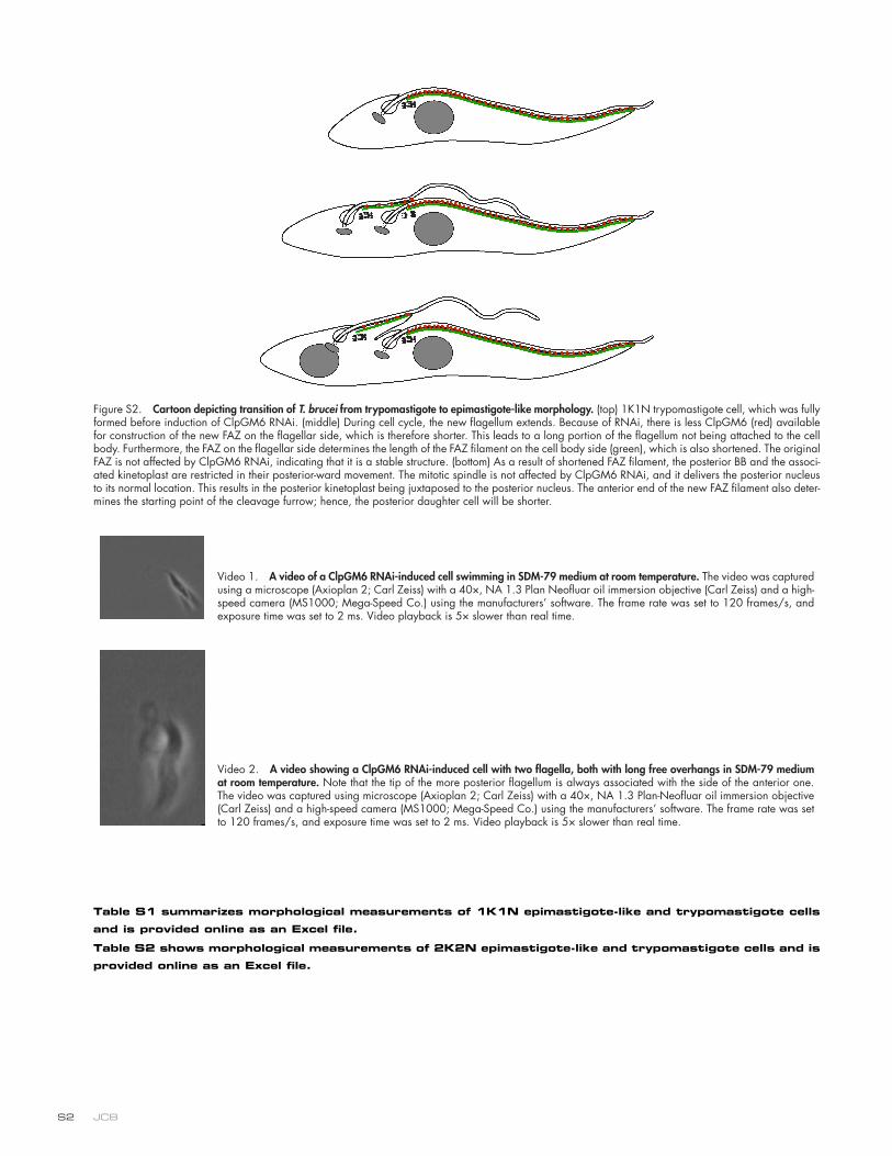
Figure S2. Cartoon depicting transition of T. brucei from trypomastigote to epimastigote-like morphology. (top) 1K1N trypomastigote cell, which was fully formed before induction of ClpGM6 RNAi. (middle) During cell cycle, the new flagellum extends. Because of RNAi, there is less ClpGM6 (red) available for construction of the new FAZ on the flagellar side, which is therefore shorter. This leads to a long portion of the flagellum not being attached to the cell body. Furthermore, the FAZ on the flagellar side determines the length of the FAZ filament on the cell body side (green), which is also shortened. The original FAZ is not affected by ClpGM6 RNAi, indicating that it is a stable structure. (bottom) As a result of shortened FAZ filament, the posterior BB and the associ-ated kinetoplast are restricted in their posterior-ward movement. The mitotic spindle is not affected by ClpGM6 RNAi, and it delivers the posterior nucleus to its normal location. This results in the posterior kinetoplast being juxtaposed to the posterior nucleus. The anterior end of the new FAZ filament also deter-mines the starting point of the cleavage furrow; hence, the posterior daughter cell will be shorter.
Video 1. A video of a ClpGM6 RNAi-induced cell swimming in SDM-79 medium at room temperature. The video was captured using a microscope (Axioplan 2; Carl Zeiss) with a 40×, NA 1.3 Plan Neofluar oil immersion objective (Carl Zeiss) and a high-speed camera (MS1000; Mega-Speed Co.) using the manufacturers’ software. The frame rate was set to 120 frames/s, and exposure time was set to 2 ms. Video playback is 5× slower than real time.
Video 2. A video showing a ClpGM6 RNAi-induced cell with two flagella, both with long free overhangs in SDM-79 medium at room temperature. Note that the tip of the more posterior flagellum is always associated with the side of the anterior one. The video was captured using microscope (Axioplan 2; Carl Zeiss) with a 40×, NA 1.3 Plan-Neofluar oil immersion objective (Carl Zeiss) and a high-speed camera (MS1000; Mega-Speed Co.) using the manufacturers’ software. The frame rate was set to 120 frames/s, and exposure time was set to 2 ms. Video playback is 5× slower than real time.
Table S1 summarizes morphological measurements of 1K1N epimastigote-like and trypomastigote cells
and is provided online as an Excel file.
Table S2 shows morphological measurements of 2K2N epimastigote-like and trypomastigote cells and is
provided online as an Excel file.