Suppl Fig1 1012-10
21
Supplementary Appendix This appendix has been provided by the authors to give readers additional information about their work. Supplement to: Hara Y, Balci-Hayta B, Yoshida-Moriguchi T, et al. A dystroglycan mutation associated with limb- girdle muscular dystrophy. N Engl J Med 2011;364:939-46.
Transcript of Suppl Fig1 1012-10

Supplementary Appendix
This appendix has been provided by the authors to give readers additional information about their work.
Supplement to: Hara Y, Balci-Hayta B, Yoshida-Moriguchi T, et al. A dystroglycan mutation associated with limb-girdle muscular dystrophy. N Engl J Med 2011;364:939-46.

SUPPLEMENTARY METHODS
Additional clinical information
Clinical data on this patient were reported previously14. Briefly, the onset of disease
occurred at approximately three years of age, shortly after the patient started to walk.
Her initial difficulties were an unsteady gait and difficulty climbing stairs. She has mild
calf enlargement and ankle contractures, as well as increased lumbar lordosis. The
patient was independently ambulant, but only for short distances (25-30 meters) at 16
years of age. Her intellectual development has been slow: she said her first few words at
7 years of age, and she used only two-word sentences at 16 years of age. At age 16 her IQ
was 50, and she was unable to count money and perform independent activities. The
creatine kinase concentration at the age of 15 was 4133 U/L. Her cranial MRI was
normal.
Cell culture
The dystroglycan-/- myoblast cell line was established as previously described26. Briefly,
Dystroglycanflox/flox mice were crossed with H-2Kb-tsA58 transgenic mice26,27, and limb
muscles from E18.5 dystroglycanflox/flox; H-2Kb-tsA58 embryos were dissociated with
0.2% trypsin and 0.01% DNase. Cells were resuspended in growth medium (DMEM, 1
mM glutamine, 4.5 mg/ml glucose, 10% FBS, 10% horse serum, 0.5% chick embryo
extract (Sera Laboratories Inc.), and gentamycin), supplemented with 20 U/ml of
recombinant mouse interferon-γ (Sigma-Aldrich, I4777, St. Louis, MO) and pre-plated
for 20 min at 33°C. Non-adherent cells were transferred to a Matrigel-coated tissue
culture dish (BD Falcon, San Jose, CA) and maintained in growth medium at 33°C/10%
CO2. Dystroglycanflox/flox myoblasts that had been screened for their ability to
differentiate into myotubes were infected with a pBabe-Cre retroviral vector, and

dystroglycan-/- myoblasts were identified by PCR. TSA201, a transformed human kidney
cell line stably expressing an SV40 T antigen, was cultured as previously described15.
Antibodies
The monoclonal antibody to the glycosylated form of α-dystroglycan (IIH6), as well as
the polyclonal antibody to β-dystroglycan (βDG; ap83), that were used had been
characterized previously3. The CORE antibody (sheep5, against the α-dystroglycan core
protein) is from sheep antiserum raised against the whole dystrophin-glycoprotein
complex and had also been characterized previously3. Anti-laminin (L9393), and anti-
myc tag (clone 4A6) antibodies were purchased from Sigma-Aldrich (St. Louis, MO),
and Millipore (Billerica, MA), respectively. Biotinylated anti-human IgG was obtained
from Vector Laboratories (Burlingame, CA). An antibody against the Laminin α1 chain
(clone 317) was a kind gift from Dr. Lydia Sorokin (Münster University, Münster,
Germany).
Vector construction
The T192M-dystroglycan mutation was introduced into expression vectors encoding
rabbit dystroglycan, using a conventional PCR method. To generate adenoviral vectors
expressing the T192M-dystroglycan mutant, we subcloned a full-length cDNA carrying
the T192M mutation into the HindIII and NotI sites of the vector pAd5RSVK-NpA
(obtained from the University of Iowa Gene Transfer Vector Core). Adenoviral vectors
were generated as described elsewhere15. Construction of expression vectors encoding
Fc2 (the dystroglycan N-terminus) or Fc5 (α-dystroglycan) were described elsewhere15.
Biochemical and ligand-binding analyses
Glycoprotein enrichment and Western blotting were performed as described previously,
with minor modifications3,15.

For the cell-surface biotinylation assay, myoblasts infected by adenoviruses
expressing WT- or T192M-dystroglycan were washed three times with ice-cold PBS (-)
and incubated with PBS (-) containing the membrane impermeable sulfo-NHS-LC-biotin
reagent (PIERCE, Rockford, IL) for one hour at 4°C. The cells were then washed twice
at room temperature with PBS (-) containing 100 mM glycine, to quench the cross-linker
and remove excess biotin. Cells were rinsed in PBS (-) and lysed in Buffer A (150 mM
NaCl, 50 mM Tris, pH 7.5) plus 1% Triton X-100 containing a cocktail of protease
inhibitors. Cell-surface proteins were immunoprecipitated from the biotin-labeled cells
using ImmunoPure Immobilized Streptavidin (PIERCE, Rockford, IL). The samples
were then analyzed by Western blotting with the IIH6, CORE, and βDG antibodies.
For the pull-down assay, fusion proteins encoding full-length α-dystroglycan and
the α-dystroglycan N-terminus bearing the T192M mutation and attached to Fc (Fc5 and
Fc2, respectively) were expressed independently in TSA201 cells and purified from the
cell lysates using Protein-A affinity beads15. The affinity bead-DGFc fusion protein
complexes were then incubated with lysates from TSA201 cells expressing myc-tagged
LARGE28. Materials bound to the beads were eluted with Laemmli sample buffer and
analyzed by Western blotting. Binding between dystroglycan and LARGE was detected
using the Odyssey infrared imaging system (LI-COR Biosciences, Lincoln, NE).
Ligand overlay and laminin solid-phase assays were performed as previously
described3,15,29.
To analyze laminin clustering, dystroglycan-null myoblast cell-line was infected
with adenoviral vectors, at an MOI of 1000, in growth medium. At 24 hours post-
infection, cells were seeded onto 8-well glass slides (BD biosciences, San Jose, CA)
coated with fibronectin (Sigma-Aldrich, St. Louis, MO). After an additional 24 hours of
incubation, the culture medium was replaced with fresh medium containing 7.5 nM
mouse EHS Laminin-I (Invitrogen, Carlsbad, CA) and the cells were incubated in this
medium for 16 hrs. After fixation (4% paraformaldehyde) and blocking, the cells were
co-stained with the anti-Laminin α1 chain (clone No. 317) and CORE antibodies.
Confocal microscopy was performed images were analyzed using FV10 ver-1.5
(Olympus, Center Valley, PA). Data show the mean ± s.e.m. for three independent
experiments.
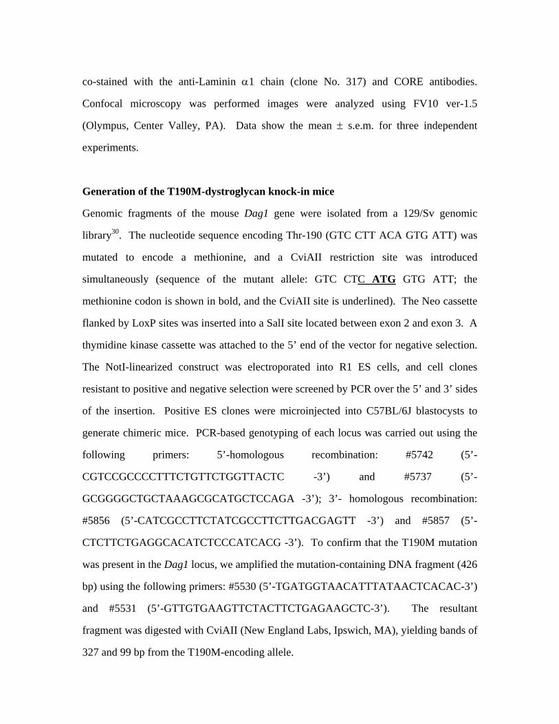
Generation of the T190M-dystroglycan knock-in mice
Genomic fragments of the mouse Dag1 gene were isolated from a 129/Sv genomic
library30. The nucleotide sequence encoding Thr-190 (GTC CTT ACA GTG ATT) was
mutated to encode a methionine, and a CviAII restriction site was introduced
simultaneously (sequence of the mutant allele: GTC CTC ATG GTG ATT; the
methionine codon is shown in bold, and the CviAII site is underlined). The Neo cassette
flanked by LoxP sites was inserted into a SalI site located between exon 2 and exon 3. A
thymidine kinase cassette was attached to the 5’ end of the vector for negative selection.
The NotI-linearized construct was electroporated into R1 ES cells, and cell clones
resistant to positive and negative selection were screened by PCR over the 5’ and 3’ sides
of the insertion. Positive ES clones were microinjected into C57BL/6J blastocysts to
generate chimeric mice. PCR-based genotyping of each locus was carried out using the
following primers: 5’-homologous recombination: #5742 (5’-
CGTCCGCCCCTTTCTGTTCTGGTTACTC -3’) and #5737 (5’-
GCGGGGCTGCTAAAGCGCATGCTCCAGA -3’); 3’- homologous recombination:
#5856 (5’-CATCGCCTTCTATCGCCTTCTTGACGAGTT -3’) and #5857 (5’-
CTCTTCTGAGGCACATCTCCCATCACG -3’). To confirm that the T190M mutation
was present in the Dag1 locus, we amplified the mutation-containing DNA fragment (426
bp) using the following primers: #5530 (5’-TGATGGTAACATTTATAACTCACAC-3’)
and #5531 (5’-GTTGTGAAGTTCTACTTCTGAGAAGCTC-3’). The resultant
fragment was digested with CviAII (New England Labs, Ipswich, MA), yielding bands of
327 and 99 bp from the T190M-encoding allele.

Analysis of the T190M-dystroglycan knock-in mice
Immunofluorescence analysis and hematoxylin-eosin staining were carried out as
described previously3. For the fore-limb grip-strength test, T190M (n=10) and littermate
control (WT; n=9) mice at 11-20 weeks of age were examined using a grip-strength meter
(Columbus Instruments, Columbus, OH, model #1027). This test was repeated five
consecutive times within the same session, and the means of all trials were recorded. For
analysis of NMJs with Alexa488-labelled α-bungarotoxin, diaphragm sections (30-40
μm) or whole diaphragm samples were prepared from adult T190M knock-in mice or
their littermate controls. Those samples were fixed with 4% paraformaldehyde in PBS
(sections: for 20 min; whole diaphragm: for three hours), and permeabilized with 0.5%
Triton X-100 in PBS for 10 min on ice. After blocking with 3% BSA in PBS, the
sections were incubated with Alexa488-labeled α-bungarotoxin (Invitrogen, Carlsbad,
CA) overnight at 4°C. Images were taken using a confocal microscope (FV10 ver-1.5,
Olympus, Center Valley, PA), and analyzed using the Image Pro plus program (Media
Cybernetics, Bethesda, MD). For Evan’s blue dye (EBD) uptake assay, adult T190M
knock-in mice and littermate controls were injected with EBD one day before exercise.
Those mice were subjected to downhill running on a treadmill with a built-in shock grid
at a 15 degree declination. After a warm-up running (3 m/min, 5 minutes), the initial
running speed of 10 m/min was increased every 5 minutes by 5 m/min until the maximal
speed (25 m/min) was reached. Exhaustion was defined as the point at which the animal
would not resume running. Tissues were harvested at one day after the exercise. In
evaluating cardiac muscle pathology, we used skeletal and cardiac muscle-specific
(MCK-cre) dystrolgycan-deficient mice27 as a control. HFaq treatment and an IMAC
bead binding assay were performed as described previously12. Rotarod performance was
tested in T190M knock-in mice and littermates, 10 to 15 weeks of age, using the
ROTOR-ROD system (San Diego Instruments, San Diego, CA). The mice were placed
on top of the beam, and the rotarod accelerated gradually, without jerks, from 0 to 35 rpm

over a two-minute trial. Latencies for the mice to fall from the rod were recorded
automatically. Each mouse was subjected to 5 trials at 15-min intertrial intervals, on each
of three consecutive days.
Molecular modeling
The N-terminal portion of the T192M mutant of α-dystroglycan was modeled using the
SWISS-MODEL program for the analysis, and the crystal structure of the WT mouse
orthologue as a template (Ref. 23, PDB accession No. 1u2c). The figure was prepared
using the program PyMOL v-0.99 (DeLano, W.L. The PyMOL Molecular Graphics
System (2002) DeLano Scientific, San Carlos, CA, http://www.pymol.org).
Author contributions
Y.H., P.D. and K.P.C. designed the study. B.B., H.G., B.T., F.M., H.T. and P.D.
diagnosed patients, collected blood from the patient, and analyzed genetic data. S.J.B.
provided the dystroglycan-null myoblast cell line from our skeletal muscle-specific
conditional knockout mice. Y.H. and M.K. performed biochemical studies, with the
assistance of T.Y.-M., T.W., S.K. and M.B.A.O. D.B., T.Y.-M., T.W., J.S.S., S.K.,
M.B.A.O. and F.M. provided critical discussion on the research. Y.H. and R.W.C.
performed the mouse studies. Y.H. and A.A. performed the structural modeling studies.
K.P.C supervised and mentored all work. Y.H. and K.P.C. wrote the initial manuscript,
and all authors contributed to the final version of the manuscript.
SUPPLEMENTARY REFERENCES
26. Herbst R, Burden SJ. The juxtamembrane region of MuSK has a critical role in
agrin-mediated signaling. Embo J 2000;19:67-77. 27. Cohn RD, Henry MD, Michele DE, et al. Disruption of Dag1 in differentiated
skeletal muscle reveals a role for dystroglycan in muscle regeneration. Cell 2002;110:639-48.
28. Rojek JM, Campbell KP, Oldstone MB, Kunz S. Old World arenavirus infection
interferes with the expression of functional alpha-dystroglycan in the host cell. Mol Biol Cell 2007;18:4493-507.
29. Sugita S, Saito F, Tang J, Satz J, Campbell K, Sudhof TC. A stoichiometric
complex of neurexins and dystroglycan in brain. J Cell Biol 2001;154:435-45. 30. Williamson RA, Henry MD, Daniels KJ, et al. Dystroglycan is essential for early
embryonic development: disruption of Reichert's membrane in Dag1-null mice. Hum Mol Genet 1997;6:831-41.
A
C
Con
trol
WT-
DG
T192
M
Lm-p
ositi
ve c
ells
(%)
0
10
20
30
40
50
*
BControl + WT-DG + T192M-DG
LamininCORE
Campbell, Supplementary Figure 1
T192M
-DG
DG (+/+)
WT-D
G
Contro
l
T192M
-DG
DG (+/+)
WT-D
G
Contro
l
207
114
78
53
35
28
207
114
78
53
53
35
IIH6 CORE (upper)βDG (lower)
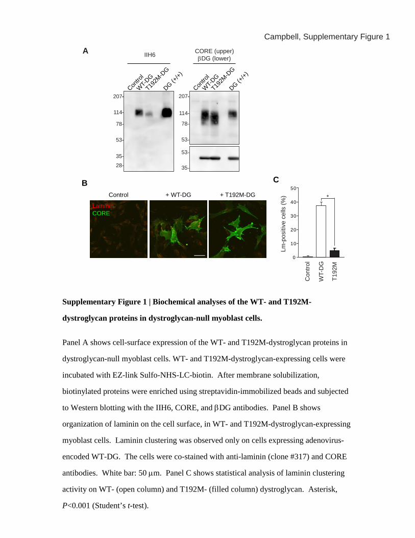
Supplementary Figure 1 | Biochemical analyses of the WT- and T192M-
dystroglycan proteins in dystroglycan-null myoblast cells.
Panel A shows cell-surface expression of the WT- and T192M-dystroglycan proteins in
dystroglycan-null myoblast cells. WT- and T192M-dystroglycan-expressing cells were
incubated with EZ-link Sulfo-NHS-LC-biotin. After membrane solubilization,
biotinylated proteins were enriched using streptavidin-immobilized beads and subjected
to Western blotting with the IIH6, CORE, and βDG antibodies. Panel B shows
organization of laminin on the cell surface, in WT- and T192M-dystroglycan-expressing
myoblast cells. Laminin clustering was observed only on cells expressing adenovirus-
encoded WT-DG. The cells were co-stained with anti-laminin (clone #317) and CORE
antibodies. White bar: 50 μm. Panel C shows statistical analysis of laminin clustering
activity on WT- (open column) and T192M- (filled column) dystroglycan. Asterisk,
P<0.001 (Student’s t-test).

WT
T190M
426-327-
99-
(bp)WT allele
T190M allele
190 GTC CTT ACA GTG ATT V L T V I
GTC CTC ATG GTG ATT V L M V I
B C
T190M
SS,E
N
Nc E
E
WT allele
Targeting vector
T190M allele
E
EE E S
EE E
Ex.2 Ex.3
SS,Etk neo
neo
A
Campbell, Supplementary Figure 2
Supplementary Figure 2 | Generation of a mouse model harboring the T190M
mutation.
Panel A shows a schematic representation of the WT Dag1 allele, the targeting vector,
and the homologously recombined allele. The open circle in exon 3 indicates T190M,
which corresponds to the human T192M mutation. The thymidine kinase and Neo
cassettes are illustrated as boxed tk and Neo, respectively. Filled arrowheads flanking the
Neo cassette indicate loxP sites. Selected restriction enzyme cleavage sites are indicated
above the gene (E: EcoRI; N: NotI; Nc: NciI; and S: SalI). Panel B shows engineered
sequence abnormalities in the T190M knock-in mice, with nucleotide sequence shown at
top and amino acid sequence shown at bottom. The knock-in sequence includes a
recognition site for the restriction enzyme CviAII (marked by arrow). Panel C shows
representive PCR genotyping analysis of the T190M-homozygous (T190M) mice. PCR
products containing the T190M mutation are cleaved by CviAII.
AWT T190M
Campbell, Supplementary Figure 3
B Brain
Rel
ativ
e bi
ndin
g
0
0.1
0.2
0 5 10Laminin (nM)
0
0.2
0.4
0.6
Laminin (nM)
SkM
WT
T190M
0 5 10
WT
T190M
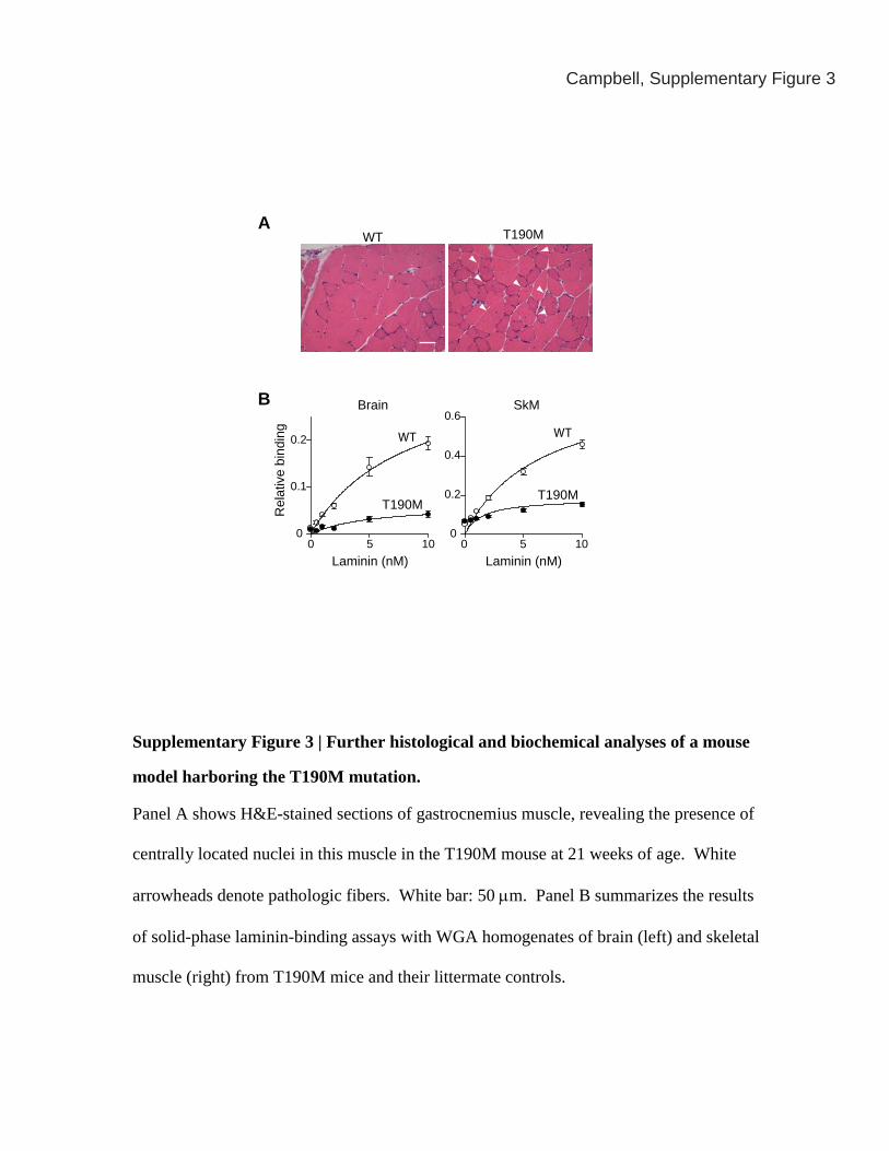
Supplementary Figure 3 | Further histological and biochemical analyses of a mouse
model harboring the T190M mutation.
Panel A shows H&E-stained sections of gastrocnemius muscle, revealing the presence of
centrally located nuclei in this muscle in the T190M mouse at 21 weeks of age. White
arrowheads denote pathologic fibers. White bar: 50 μm. Panel B summarizes the results
of solid-phase laminin-binding assays with WGA homogenates of brain (left) and skeletal
muscle (right) from T190M mice and their littermate controls.

WT T190MHet
Campbell, Supplementary Figure 4
Supplementary Figure 4 | Histological analysis of one-year-old T190M mice.
H&E staining of wild-type (WT), heterozygous (Het), and T190M iliopsoas muscle taken
from one-year-old mice. Myofibers with centrally localized nuclei were only observed in
the homozygous mouse. Scale bar: 50 μm.

Campbell, Supplementary Figure 5
WT
T190M
250
150
100
75
50
Neurexin overlay
Brain Sk. muscle
WT
T190M
Supplementary Figure 5 | Neurexin overlay assay with WT- and T190M-
dystroglycan
Representative image of neurexin overlay assay with WT- and T190M-dystroglycan.
The membrane of WGA-enriched fractions prepared from WT and T190M mice were
subject to the ligand overlay assay, using neurexin-immunoglobulin fusion protein3,29.
Neurexin binding activity was significantly reduced on T190M-dystroglycan.
250
150
100
75
HetWT
T190M
Brain Sk. muscle
HetWT
T190M
HetWT
T190M
HetWT
T190M
IIH6 Laminin overlay
HetWT
T190M
HetWT
T190M
CORE & βDG
Brain Sk. muscle
Brain Sk. muscle
250
150
100
75
250
150
100
75
50
37
HetWT
T190M
200
150
100
50
0
A
B
Grip
stre
ngth
(g)
Campbell, Supplementary Figure 6
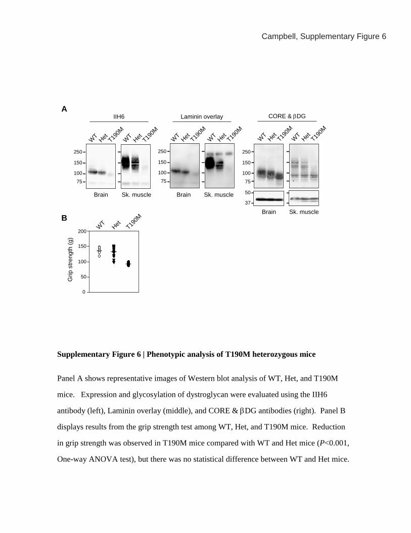
Supplementary Figure 6 | Phenotypic analysis of T190M heterozygous mice
Panel A shows representative images of Western blot analysis of WT, Het, and T190M
mice. Expression and glycosylation of dystroglycan were evaluated using the IIH6
antibody (left), Laminin overlay (middle), and CORE & βDG antibodies (right). Panel B
displays results from the grip strength test among WT, Het, and T190M mice. Reduction
in grip strength was observed in T190M mice compared with WT and Het mice (P<0.001,
One-way ANOVA test), but there was no statistical difference between WT and Het mice.
0 20 40 60 80 100%
WT
T190M
Normal
Immature
Fragmented
WT T190MA
B
Campbell, Supplementary Figure 7
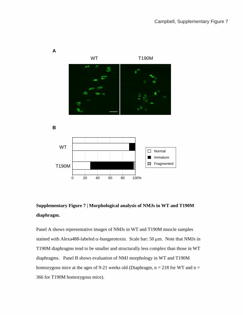
Supplementary Figure 7 | Morphological analysis of NMJs in WT and T190M
diaphragm.
Panel A shows representative images of NMJs in WT and T190M muscle samples
stained with Alexa488-labeled α-bungarotoxin. Scale bar: 50 μm. Note that NMJs in
T190M diaphragms tend to be smaller and structurally less complex than those in WT
diaphragms. Panel B shows evaluation of NMJ morphology in WT and T190M
homozygous mice at the ages of 9-21 weeks old (Diaphragm, n = 218 for WT and n =
366 for T190M homozygous mice).

WT T190M
Campbell, Supplementary Figure 8
Supplementary Figure 8 | The T190M mouse model displays neurological
phenotypes.
Top panels show representative WT (left) and T190M (right) mice suspended by the tail.
Only mutant mice displayed a clasping phenotype (T190M: 13 out of 19 mice). Middle
panels show gross structures of the control and T190M mutant brains. No structural
abnormality was evident in the mutant mice. Bottom panels show histological sections of
cerebral cortices of WT and T190M mice. H&E staining of the brain sections reveals
that neuronal migration in the T190M brain was normal. White bar: 200 μm.
H&E
WT T190M Dystroglycan-null
Campbell, Supplementary Figure 9
EBD
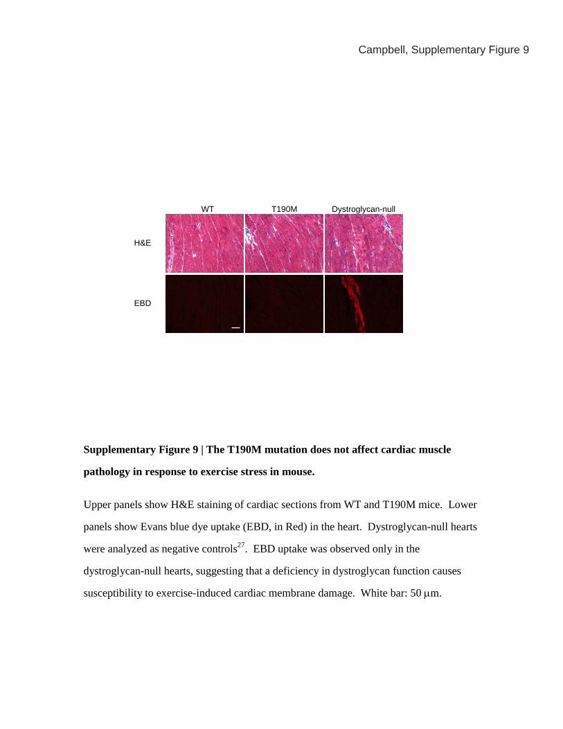
Supplementary Figure 9 | The T190M mutation does not affect cardiac muscle
pathology in response to exercise stress in mouse.
Upper panels show H&E staining of cardiac sections from WT and T190M mice. Lower
panels show Evans blue dye uptake (EBD, in Red) in the heart. Dystroglycan-null hearts
were analyzed as negative controls27. EBD uptake was observed only in the
dystroglycan-null hearts, suggesting that a deficiency in dystroglycan function causes
susceptibility to exercise-induced cardiac membrane damage. White bar: 50 μm.
250
150
10075
50
37
25
250
150
10075
50
37
25
Campbell, Supplementary Figure 10
WT T190M myd WT T190M myd
Bound
Void Bound
Void Bound
Void Bound
Void Bound
Void Bound
Void
IIH6 CORE
Bound
Void Bound
Void Bound
Void
WT T190M myd WT T190M myd
Bound
Void Bound
Void Bound
Void
IIH6 CORE
250
150
10075
50
37
25
250
150
10075
50
37
25
HFaq. - + - +
WT T190M
CORE
A B
198
116
84
53
3729
(kDa)
C
Campbell, Supplementary Figure 10
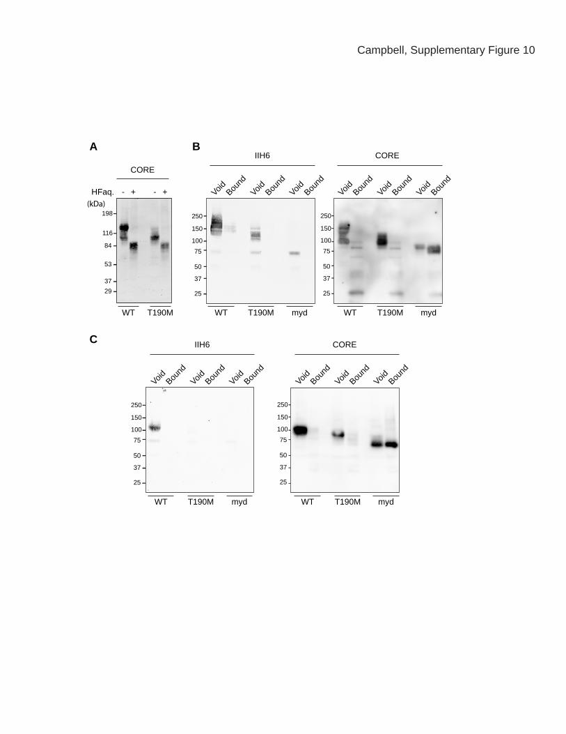
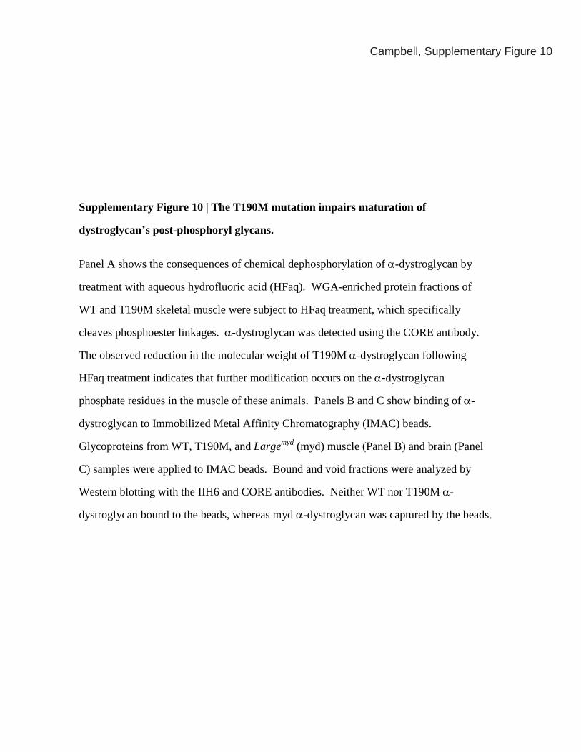
Supplementary Figure 10 | The T190M mutation impairs maturation of
dystroglycan’s post-phosphoryl glycans.
Panel A shows the consequences of chemical dephosphorylation of α-dystroglycan by
treatment with aqueous hydrofluoric acid (HFaq). WGA-enriched protein fractions of
WT and T190M skeletal muscle were subject to HFaq treatment, which specifically
cleaves phosphoester linkages. α-dystroglycan was detected using the CORE antibody.
The observed reduction in the molecular weight of T190M α-dystroglycan following
HFaq treatment indicates that further modification occurs on the α-dystroglycan
phosphate residues in the muscle of these animals. Panels B and C show binding of α-
dystroglycan to Immobilized Metal Affinity Chromatography (IMAC) beads.
Glycoproteins from WT, T190M, and Largemyd (myd) muscle (Panel B) and brain (Panel
C) samples were applied to IMAC beads. Bound and void fractions were analyzed by
Western blotting with the IIH6 and CORE antibodies. Neither WT nor T190M α-
dystroglycan bound to the beads, whereas myd α-dystroglycan was captured by the beads.

Fc5 *
Fc2 *
Fc5 T19
2M
Fc5 W
T
Fc2 T19
2M
Fc2 W
T
Fc
199
11697
50
N-terminal mucin-like C-terminalSP
α-dystroglycan β-dystroglycan
291 316 485 653 895
Full lengthDystroglycan
B
C
FcN
orm
aliz
ed b
indi
ng (%
)
100
50
0
Fc2 Fc5* *
Campbell, Supplementary Figure 11
WT Mouse T190MA
T192
MW
T
T192
MW
Tα-myc Ab

Campbell, Supplementary Figure 11(Continued)
Supplementary Figure 11 | The T192M mutation disrupts the molecular interaction
between dystroglycan and LARGE.
Panel A shows models of the RNA binding protein-like domain of the mouse
dystroglycan N-terminus, based on the crystal structure of the WT dystroglycan N-
terminus23. The model of the WT protein is shown on the left, and an in silico model of
the mouse T190M mutant is shown on the right. Thr-190 and the mutated residues are
indicated in yellow. The domain is displayed in surface representation, with ribbon
diagrams superimposed (cyan). The crystal structure of the WT mouse dystroglycan N-
terminus shows that Thr-190 is located in the middle of a cleft, with its side chain
exposed23. In the in-silico model of the mouse T190M mutant protein, the overall fold is
conserved but the bulky methionine side chain protrudes into the cleft, partially occluding
it. Panel B shows a schematic representation of the α-dystroglycan:Fc fusion proteins.
Fc5 (α-dystroglycan-Fc) and Fc2 (the dystroglycan N-terminus-Fc) were used for pull-
down assays with LARGE. The position of residue Thr-192 is indicated by an asterisk.
Panel C demonstrates that the dystroglycan N-terminus interacts molecularly with
LARGE. (Left) A representative result from the pull-down assay, carried out with myc-
tagged LARGE and an anti-myc antibody. (Right) Quantification of LARGE-binding
activity by WT (open columns) and mutant (T192M, filled columns) dystroglycan-Fc
proteins. Asterisk, P<0.001 (Student’s t-test).


![1012 winglee[1]](https://static.fdocuments.us/doc/165x107/55842288d8b42a79568b4683/1012-winglee1.jpg)