
SUMMARY BENCHMARKS FOR PREFERRED PRACTICE …
32
© 2016 American Academy of Ophthalmology SUMMARY BENCHMARKS FOR PREFERRED PRACTICE PATTERN® GUIDELINES TABLE OF CONTENTS Summary Benchmarks for Preferred Practice Pattern Guidelines Introduction 1 Glaucoma Primary Open-Angle Glaucoma (Initial Evaluation) 3 Primary Open-Angle Glaucoma (Follow-up Evaluation) 4 Primary Open-Angle Glaucoma Suspect (Initial and Follow-up Evaluation) 5 Primary Angle Closure (Initial Evaluation and Therapy) 6 Retina Age-Related Macular Degeneration (Initial and Follow-up Evaluation) 7 Age-Related Macular Degeneration (Management Recommendations) 8 Diabetic Retinopathy (Initial and Follow-up Evaluation) 9 Diabetic Retinopathy (Management Recommendations) 10 Idiopathic Epiretinal Membrane and Vitreomacular Traction (Initial Evaluation and Therapy) 11 Idiopathic Macular Hole (Initial Evaluation and Therapy) 12 Posterior Vitreous Detachment, Retinal Breaks, and Lattice Degeneration (Initial and Follow-up Evaluation) 13 Retinal and Ophthalmic Artery Occlusions (Initial Evaluation and Therapy) 14 Retinal Vein Occlusions (Initial Evaluation and Therapy) 15 Cataract/Anterior Segment Cataract (Initial and Follow-up Evaluation) 16 Cornea/External Disease Bacterial Keratitis (Initial Evaluation) 18 Bacterial Keratitis (Management Recommendations) 19 Blepharitis (Initial and Follow-up Evaluation) 20 Conjunctivitis (Initial Evaluation) 21 Conjunctivitis (Management Recommendations) 22 Corneal Ectasia (Initial Evaluation and Follow-up) 23 Corneal Edema and Opacification (Initial Evaluation) 24 Corneal Edema and Opacification (Management Recommendations) 25 Dry Eye Syndrome (Initial Evaluation) 26 Dry Eye Syndrome (Management Recommendations) 27 Pediatric Ophthalmology/Strabismus Amblyopia (Initial and Follow-up Evaluation) 28 Esotropia (Initial and Follow-up Evaluation) 29 Exotropia (Initial and Follow-up Evaluation) 30 Refractive Management/Intervention Keratorefractive Surgery (Initial and Follow-up Evaluation) 31 October 2016 aaoorg
Transcript of SUMMARY BENCHMARKS FOR PREFERRED PRACTICE …

© 2016 American Academy of Ophthalmology
SUMMARY BENCHMARKS FOR PREFERRED PRACTICE PATTERN® GUIDELINES
TABLE OF CONTENTS
Summary Benchmarks for Preferred Practice Pattern GuidelinesIntroduction . . . . . . . . . . . . . . . . . . . . . . . . . . . . . . . . . . . . . . . . . . . . . . . . . . . . . . . . . . . . . . . . . . . . . . . . . . . . . . . . . . . . . . . . . . . . . . . . . . . . 1
GlaucomaPrimary Open-Angle Glaucoma (Initial Evaluation) . . . . . . . . . . . . . . . . . . . . . . . . . . . . . . . . . . . . . . . . . . . . . . . . . . . . . . . . . . . . . . . 3Primary Open-Angle Glaucoma (Follow-up Evaluation) . . . . . . . . . . . . . . . . . . . . . . . . . . . . . . . . . . . . . . . . . . . . . . . . . . . . . . . . . . . 4Primary Open-Angle Glaucoma Suspect (Initial and Follow-up Evaluation) . . . . . . . . . . . . . . . . . . . . . . . . . . . . . . . . . . . . . . . . 5Primary Angle Closure (Initial Evaluation and Therapy) . . . . . . . . . . . . . . . . . . . . . . . . . . . . . . . . . . . . . . . . . . . . . . . . . . . . . . . . . . . 6
RetinaAge-Related Macular Degeneration (Initial and Follow-up Evaluation) . . . . . . . . . . . . . . . . . . . . . . . . . . . . . . . . . . . . . . . . . . . . . 7Age-Related Macular Degeneration (Management Recommendations) . . . . . . . . . . . . . . . . . . . . . . . . . . . . . . . . . . . . . . . . . . . . 8Diabetic Retinopathy (Initial and Follow-up Evaluation) . . . . . . . . . . . . . . . . . . . . . . . . . . . . . . . . . . . . . . . . . . . . . . . . . . . . . . . . . . . 9Diabetic Retinopathy (Management Recommendations) . . . . . . . . . . . . . . . . . . . . . . . . . . . . . . . . . . . . . . . . . . . . . . . . . . . . . . . . 10Idiopathic Epiretinal Membrane and Vitreomacular Traction (Initial Evaluation and Therapy) . . . . . . . . . . . . . . . . . . . . . . . . 11Idiopathic Macular Hole (Initial Evaluation and Therapy) . . . . . . . . . . . . . . . . . . . . . . . . . . . . . . . . . . . . . . . . . . . . . . . . . . . . . . . . . . 12Posterior Vitreous Detachment, Retinal Breaks, and Lattice Degeneration (Initial and Follow-up Evaluation) . . . . . . . . . . . . . . . . . . . . . . . . . . . . . . . . . . . . . . . . . . . . . . . . . . . . . . . . . . . . . . . . . . . . . . . . . . . 13Retinal and Ophthalmic Artery Occlusions (Initial Evaluation and Therapy) . . . . . . . . . . . . . . . . . . . . . . . . . . . . . . . . . . . . . . . 14Retinal Vein Occlusions (Initial Evaluation and Therapy) . . . . . . . . . . . . . . . . . . . . . . . . . . . . . . . . . . . . . . . . . . . . . . . . . . . . . . . . . . 15
Cataract/Anterior SegmentCataract (Initial and Follow-up Evaluation) . . . . . . . . . . . . . . . . . . . . . . . . . . . . . . . . . . . . . . . . . . . . . . . . . . . . . . . . . . . . . . . . . . . . . . . 16
Cornea/External DiseaseBacterial Keratitis (Initial Evaluation) . . . . . . . . . . . . . . . . . . . . . . . . . . . . . . . . . . . . . . . . . . . . . . . . . . . . . . . . . . . . . . . . . . . . . . . . . . . . 18Bacterial Keratitis (Management Recommendations) . . . . . . . . . . . . . . . . . . . . . . . . . . . . . . . . . . . . . . . . . . . . . . . . . . . . . . . . . . . . . 19Blepharitis (Initial and Follow-up Evaluation) . . . . . . . . . . . . . . . . . . . . . . . . . . . . . . . . . . . . . . . . . . . . . . . . . . . . . . . . . . . . . . . . . . . . 20Conjunctivitis (Initial Evaluation) . . . . . . . . . . . . . . . . . . . . . . . . . . . . . . . . . . . . . . . . . . . . . . . . . . . . . . . . . . . . . . . . . . . . . . . . . . . . . . . . 21Conjunctivitis (Management Recommendations) . . . . . . . . . . . . . . . . . . . . . . . . . . . . . . . . . . . . . . . . . . . . . . . . . . . . . . . . . . . . . . . . 22Corneal Ectasia (Initial Evaluation and Follow-up) . . . . . . . . . . . . . . . . . . . . . . . . . . . . . . . . . . . . . . . . . . . . . . . . . . . . . . . . . . . . . . . 23Corneal Edema and Opacification (Initial Evaluation) . . . . . . . . . . . . . . . . . . . . . . . . . . . . . . . . . . . . . . . . . . . . . . . . . . . . . . . . . . . . 24Corneal Edema and Opacification (Management Recommendations) . . . . . . . . . . . . . . . . . . . . . . . . . . . . . . . . . . . . . . . . . . . . 25Dry Eye Syndrome (Initial Evaluation) . . . . . . . . . . . . . . . . . . . . . . . . . . . . . . . . . . . . . . . . . . . . . . . . . . . . . . . . . . . . . . . . . . . . . . . . . . 26Dry Eye Syndrome (Management Recommendations) . . . . . . . . . . . . . . . . . . . . . . . . . . . . . . . . . . . . . . . . . . . . . . . . . . . . . . . . . . . .27
Pediatric Ophthalmology/StrabismusAmblyopia (Initial and Follow-up Evaluation) . . . . . . . . . . . . . . . . . . . . . . . . . . . . . . . . . . . . . . . . . . . . . . . . . . . . . . . . . . . . . . . . . . . 28Esotropia (Initial and Follow-up Evaluation) . . . . . . . . . . . . . . . . . . . . . . . . . . . . . . . . . . . . . . . . . . . . . . . . . . . . . . . . . . . . . . . . . . . . . 29Exotropia (Initial and Follow-up Evaluation) . . . . . . . . . . . . . . . . . . . . . . . . . . . . . . . . . . . . . . . . . . . . . . . . . . . . . . . . . . . . . . . . . . . . . 30
Refractive Management/InterventionKeratorefractive Surgery (Initial and Follow-up Evaluation) . . . . . . . . . . . . . . . . . . . . . . . . . . . . . . . . . . . . . . . . . . . . . . . . . . . . . . . 31
October 2016 aao .org
© 2016 American Academy of Ophthalmology
IntroductionThese are summary benchmarks for the Academy’s Preferred Practice Pattern® (PPP) guidelines . The Preferred Practice Pattern series of guidelines has been written on the basis of three principles .
• Each Preferred Practice Pattern should be clinically relevant and specific enough to provide useful information to practitioners .
• Each recommendation that is made should be given an explicit rating that shows its importance to the care process .
• Each recommendation should also be given an explicit rating that shows the strength of evidence that supports the recommendation and reflects the best evidence available .
Preferred Practice Patterns provide guidance for the pattern of practice, not for the care of a particular individual. While they should generally meet the needs of most patients, they cannot possibly best meet the needs of all patients . Adherence to these Preferred Practice Patterns will not ensure a successful outcome in every situation . These practice patterns should not be deemed inclusive of all proper methods of care or exclusive of other methods of care reasonably directed at obtaining the best results . It may be necessary to approach different patients’ needs in different ways . The physician must make the ultimate judgment about the propriety of the care of a particular patient in light of all of the circumstances presented by that patient . The American Academy of Ophthalmology is available to assist members in resolving ethical dilemmas that arise in the course of ophthalmic practice .
The Preferred Practice Pattern® guidelines are not medical standards to be adhered to in all individual situations. The Academy specifically disclaims any and all liability for injury or other damages of any kind, from negligence or otherwise, for any and all claims that may arise out of the use of any recommendations or other information contained herein .
For each major disease condition, recommendations for the process of care, including the history, physical exam and ancillary tests, are summarized, along with major recommendations for the care management, follow-up, and education of the patient . For each PPP, a detailed literature search of PubMed and the
Cochrane Library for articles in the English language is conducted . The results are reviewed by an expert panel and used to prepare the recommendations, which are then given a rating that shows the strength of evidence when sufficient evidence exists .
To rate individual studies, a scale based on the Scottish Intercollegiate Guideline Network (SIGN) is used . The definitions and levels of evidence to rate individual studies are as follows:
• I++: High-quality meta-analyses, systematic reviews of randomized controlled trials (RCTs), or RCTs with a very low risk of bias
• I+: Well-conducted meta-analyses, systematic reviews of RCTs, or RCTs with a low risk of bias
• I–: Meta-analyses, systematic reviews of RCTs, or RCTs with a high risk of bias
• II++: High-quality systematic reviews of case-control or cohort studies; high-quality case-control or cohort studies with a very low risk of confounding or bias and a high probability that the relationship is causal
• II+: Well-conducted case-control or cohort studies with a low risk of confounding or bias and a moderate probability that the relationship is causal
• II–: Case-control or cohort studies with a high risk of confounding or bias and a significant risk that the relationship is not causal
• III: Nonanalytic studies (e .g ., case reports, case series)
Recommendations for care are formed based on the body of the evidence . The body of evidence quality ratings are defined by Grading of Recommendations Assessment, Development and Evaluation (GRADE) as follows:
• Good quality (GQ): Further research is very unlikely to change our confidence in the estimate of effect
• Moderate quality (MQ): Further research is likely to have an important impact on our confidence in the estimate of effect and may change the estimate
• Insufficient quality (IQ): Further research is very likely to have an important impact on our confidence in the estimate of effect and is likely to change the estimate; any estimate of effect is very uncertain
SUMMARY BENCHMARKS FOR PREFERRED PRACTICE PATTERN® GUIDELINES
October 2016 aao .org 1
© 2016 American Academy of Ophthalmology
Introduction (continued)
Key recommendations for care are defined by GRADE as follows:
• Strong recommendation (SR): Used when the desirable effects of an intervention clearly outweigh the undesirable effects or clearly do not
• Discretionary recommendation (DR): Used when the trade-offs are less certain—either because of low- quality evidence or because evidence suggests that desirable and undesirable effects are closely balanced
In PPPs prior to 2011, the panel rated recommendations according to its importance to the care process . This “importance to the care process” rating represents care that the panel thought would improve the quality of the patient’s care in a meaningful way . The ratings of importance are divided into three levels .
• Level A, defined as most important
• Level B, defined as moderately important
• Level C, defined as relevant but not critical
The panel also rated each recommendation on the strength of evidence in the available literature to support the recommendation made . The “ratings of strength of evidence” also are divided into three levels .
• Level I includes evidence obtained from at least one properly conducted, well-designed randomized controlled trial . It could include meta-analyses of randomized controlled trials .
• Level II includes evidence obtained from the following:
• Well-designed controlled trials without randomization
• Well-designed cohort or case-control analytic studies, preferably from more than one center
• Multiple-time series with or without the intervention
• Level III includes evidence obtained from one of the following:
• Descriptive studies
• Case reports
• Reports of expert committees/organizations (e .g ., PPP panel consensus with external peer review)
This former approach, however, will eventually be phased out as the AAO adopted the SIGN and GRADE rating and grading systems .
The PPPs are intended to serve as guides in patient care, with greatest emphasis on technical aspects . In applying this knowledge, it is essential to recognize that true medical excellence is achieved only when skills are applied in a such a manner that the patients’ needs are the foremost consideration . The AAO is available to assist members in resolving ethical dilemmas that arise in the course of practice . (AAO Code of Ethics)
SUMMARY BENCHMARKS FOR PREFERRED PRACTICE PATTERN® GUIDELINES
October 2016 aao .org 2
October 2016 aao .org 3© 2016 American Academy of Ophthalmology
GLAUCOMA
Initial Exam History (Key elements)• Ocular history• Race/ethnicity• Family history• Systemic history• Review of pertinent records• Current medications• Ocular surgery
Initial Physical Exam (Key elements)• Visual acuity measurement• Pupil examination• Slit-lamp biomicroscopy of anterior segment• Measurement of IOP• Central corneal thickness• Gonioscopy• Evaluation of optic nerve head and retinal nerve fiber
layer using magnified stereoscopic visualization with slit-lamp biomicroscope and through a dilated pupil (I+, MQ, SR)
• Examination of optic nerve head appearance by color stereophotography or computer-based image analysis should be serially documented (I+, MQ, SR)
• Evaluation of the fundus (through a dilated pupil whenever feasible)
• Visual field evaluation, preferably by automated static threshold perimetry
• Evaluation of the optic disc• Thinning of the inferior and/or superior neuroretinal
rim
Management Plan for Patients in Whom Therapy is Indicated• Set an initial target pressure of at least 25% lower
than pretreatment IOP . Choosing a lower target IOP can be justified if there is more severe optic nerve damage .
• Target pressure is an estimate and must be individualized and/or adjusted during the course of the disease (III, IQ, DR)
• The goal of treatment is to maintain the IOP in a range at which visual field loss is unlikely to significantly reduce a patient’s health-related quality of life over his/her lifetime (II+, MQ, DR)
• Medical therapy is presently the most common initial intervention to lower IOP; consider balance between side effects and effectiveness in choosing a regimen of maximal effectiveness and tolerance to achieve the desired IOP reduction for each patient
• If progression occurs at the target pressure, undetected IOP fluctuations and adherence to therapy should be re-evaluated before adjusting target IOP downward
• Assess the patient who is being treated with glaucoma medication for local ocular and systemic side effects and toxicity
• Laser trabeculoplasty can be considered as initial therapy in selected patients or an alternative for patients at high risk for nonadherence to medical therapy who cannot or will not use medications reliably due to cost, memory problems, difficulty with instillation, or intolerance to medication (I+, GQ, DR)
• Trabeculectomy is effective in lowering IOP; it is gen-erally indicated when medications and appropriate laser therapy are insufficient to control disease and can be considered in selected cases as initial therapy (I+, GQ, SR)
Surgery and Postoperative Care for Laser Trabeculoplasty Patients• The ophthalmologist who performs surgery has the
following responsibilities: - Obtain informed consent - Ensure that the preoperative evaluation confirms
the need for surgery - At least one IOP check within 30 minutes to 2
hours of surgery - Follow-up examination within 6 weeks of surgery
or sooner if concern about IOP-related optic nerve damage
Surgery and Postoperative Care for Incisional Glaucoma Surgery Patients• The ophthalmologist who performs surgery has the
following responsibilities: - Obtain informed consent - Ensure that the preoperative evaluation accurately
documents findings and indications for surgery - Prescribe topical corticosteroids in the
postoperative period - Follow-up evaluation on the first postoperative day
(12 to 36 hours after surgery) and at least once during the first 1 to 2 weeks
- In the absence of complications, perform additional postoperative visits during a 6-week period
- Schedule more frequent visits, as necessary, for patients with postoperative complications
- Additional treatments as necessary to maximize the chances for a successful long-term result
Patient Education for Patients with Medical Therapy• Discuss diagnosis, severity of the disease, prognosis
and management plan, and likelihood of lifelong therapy
• Educate about eyelid closure or nasolacrimal occlusion when applying topical medications to reduce systemic absorption
• Encourage patients to alert their ophthalmologist to physical or emotional changes that occur when taking glaucoma medications
Primary Open-Angle Glaucoma (Initial Evaluation)
October 2016 aao .org 4© 2016 American Academy of Ophthalmology
GLAUCOMAPrimary Open-Angle Glaucoma (Follow-up Evaluation)
Exam History• Interval ocular history
• Interval systemic medical history
• Side effects of ocular medications
• Frequency and time of last IOP-lowering medications, and review of medication use
Physical Exam• Visual acuity measurement
• Slit-lamp biomicroscopy
• Measurement of IOP
• Evaluation of optic nerve head and visual fields (see table below)
• Measurement of central corneal thickness should be repeated after any event that may alter it (e .g ., refractive surgery)
Management Plan For Patients On Medical Therapy• At each exam, record dosage and frequency of use,
discuss adherence to the therapeutic regimen and patient’s response to recommendations for therapeutic alternatives or diagnostic procedures
• Perform gonioscopy if there is a suspicion of angle closure, anterior-chamber shallowing or anterior-chamber angle abnormalities or if there is an unexplained change in IOP . Perform gonioscopy periodically .
• Reassess treatment regimen if target IOP is not achieved and benefits of a change in therapy outweigh the risk
• Adjust target pressure downward if optic disc, retinal nerve fiber layer, or visual field change is progressive
• Within each of the recommended intervals, factors that determine frequency of evaluation include the severity of damage, the rate of progression, the extent to which the IOP exceeds the target pressure and the number and significance of other risk factors for damage to the optic nerve
Patient Education• Educate about the disease process, rationale and
goals of intervention, status of their condition, and relative benefits and risks of alternative interventions so that patients can participate meaningfully in developing an appropriate plan of action
• Refer for or encourage patients with significant visual impairment or blindness to use appropriate vision rehabilitation and social services
• Patients considering keratorefractive surgery should be informed about the possible impact laser vision correction has on reducing contrast sensitivity and decreasing the accuracy of IOP measurements
Follow-Up:
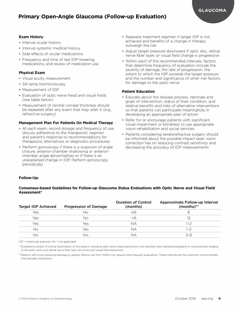
Consensus-based Guidelines for Follow-up Glaucoma Status Evaluations with Optic Nerve and Visual Field Assessment*
Duration of Control Approximate Follow-up Interval Target IOP Achieved Progression of Damage (months) (months)** Yes No 6 6
Yes No 6 12
Yes Yes NA 1–2
No Yes NA 1–2
No No NA 3–6
IOP = intraocular pressure; NA = not applicable
** Evaluations consist of clinical examination of the patient, including optic nerve head assessment (with periodic color stereophotography or computerized imaging of the optic nerve and retinal nerve fiber layer structure) and visual field assessment .
** Patients with more advanced damage or greater lifetime risk from POAG may require more frequent evaluations . These intervals are the maximum recommended time between evaluations .
October 2016 aao .org 5© 2016 American Academy of Ophthalmology
GLAUCOMA
Initial Exam History (Key elements)• Ocular history
• Family history
• Systemic history
• Review of pertinent records
• Current medications
• Ocular surgery
Initial Physical Exam (Key elements)• Visual acuity measurement
• Pupil examination
• Slit-lamp biomicroscopy of anterior segment
• Measurement of IOP
• Central corneal thickness
• Gonioscopy
• Evaluation of optic nerve head and retinal nerve fiber layer using magnified stereoscopic visualization with slit-lamp biomicroscope and through a dilated pupil
• Appearance of the optic nerve head and, if possible, the RNFL should be documented (II++, GQ, SR)
• Evaluation of the fundus (through a dilated pupil whenever feasible)
• Visual field evaluation, preferably by automated static threshold perimetry
• Excavation of the optic cup
• Thinning of the inferior and/or superior neuroretinal rim
Management Plan for Patients in Whom Therapy is Indicated• A reasonable initial goal is to set a target pressure
20% less than mean of several baseline IOP measurements based on criteria from the Ocular Hypertension Study (I+, MQ, DR)
• The goal of treatment is to maintain the IOP in a range at which visual field loss is unlikely to significantly affect a patient’s health related quality of life over his/her lifetime (II+, MQ, DR)
• If visual field glaucomatous damage is newly detected in a glaucoma suspect patient, it is best to repeat testing (II++, GQ, SR)
• Clinicians should include all perimetric and other structural information in addition to digital imaging technology when formulating patient management decisions (III, IQ, SR)
Follow-up Exam History• Interval ocular history
• Interval systemic medical history and any change of systemic medications
• Side effects of ocular medications if patient is being treated
• Frequency and time of last glaucoma medications, and review of use, if patient is being treated
Follow-up Physical Exam• Visual acuity
• Slit-lamp biomicroscopy
• Measurement of IOP
• Gonioscopy is indicated when there is a suspicion of an angle-closure component, anterior chamber shallowing or unexplained change in IOP
Follow-up Intervals• Visit intervals depend on the interaction between
patient and disease, which is unique for every patient
• Frequency of periodic optic nerve head and visual field evaluation is based on risk assessment . Patients with thinner corneas, higher IOPs, disc hemorrhage, larger cup-to-disc, larger mean pattern standard deviation, or family history of glaucoma may warrant closer follow-up .
Patient Education for Patients with Medical Therapy• Discuss diagnosis, number and severity of risk
factors, prognosis, management plan and likelihood that therapy, once started, will be long term
• Educate about disease process, rationale and goals of intervention, status of their condition, and relative benefits and risks of alternative interventions
• Educate about eyelid closure and nasolacrimal occlusion when applying topical medications to reduce systemic absorption
• Encourage patients to alert their ophthalmologist to physical or emotional changes that occur when taking glaucoma medications
Primary Open-Angle Glaucoma Suspect (Initial and Follow-up Evaluation)
October 2016 aao .org 6© 2016 American Academy of Ophthalmology
GLAUCOMAPrimary Angle Closure (Initial Evaluation and Therapy)
Initial Exam History (Key elements)• Ocular history (symptoms suggestive of intermittent
angle-closure attacks)
• Family history of acute angle-closure glaucoma
• Systemic history (e .g ., use of topical or systemic medications)
Initial Physical Exam (Key elements)• Refractive status
• Pupil
• Slit-lamp biomicroscopy
- Conjunctival hyperemia (in acute cases)
- Central and peripheral anterior chamber depth narrowing
- Anterior chamber inflammation suggestive of a recent or current attack
- Corneal swelling . (Microcystic edema and stromal edema are common in acute cases .)
- Iris abnormalities, including diffuse or focal atrophy, posterior synechiae, abnormal pupillary function, irregular pupil shape, and a mid-dilated pupil (suggestive of a recent or current attack)
- Lens changes, including cataract and glaukomflecken
- Corneal endothelial cell loss
• Measurement of IOP
• Gonioscopy and/or anterior segment imaging of both eyes
• Evaluation of fundus and optic nerve head using direct ophthalmoscope or slit-lamp biomicroscope with an indirect lens
Management Plan for Patients in Whom Iridotomy is Indicated• Iridotomy is indicated for eyes with PAC or primary
angle-closure glaucoma (I++, GQ, SR)
• Laser iridotomy is the preferred surgical treatment for acute angle-closure crisis (AACC) because it has a favorable risk-benefit ratio (II+, MQ, SR)
• In AACC, use medical therapy first to lower the IOP to reduce pain and clear corneal edema . Iridotomy should then be performed as soon as possible . (III, GQ, SR)
• Perform prophylactic iridotomy in fellow eye if chamber angle is anatomically narrow, as nearly half of fellow eyes can develop AACC within 5 years (II++, GQ, SR)
Surgery and Postoperative Care for Iridotomy Patients• The ophthalmologist who performs surgery has the
following responsibilities:
- Obtain informed consent
- Ensure that preoperative evaluation confirms the need for surgery
- Perform at least one IOP check immediately prior to surgery and within 30 minutes to 2 hours following surgery
- Prescribe topical cortico steroids in the postoperative period
- Ensure that the patient receives adequate postoperative care
• Follow-up evaluations include:
- Evaluation of patency of iridotomy by visualizing the anterior lens capsule
- Measurement of IOP
- Gonioscopy with compression/indentation, if not performed immediately after iridotomy
- Pupil dilation to reduce risk of posterior synechiae formation
- Fundus examination as clinically indicated
• Prescribe medications perioperatively to avert sudden IOP elevation, particularly in patients with severe disease
Follow-up of Patients with Iridotomy• After iridotomy, follow patients with glaucomatous
optic neuropathy as specified in the Primary Open-Angle Glaucoma PPP
• After iridotomy, patients with a residual open angle or a combination of open angle and some PAS with or without glaucomatous optic neuropathy should be followed at least annually, with special attention to repeat gonioscopy
Education For Patients if Iridotomy is Not Performed• Patients with primary angle-closure suspect who
have not had an iridotomy should be warned that they are at risk for AACC and that certain medications cause pupil dilation and include AACC (III, MQ, DR)
• Patients should be informed about the symptoms of AACC and instructed to notify their ophthalmologist immediately if symptoms occur (III, MQ, SR)
October 2016 aao .org 7© 2016 American Academy of Ophthalmology
RETINAAge-Related Macular Degeneration (Initial and Follow-up Evaluation)
Initial Exam History (Key elements)• Symptoms (metamorphopsia, decreased vision,
scotoma, photopsia, difficulties in dark adaptation) (II–, GQ, SR)
• Medications and nutritional supplements (II+, GQ, SR)
• Ocular history (II+, GQ, SR)
• Systemic history (any hypersensitivity reactions)• Family history, especially family history of AMD
(II+, GQ, SR)
• Social history, especially smoking (III, GQ, SR)
Initial Physical Exam (Key elements)• Comprehensive eye examination (II++, GQ, SR)
• Stereo biomicroscopic examination of the macula (III, GQ, SR)
Diagnostic TestsOptical coherence tomography is important in diagnosing and managing AMD, particularly with respect to determining the presence of subretinal fluid and in documenting the degree of retinal thickening . (III, GQ, SR) Optical coherence tomography defines the cross sectional architecture of the retina in a manner that is not possible with any other imaging technology . It may reveal the presence of fluid that is not apparent on biomicroscopy alone . It also assists in evaluating the response of the retina and RPE to therapy by allowing structural changes to be followed accurately . (II+, GQ, SR)
Intravenous fundus fluorescein angiography in the clinical setting of AMD is indicated:• when patient complains of new metamorphopsia• when patient has unexplained blurred vision• when clinical exam reveals elevation of the RPE
or retina, subretinal blood, hard exudates or subretinal fibrosis (II–, GQ, SR)
• to detect the presence of and determine the extent, type, size, and location of CNV and to calculate the percentage of the lesion composed of or consisting of classic CNV (III, IQ, DR)
• to guide treatment (laser photocoagulation surgery or verteporfin PDT) (III, IQ, DR)
• to detect persistent or recurrent CNV following treatment (III, IQ, DR)
• to assist in determining the cause of visual loss that is not explained by clinical exam (III, IQ, DR)
Each angiographic facility must have a care plan or an emergency plan and a protocol to minimize the risk and manage any complications . (III, GQ, SR)
Follow-up Exam History• Visual symptoms, including decreased vision and
metamorphopsia (II–, GQ, SR)
• Changes in medications and nutritional supplements (III, GQ, SR)
• Changes in ocular history and systemic history (II+, GQ, SR)
• Changes in social history, especially smoking (III, GQ, SR)
Follow-up Physical Exam• Visual acuity (III, GQ, SR)
• Stereo biomicroscopic examination of the fundus (III, GQ, SR)
Follow-up after Treatment for Neovascular AMD• Examine patients treated with intravitreal injections
of aflibercept, bevacizumab, or ranibizumab approximately 4 weeks after treatment (III, GQ, SR)
• Examine and perform fluorescein angiography at least every 3 months until stable after verteporfin PDT
• Examine patients treated with thermal laser photo coagulation via fluorescein angiography approximately 2 to 4 weeks after treatment and then at 4 to 6 weeks (III, GQ, SR)
• Subsequent examinations, OCT, and fluorescein angiography should be performed as indicated depending on the clinical findings and the judgment of the treating ophthalmologist (III, GQ, SR)
Patient Education• Educate patients about the prognosis and potential
value of treatment as appropriate for their visual and functional status (III, GQ, SR)
• Encourage patients with early AMD to assess their own visual acuity and to have regular dilated eye exams for early detection of intermediate AMD
• Educate patients with a high-risk AMD phenotype about methods of detecting new symptoms of CNV and about the need for prompt notification to an ophthalmologist (III, GQ, SR)
• Instruct patients with unilateral disease to monitor their vision in their fellow eye and to return periodically even in absence of symptoms, but promptly after onset of new or significant visual symptoms (III, GQ, SR)
• Instruct patients to report symptoms suggestive of endophthalmitis, including eye pain or increased discomfort, worsening eye redness, blurred or decreased vision, increased sensitivity to light, or increased number of floaters promptly (III, GQ, SR)
• Encourage patients who are currently smoking to stop because there are observational data that support a causal relationship between smoking and AMD and other considerable health benefits of smoking cessation (I++, GQ, SR)
• Refer patients with reduced visual function for vision rehabilitation (see www .aao .org/smart-sight-low-vision) and social services (III, GQ, SR)

October 2016 aao .org 8© 2016 American Academy of Ophthalmology
RETINAAge-Related Macular Degeneration (Management Recommendations)
Treatment Recommendations and Follow-up Plans for Age-Related Macular Degeneration
Recommended TreatmentObservation with no medical or surgical therapies
Antioxidant vitamin and mineral supplements as recommended in the original AREDS and AREDS2 reports
Aflibercept intravitreal injection 2 .0 mg as described in published reports
Bevacizumab intravitreal injection 1 .25 mg as described in published reports
The ophthalmologist should provide appropriate informed consent with respect to the off-label status
Ranibizumab intravitreal injection 0 .5 mg as recommended in ranibizumab literature
PDT with verteporfin as recommended in the TAP and VIP reports
Thermal laser photocoagulation surgery as recommended in the MPS reports
Diagnoses Eligible for TreatmentNo clinical signs of AMD (AREDS category 1)
Early AMD (AREDS category 2)
Advanced AMD with bilateral subfoveal geographic atrophy or disciform scars
Intermediate AMD (AREDS category 3)
Advanced AMD in one eye (AREDS category 4)
Macular CNV
Macular CNV
Macular CNV
Macular CNV, new or recurrent, where the classic component is >50% of the lesion and the entire lesion is 5400 microns in greatest linear diameter
Occult CNV may be considered for PDT with vision <20/50 or if the CNV is <4 MPS disc areas in size when the vision is >20/50
Juxtafoveal CNV is an off-label indication for PDT, but may be considered in select cases .
May be considered for extrafoveal classic CNV, new or recurrent
May be considered for juxtapapillary CNV
Follow-up RecommendationsAs recommended in the Comprehensive Adult Medical Eye Evaluation PPP
Return exam at 6 to 24 months if asymptomatic or prompt exam for new symptoms suggestive of CNV
OCT, fluorescein angiography, or fundus photos as appropriate
Return exam at 6 to 24 months if asymptomatic or prompt exam for new symptoms suggestive of CNV
Fundus photos or fluorescein angiography as appropriate
Monitoring of monocular near vision (reading/Amsler grid)
Return exam at 6 to 18 months if asymptomatic or prompt exam for new symptoms suggestive of CNV
Fundus photography and/or fundus autofluorescence as appropriate
Fluorescein angiography and/or OCT for suspicion of CNV
Patients should be instructed to report promptly symptoms suggestive of endophthalmitis, including eye pain or increased discomfort, worsening eye redness, blurred or decreased vision, increased sensitivity to light, or increased number of floaters
Return examination approximately 4 weeks after treatment initially; subsequent follow-up and treatment depends on the clinical findings and judgment of the treating ophthalmologist . An every 8-week maintenance treatment regimen has been shown to have comparable results to every 4 weeks in the first year of therapy .
Monitoring of monocular near vision (reading/Amsler grid)
Patients should be instructed to report any symptoms suggestive of endophthalmitis promptly, including eye pain or increased discomfort, worsening eye redness, blurred or decreased vision, increased sensitivity to light, or increased number of floaters
Return exam approximately 4 weeks after treatment; subsequent follow-up depends on the clinical findings and judgment of the treating ophthalmologist
Monitoring of monocular near vision (reading/Amsler grid)
Return exam approximately every 3 months until stable, with retreatments as indicated
Monitoring of monocular near vision (reading/Amsler grid)
Return exam with fluorescein angiography approximately 2 to 4 weeks after treatment, and then at 4 to 6 weeks and thereafter depending on the clinical and angiographic findings
Retreatments as indicated
Monitoring of monocular near vision (reading/Amsler grid)
AMD = age-related macular degeneration; AREDS = Age-Related Eye Disease Study; CNV = choroidal neovascularization; MPS = Macular Photocoagulation Study; OCT = optical coherence tomography; PDT = photodynamic therapy; TAP = Treatment of Age-Related Macular Degeneration with Photodynamic Therapy; VIP = Verteporfin in Photodynamic Therapy
October 2016 aao .org 9© 2016 American Academy of Ophthalmology
RETINADiabetic Retinopathy (Initial and Follow-up Evaluation)
Initial Exam History (Key elements)• Duration of diabetes (II++, GQ, SR)
• Past glycemic control (hemoglobin A1c) (II++, GQ, SR)
• Medications (III, GQ, SR)
• Medical history (e .g ., obesity, renal disease, systemic hypertension, serum lipid levels, pregnancy) (II++, GQ, SR)
• Ocular history (III, GQ, SR)
Initial Physical Exam (Key elements)• Visual acuity (III, GQ, SR)
• Slit-lamp biomicroscopy (III, GQ, SR)
• Measurement of IOP (III, GQ, SR)
• Gonioscopy before dilation when indicated (for neovascularization of the iris or increased IOP) (III, GQ, SR)
• Pupillary assessment for optic nerve dysfunction
• Thorough funduscopy including stereoscopic examination of the posterior pole (III, GQ, SR)
• Examination of the peripheral retina and vitreous, best performed with indirect ophthalmoscopy or with slit-lamp biomicroscopy (III, GQ, SR)
Diagnosis• Classify both eyes as to category and severity of
diabetic retinopathy and macular edema . (III, GQ, SR) Each category has an inherent risk for progression and is dependent on adherence to overall diabetes control .
Follow-up History• Visual symptoms (II+, GQ, SR)
• Systemic status (pregnancy, blood pressure, serum cholesterol, renal status) (III, GQ, SR)
• Glycemic status (hemoglobin A1c) (III, GQ, SR)
Follow-up Physical Exam• Visual acuity (III, GQ, SR)
• Measurement of IOP (III, GQ, SR)
• Slit-lamp biomicroscopy with iris examination (III, GQ, SR)
• Gonioscopy (preferably before dilation when iris neovascularization is suspected or if IOP is elevated) (III, GQ, SR)
• Stereoscopic examination of the posterior pole after dilation of the pupils (III, GQ, SR)
• Examination of the peripheral retina and vitreous when indicated (III, GQ, SR)
• OCT imaging when appropriate (III, GQ, SR)
Ancillary Tests• Optical coherence tomography can be used to
quantify retinal thickness, monitor macular edema, identify vitreomacular traction, and detect other forms of macular disease in patients with diabetic macular edema . (III, IQ, DR) Decisions to repeat anti-VEGF injections, change therapeutic agents (e .g ., use of intraocular corticosteroids), initiate laser treatment, or even consider vitrectomy surgery are often based in part on OCT findings .
• Fundus photography may be useful for documenting the presence of NVE and NVD, the response to treatment, and the need for additional treatment at future visits . (III, IQ, DR)
• Fluorescein angiography is used as a guide for laser treatment of CSME and as a means of evaluating the cause(s) of unexplained decreased visual acuity . (III, IQ, DR) Angiography can identify macular capillary nonperfusion or sources of capillary leakage resulting in macular edema as possible explanations for visual loss . (III, IQ, DR)
• Fluorescein angiography is not routinely indicated as a part of the examination of patients with diabetes . (III, GQ, SR)
• Ultrasonography enables assessment of the status of the retina in the presence of a vitreous hemorrhage or other media opacity, and may be helpful to define the extent and severity of vitreoretinal traction, especially on the macula of diabetic eyes . (III, GQ, SR)
Patient Education• Discuss results of exam and implications
• Encourage patients with diabetes but without dia- betic retinopathy to have annual dilated eye exams (II++, GQ, SR)
• Inform patients that effective treatment for diabetic retinopathy depends on timely intervention, despite good vision and no ocular symptoms
• Educate patients about the importance of maintaining near-normal glucose levels and near-normal blood pressure and lowering serum lipid levels (III, GQ, SR)
• Communicate with the attending physician, e .g ., family physician, internist, or endocrinologist, regarding eye findings (III, GQ, SR)
• Provide patients whose conditions fail to respond to surgery and for whom further treatment is unavailable with proper professional support and offer referral for counseling, rehabilitative, or social services as appropriate (III, GQ, SR)
• Refer patients with functionally limiting postoperative visual impairment for vision rehabilitation (see www .aao .org/smart-sight- low-vision) and social services (III, GQ, SR)
October 2016 aao .org 10© 2016 American Academy of Ophthalmology
RETINADiabetic Retinopathy (Management Recommendations)
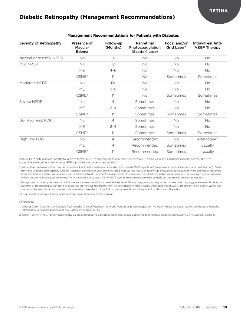
Management Recommendations for Patients with Diabetes
Severity of Retinopathy Presence of Follow-up Panretinal Focal and/or Intravitreal Anti- Macular (Months) Photocoagulation Grid Laser* VEGF Therapy Edema (Scatter) Laser
Normal or minimal NPDR No 12 No No No
Mild NPDR No 12 No No No
ME 4–6 No No No
CSME† 1* No Sometimes Sometimes
Moderate NPDR No 12‡ No No No
ME 3–6 No No No
CSME† 1* No Sometimes Sometimes
Severe NPDR No 4 Sometimes No No
ME 2–4 Sometimes No No
CSME† 1* Sometimes Sometimes Sometimes
Non-high-risk PDR No 4 Sometimes No No
ME 2–4 Sometimes No No
CSME† 1* Sometimes Sometimes Sometimes
High-risk PDR No 4 Recommended No Alternative1,2
ME 4 Recommended Sometimes Usually
CSME† 1* Recommended Sometimes Usually
Anti-VEGF = anti-vascular endothelial growth factor; CSME = clinically significant macular edema; ME = non-clinically significant macular edema; NPDR = nonproliferative diabetic retinopathy; PDR = proliferative diabetic retinopathy
* Adjunctive treatments that may be considered include intravitreal corticosteroids or anti-VEGF agents (off-label use, except aflibercept and ranibizumab) . Data from the Diabetic Retinopathy Clinical Research Network in 2011 demonstrated that, at two years of follow-up, intravitreal ranibizumab with prompt or deferred laser resulted in greater visual acuity gain and intravitreal triamcinolone acetonide plus laser also resulted in greater visual gain in pseudophakic eyes compared with laser alone . Individuals receiving the intravitreal injections of anti-VEGF agents may be re-examined as early as one month following injection .
† Exceptions include hypertension or fluid retention associated with heart failure, renal failure, pregnancy, or any other causes that may aggravate macular edema . Deferral of photocoagulation for a brief period of medical treatment may be considered in these cases . Also, deferral of CSME treatment is an option when the center of the macula is not involved, visual acuity is excellent, close follow-up is possible, and the patient understands the risks .
‡ Or at shorter intervals if signs approaching those of severe NPDR appear .
References:
1 . Writing Committee for the Diabetic Retinopathy Clinical Research Network . Panretinal photocoagulation vs intravitreous ranibizumab for proliferative diabetic retinopathy: a randomized clinical trial . JAMA 2015;314:2137–46 .
2 . Olsen TW . Anti-VEGF pharmacotherapy as an alternative to panretinal laser photocoagulation for proliferative diabetic retinopathy . JAMA 2015;314:2135–6 .
October 2016 aao .org 11© 2016 American Academy of Ophthalmology
RETINAIdiopathic Epiretinal Membrane and Vitreomacular Traction (Initial Evaluation and Therapy)
Initial Exam (Key elements)• Ocular history (e .g ., posterior vitreous detachment,
uveitis, retinal breaks, retinal vein occlusions, proliferative diabetic retinopathy, ocular inflammatory diseases, recent wound healing)
• Duration of symptoms (e .g ., metamorphopsia, difficulty using both eyes together, and diplopia)
• Race/ethnicity
• Systemic history
Physical Exam (Key elements)• Visual acuity
• Measurement of IOP
• Slit-lamp biomicroscopy of the anterior segment
• Spectral domain OCT to diagnose macula and retinal changes (e .g ., proliferation of retinal pigment epithelial cells and/or retinal glial cells) (III, GQ, SR)
• Identify presence of extracellular matrix material, laminocytes, and/or vitreous cells
• ERMs and VMTs often occur together (OCT findings of VMT are similar, but posterior hyaloid remains partially attached to macula)
• Fluorescein angiogram may be helpful in evaluating ERMs and/or VMTs and associated retinal pathologies
Management Plan• The decision to intervene surgically in patients with
ERM/VMT usually depends upon the severity of symptoms, especially the impact on daily activities
• Patients should be informed that the majority of ERMs will remain stable and do not require therapy (GQ, SR)
• Patients should be reassured that there is a very successful surgical procedure that could address worsening symptoms or decreasing visual acuity (GQ, SR)
• Risks versus benefits of vitrectomy surgery should be discussed . Risks include cataract, retinal tears, retinal detachment, and endophthalmitis
Surgery and Postoperative Care• Vitrectomy surgery is often indicated in patients
who are affected with a decrease in visual acuity, metamorphopsia, and double vision (II, MQ, DR)
• Patients do not typically improve without vitrectomy surgery when the area of VMT is broad (>1500 µm), when there is an accompanying pathologic detachment of the macula, or when the presenting visual acuity is poor (III, IQ, DR)
• Vitrectomy surgery for ERM or VMT usually leads to an improvement in visual acuity since the outer retina, ellipsoid zone, and the photoreceptors outer segment length may improve or even normalize after surgery (III, IQ, DR)
• A patient with an ERM should be informed that it is unlikely that intravitreal ocriplasmin will lead to effective treatment (III, GQ, SR)
• Hypotony and elevated IOPs are a well-known risk of vitrectomy surgery and should be monitored post operatively
• Patients should be examined postoperatively day 1, and again 1 to 2 weeks following surgery, or sooner depending upon the development of new symptoms or new findings during early postoperative examination (GQ, SR)
Patient Education and Follow-up• Comparing OCT images in the abnormal versus
normal eye can aid patient understanding
• Patients should be encouraged to periodically test their central vision monocularly to detect changes that may occur over time, like small central scotoma (GQ, SR)
• Patients should be informed to notify their ophthalmologist promptly if they have symptoms such as an increase of floaters, loss of visual field, metamorphopsia, or a decrease in visual acuity (III, GQ, SR)
October 2016 aao .org 12© 2016 American Academy of Ophthalmology
RETINAIdiopathic Macular Hole (Initial Evaluation and Therapy)
Initial Exam History (Key elements)• Duration of symptoms (III, GQ, DR)
• Ocular history: glaucoma, retinal detachment or tear, other prior eye diseases or injuries, ocular surgery, or prolonged sun or eclipse gazing (III, GQ, DR)
• Medications that may be related to macular cystoid edema (III, GQ, DR)
Initial Physical Exam (Key elements)• Visual acuity (III, GQ, SR)
• Slit-lamp biomicroscopic examination of the macula and the vitreoretinal interface, and the optic disc (III, GQ, SR)
• Indirect peripheral retinal examination (III, GQ, SR)
Surgical and Postoperative Care if Patient Receives Treatment• Inform the patient about relative risks, benefits,
and alternatives to surgery, and the need for use of expansile intraocular gas or facedown positioning postoperatively (III, GQ, SR)
• Formulate a postoperative care plan and inform the patient of these arrangements (III, GQ, SR)
• Inform patients with glaucoma of possible postoperative increase in IOP (III, GQ, SR)
• Examine postoperatively within 1 or 2 days and again 1 to 2 weeks after surgery (III, GQ, DR)
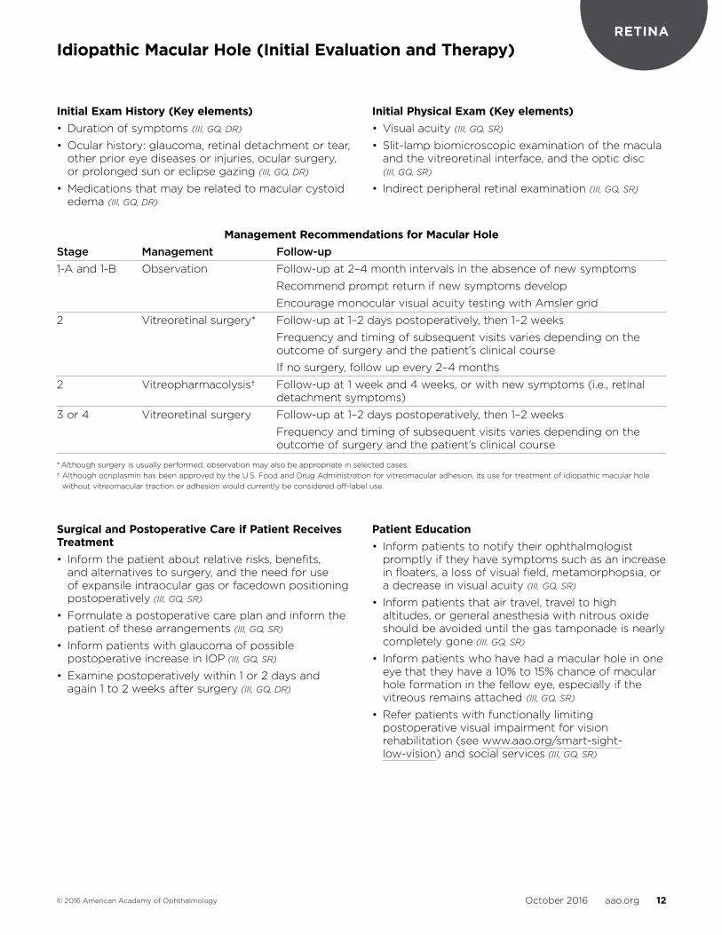
Management Recommendations for Macular HoleStage Management Follow-up1-A and 1-B Observation Follow-up at 2–4 month intervals in the absence of new symptoms
Recommend prompt return if new symptoms develop
Encourage monocular visual acuity testing with Amsler grid
2 Vitreoretinal surgery* Follow-up at 1–2 days postoperatively, then 1–2 weeks
Frequency and timing of subsequent visits varies depending on the outcome of surgery and the patient’s clinical course
If no surgery, follow up every 2–4 months
2 Vitreopharmacolysis† Follow-up at 1 week and 4 weeks, or with new symptoms (i .e ., retinal detachment symptoms)
3 or 4 Vitreoretinal surgery Follow-up at 1–2 days postoperatively, then 1–2 weeks
Frequency and timing of subsequent visits varies depending on the outcome of surgery and the patient’s clinical course
* Although surgery is usually performed, observation may also be appropriate in selected cases .† Although ocriplasmin has been approved by the U .S . Food and Drug Administration for vitreomacular adhesion, its use for treatment of idiopathic macular hole
without vitreomacular traction or adhesion would currently be considered off-label use .
Patient Education• Inform patients to notify their ophthalmologist
promptly if they have symptoms such as an increase in floaters, a loss of visual field, metamorphopsia, or a decrease in visual acuity (III, GQ, SR)
• Inform patients that air travel, travel to high altitudes, or general anesthesia with nitrous oxide should be avoided until the gas tamponade is nearly completely gone (III, GQ, SR)
• Inform patients who have had a macular hole in one eye that they have a 10% to 15% chance of macular hole formation in the fellow eye, especially if the vitreous remains attached (III, GQ, SR)
• Refer patients with functionally limiting postoperative visual impairment for vision rehabilitation (see www .aao .org/smart-sight- low-vision) and social services (III, GQ, SR)
October 2016 aao .org 13© 2016 American Academy of Ophthalmology
RETINAPosterior Vitreous Detachment, Retinal Breaks, and Lattice Degeneration (Initial and Follow-up Evaluation)
Initial Exam History (Key elements)• Symptoms of PVD (II+, GQ, SR)
• Family history of RD, related genetic disorders (II–, GQ, SR)
• Prior eye trauma (III, GQ, SR)
• Myopia (II+, GQ, SR)
• History of ocular surgery including refractive lens exchange and cataract surgery (II++, GQ, SR)
Initial Physical Exam (Key elements)• Confrontation visual field examination, and assessing
for the presence of a relative afferent pupillary defect (III, GQ, SR)
• Examination of the vitreous for hemorrhage, detachment, and pigmented cells (II+, GQ, SR)
• Examination of the peripheral fundus with scleral depression . The preferred method of evaluating peripheral vitreoretinal pathology is with indirect ophthalmoscopy combined with scleral depression . (III, GQ, SR)
Ancillary Tests• Optical coherence tomography may be helpful to
evaluate and stage the PVD (II+, MQ, DR)
• Perform B-scan ultrasonography if peripheral retina cannot be evaluated . If no abnormalities are found, frequent follow-up examinations are recommended . (III, IQ, DR)
Surgical and Postoperative Care if Patient Receives Treatment:• Inform patient about the relative risks, benefits, and
alternatives to surgery (III, GQ, SR)
• Formulate a postoperative care plan and inform patient of these arrangements (III, GQ, SR)
• Advise patient to contact ophthalmologist promptly if they have a substantial change in symptoms such as floaters, visual field loss, or decreased visual acuity (II+, GQ, SR)
Follow-up History• Visual symptoms (III, GQ, SR)
• Interval history of eye trauma or intraocular surgery (III, GQ, SR)
Follow-up Physical Exam• Visual acuity (III, GQ, SR)
• Evaluation of the status of the vitreous, with attention to the presence of pigment, hemorrhage, or syneresis (III, GQ, SR)
• Examination of the peripheral fundus with scleral depression (III, GQ, SR)
• Optical coherence tomography if vitreomacular traction is present (III, GQ, SR)
• B-scan ultrasonography if the media are opaque (III, GQ, SR)
Patient Education• Educate patients at high risk of developing retinal
detachment about the symptoms of PVD and retinal detachment and the value of periodic follow-up exams (III, GQ, SR)
• Instruct all patients at increased risk of retinal detachment to notify their ophthalmologist promptly if they have a substantial change in symptoms such as increase in floaters, loss of visual field, or decrease in visual acuity (II+, GQ, SR)
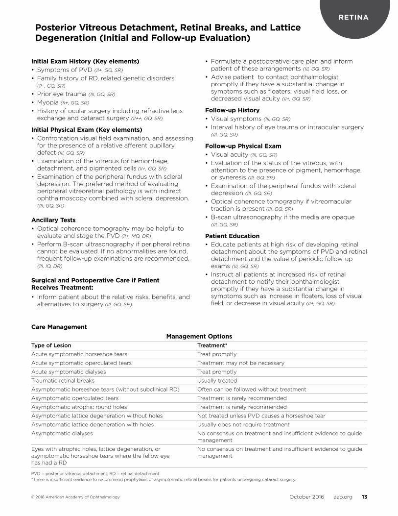
Care ManagementManagement Options
Type of Lesion Treatment*Acute symptomatic horseshoe tears Treat promptly
Acute symptomatic operculated tears Treatment may not be necessary
Acute symptomatic dialyses Treat promptly
Traumatic retinal breaks Usually treated
Asymptomatic horseshoe tears (without subclinical RD) Often can be followed without treatment
Asymptomatic operculated tears Treatment is rarely recommended
Asymptomatic atrophic round holes Treatment is rarely recommended
Asymptomatic lattice degeneration without holes Not treated unless PVD causes a horseshoe tear
Asymptomatic lattice degeneration with holes Usually does not require treatment
Asymptomatic dialyses No consensus on treatment and insufficient evidence to guide management
Eyes with atrophic holes, lattice degeneration, or No consensus on treatment and insufficient evidence to guide asymptomatic horseshoe tears where the fellow eye management has had a RD
PVD = posterior vitreous detachment; RD = retinal detachment*There is insufficient evidence to recommend prophylaxis of asymptomatic retinal breaks for patients undergoing cataract surgery .

October 2016 aao .org 14© 2016 American Academy of Ophthalmology
RETINA
Initial Exam (Key elements)• Initial exam should include all aspects of a
comprehensive adult medical eye evaluation (see Comprehensive Adult Medical Eye Evaluation PPP for details) with special attention paid to retinal vascular disease elements (II+, MQ, SR)
• Medical history should include a careful review of systems for embolic disease (e .g ., transient ischemic symptoms, lateralizing weakness, paresthesias)
• GCA symptoms (e .g ., headaches, scalp tenderness, malaise, fatigue, temporal tenderness, fever, history of polymyalgia rheumatic) must be recognized
Physical Exam (Key elements)• Visual acuity
• Measurement of IOP
• Slit-lamp biomicroscopy
• Dilated examination of the far peripheral retina with indirect ophthalmoscopy
• Gonioscopy when IOP is elevated or when iris neovascularization risk is suspected (prior to dilation)
• Funduscopy
• Relative afferent pupil defect assessment
• Slit-lamp biomicroscopy of the posterior pole
• Examination of the peripheral retina using indirect ophthalmoscopy through a dilated pupil to assess: retinal hemorrhages, cotton-wool spots, retinal emboli, retinal vascular “boxcarring,” and optic disc neovascularization
Diagnostic Tests• Color and red-free fundus photography
• Fluorescein angiogram
• Optical coherence tomography
• Ultrasonography in the setting of significant media
opacity
Care Management• Acute symptomatic OAO, CRAO, or BRAO represent
urgent ophthalmic conditions and require prompt evaluation
• Physicians should immediately consider GCA in patients 50 years of age or older
• In cases of GCA, physicians should initiate urgent systemic corticosteroid therapy to prevent vision loss in the fellow eye or vascular occlusion elsewhere (I-/I+, GQ, SR)
• Diabetics should be carefully monitored since systemic corticosteroid treatment may destabilize glucose control
Ophthalmologists should refer patients with retinal vascular disease to the appropriate setting, depending on the nature of the retinal occlusion.
• Acute symptomatic OAOs or CRAOs from embolic etiologies should prompt an immediate referral to the nearest stroke center
• At present there is no evidence in support of treating asymptomatic patients who have a BRAO with an expedited stroke work-up
Patient Follow-up• Follow-up should consider the extent of retinal or
ocular ischemia neovascularization . Patients with greater ischemia require more frequent follow-up
• Many patients with retinal vascular disease will lose substantial vision despite various treatment options and should be referred for appropriate social services and vision rehabilitation
Retinal and Ophthalmic Artery Occlusions (Initial Evaluation and Therapy)
October 2016 aao .org 15© 2016 American Academy of Ophthalmology
RETINARetinal Vein Occlusions (Initial Evaluation and Therapy)
Initial Exam (Key elements)• Ocular history (e .g ., glaucoma, other ophthalmologic
disorders, ocular injections, surgery, including retinal laser treatment, cataract surgery, refractive surgery)
• Location and duration of vision loss
• Current medications
• Systemic history (e .g ., systemic hypertension, diabetes, hyperlipidemia, cardiovascular disease, sleep apnea, coagulopathies, thrombotic disorders, and pulmonary embolus)
Physical Exam (Key elements)• Visual acuity
• Measurement of IOP
• Slit-lamp biomicroscopy to detect fine abnormal new iris vessels
• Dilated examination of the far peripheral retina with indirect ophthalmoscopy
• Gonioscopy prior to pupil dilation; especially in cases of an ischemic CRVO, when IOP is elevated, or when iris neovascularization risk is high
• Binocular funduscopic evaluation of the posterior pole
Diagnostic Tests• Color fundus photography to document retinal
findings
• Fluorescein angiogram to evaluate the degree of vascular occlusion
• Optical coherence tomography to detect macular disease
• Ultrasonography (e .g ., when vitreous hemorrhage is present)
Care Management• Best prevention is to manage risk factors
aggressively by optimizing control of diabetes mellitus, hypertension, and hyperlipidemia (I+, GQ, SR)
• Participants who received a 4-mg corticosteroid treatment dose had higher rates of cataract formation, cataract surgery, and elevated IOP, indicating a preference for a 1-mg dose (I++, GQ, SR)
• Multiple studies have demonstrated the efficacy of anti-VEGF agents in the treatment of macular edema associated with BRVO (I++, GQ, SR)
• Randomized controlled studies have shown the efficacy of anti-VEGF agents in treating macular edema related to CRVO (I++, GQ, SR)
• Betadine antiseptic drops and a lid speculum are recommended during all intravitreal injections (III, MQ, DR)
• Intravitreal triamcinolone, dexamethasone, and other corticosteroids have been shown to be efficacious for macular edema associated with CRVO, yet there are known associated risks of cataracts and glaucoma (I+, GQ, SR)
• Laser treatment remains a viable treatment in eyes with BRVO, even if the duration of the disease is greater than 12 months (I+, GQ, SR)
• Sectoral pan retinal photocoagulation is still recommended for neovascularization when complications such as vitreous hemorrhage or iris neovascularization occur (I+, GQ, SR)
• Ophthalmologists caring for patients with retinal vascular occlusion should be familiar with specific recommendations of relevant clinical trials due to the complexity of diagnosis and treatment (I++, GQ, SR)
Patient Follow-up• Ophthalmologist should refer patients with an RVO
to a primary care physician for appropriate management of their systemic condition and communicate results to the physician managing the patient’s ongoing care (I+, GQ, SR)
• Risk to the fellow eye should be communicated to both the primary care provider and the patient (I+, MQ, SR)
• Patients whose conditions fail to respond to therapy and when further treatment is unavailable should be provided with professional support and offered a referral for counseling, vision rehabilitation, or social services as appropriate (I++, GQ, SR)
October 2016 aao .org 16© 2016 American Academy of Ophthalmology
CATARACTCataract (Initial and Follow-up Evaluation)
Initial Exam History (Key elements)• Symptoms
• Ocular history
• Systemic history
• Assessment of visual function status
• Medications currently used
Initial Physical Exam (Key elements)• Visual acuity with current correction
• Measurement of BCVA (with refraction when indicated)
• External examination
• Ocular alignment and motility
• Glare testing when indicated
• Pupil reactivity and function
• Measurement of IOP
• Slit-lamp biomicroscopy, including gonioscopy
• Dilated examination of the lens, macula, peripheral retina, optic nerve, and vitreous through a dilated pupil
• Assessment of relevant aspects of the patient’s medical and physical status
Care Management• Treatment is indicated when visual function no
longer meets the patient’s needs and cataract surgery provides a reasonable likelihood of quality-of-life improvement
• Cataract removal is also indicated when there is evidence of lens-induced disease or when it is necessary to visualize the fundus in an eye that has the potential for sight
• Surgery should not be performed under the following circumstances:
- Tolerable refractive correction provides vision that meets the patient’s needs and desires; surgery is not expected to improve visual function, and no other indication for lens removal exists
- The patient cannot safely undergo surgery because of coexisting medical or ocular conditions
- Appropriate postoperative care cannot be arranged
- Patient or patient’s surrogate decision maker is unable to give informed consent for nonemergent surgery
• Indications for second eye surgery are the same as for the first eye (with considerations given to needs for binocular function)
• The standard of care in the United States is a small-incision phacoemulsification with foldable IOL implantation with either biaxial or coaxial approach (I+, GQ, SR)
Preoperative CareThe ophthalmologist who is to perform the surgery has the following responsibilities:
• Examine the patient preoperatively• Ensure that the evaluation accurately documents
symptoms, findings, and indications for treatment
• Inform the patient about the risks, benefits, and expected outcomes of surgery, including the anticipated refractive outcome or surgical experience
• Formulate surgical plan, including selection of IOL and anesthesia
• Review results of presurgical and diagnostic evaluations with the patient
• Inform the patient about the possibility of visual impairment continuing after cataract surgery, and the potential for rehabilitation (III, GQ, SR)
• Formulate postoperative plans and inform patient of arrangements
• Answer patient’s questions regarding surgery, care, and cost
• Routine preoperative laboratory testing in association with the history and physical examination is not indicated (I+, GQ, SR)
Follow-up Evaluation• High-risk patients should be seen within 24 hours of
surgery• Routine patients should be seen within 48 hours of
surgery• Frequency and timing of subsequent visits depend
on refraction, visual function, and medical condition of the eye
• More frequent follow-up usually necessary for high risk patients
• Components of each postoperative exam should include:- Interval history, including new symptoms and use
of postoperative medications- Patient’s assessment of visual function status- Measurement of IOP- Slit-lamp biomicroscopy- Operating ophthalmologist should provide
postoperative care that is within the unique competence of the ophthalmologist (III, GQ, SR)
October 2016 aao .org 17© 2016 American Academy of Ophthalmology
CATARACT
Nd:YAG Laser Capsulotomy• Treatment is indicated when vision impaired by
posterior capsular opacification does not meet the patient’s functional needs or when it critically interferes with visualization of the fundus
• Educate about the symptoms of posterior vitreous detachment, retinal tears, and detachment and the need for immediate examination if these symptoms are noticed
• The decision to perform capsulotomy should take into account the benefits and risks of the laser surgery . Laser posterior capsulotomy should not be performed prophylactically (i .e ., when the capsule remains clear) . The should be inflammatory-free and the IOL stable prior to performing Nd:YAG laser capsulotomy . (III, GQ, SR)
Cataract (Initial and Follow-up Evaluation) (continued)
October 2016 aao .org 18© 2016 American Academy of Ophthalmology
CORNEABacterial Keratitis (Initial Evaluation)
Initial Exam History• Ocular symptoms (e .g ., degree of pain, redness,
discharge, blurred vision, photophobia, duration of symptoms, circumstances surrounding the onset of symptoms) (III, GQ, SR)
• Contact lens history (e .g ., wearing schedule, overnight wear, type of contact lenses, contact lens solution, contact lens hygiene protocol, tap-water rinse of contact lenses, swimming, using a hot tub, or showering while wearing contact lenses) (II+, GQ, SR)
• Review of other ocular history, including risk factors such as herpes simplex virus keratitis, varicella zoster virus keratitis, previous bacterial keratitis, trauma, dry eye, and previous ocular surgery, including refractive surgery (III, GQ, SR)
• Review of other medical problems (III, GQ, SR)
• Current and recently used ocular medications (III, GQ, SR)
• Medication allergies (III, GQ, SR)
Initial Physical Exam• Visual acuity (III, GQ, SR)
• General appearance of patient, including skin conditions (III, GQ, SR)
• Facial examination (III, GQ, SR)
• Globe position (III, GQ, SR)
• Eyelids and eyelid closure (III, GQ, SR)
• Conjunctiva (III, GQ, SR)
• Nasolacrimal apparatus (III, GQ, SR)
• Corneal sensation (III, GQ, SR)
• Slit-lamp biomicroscopy (III, GQ, SR)
- Eyelid margins (III, GQ, SR)
- Conjunctiva (III, GQ, SR)
- Sclera (III, GQ, SR)
- Cornea (III, GQ, SR)
- Anterior chamber for depth and the presence of inflammation, including cell and flare, hypopyon, fibrin, hyphema (III, GQ, SR)
- Anterior vitreous (III, GQ, SR)
- Contralateral eye for clues to etiology as well as possible similar underlying pathology (III, GQ, SR)
Diagnostic Tests• Manage majority of community-acquired cases
with empiric therapy and without smears or cultures . (III, IQ, DR)
• Indications for smears and cultures:
- Sight-threatening or severe keratitis of suspected micro bial origin prior to initiating therapy . (III, IQ, DR)
- A large central corneal infiltrate that extends to the middle to deep stroma . (III, IQ, DR)
- Chronic in nature . (III, IQ, DR)
- Unresponsive to broad spectrum antibiotic therapy . (III, IQ, DR)
- Clinical features suggestive of fungal, amœbic, or mycobacterial keratitis . (III, IQ, DR)
• The hypopyon that occurs in eyes with bacterial keratitis is usually sterile, and aqueous or vitreous taps should not be performed unless there is a high suspicion of microbial endophthalmitis . (III, IQ, DR)
• Corneal scrapings for culture should be inoculated directly onto appropriate culture media to maximize culture yield . (III, IQ, DR) If this is not feasible, place specimens in transport media . (II+, MQ, DR) In either case, immediately incubate cultures or take promptly to the laboratory . (III, GQ, SR)
Care Management• Topical antibiotic eye drops are preferred method in
most cases . (III, GQ, SR)
• Use topical broad-spectrum antibiotics initially in the empiric treatment of presumed bacterial keratitis. (III, IQ, DR)
• For central or severe keratitis (e .g ., deep stromal involvement or an infiltrate larger than 2 mm with extensive suppuration), use a loading dose (e .g ., every 5 to 15 minutes for the first 30 to 60 minutes), followed by frequent applications (e .g ., every 30 minutes to 1 hour around the clock) . (III, IQ, DR) For less severe keratitis, a regimen with less frequent dosing is appropriate . (III, IQ, DR)
• Use systemic therapy for gonococcal keratitis . (III, IQ, DR)
• For patients treated with ocular topical corticosteroids at time of presentation of suspected bacterial keratitis, reduce or eliminate corticosteroids until infection has been controlled . (III, GQ, SR)
• When the corneal infiltrate compromises the visual axis, may add topical corticosteroid therapy following at least 2 to 3 days of progressive improvement with treatment with topical antibiotics . (III, IQ, DR) Continue topical antibiotics at high levels with gradual tapering . (III, IQ, DR)
• Examine patients within 1 to 2 days after initiation of topical corticosteroid therapy . (III, IQ, DR)
October 2016 aao .org 19© 2016 American Academy of Ophthalmology
CORNEABacterial Keratitis (Management Recommendations)
Patient Education• Inform patients with risk factors predisposing them
to bacterial keratitis of their relative risk, the signs and symptoms of infection, and to consult an ophthalmologist promptly if they experience such warning signs or symptoms (III, GQ, SR)
• Educate about the destructive nature of bacterial keratitis and need for strict compliance with therapy (III, GQ, SR)
• Discuss possibility of permanent visual loss and need for future visual rehabilitation (III, GQ, SR)
• Educate patients with contact lenses about increased risk of infection associated with contact lens, overnight wear, and importance of adherence to techniques to promote contact lens hygiene (II+, GQ, SR)
• Refer patients with significant visual impairment or blindness for vision rehabilitation if they are not surgi cal candidates (see www .aao .org/smart-sight-low-vision)
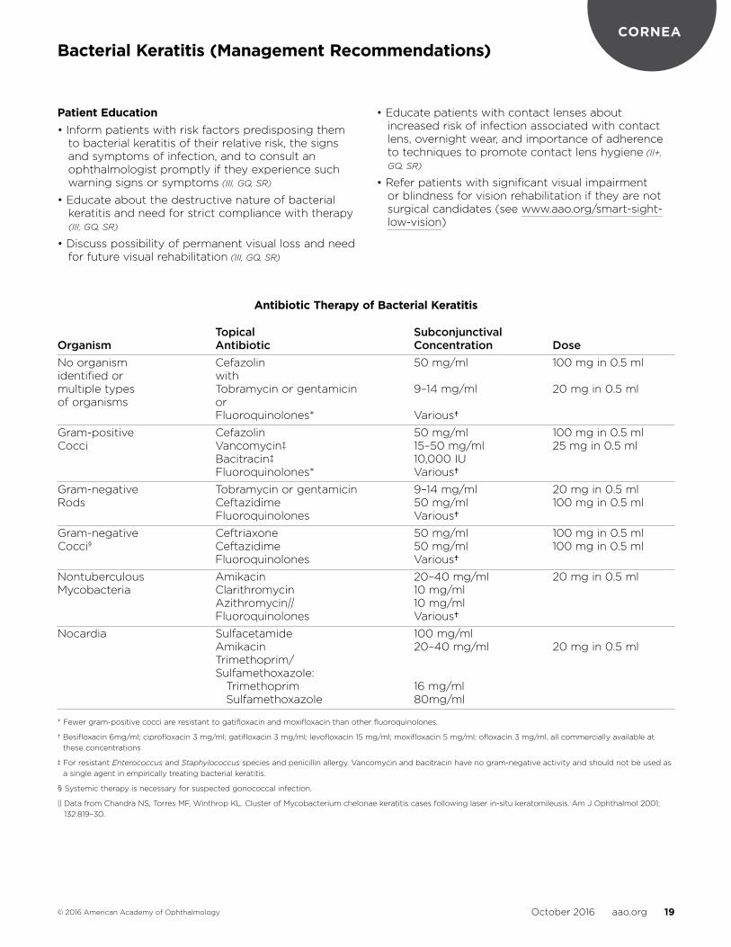
Antibiotic Therapy of Bacterial Keratitis
Topical Subconjunctival Organism Antibiotic Concentration DoseNo organism Cefazolin 50 mg/ml 100 mg in 0 .5 ml identified or with multiple types Tobramycin or gentamicin 9–14 mg/ml 20 mg in 0 .5 ml of organisms or Fluoroquinolones* Various†Gram-positive Cefazolin 50 mg/ml 100 mg in 0 .5 ml Cocci Vancomycin‡ 15–50 mg/ml 25 mg in 0 .5 ml Bacitracin‡ 10,000 IU Fluoroquinolones* Various†Gram-negative Tobramycin or gentamicin 9–14 mg/ml 20 mg in 0 .5 ml Rods Ceftazidime 50 mg/ml 100 mg in 0 .5 ml Fluoroquinolones Various†Gram-negative Ceftriaxone 50 mg/ml 100 mg in 0 .5 ml Cocci§ Ceftazidime 50 mg/ml 100 mg in 0 .5 ml Fluoroquinolones Various† Nontuberculous Amikacin 20–40 mg/ml 20 mg in 0 .5 ml Mycobacteria Clarithromycin 10 mg/ml Azithromycin|| 10 mg/ml Fluoroquinolones Various† Nocardia Sulfacetamide 100 mg/ml Amikacin 20–40 mg/ml 20 mg in 0 .5 ml Trimethoprim/ Sulfamethoxazole: Trimethoprim 16 mg/ml Sulfamethoxazole 80mg/ml
* Fewer gram-positive cocci are resistant to gatifloxacin and moxifloxacin than other fluoroquinolones .
† Besifloxacin 6mg/ml; ciprofloxacin 3 mg/ml; gatifloxacin 3 mg/ml; levofloxacin 15 mg/ml; moxifloxacin 5 mg/ml; ofloxacin 3 mg/ml, all commercially available at these concentrations
‡ For resistant Enterococcus and Staphylococcus species and penicillin allergy . Vancomycin and bacitracin have no gram-negative activity and should not be used as a single agent in empirically treating bacterial keratitis .
§ Systemic therapy is necessary for suspected gonococcal infection .
|| Data from Chandra NS, Torres MF, Winthrop KL . Cluster of Mycobacterium chelonae keratitis cases following laser in-situ keratomileusis . Am J Ophthalmol 2001; 132:819–30 .
October 2016 aao .org 20© 2016 American Academy of Ophthalmology
CORNEABlepharitis (Initial and Follow-up Evaluation)
Initial Exam History
• Ocular symptoms and signs (e .g ., redness, irritation, burning, tearing, itching, crusting of eyelashes, eyelid sticking, contact lens intolerance, photophobia, increased frequency of blinking) (III, GQ, SR)
• Time of day when symptoms are worse
• Duration of symptoms
• Unilateral or bilateral presentation
• Exacerbating conditions (e .g ., smoke, allergens, wind, contact lenses, low humidity, retinoids, diet and alcohol consumption, eye makeup)
• Symptoms related to systemic diseases (e .g ., rosacea, allergy) (III, IQ, DR)
• Current and previous systemic and topical medications (e .g ., antihistamines or drugs with anticholinergic effects, or drugs used in the past that might have an effect on the ocular surface [e .g ., isotretinoin]) (III, GQ, SR)
• Recent exposure to an infected individual (e .g ., pediculosis palpebrarum [Pthirus pubis])
• Ocular history (e .g ., previous intraocular and eyelid surgery, local trauma, including mechanical, thermal, chemical, and radiation injury, history of cosmetic blepharoplasty, history of styes and/or chalazia) (III, GQ, SR)
Initial Physical Exam• Visual acuity (III, GQ, SR)
• External examination
- Skin (III, GQ, SR)
- Eyelids (III, GQ, SR)
• Slit-lamp biomicroscopy
- Tear film (III, GQ, SR)
- Anterior eyelid margin (III, GQ, SR)
- Eyelashes (III, GQ, SR)
- Posterior eyelid margin (III, GQ, SR)
- Tarsal conjunctiva (everting eyelids) (III, GQ, SR)
- Bulbar conjunctiva (III, GQ, SR)
- Cornea (III, GQ, SR)
Diagnostic Tests• Cultures may be indicated for patients with
recurrent anterior blepharitis with severe inflammation as well as for patients who are not responding to therapy . (III, IQ, DR)
• Biopsy of the eyelid to exclude the possibility of carcinoma may be indicated in cases of marked asymmetry, resistance to therapy or unifocal recurrent chalazia that do not respond well to therapy . (III, IQ, DR)
• Consult with the pathologist prior to obtaining the biopsy if sebaceous cell carcinoma is suspected . (III, GQ, SR)
Care Management• Treat patients with blepharitis initially with a
regimen of warm compresses and eyelid hygiene . (III, IQ, DR)
• A topical antibiotic such as bacitracin or erythromycin can be prescribed to be applied one or more times daily or at bedtime on the eyelids for one or more weeks . (III, IQ, DR)
• For patients with meibomian gland dysfunction, whose chronic symptoms and signs are not adequately controlled with eyelid hygiene, oral tetracyclines and topical antibiotics can be prescribed . (I–, MQ, DR)
• A brief course of topical corticosteroids may be helpful for eyelid or ocular surface inflammation . The minimal effective dose of corticosteroid should be utilized and long-term corticosteroid therapy should be avoided if possible . (III, GQ, SR)
Follow-Up Evaluation• Follow-up visits should include:
- Interval history (III, GQ, SR)
- Measurement of visual acuity (III, GQ, SR)
- External examination (III, GQ, SR)
- Slit-lamp biomicroscopy (III, GQ, SR)
• If corticosteroid therapy is prescribed, re-evaluate patient within a few weeks to determine the response to therapy, measure intraocular pressure, and assess treatment compliance (III, GQ, SR)
Patient Education• Counsel patients about the chronicity and
recurrence of the disease process . (III, GQ, SR)
• Inform patients that symptoms can frequently be improved but are rarely eliminated . (III, GQ, SR)
• Patients with an inflammatory eyelid lesion that appears suspicious for malignancy should be referred to an appropriate specialist . (III, GQ, SR)
October 2016 aao .org 21© 2016 American Academy of Ophthalmology
CORNEAConjunctivitis (Initial Evaluation)
Initial Exam History• Ocular symptoms and signs (e .g ., itching, discharge,
irritation, pain, photophobia, blurred vision)
• Duration of symptoms and time course
• Exacerbating factors
• Unilateral or bilateral presentation
• Character of discharge
• Recent exposure to an infected individual
• Trauma (mechanical, chemical, ultraviolet)
• Mucus fishing
• Contact lens wear (lens type, hygiene and use regimen)
• Symptoms and signs potentially related to systemic diseases (e .g ., genitourinary discharge, dysuria, dysphagia, upper respiratory infection, skin and mucosal lesions)
• Allergy, asthma, eczema
• Use of topical and systemic medications
• Ocular history (e .g ., previous episodes of conjunctivitis and previous ophthalmic surgery)
• Compromised immune status
• Current and prior systemic diseases
• Social history (e .g ., smoking, occupation and hobbies, travel and sexual activity)
Initial Physical Exam• Visual acuity (III, IQ, DR)
• External examination (III, IQ, DR)
- Skin (signs of rosacea, eczema, seborrhea) (III, IQ, DR)
- Abnormalities of the eyelids and adnexae (swelling, discoloration, malposition, laxity, ulceration, nodules, ecchymosis, neoplasia) (III, IQ, DR)
- Conjunctiva (pattern of injection, subconjunctival hemorrhage, chemosis, cicatricial change, symbleph aron, masses, discharge) (III, IQ, DR)
• Slit-lamp biomicroscopy (III, IQ, DR)
- Eyelid margins (inflammation, ulceration, discharge, nodules or vesicles, blood-tinged debris, keratinization) (III, IQ, DR)
- Eyelashes (loss of lashes, crusting, scurf, nits, lice, trichiasis) (III, IQ, DR)
- Lacrimal puncta and canaliculi (pouting, discharge) (III, IQ, DR)
- Tarsal and forniceal conjunctiva (III, IQ, DR)
- Bulbar conjunctiva/limbus (follicles, edema, nodules, chemosis, laxity, papillae, ulceration, scarring, phlyctenules, hemorrhages, foreign material, keratinization) (III, IQ, DR)
- Cornea (III, IQ, DR)
- Anterior chamber/iris (inflammation reaction, synechiae, transillumination defects) (III, IQ, DR)
- Dye-staining pattern (conjunctiva and cornea) (III, IQ, DR)
Diagnostic Tests• Cultures, smears for cytology and special stains are
indicated in cases of suspected infectious neonatal conjunctivitis . (II–, IQ, DR)
• Smears for cytology and special stains are recommended in cases of suspected gonococcal conjunctivitis . (II–, IQ, DR)
• Confirm diagnosis of adult and neonate chlamydial conjunctivitis with immunodiagnostic test and/or culture .
• Biopsy the bulbur conjunctiva and take a sample from an uninvolved area adjacent to the limbus in an eye with active inflammation when ocular mucous membrane pemphigoid is suspected . (II–, IQ, DR)
• A full-thickness lid biopsy is indicated in cases of suspected sebaceous carcinoma . (III, IQ, DR)
• Confocal microscopy may be helpful to evaluate some forms of conjunctivitis (e .g ., atopic, SLK) . (II–, MQ, DR)
• Thyroid function tests are indicated for patients with SLK who do not have known thyroid disease . (III, IQ, DR)
October 2016 aao .org 22© 2016 American Academy of Ophthalmology
CORNEAConjunctivitis (Management Recommendations)
Care Management• Avoid indiscriminate use of topical antibiotics or
cor ticosteroids because antibiotics can induce toxicity and corticosteroids can potentially prolong adenoviral infections and worsen herpes simplex virus infections (III, GQ, SR)
• Treat mild allergic conjunctivitis with an over-the-counter antihistamine/vasoconstrictor agent or second-generation topical histamine H1-receptor antagonists . If the condition is frequently recurrent or persistent, use mast-cell stabilizers (I++, GQ, SR)
• For contact lens-related keratoconjunctivitis, discontinue contact lens wear for 2 or more weeks (III, IQ, DR)
• If corticosteroids are indicated, prescribe the lowest potency and frequency based on patient response and tolerance (III, IQ, DR)
• If corticosteroids are used, perform baseline and periodic measurement of intraocular pressure and pupillary dilation (III, IQ, DR)
• Use systemic antibiotic treatment for conjunctivitis due to Neisseria gonorrhoeae or Chlamydia trachomatis (III, IQ, DR)
• Treat sexual partners to minimize recurrence and spread of disease when conjunctivitis is associated with sexually transmitted diseases and refer patients and their sexual partners to an appropiate medical specialist (III, GQ, SR)
• Refer patients with manifestation of a systemic disease to an appropriate medical specialist (III, GQ, SR)
Follow-Up Evaluation• Follow-up visits should include
- Interval history (III, IQ, DR)
- Visual acuity (III, IQ, DR)
- Slit-lamp biomicroscopy (III, IQ, DR)
• If corticosteroids are used, perform periodic measurement of intraocular pressure and pupillary dilation to evaluate for cataract and glaucoma (III, IQ, DR)
Patient Education• Counsel patients with contagious varieties to mini-
mize or prevent spread of diseases in the community (III, IQ, DR)
• Inform patients who may require repeat short-term therapy with topical corticosteroid of potential complications of corticosteroid use
• Advise patients with allergic conjunctivitis that frequent clothes washing and bathing/showering before bedtime may be helpful (III, IQ, DR)
October 2016 aao .org 23© 2016 American Academy of Ophthalmology
CORNEACorneal Ectasia (Initial and Follow-up Evaluation)
Initial Exam History• Disease onset and course
• Vision impairment
• Ocular, medical, and family history
Initial Physical Exam• Visual function assessment
• External examination
- Corneal protrusion
- Eyelids and periorbital skin
• Slit-lamp biomicroscopy
- Presence, extent, and location of the corneal thinning or protrusion
- Indication of previous ocular surgery
- Presence of Vogt striae, prominent corneal nerves, Fleischer ring, or other iron deposition
- Evidence of corneal scarring or previous hydrops, and presence of prominent corneal nerves
• IOP measurement (III, IQ, DR)
• Fundus examination: assessment of red reflex for dark area, and retina for tapetoretinal degenerations (III, IQ, DR)
Diagnostic Tests• Keratometry (II+, MQ, DR)
• Corneal topography (II–, MQ, SR)
• Topographic power map
• Topographic elevation map (II+, MQ, DR)
• Corneal pachymetry (II++, GQ, SR)
Care Management• Therapy is tailored to the individual patient,
depending on the visual impairment and treatment option(s) .
• Vision can be corrected with eyeglasses, but contact lenses may be required as keratoconus progresses .
• Rigid corneal gas permeable contact lenses can mask corneal irregularities . New hybrid contact lenses provide higher oxygen permeability and greater RGP/hydrogel junction strength . Piggyback contact lenses may be employed in cases of corneal scaring or decentered cones . Scleral lenses may be indicated when RGP and/or hybrid contact lenses fail .
• Intrastromal corneal ring segment implantation can improve contact lens tolerance and BCVA for patients with corneal ectasia, a clear cornea, and contact lens intolerance . (II–, MQ, DR)
• Collagen crosslinking can improve corneal rigidity by increasing bonds between fibers .
• Lamellar keratoplasty using DALK techniques can be considered for progressive keratoconus without significant scarring or hydrops . (II++, MQ, DR) Crescentic lamellar keratoplasty is an option when maximal thinning is in the cornea’s periphery . (III, IQ, DR)
• Peripheral thinning and ectasia can be managed by a standard decentered lamellar procedure for tectonic support, followed by a central penetrating keratoplasty later . (III, IQ, DR)
• Penetrating keratoplasty is indicated when a patient can no longer achieve functional vision with eyeglasses or contact lenses, or when persistent corneal edema occurs following hydrops . (III, IQ, DR) Descemet stripping endothelial keratoplasty cannot correct ectatic disorder . (III, IQ, DR)
• Penetrating keratoplasty is preferred over DALK in cases of deep stromal scarring . (III, IQ, DR)
• A lamellar graft can be performed for tectonic support when ectasia occurs in the far periphery of the cornea . (III, IQ, DR)
Follow-Up Evaluation• Follow-up evaluation and visit intervals are dictated
by treatment and disease progression . (III, IQ, DR)
• Annual follow up is recommended for cases of ectasia unless the patient has significant changes in visual function . (III, IQ, DR)
• Patients should be made aware of the warning signs of rejection and should seek medical attention promptly if symptoms occur . (III, GQ, SR) The practitioner should be aware of the slit-lamp biomicroscopic findings of epithelial, stromal, and endothelial rejection . (III, GQ, SR)
Counseling and Referral• When medical therapy with eyeglasses and/or
contact lenses cannot improve visual function, a referral to an ophthalmologist trained in surgical treatments for corneal ectasia is indicated (III, GQ, SR)
• Patients with a history of allergy and atopy may require a referral to a dermatologist or allergist (III, GQ, SR)
• Patients with floppy eyelid disease may be best managed by an oculoplastics specialist and referrals to other medical specialists may also be needed (III, GQ, SR)
October 2016 aao .org 24© 2016 American Academy of Ophthalmology
CORNEACorneal Edema and Opacification (Initial Evaluation)
Initial Exam History• Symptoms: blurred or variable vision; photophobia;
redness; tearing; intermittent foreign body sensation; pain
• Age of onset
• Rapidity of onset
• Persistence
• Unilateral or bilateral presentation
• Moderating factors, like visual improvement related to environmental factors
• Past ocular and medical history
• Topical and systemic medications
• Trauma
• Contact lens wear
• Family and social history
Initial Physical Exam• Visual function assessment
• External examination
- Evidence of proptosis, ptosis, lagophthalmos, or floppy eyelid syndrome
- Eyelid or facial asymmetry, scarring, and malfunction
• Slit-lamp biomicroscopy (III, IQ, DR)
- Unilateral or bilateral signs
- Diffuse or localized edema
- Primarily epithelial or stromal edema
- Evidence of epithelial breakdown, stromal infiltration, epithelial ingrowth, striae, focal thickening, thinning, scarring, interface haze, striae or inflammation, or stromal vascularization
- Evidence of guttae, Descemet’s membrane tear or detachment, endothelial vesicles, keratic precipitates (KP), pigment peripheral anterior synechiae
- Involvement of host or donor tissue
- Evidence of sectoral corneal edema and KPs, or an anterior chamber reaction
- Status, shape, and position of the pupil and iris
- Evidence of vitreous strands or pigment dusting
- Status and position of the lens
• IOP measurement
• Fundus examination
• Gonioscopy
Diagnostic Tests• Potential acuity meter
• Rigid contact lens overrefraction
• Pachymetry (III, IQ, DR)
• Scheimpflug imaging
• Specular and confocal microscopy (III, IQ, DR)
• Anterior segment optical coherence tomography (III, IQ, DR)
• Ultrasound biomicroscopy
October 2016 aao .org 25© 2016 American Academy of Ophthalmology
CORNEACorneal Edema and Opacification (Management Recommendations)
Care Management• Therapeutic goal is to control the cause of corneal
edema or opacity and enhance a patient’s quality of life by improving visual acuity and comfort
• Treatment starts with medical management, but surgery may be ultimately required
• Corneal edema: medical management
- Lowering an elevated IOP is helpful
- Topical carbonic anhydrase inhibitors should not be the first line of therapy when endothelial dysfunction is suspected (II–, MQ, SR)
- Topical corticosteroid can control inflammation once infection has been ruled out (III, GQ, SR)
- Microcystic or bullous epithelial disease may produce discomfort or pain necessitating the placement of a bandage contact . (III, GQ, SR) Periodic lens exchange is advised for longer-term use . (III, IQ, DR)
• Corneal edema: surgical management
- Patients with corneal edema and persistent discomfort, but limited or no visual potential, are generally better candidates for the following procedures:
° Phototherapeutic keratectomy (III, IQ, DR)
° Conjunctival flap of Gunderson (III, IQ, DR)
° Corneal transplantation
° Endothelial keratoplasty
° Penetrating keratoplasty (III, GQ, SR)
• Corneal opacification: medical management
- Corneal opacity treatment can be divided into two phases: a) management of the principal, initiating process (i .e ., infection, trauma), and b) management of the resulting problems (i .e ., surface erosions and irregularity, scarring, thinning, and vascularization)
- Conventional treatment involves an antibiotic drop or ointment to protect against secondary bacterial infection (III, IQ, DR)
- Temporary glue, suture tarsorraphy, or lid splints can be helpful when blinking or lid closure is inadequate (III, IQ, DR)
- A bandage contact lens may be useful in cases of delayed healing (III, GQ, SR)
- A rigid gas permeable lens — or hybrid or scleral lens when greater stability is needed — will often improve vision when surface irregularity is a factor; such lenses may preclude the need for more invasive procedures (III, IQ, DR)
• Corneal opacification: surgical management
- Surgical strategy for managing corneal opacities depends on the tissue layer(s) involved:
° Epithelial debridement is most helpful with lesions anterior to Bowman’s layer (III, IQ, DR)
° Ethylenediaminetetraacetic acid (EDTA) may be used to remove calcific band keratopathy (III, IQ, DR)
° Mitomycin-C for subepithelial, Bowman’s layer, and anterior stromal scarring may help in cases of possible recurrence (III, IQ, DR)
° Corneal tattooing can mask cosmetically objectionable corneal leukomas
° Anterior corneal lesions, extending beyond Bowman’s layer into the anterior and mid-stroma, require more extensive treatment, such as superficial keratectomy, lamellar or penetrating keratoplasty, and keratoprosthesis (III, GQ, SR)
Follow-Up Evaluation
• In the management of corneal edema, follow up is essential to monitor endothelial dysfunction
• In the management of corneal opacification, follow up to monitor corneal clarity and surface irregularity is necessary (III, GQ, SR)
• Coexisting problems, particularly intraocular inflammation and IOP, need regular reassessment (III, GQ, SR)
Counseling and Referral
• Detailed discussion of the causes of edema or opacity, and various treatment options, is important . (III, GQ, SR)
• Referral to a corneal subspecialist is recommended when sophisticated diagnostic or medical management approaches are required (i .e ., in cases exceeding the training of the treating physician) . (III, GQ, SR) Referrals to retina, glaucoma, or pediatric ophthalmic subspecialists may also be needed . (III, GQ, SR) Once the condition has been resolved, or has stabilized, referral back to the comprehensive ophthalmologist is appropriate . (III, GQ, SR)
• When the disease process or management is complex, every effort should be made to counsel the patient regarding such challenges to allow for appropriate expectations and informed decision-making . (III, GQ, SR)
October 2016 aao .org 26© 2016 American Academy of Ophthalmology
CORNEA
Initial Exam History• Ocular symptoms and signs (e .g ., irritation, tearing,
burning, stinging, dry or foreign body sensation, mild itching, photophobia, blurry vision, contact lens intolerance, redness, mucous discharge, increased frequency of blinking, eye fatigue, diurnal fluctuation, symptoms that worsen later in the day) (III, GQ, SR)
• Exacerbating conditions (e .g ., wind, air travel, decreased humidity, prolonged visual efforts associated with decreased blink rate such as reading and computer use) (III, GQ, SR)
• Duration of symptoms (III, GQ, SR)
• Ocular history, including - Topical medications used and their effect on
symptoms (e .g ., artificial tears, “eyewash,” antihistamines, glaucoma medications, vasoconstrictors, corticosteroids, homeopathic or herbal preparations) (III, GQ, SR)
- Contact lens wear, schedule and care (III, GQ, SR) - Allergic conjunctivitis (III, GQ, SR) - Ocular surgical history (e .g ., prior keratoplasty,
cataract surgery, keratorefractive surgery) (III, GQ, SR)
- Ocular surface disease (e .g ., herpes simplex virus, varicella zoster virus, ocular mucous membrane pemphigoid, Stevens-Johnson syndrome, aniridia, graft-versus-host disease) (III, GQ, SR)
- Punctal surgery (III, GQ, SR) - Eyelid surgery (e .g . prior ptosis repair,
blepharoplasty, entropion/ectropion repair) (III, GQ, SR)
- Bell palsy (III, GQ, SR)
• Medical history, including - Smoking or exposure to second-hand smoke
(II+, GQ, SR) - Dermatological diseases (e .g ., rosacea, psoriasis)
(II++, GQ, SR) - Technique and frequency of facial washing
including eyelid and eyelash hygiene (II++, GQ, SR) - Atopy (II++, GQ, SR) - Menopause (II++, GQ, SR) - Systemic inflammatory diseases (e .g ., Sjögren
syndrome, graft-versus-host disease, rheumatoid arthritis, systemic lupus erythematosus, scleroderma) (II++, GQ, SR)
- Other systemic conditions (e .g ., lymphoma, sarcoidosis) (II++, GQ, SR)
- Systemic medications (e .g ., antihistamines, diuretics, hormones and hormonal antagonists, antidepressants, cardiac antiarrhythmic drugs, isotretinoin, diphenoxylate/atropine, beta-adrenergic antagonists, chemotherapy agents, any other drug with anticholinergic effects) (II++, GQ, SR)
- Trauma (e .g ., mechanical, chemical, thermal) (II++, GQ, SR)
- Chronic viral infections (e .g ., hepatitis C, human immunodeficiency virus) (II++, GQ, SR)
- Nonocular surgery (e .g ., bone marrow transplant, head and neck surgery, trigeminal neuralgia surgery) (II++, GQ, SR)
- Radiation of orbit (II++, GQ, SR) - Neurological conditions (e .g ., Parkinson disease,
Bell palsy, Riley-Day syndrome, trigeminal neural- gia) (II++, GQ, SR)
- Dry mouth, dental cavities, oral ulcers (II++, GQ, SR) - Fatigue (II++, GQ, SR) - Joint pain, muscle aches (II++, GQ, SR)
Initial Physical Exam• Visual acuity• External examination - Skin (e .g ., scleroderma, facial changes consistent
with rosacea, seborrhea) - Eyelids (incomplete closure/malposition,
incomplete or infrequent blink, eyelid lag, erythema of eyelid margins, abnormal deposits or secretions, entropion, ectropion)
- Adnexa (enlargement of the lacrimal glands) - Proptosis - Cranial nerve function (e .g ., cranial nerve V
[trigeminal], cranial nerve VII [facial]) - Hands (joint deformities characteristic of
rheumatoid arthritis, Raynaud phenomenon, splinter hemorrhage underneath nails)
• Slit-lamp biomicroscopy - Tear film (height of the meniscus, debris, increased
viscosity, mucus strands, and foam break-up time and pattern)
- Eyelashes (trichiasis, distichiasis, madarosis, deposits)
- Anterior and posterior eyelid margins (abnormalities of meibomian glands [e .g ., orifice metaplasia, reduced expressible meibum, atrophy], character of meibomian gland secretions [e .g ., turbid, thickened, foamy, deficient], vascularization crossing the mucocutaneous junction, keratinization, scarring
- Puncta (patency, position, presence, and position of plugs)
- Inferior fornix and tarsal conjunctiva (e .g ., mucous threads, scarring, erythema, papillary reaction, follicle enlargement, keratinization, foreshortening, symblepharon)
- Bulbar conjunctiva (e .g ., punctate staining with rose bengal, lissamine green, or fluorescein dyes; hyperemia; localized drying; keratinization, chemosis, chalosis, follicles)
- Cornea (localized interpalpebral drying, punctate epithelial erosions, punctate staining with rose bengal or fluorescein dyes, filaments, epithelial defects, basement membrane irregularities, mucous plaques, keratinization, pannus formation, thinning, infiltrates, ulceration, scarring, neovascularization, evidence of corneal or refractive surgery)
Dry Eye Syndrome (Initial Evaluation)
October 2016 aao .org 27© 2016 American Academy of Ophthalmology
CORNEADry Eye Syndrome (Management Recommendations)
Care Management• Treat any causative factors that are amenable to
treatment as patients with dry eye symptoms often have many contributory factors
• Sequence and combination of therapies is determined based on the patient’s needs and preferences and the treating ophthalmologist’s medical judgment (III, GQ, SR)
• For mild dry eye, the following measures are appropriate:
- Education and environmental modifications (III, GQ, SR)
- Elimination of offending topical or systemic medications (III, IQ, DR)
- Aqueous enhancement using artificial tear substitutes, gels/ointments (III, IQ, DR)
- Eyelid therapy (warm compresses and eyelid hygiene) (III, IQ, DR)
- Treatment of contributing ocular factors such as blepharitis or meibomianitis (II++, GQ, DR)
- Correction of eyelid abnormalities (II++, MQ, DR)
• For moderate dry eye, in addition to above treatments, the following measures are appropriate:
- Anti-inflammatory agents (topical cyclosporine and corticosteroids, systemic omega-3 fatty acids supplements)
- Punctal plugs (I++, GQ, SR)
- Spectacle side shields and moisture chambers (III, GQ, SR)
• For severe dry eye, in addition to above treatments, the following measures are appropriate:
- Systemic cholinergic agonists
- Systemic anti-inflammatory agents
- Mucolytic agents (III, IQ, DR)
- Autologous serum tears
- Contact lenses
- Correction of eyelid abnormalities
- Permanent punctal occlusion (III, IQ, DR)
- Tarsorrhaphy (III, IQ, DR)
• Monitor patients prescribed corticosteroids for adverse effects such as increased intraocular pressure, corneal melting, and cataract formation (III, GQ, SR)
Patient Education• Counsel patients about the chronic nature of dry
eye and its natural history . (III, GQ, SR)
• Provide specific instructions for therapeutic regimens . (III, GQ, SR)
• Reassess periodically the patient’s compliance and understanding of the disease, risks for associated structural changes and realistic expectations for effective management, and reinforce education . (III, GQ, SR)
• Refer patients with manifestation of a systemic disease to an appropriate medical specialist . (III, GQ, SR)
• Caution patients with pre-existing dry eye that kerato refractive surgery, particularly LASIK, may worsen their dry eye condition . (III, GQ, SR)

October 2016 aao .org 28© 2016 American Academy of Ophthalmology
PEDIATRICSAmblyopia (Initial and Follow-up Evaluation)
Initial Exam History (Key elements)• Ocular symptoms and signs [A:III]
• Ocular history [A:III]
• Systemic history, including review of prenatal, peri-natal, and postnatal medical factors [A:III]
• Family history, including eye conditions and relevant systemic diseases [A:III]
Initial Physical Exam (Key elements)
• Assessment of fixation pattern and visual acuity [A:III]
• Binocular alignment and ocular motility [A:III]
• Binocular red reflex (Brückner) test [A:III]
• Pupillary examination [A:III]
• External examination [A:III]
• Anterior segment examination [A:III]
• Cycloplegic retinoscopy/refraction [A:III]
• Funduscopic examination [A:III]
Care Management
• All children with amblyopia should be offered an attempt at treatment regardless of age [A:III]
• Choose treatment based on patient’s age; visual acuity; adherence with previous treatment; and physical, social, and psychological status [A:III]
• Treatment goal is equal visual acuity between the two eyes [A:III]
• Once maximal visual acuity has been obtained, treatment should be tapered and eventually stopped [A:III]
Follow-Up Evaluation• Follow-up visits should include:
- Interval history [A:III]
- Adherence to treatment plan [A:III]
- Side effects of treatment [A:III]
- Visual acuity of each eye [A:III]
• Follow-up examination generally arranged 2 to 3 months after initiation of treatment [A:III]
• Timing varies according to intensity of treatment and age of child [A:III]
• Continued monitoring required because about one-fourth of children successfully treated experience a recurrence within the first year after treatment has stopped [A:III]
Patient Education• Discuss diagnosis, severity of disease, prognosis
and treatment plan with patient, parents and/or caregivers [A:III]
• Explain the disorder and recruit the family in a collaborative approach to therapy [A:III]
October 2016 aao .org 29© 2016 American Academy of Ophthalmology
PEDIATRICSEsotropia (Initial and Follow-up Evaluation)
Initial Exam History (Key elements)• Ocular symptoms and signs [A:III]
• Ocular history (date of onset and frequency of the deviation, presence or absence of diplopia) [A:III]
• Systemic history (review of prenatal, perinatal and postnatal medical factors) [A:III]
• Family history (strabismus, amblyopia, type of eyeglasses and history of wear, extraocular muscle surgery, genetic diseases) [A:III]
Initial Physical Exam (Key elements)• Fixation pattern and visual acuity [A:III]
• Binocular alignment (at distance and near) [A:III]
• Extraocular muscle function [A:III]
• Monocular and binocular optokinetic nystagmus testing for nasal-temporal pursuit asymmetry [A:III]
• Detection of latent or manifest nystagmus [A:III]
• Sensory testing [A:III]
• Cycloplegic retinoscopy/refraction [A:III]
• Fundoscopic examination [A:III]
Care Management
• Consider all forms of esotropia for treatment and re-establish ocular alignment as soon as possible [A:III]
• Prescribe corrective lenses for any clinically significant refractive error [A:I]
• If eyeglasses and amblyopia management are inef- fective in aligning the eyes, then surgical correction is indicated [A:III]
• Start amblyopia treatment before surgery to alter angle of strabismus and/or increase likelihood of binocularity [A:III]
Follow-Up Evaluation• Periodic evaluations necessary because of risk of
developing amblyopia losing binocular vision, and recurrence [A:II]
• Children who are well-aligned and do not have amblyopia may be followed every 4 to 6 months [A:III]
• Frequency of follow-up visits can be reduced as child matures [A:II]
• New or changing findings may indicate need for more frequent follow-up examinations [A:III]
• Hyperopia should be assessed at least annually and more frequently if visual acuity decreases or esotropia increases [A:III]
• Repeat cyclopegic refraction is indicated when esotropia does not respond to initial prescription of hyperopic refraction or when esotropia recurs after surgery [A:II]
Patient Education• Discuss findings with the patient when
appropriate and/or parents/caregivers to enhance understanding of disorder and to recruit them in a collaborative approach to therapy [A:III]
• Formulate treatment plans in consultation with the patient and/or family/caregivers [A:III]
October 2016 aao .org 30© 2016 American Academy of Ophthalmology
PEDIATRICSExotropia (Initial and Follow-up Evaluation)
Initial Exam History (Key elements)• Ocular symptoms and signs [A:III]
• Ocular history (date of onset and frequency of the deviation, presence or absence of diplopia) [A:III]
• Systemic history (review of prenatal, perinatal and postnatal medical factors) [A:III]
• Family history (strabismus, amblyopia, type of eyeglasses and history of wear, extraocular muscle surgery, genetic diseases) [A:III]
Initial Physical Exam (Key elements)• Fixation pattern and visual acuity [A:III]
• Binocular alignment (at distance and near) [A:III]
• Extraocular muscle function [A:III]
• Detection of latent or manifest nystagmus [A:III]
• Sensory testing [A:III]
• Cycloplegic retinoscopy/refraction [A:III]
• Fundoscopic examination [A:III]
Care Management• All forms of exotropia should be monitored and
some will require treatment [A:III]
• Young children with intermittent exotropia and good fusional control can be followed without surgery [A:II]
• Deviations that are present most or all of the time require treatment [A:III]
• Prescribe corrective lenses for any clinically significant refractive error [A:III]
• Optimal modes of therapy are not well established
Follow-up Evaluation• Frequency of follow-up evaluations is based on age
of child, ability to obtain an accurate visual acuity, and control of the deviation [A:III]
• Children with good fusional control of intermittent exotropia and without amblyopia are typically examined every 6 to 12 months [A:III]
• Intervals are reduced once visual maturity is reached [A:III]
• Includes interval history, adherence to treatment (if any), and assessment of ocular motility [A:III]
Patient Education• Discuss findings with the patient when appropriate
and/or parents/caregivers to enhance understanding of disorder and recruit them in a collaborative approach to therapy [A:III]
• Formulate treatment plans in consultation with the patient and/or family/caregivers [A:III]
October 2016 aao .org 31© 2016 American Academy of Ophthalmology
REFRACTIVEKeratorefractive Surgery (Initial and Follow-up Evaluation)
Initial Exam History• Present status of visual function [A:III]
• Ocular history [A:III]
• Systemic history [A:III]
• Medications [A:III]
Initial Physical Exam• Distance visual acuity with and without correction
[A:III]
• Manifest, and when appropriate, cycloplegic refrac- tion [A:III]
• Computerized corneal topography [A:III]
• Central corneal thickness measurement [A:III]
• Evaluation of tear film and ocular surface [A:III]
• Evaluation of ocular motility and alignment [A:III]
Care Management• Discontinue contact lenses before preoperative
exam and procedure [A:III]
• Inform patient of the potential risks, benefits, and alternatives to and among the different refractive procedures [A:III]
• Document informed consent process; patient should be given an opportunity to have all questions answered before surgery [A:III]
• Check and calibrate instrumentation before the procedure [A:III]
• Surgeon confirms the identity of the patient, the operative eye, and that the parameters are correctly entered into the laser’s computer [A:III]
Postoperative Care• Operating surgeon is responsible for postoperative
management [A:III]
• For surface ablation techniques, examination on the day following surgery is advisable and every 2 to 3 days thereafter until the epithelium is healed [A:III]
• For uncomplicated LASIK, examine within 36 hours following surgery, a second visit 1 to 4 weeks post-operatively, and further visits thereafter as appropri- ate [A:III]
Patient EducationDiscuss the risks and benefits of the planned procedure with the patient . [A:III] Elements of the discussion include the following:
• Range of expected refractive outcomes
• Residual refractive error
• Reading and/or distance correction postoperatively
• Loss of best-corrected visual acuity
• Side effects and complications (e .g ., microbial keratitis, sterile keratitis, keratectasia)
• Changes in visual function not necessarily measured by visual acuity testing, including glare and function under low-light conditions
• Night vision symptoms (e .g ., glare, haloes) developing or worsening; careful consideration should be given to this issue for patients with high degrees of ametropia or for individuals who require a high level of visual function in low-light conditions
• Effect on ocular alignment
• Dry eye symptoms developing or worsening
• Recurrent erosion syndrome
• The limitations of keratorefractive surgery with respect to presbyopia and the potential loss of uncorrected near visual function that accompanies myopia correction
• Monovision advantages and disadvantages (for patients of presbyopic age)
• Conventional and advanced ablations advantages and disadvantages
• Advantages and disadvantages of same-day bilateral keratorefractive surgery versus sequential surgery . Because vision might be poor for some time after bilateral same-day photorefractive keratectomy, the patient should be informed that activities such as driving might not be possible for weeks .
• May influence predictive accuracy of IOL calculations for subsequent cataract surgery
• Postoperative care plans (setting of care, providers of care)


![Benchmarks - May, 2011 | Benchmarks Onlineit.unt.edu/sites/default/files/benchmarks-05-2011.pdf · Benchmarks - May, 2011 | Benchmarks Online 4/28/16, 9:13:42 AM] By Patrick McLoud,](https://static.fdocuments.us/doc/165x107/5fe545814aa19825752e7bae/benchmarks-may-2011-benchmarks-benchmarks-may-2011-benchmarks-online-42816.jpg)

