SUMÁRIO / CONTENTS - CBC · SUMÁRIO / CONTENTS EDITORIAL Qual o maior problema de saúde...
72
SUMÁRIO / CONTENTS SUMÁRIO / CONTENTS SUMÁRIO / CONTENTS SUMÁRIO / CONTENTS SUMÁRIO / CONTENTS EDITORIAL EDITORIAL EDITORIAL EDITORIAL EDITORIAL Qual o maior problema de saúde pública: a obesidade mórbida ou a cirurgia bariátrica no Sistema Único de Saúde? (Parte I) What is the major public health problem: the morbid obesity or bariatric surgery coordinated for health system single? Fernando de Barros ...................................................................................................................................................................................... 069 ARTIGOS ORIGINAIS ARTIGOS ORIGINAIS ARTIGOS ORIGINAIS ARTIGOS ORIGINAIS ARTIGOS ORIGINAIS Avaliação da alta ambulatorial em pacientes com melanoma cutâneo Evaluation of outpatient discharge in patients with cutaneous melanoma Nurimar C. Fernandes; Flauberto de Sousa Marinho ................................................................................................................................... 070 Importância da broncoscopia flexível na decanulação dos pacientes traqueostomizados Importance of flexible bronchoscopy in decannulation of tracheostomy patients who Leonardo Brand Rodrigues; Tarcizo Afonso Nunes ...................................................................................................................................... 075 As alterações ultrassonográficas na veia axilar de portadoras de linfedema pós-mastectomia Ultrasonografic changes in the axillary vein of patients with lymphedema after mastectomy Gilberto Ferreira de Abreu Junior; Guilherme Benjamin Brandão Pitta; Marcelo Araújo; Aldemar de Araújo Castro; Walter Ferreira de Azevedo Junior; Fausto Miranda Junior ......................................................................................................................... 081 Abdômen aberto: experiência em uma única instituição Open abdomen management: single institution experience Adilson Costa Rodrigues Junior; Fernando da Costa Ferreira Novo; Rafael de Castro Santana Arouca; Francisco de Salles Collet e Silva; Edna Frasson de Souza Montero; Edivaldo Massazo Utiyama .................................................................................................................... 093 Caracterização de pacientes operados por doença de Crohn pela classificação de Montreal e identificação de fatores preditores de sua recorrência cirúrgica Montreal classification of patient operated for crohn’s disease and identification of surgical recurrence predictors Cristiane de Souza Bechara; Antonio Lacerda Filho; Maria de Lourdes Abreu Ferrari; Déborah Almeida Roquette Andrade; Magda Maria Profeta da Luz; Rodrigo Gomes da Silva ............................................................................................................................... 097 Perfil dos pacientes submetidos à artroplastia do quadril em hospital de ensino Profile of hip arthroplasty patients in a teaching hospital Vania Regina Goveia; Isabel Yovana Quispe Mendoza; Bráulio Roberto Gonçalves Marinho Couto; Jose Antonio Guimarães Ferreira; Edson Barreto Paiva; Gilberto Lima Guimarães; Maria Aparecida Resende Stoianoff ................................................................................ 106 Eficácia do tratamento cirúrgico das varizes com preservação de veia safena interna Efficacy of varicose vein surgery with preservation of the great safenous vein Bernardo Cunha Senra Barros; Antonio Luiz de Araujo; Carlos Eduardo Virgini Magalhães; Raimundo Luiz Senra Barros; Stenio Karlos Alvim Fiorelli; Raphaella Ferreira Gatts ................................................................................................................................... 111 Influência da suplementação pré-operatória com ácido graxo ômega-3 na cicatrização das anastomoses colônicas em ratos desnutridos que receberam paclitaxel Influence of preoperative supplementation of omega-3 fatty acid in the healing of colonic anastomoses in malnourished rats receiving paclitaxel Alvo Orlando Vizzotto Junior; Antonio Carlos Ligocki Campos; Eneri Vieira de Souza Leite Mello; Tiago Jacometo Castilho .................... 116 Rev Col Bras Cir 2015; 42(2) Rev Col Bras Cir 2015; 42(2) Rev Col Bras Cir 2015; 42(2) Rev Col Bras Cir 2015; 42(2) Rev Col Bras Cir 2015; 42(2) Rev. Col. Bras. Cir. Rio de Janeiro Vol 42 Nº 2 p 069 / 135 mar/abr 2015
Transcript of SUMÁRIO / CONTENTS - CBC · SUMÁRIO / CONTENTS EDITORIAL Qual o maior problema de saúde...

SUMÁRIO / CONTENTSSUMÁRIO / CONTENTSSUMÁRIO / CONTENTSSUMÁRIO / CONTENTSSUMÁRIO / CONTENTS
EDITORIALEDITORIALEDITORIALEDITORIALEDITORIAL
Qual o maior problema de saúde pública: a obesidade mórbida ou a cirurgia bariátrica no Sistema Único de Saúde? (Parte I)What is the major public health problem: the morbid obesity or bariatric surgery coordinated for health system single?
Fernando de Barros ...................................................................................................................................................................................... 069
ARTIGOS ORIGINAISARTIGOS ORIGINAISARTIGOS ORIGINAISARTIGOS ORIGINAISARTIGOS ORIGINAIS
Avaliação da alta ambulatorial em pacientes com melanoma cutâneoEvaluation of outpatient discharge in patients with cutaneous melanoma
Nurimar C. Fernandes; Flauberto de Sousa Marinho ................................................................................................................................... 070
Importância da broncoscopia flexível na decanulação dos pacientes traqueostomizadosImportance of flexible bronchoscopy in decannulation of tracheostomy patients who
Leonardo Brand Rodrigues; Tarcizo Afonso Nunes ...................................................................................................................................... 075
As alterações ultrassonográficas na veia axilar de portadoras de linfedema pós-mastectomiaUltrasonografic changes in the axillary vein of patients with lymphedema after mastectomy
Gilberto Ferreira de Abreu Junior; Guilherme Benjamin Brandão Pitta; Marcelo Araújo; Aldemar de Araújo Castro;Walter Ferreira de Azevedo Junior; Fausto Miranda Junior ......................................................................................................................... 081
Abdômen aberto: experiência em uma única instituiçãoOpen abdomen management: single institution experience
Adilson Costa Rodrigues Junior; Fernando da Costa Ferreira Novo; Rafael de Castro Santana Arouca; Francisco de Salles Collet e Silva;Edna Frasson de Souza Montero; Edivaldo Massazo Utiyama .................................................................................................................... 093
Caracterização de pacientes operados por doença de Crohn pela classificação de Montreal e identificação de fatores preditores de suarecorrência cirúrgicaMontreal classification of patient operated for crohn’s disease and identification of surgical recurrence predictors
Cristiane de Souza Bechara; Antonio Lacerda Filho; Maria de Lourdes Abreu Ferrari; Déborah Almeida Roquette Andrade;Magda Maria Profeta da Luz; Rodrigo Gomes da Silva ............................................................................................................................... 097
Perfil dos pacientes submetidos à artroplastia do quadril em hospital de ensinoProfile of hip arthroplasty patients in a teaching hospital
Vania Regina Goveia; Isabel Yovana Quispe Mendoza; Bráulio Roberto Gonçalves Marinho Couto; Jose Antonio Guimarães Ferreira;Edson Barreto Paiva; Gilberto Lima Guimarães; Maria Aparecida Resende Stoianoff ................................................................................ 106
Eficácia do tratamento cirúrgico das varizes com preservação de veia safena internaEfficacy of varicose vein surgery with preservation of the great safenous vein
Bernardo Cunha Senra Barros; Antonio Luiz de Araujo; Carlos Eduardo Virgini Magalhães; Raimundo Luiz Senra Barros;Stenio Karlos Alvim Fiorelli; Raphaella Ferreira Gatts ................................................................................................................................... 111
Influência da suplementação pré-operatória com ácido graxo ômega-3 na cicatrização das anastomoses colônicas em ratos desnutridos quereceberam paclitaxelInfluence of preoperative supplementation of omega-3 fatty acid in the healing of colonic anastomoses in malnourished rats receivingpaclitaxel
Alvo Orlando Vizzotto Junior; Antonio Carlos Ligocki Campos; Eneri Vieira de Souza Leite Mello; Tiago Jacometo Castilho .................... 116
Rev Col Bras Cir 2015; 42(2)Rev Col Bras Cir 2015; 42(2)Rev Col Bras Cir 2015; 42(2)Rev Col Bras Cir 2015; 42(2)Rev Col Bras Cir 2015; 42(2)
Rev. Col. Bras. Cir. Rio de Janeiro Vol 42 Nº 2 p 069 / 135 mar/abr 2015

Ba r ro sBa r ro sBa r ro sBa r ro sBa r ro sWhat is the major public health problem: the morbid obesity or bariatric surgery coordinated for health system single? (Part I) 69
Rev. Col. Bras. Cir. 2015; 42(2): 069
EditorialEditorialEditorialEditorialEditorial
What is the major public health problem: the morbid obesity orWhat is the major public health problem: the morbid obesity orWhat is the major public health problem: the morbid obesity orWhat is the major public health problem: the morbid obesity orWhat is the major public health problem: the morbid obesity orbariatric surgery coordinated for health system single? (Part I)bariatric surgery coordinated for health system single? (Part I)bariatric surgery coordinated for health system single? (Part I)bariatric surgery coordinated for health system single? (Part I)bariatric surgery coordinated for health system single? (Part I)
Qual o maior problema de saúde pública: a obesidade mórbida ou a cirurgiaQual o maior problema de saúde pública: a obesidade mórbida ou a cirurgiaQual o maior problema de saúde pública: a obesidade mórbida ou a cirurgiaQual o maior problema de saúde pública: a obesidade mórbida ou a cirurgiaQual o maior problema de saúde pública: a obesidade mórbida ou a cirurgiabariátrica no Sistema Único de Saúde? (Parte I)bariátrica no Sistema Único de Saúde? (Parte I)bariátrica no Sistema Único de Saúde? (Parte I)bariátrica no Sistema Único de Saúde? (Parte I)bariátrica no Sistema Único de Saúde? (Parte I)
FERNANDO DE BARROS – TCBC-RJ
Today we are witnessing a real pandemic of overweight,morbidly obese (MO) and metabolic syndrome. According
to the World Health Organization (WHO), the prevalenceof overweight patients is 1.9 billion, and of obese ones,600 million1. Currently, MO is the second factor ofpreventable death in Brazil, surpassed only by smoking.The way of life of our contemporary world certainly has abig share of the blame. We cannot, however, fail to reflecton the health policies and the current model of publicmanagement assistance created to reference centers forthe treatment of MO in the country. We note that policiesand guidelines are much more focused on solving thisproblem through bureaucratic measures, inefficient anddifficult to perform in practice, rather than developingeffective preventive and care actions that render thetreatment of obesity feasible. We do not underestimatethe scale of the problem, which is undoubtedly a majorchallenge for managers and specialists of our country publichealth, nor is our intention to point the way to “win” thisbattle, but we believe that, as doctors, is our duty andcommitment to analyze some important mechanismscurrently in the system for the comprehensive care of themorbidly obese patient.
According to VIGITEL (Risk and Protective Factorsfor Chronic Diseases Surveillance Through Telephone), forthe first time in Brazil more than half of the population over18 has a diagnosis of overweight (51%)2. Should nothingbe done, there are going to be, in 2030, amazing threebillion morbidly obese in the world.
Let us stop and think, we are discussing a poorlycontrolled epidemic, of significant number, which does notdistinguish race, economic status, gender, age, ethnicity orlevel of education. It affects everyone gradually, withoutmercy, chronically, deleteriously, overwhelmingly and, tomake matters worse, there is a complex understanding ofthe health-disease process. Further compounding thedisaster framework, treatment of MO requires qualified staff,adapted infrastructure, high cost and the recognition as anurgent public health matter. The issue is the complexity
involved in the situation of being obese. The lack of adequateinformation, prejudice, stigma – individual, social and cul-tural barriers – are undoubtedly the first aspect to face;often the obese patient is seen in a distorted way by thewhole society. The population, the media and even somehealthcare components do not see the morbidly obese as asick person, but as a sedentary, gluttonous and undisciplinedindividual. The result often is a refusal to host these patientsin the public hospital. Other barriers add up, this timestructural and physical. On the day-to-day of public service,it is common to find the following limiting situations to theattention that an obese patient requires: overcrowded clinics,emergencies and image sectors; lack of adequate facilities;inefficient reference and counter reference; lack ofadequate staff; lack of knowledge about the disease;prejudice on the patients’ condition; and ineffectivemanagement priorities.
It is interesting to note at this time how thecontemporary world we live in is paradoxical, especiallyBrazil. We exhaustively watched the employment ofgovernment policies of “Zero Hunger” while our populationreaches record statistics of overweight and obesity in recentyears. We live in a culture of sculptural perfection – a bodyworshiping era – idealizing the perfect “contours”, searchingfor numerous aesthetic resources, without worrying aboutthe “base” of this iceberg: the metabolic syndrome.
Have not we reached the time to be concernedabout the obesity health-disease process?
REFERENCESREFERENCESREFERENCESREFERENCESREFERENCES
1. World Health Organization. Obesity and overweight fact sheet no
311, March 2013. Acessado em 23 de novembro de 2013. Dispo-nível em: http://www.who.int/mediacentre/factsheets/fs311/en/
2. Vigitel Brasil 2012 – Vigilância de Fatores de Risco e Proteção paraDoenças Crônicas por Inquérito Telefônico. Acessado em 02 dedezembro de 2013. Disponível em: http://portalsaude.saude.gov.br/portalsaude/arquivos/pdf/2013/Nov/26/Relatorios_Vigitel_2012.pdf
DOI: 10.1590/0100-69912015002001

Rev. Col. Bras. Cir. Rio de Janeiro Vol 42 Nº 2 p 069 / 135 mar/abr 2015
REVISÃOREVISÃOREVISÃOREVISÃOREVISÃO
Recrutamento pulmonar na síndrome do desconforto respiratório agudo. Qual a melhor estratégia?Pulmonar recruitment in acute respiratory distress syndrome. What is the best strategy?
Cíntia Lourenço Santos; Cynthia dos Santos Samary; Pedro Laurindo Fiorio Júnior; Bruna Lourenço Santos; Alberto Schanaider ........... 125
ENSINOENSINOENSINOENSINOENSINO
Desempenho profissional, em longo prazo, dos egressos do programa de pós graduaçåo em cirurgia minimamente invasivaLong-term professional performance of minimally invasive surgery post- graduates
Marcelo de Paula Loureiro; Christiano Maggi Claus; Eduardo Aimoré Bonin; Antonio Cury Filho; Danielson Dimbarre;Pedro Trauczinski; Lee Swanstrom .............................................................................................................................................................. 130
CirurgiõesRevista do Colégio Brasileiro de
Órgão Oficial do Colégio Brasileiro de Cirurgiões
EDITORES ASSOCIADOSEDITORES ASSOCIADOSEDITORES ASSOCIADOSEDITORES ASSOCIADOSEDITORES ASSOCIADOS
JUAN MIGUEL RENTERÍA
TCBC - RJ
CARLOS ALBERTO GUIMARÃES
TCBC - RJ
JÚLIO CÉSAR BEITLER
TCBC - RJ
RODRIGO MARTINEZ
TCBC - RJ
ASSISTENTE DE PUBLICAÇÕESASSISTENTE DE PUBLICAÇÕESASSISTENTE DE PUBLICAÇÕESASSISTENTE DE PUBLICAÇÕESASSISTENTE DE PUBLICAÇÕESMARIA RUTH MONTEIRO
JORNALISTA RESPONSÁVELJORNALISTA RESPONSÁVELJORNALISTA RESPONSÁVELJORNALISTA RESPONSÁVELJORNALISTA RESPONSÁVELJOÃO MAURÍCIO CARNEIRO RODRIGUES
Mtb 18.552
ED ITORED ITORED ITORED ITORED ITOR
JOSÉ EDUARDO FERREIRA MANSO
TCBC - Rio de Janeiro
CONSULTORES NACIONAISCONSULTORES NACIONAISCONSULTORES NACIONAISCONSULTORES NACIONAISCONSULTORES NACIONAIS
ADIB DOMINGOS JATENE – ECBC-SPALCINO LÁZARO DA SILVA, ECBC-MGALUIZIO SOARES DE SOUZA RODRIGUES, ECBC-RJANTONIO LUIZ DE MEDINA, TCBC-RJANTONIO PELOSI DE MOURA LEITE, ECBC-SPDARIO BIROLINI, ECBC-SPFARES RAHAL, ECBC-SPFERNANDO MANOEL PAES LEME, ECBC-RJFERNANDO LUIZ BARROSO, ECBC-RJISAC JORGE FILHO, ECBC-SP
IVO H. J. CAMPOS PITANGUY, TCBC-RJMARCOS F. MORAES, ECBC-RJSAUL GOLDENBERG, ECBC-SP
ARNULF THIEDEARNULF THIEDEARNULF THIEDEARNULF THIEDEARNULF THIEDEDepartment of Surgery, University of WürzburgHospital, Oberdürrbacher Str. 6, D-97080Würzburg, GermanyMURRAY BRENNANMURRAY BRENNANMURRAY BRENNANMURRAY BRENNANMURRAY BRENNANHeCBC Department of Surgery, Memorial Sloan-Kettering Cancer Center, New York NY, USA
CONSELHO DE REVISORESCONSELHO DE REVISORESCONSELHO DE REVISORESCONSELHO DE REVISORESCONSELHO DE REVISORES
ABRAO RAPOPORT – ECBC-SP- HOSPHEL- SP-BR
ADAMASTOR HUMBERTO PEREIRA- TCBC-RS- UFRS-BR
ADEMAR LOPES – TCBC-SP – UMG-SP-BR
ALBERTO GOLDENBERG – TCBC-SP- UNIFESP- BR
ALBERTO SCHANAIDER – TCBC-RJ - UFRJ-BR
ALDO DA CUNHA MEDEIROS- TCBC-RN-UFRN-BR
ALESSANDRO BERSCH OSVALDT – TCBC-RS- UFRGS-BR
ÁLVARO ANTONIO BANDEIRA FERRAZ – TCBC-PE -UFPE-BR
ANDY PETROIANU- TCBC-MG - UFMG-BR
ANGELITA HABR-GAMA – TCBC-SP- USP-BR
ANTONIO JOSÉ GONÇALVES – TCBC-SP - FCMSCSP-BR
ANTONIO NOCCHI KALIL – TCBC-RS - UFCSPA-BR
ANTONIO PEDRO FLORES AUGE - SP - FCMSCSP-BR
ARTHUR BELARMINO GARRIDO JUNIOR – TCBC-SP - USP-BR
AUGUSTO DIOGO FILHO – TCBC-MG- UFU-BR
CARLOS ALBERTO MALHEIROS- TCBC- SP-FCMSC-SP-BR
CLEBER DARIO KRUEL – TCBC-RS - UFRGS-BR
DAN LINETZKY WAITZBERG – TCBC-SP- USP-BR
DANILO NAGIB SALOMÃO PAULO – TCBC-ES- EMESCAM-BR
DIOGO FRANCO – TCBC-RJ- UFRJ-BR
DJALMA JOSE FAGUNDES – TCBC-SP- UNIFESP-BR
EDMUND CHADA BARACAT – TCBC – SP- UNIFESP-BR
EDNA FRASSON DE SOUZA MONTERO – TCBC-SP- UNIFESP-BR
EDUARDO CREMA – TCBC-MG- UFTM-UBERABA-MG-BR
FABIO BISCEGLI JATENE- TCBC-SP- USP-BR
FRANCISCO SÉRGIO PINHEIRO REGADAS-TCBC-CE-UFCE-BR
FERNANDO QUINTANILHA RIBEIRO – SP- FCMSC-SP-BR
GASPAR DE JESUS LOPES FILHO –TCBC-SP – UNIFESP
GUILHERME PINTO BRAVO NETO, TCBC-RJ- UFRJ-BR
GUSTAVO PEREIRA FRAGA – TCBC-SP- UNICAMP - BR
HAMILTON PETRY DE SOUZA – TCBC-RS- PUCRS-BR
IVAN CECCONELLO – TCBC-SP- USP-BR
JOÃO GILBERTO MAKSOUD- ECBC-SP- USP-BR
JOÃO GILBERTO MAKSOUD FILHO- USP-BR
JOAQUIM RIBEIRO FILHO – TCBC-RJ-UFRJ-BR
JOSÉ IVAN DE ANDRADE- TCBC-SP- FMRP- SP-BR
JOSÉ EDUARDO DE AGUILAR-NASCIMENTO – TCBC –MT- UFMT-BR
JOSÉ EDUARDO P. MONTEIRO DA CUNHA – ECBC-SP- USP-BR
JÚLIO CEZAR WIERDERKEHR- TCBC-PR- UFPR-BR
JÚLIO CEZAR UILI COELHO- TCBC-PR - UFPR-BR
LISIEUX EYER DE JESUS- TCBC-RJ- UFF-BR
LUCIANO ALVES FAVORITO- TCBC-RJ- UERJ-BR
LUIS CARLOS FEITOSA TAJRA- TCBC-PI- UFPI-BR
LUIZ CARLOS VON BAHTEN- TCBC-PR- UFPR-BR
LUÍS FELIPE DA SILVA, TCBC-RJ - UFRJ - BR
MANOEL XIMENES NETO- ECBC-DF - UNB-DF-BR
MANUEL DOMINGOS DA CRUZ GONÇALVES – TCBC-RJ- UFRJ-BR
MARIA DE LOURDES P. BIONDO SIMOES – TCBC-PR – PUCPR-BR
CONSULTORES CONSULTORES CONSULTORES CONSULTORES CONSULTORES ESTRANGEIROSESTRANGEIROSESTRANGEIROSESTRANGEIROSESTRANGEIROS
KARL H. FUCHSKARL H. FUCHSKARL H. FUCHSKARL H. FUCHSKARL H. FUCHS
Markus-Krankenhaus Frankfurter Diakonie-
Kliniken, Wilhelm-Epstein-Straße 4, 60435
Frankfurt am Main
ULRICH ANDREAS DIETZULRICH ANDREAS DIETZULRICH ANDREAS DIETZULRICH ANDREAS DIETZULRICH ANDREAS DIETZ
Department of Surgery I, University of Würzburg,
Medical School, Würzburg, Germany
PROF. W. WEDERPROF. W. WEDERPROF. W. WEDERPROF. W. WEDERPROF. W. WEDER
Klinikdirektor- UniversitätsSpital Zürich,
Switzerland
CLAUDE DESCHAMPSCLAUDE DESCHAMPSCLAUDE DESCHAMPSCLAUDE DESCHAMPSCLAUDE DESCHAMPS
M.D - The Mayo Clinic, MN,USA
MARCEL C. C. MACHADO – TCBC-SP- USP-BR
MARCEL A. C. MACHADO – TCBC-SP- USP-BR
NELSON ADAMI ANDREOLLO – TCBC-SP - UNICAMP-SP-BR
NELSON FONTANA MARGARIDO – TCBC-SP - USP-BR
MAURO DE SOUZA LEITE PINHO – TCBC-SC - HOSPITAL
MUNICIPAL SÃO JOSÉ- SC-BR
ORLANDO JORGE MARTINS TORRES- TCBC-MA- UFMA - BR
OSVALDO MALAFAIA – TCBC-PR- UFPR-BR
OSMAR AVANZI – SP - FCMSC-SP-BR
PAULO FRANCISCO GUERREIRO CARDOSO – ACBC-RS-
FFFCMPA-BR
PAULO GONÇALVES DE OLIVEIRA – TCBC-DF- UNB-DF-BR
PAULO LEITÃO DE VASCONCELOS – CE- UFC- BR
PAULO ROBERTO SAVASSI ROCHA – TCBC-MG- UFMG-BR
RAUL CUTAIT – TCBC-SP- USP-BR
RICHARD RICACHENEVSKY GURSKI – TCBC-RS- UFRGS-BR
RODRIGO ALTENFELDER SILVA – TCBC-SP- FCMSC-SP-BR
RUFFO DE FREITAS JÚNIOR- TCBC-GO- UFGO-BR
RUY GARCIA MARQUES – TCBC-RJ - UERJ –BR
RUI HADDAD – TCBC-RJ- UFRJ-BR
SÉRGIO MIES - TCBC-SP- USP- BR
SILVIA CRISTINE SOLDÁ- TCBC-SP- FCMSC-SP-BR
TALITA ROMERO FRANCO- ECBC-RJ- UFRJ-BR
WILLIAM ABRÃO SAAD- ECBC-SP- USP -BR
REDAÇÃO, ASSINATURAS e ADMINISTRAÇÃOREDAÇÃO, ASSINATURAS e ADMINISTRAÇÃOREDAÇÃO, ASSINATURAS e ADMINISTRAÇÃOREDAÇÃO, ASSINATURAS e ADMINISTRAÇÃOREDAÇÃO, ASSINATURAS e ADMINISTRAÇÃO
Rua Visconde de Silva, 52 - 3° andar - Botafogo - 22271-092 - Rio de Janeiro - RJ - BrasilTel.: + 55 21 2138-0659; Fax: + 55 21 2286-2595; E-mail: [email protected]
http//www.cbc.org.br
Preço da assinatura anual: a vista, R$ 150,00ou três parcelas de R$ 60,00
Números avulsos e/ou atrasados: R$ 40,00Preço da assinatura para o exterior: US$ 248,00
Tiragem: 5.000 exemplares
International Standard Serial NumberISSN 0100-6991ISSN 0100-6991ISSN 0100-6991ISSN 0100-6991ISSN 0100-6991
IMPRESSÃO e ACABAMENTOIMPRESSÃO e ACABAMENTOIMPRESSÃO e ACABAMENTOIMPRESSÃO e ACABAMENTOIMPRESSÃO e ACABAMENTOGráfica e Editora Prensa Ltda
Rua João Alvares, 27Saúde - Rio de Janeiro - RJ
Tel.: (21) 2253-8343
PROJETO GRÁFICOPROJETO GRÁFICOPROJETO GRÁFICOPROJETO GRÁFICOPROJETO GRÁFICOMárcio Alvim de Almeida
PROJETO GRÁFICO - CAPAPROJETO GRÁFICO - CAPAPROJETO GRÁFICO - CAPAPROJETO GRÁFICO - CAPAPROJETO GRÁFICO - CAPATasso
REVISTA DO COLÉGIO BRASILEIRO DE CIRURGIÕES
Indexada no Latindex, LILACS e SciELO, Medline/PubMed, Scopus, DOAJ e Free Medical Journals
A REVISTA DO COLÉGIO BRASILEIRO DE CIRURGIÕES A REVISTA DO COLÉGIO BRASILEIRO DE CIRURGIÕES A REVISTA DO COLÉGIO BRASILEIRO DE CIRURGIÕES A REVISTA DO COLÉGIO BRASILEIRO DE CIRURGIÕES A REVISTA DO COLÉGIO BRASILEIRO DE CIRURGIÕES é indexada no Latindex, Lilacs e Scielo, Scopus, Medline/PubMed, DOAJ,Free Medical Journals e enviada bimestralmente a todos os membros do CBC, aos seus assinantes, a entidades médicas, bibliotecas,hospitais, e centros de estudos, publicações com as quais mantém permuta, e aos seus anunciantes.
EDITORES DA REVISTA DO CBCEDITORES DA REVISTA DO CBCEDITORES DA REVISTA DO CBCEDITORES DA REVISTA DO CBCEDITORES DA REVISTA DO CBC
1967 - 1969 1973 - 1979 1983 - 1985 1992 - 1999JÚLIO SANDERSON HUMBERTO BARRETO JOSÉ LUIZ XAVIER PACHECO MERISA GARRIDO
1969 - 1971 1980 - 1982 1986 - 1991 2000 - 2001JOSÉ HILÁRIO EVANDRO FREIRE MARCOS MORAES JOSÉ ANTÓNIO GOMES DE SOUZA
2002 - 2005GUILHERME PINTO BRAVO NETO
PUBLICIDADEPUBLICIDADEPUBLICIDADEPUBLICIDADEPUBLICIDADE
Tel.: (21) 3116-8300E-mail: [email protected]
70
Rev. Col. Bras. Cir. 2015; 42(2): 070-074
Fe rnandesFe rnandesFe rnandesFe rnandesFe rnandesEvaluation of outpatient discharge in patients with cutaneous melanomaOriginal ArticleOriginal ArticleOriginal ArticleOriginal ArticleOriginal Article
Evaluation of outpatient discharge in patients with cutaneousEvaluation of outpatient discharge in patients with cutaneousEvaluation of outpatient discharge in patients with cutaneousEvaluation of outpatient discharge in patients with cutaneousEvaluation of outpatient discharge in patients with cutaneousmelanomamelanomamelanomamelanomamelanoma
Avaliação da alta ambulatorial em pacientes com melanoma cutâneoAvaliação da alta ambulatorial em pacientes com melanoma cutâneoAvaliação da alta ambulatorial em pacientes com melanoma cutâneoAvaliação da alta ambulatorial em pacientes com melanoma cutâneoAvaliação da alta ambulatorial em pacientes com melanoma cutâneo
NURIMAR C. FERNANDES1; FLAUBERTO DE SOUSA MARINHO1
A B S T R A C TA B S T R A C TA B S T R A C TA B S T R A C TA B S T R A C T
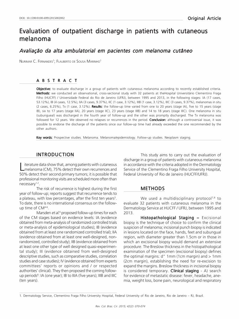
Objective:Objective:Objective:Objective:Objective: to evaluate discharge in a group of patients with cutaneous melanoma according to recently established criteria.
Methods:Methods:Methods:Methods:Methods: we conducted an observational, cross-sectional study with 32 patients at theHospital Universitário Clementino Fraga
Filho (HUCFF) / Universidade Federal do Rio de Janeiro (UFRJ), between 1995 and 2013, in the following stages: IA (17 cases,
53.12%), IB (4 cases, 12.5%), IIA (3 cases, 9.37%), IIC (1 case, 3.12%), IIIB (1 case, 3.12%), IIIC (3 cases, 9.37%), melanomas in situ
(2 cases, 6.25%), Tx (1 case, 3.12%). Results:Results:Results:Results:Results: the follow-up time varied from one to 20 years (stage IA), five to 15 years (stage
IB), six to 17 years (stage IIA), 20 years (stage IIC), 23 years (stage IIIB) and 14 to 18 years (stage IIIC). One melanoma in situ
(subungueal) was discharged in the fourth year of follow-up and the other was promptly discharged. The Tx melanoma was
followed for 12 years. We observed no relapses or recurrences in the period. Conclusion:Conclusion:Conclusion:Conclusion:Conclusion: although a controversial issue, it was
possible to endorse the discharge of the patients since our follow-up time had already exceeded the one recommended by the
other authors.
Key words:Key words:Key words:Key words:Key words: Prospective studies. Melanoma. Melanoma/epidemiology. Follow-up studies. Neoplasm staging.
1. Dermatology Service, Clementino Fraga Filho University Hospital, Federal University of Rio de Janeiro, Rio de Janeiro – RJ, Brazil.
INTRODUCTIONINTRODUCTIONINTRODUCTIONINTRODUCTIONINTRODUCTION
Literature data show that, among patients with cutaneousmelanoma (CM), 75% detect their own recurrences and
50% detect their second primary tumors; it is possible thatprofessional monitoring visits are scheduled more often thannecessary1.2.
The risk of recurrence is highest during the firstyear of follow-up; reports suggest that recurrence tends toa plateau, with low percentages, after the first ten years3.To date, there is no international consensus on the follow-up time of CM4-6.
Marsden et al.6 proposed follow-up times for eachof the CM stages based on evidence levels: IA (evidenceobtained from meta-analysis of randomized controlled trialsor meta-analysis of epidemiological studies); IB (evidenceobtained from at least one randomized controlled trial); IIA(evidence obtained from at least one well-designed, non-randomized, controlled study); IIB (evidence obtained fromat least one other type of well designed quasi-experimen-tal study); III (evidence obtained from well-designeddescriptive studies, such as comparative studies, correlationstudies and case studies); IV (evidence obtained from expertscommittees’ reports or opinions and / or respectedauthorities’ clinical). They then proposed the coming follow-up periods6: IA (one year); IB to IIIA (five years); IIIB and IIIC(ten years).
This study aims to carry out the evaluation ofdischarge in a group of patients with cutaneous melanomain accordance with the criteria adopted in the DermatologyService of the Clementino Fraga Filho University Hospital,Federal University of Rio de Janeiro (HUCFF/UFRJ).
METHODSMETHODSMETHODSMETHODSMETHODS
We used a multidisciplinary protocol7,8 toevaluate 32 patients with cutaneous melanoma in theDermatology Service at HUCFF / UFRJ, between 1995 and2013.
Histopathological Staging –Histopathological Staging –Histopathological Staging –Histopathological Staging –Histopathological Staging – Excisionalbiopsy is the technique of choice to confirm the clinicalsuspicion of melanoma; incisional punch biopsy is indicatedin lesions located on the face, hands, feet and subungualregion, with diameter greater than 1.5cm or in those inwhich an excisional biopsy would demand an extensiveprocedure. The Breslow thickness in the histopathologicalexamination of the specimen (excisional biopsy) definesthe optimal margins: d” 1mm (1cm margin) and > 1mm(2cm margin), establishing the need for re-excision toexpand the margins. Breslow thickness in incisional biopsyis considered temporary. Clinical stagingClinical stagingClinical stagingClinical stagingClinical staging - A) searchfor evidence of metastatic disease: fever, headache, ane-mia, weight loss, bone pain, neurological and respiratory
DOI: 10.1590/0100-69912015002002

Fe rnandesFe rnandesFe rnandesFe rnandesFe rnandesEvaluation of outpatient discharge in patients with cutaneous melanoma 71
Rev. Col. Bras. Cir. 2015; 42(2): 070-074
signs and symptoms. B) physical examination of the skin(transit metastasis: lesions in the lymph drainage area,more than 5 cm distant from the origin of the primarytumor; satellitosis - lesions around the tumor at a 5 cmradius C) physical examination of regional lymph nodes:.Impalpable (clinically hidden) – tomographic evaluationand / or ultrasound of the lymph node is performed whendoubts arise on palpation; palpable: macrometastasis,clinically detectable, is confirmed by therapeutic lymphnode dissection; the commitment is classified accordingto the number of metastatic lymph nodes, 1, 2-3 and e”4being the cutt-off limits. D) general physical exam: liver,spleen and especially the central nervous system; andlaboratory tests – in the absence of signs and symptomsof metastasis: blood count, ESR, glucose, urea, creatinineand lactate dehydrogenase (LDH), liver function tests, x-ray of pleuropulmonary fields; in patients with metastasesdetected by clinical examination, the following tests areadded: CT scan of chest, abdomen, pelvic cavity, andbone scintigraphy. Skeletal radiography is added if thebone scan reveals changes.
RESULTSRESULTSRESULTSRESULTSRESULTS
We evaluated 32 patients, predominantly female(21) and white (28), aged between 40 and 70 years old(29). The lesions were most commonly found in the head(8 cases) and trunk (11) (Table 1).
The cases were grouped into the following stages:IA (17 cases, 53.12%); IB (4 cases, 12.5%); IIA (3 cases,9.37%); IIC (1 case, 3.12%); IIIB (1 case, 3.12%); IIIC (3cases, 9.37%) (Tables 2 and 3).
Outpatient follow-up ranged from one to 20 years(stage IA), five to 15 years (stage IB), six to 17 years (stageIIA), 20 years (stage IIC), 23 years (stage IIIB) and 14-18years (stage IIIC).
DISCUSSIONDISCUSSIONDISCUSSIONDISCUSSIONDISCUSSION
The routine for CM used to be based in the stagingand follow-up indefinitely, except for in situ melanomasthat were discharged (Table 2). In 2013, the retrospectiveanalysis showed: the Breslow thickness was not determined(Tx) in one patient. In some circumstances, this evaluationindex is partially damaged or becomes less accurate. TheAJCC 2002/2009 does not define the conduct in melanomasTx9. Follow-up lasted 12 years (Table 2).
According to our protocol7.8, those with in situmelanoma (isM) are discharged, a conduct alsorecommended by Marsden et al.6. We observed two suchpatients (6.25%): one white, 46 years old, with a lesionlocated on the back; and one black, 40 years old, withdystrophy and overall darkening of the nail plate of thesecond right finger (subungual melanoma), evolving froma striated melanonychia; this patient underwent biopsy ofthe proximal nail fold and nail matrix. Amputation wasindicated, since the excision with safety margins and thepreservation of the finger functionality are not alwaysfeasible. The surgical specimen revealed melanoma in situ.Although our conduct in patients with melanoma in situ isdischarge, we followed the observations of Tan et al.10,who studied the initial stage of the subungual melanomain 121/124 cases: 11 (9%), stage 0 ; 16 (14%), stage I; 50(41%), stage II; 30 (32%), stage III; and five (4%) stage IV.Nine of the 11 patients with isM were followed on averagefor 35 months. Our patient was discharged on the fourthyear of monitoring (Table 2). Tan et al.10 pointed out thatthe accurate measurement of Breslow thickness may bedifficult in acral melanoma and, in particular, the subungualmelanoma, since the healthy nail matrix does not have agranular layer and the subcutaneous fat may be absent inthe subungual area.
Among the 17 stage IA patients, five had newisM after one, two, four, six and 11 years of the initial
Table 1 Table 1 Table 1 Table 1 Table 1 - Distribution of cutaneous melanoma by age group, sex, color and location.
Age GroupAge GroupAge GroupAge GroupAge Group GenderGenderGenderGenderGender Co lo rCo lo rCo lo rCo lo rCo lo r Locat ionLocat ionLocat ionLocat ionLocat ion
FFFFF MMMMM WWWWW NWNWNWNWNW HeadHeadHeadHeadHead TrunkTrunkTrunkTrunkTrunk UpperUpperUpperUpperUpper LowerLowerLowerLowerLower FootFootFootFootFoot HandHandHandHandHand
L imbL imbL imbL imbL imb L imbL imbL imbL imbL imb
20-30 years old - 1 1 - - 1 - - - -
31-40 years old 2 - 1 1 1 1 - - - -
41-50 years old 4 4 7 1 2 3 - 2 - 1
51-60 years old 6 2 6 2 1 2 1 1 3 -
61-70 years old 6 1 7 - 3 1 1 1 1 -
71-80 years old 2 3 5 - 1 2 1 - 1 -
81-90 years old 1 - 1 - 0 1 - - - -
TOTAIS 21 11 28 4 8 11 3 4 5 1
Source: HUCFF/UFRJ (1995-2013)Conventions: W - white; NW - not white; F - female; M - male.

72
Rev. Col. Bras. Cir. 2015; 42(2): 070-074
Fe rnandesFe rnandesFe rnandesFe rnandesFe rnandesEvaluation of outpatient discharge in patients with cutaneous melanoma
diagnosis, respectively. The follow-up ranged from one to20 years (Table 2). One patient (stage IB) submitted a newcutaneous melanoma two years after the first diagnosis.The four patients with stage IB were followed for five years(Table 2).
Considering the three patients included in theIIA stage, there were two acral lentiginous melanomas andone located in the chest. One of the acral lentiginousmelanomas showed a neurotropic histological type – whitefemale patient, 80 years old, left plantar region, evolutionof 30 years. The neurotropic variant is composed of spindlecells with a pattern like a neuroma and tendency forcircumferential distribution around small nerve fibers in thedeep dermis and hypodermis. As a uniqueclinicopathological variant, it presents in the form of apigmented or amelanotic nodule of rapid growth. Thefollow-up ranged from six to 17 years (Table 2).
One IIC patient(white, male, 50 years old, withnodular melanoma in his left knee and positive left inguinalsentinel lymph node) underwent lymph node dissection andthen followed for 20 years without recurrence or relapse(Table 2). One IIIB patient (white, male, 26 year old, with
nodular melanoma on the back) was followed for 23 yearswithout recurrence or relapse (Table 2).
Stage III melanoma is associated with high riskof recurrence and mortality. A retrospective study showedfive-year survival without disease in the percentages of 63%(IIIA) and 32% (IIIB)11. Early recurrence sites were: local /regional transit (28%), regional lymph node (21%) andsystemic (51%).
Three patients were classified as IIIC: a) non-white, female, 63 years old, with acral lentiginous melanomain the left plantar region, positive sentinel lymph node,submitted to inguinal lymph node dissection, monitored for14 years without recurrence or relapse (Table 2); b) white,male, 76 years old, melanoma in the sternal region withmetastatic lymph node in the right axillary region; lymphnode dissection, followed for 14 years without recurrenceor emergence of a new tumor (Table 2); c) white, male, 51year old, acral lentiginous melanoma on the right heel. Heshowed two lymph node metastasis in the right inguinallymph node chain and one in the right aortoiliac chain,having been followed for 18 years, without recurrence orappearance of new tumors (Table 2).
Table 2 Table 2 Table 2 Table 2 Table 2 - Distribution of cases according to staging and follow-up (HUCFF).
StageStageStageStageStage NumberNumberNumberNumberNumber Outpatient Follow-upOutpatient Follow-upOutpatient Follow-upOutpatient Follow-upOutpatient Follow-up T imeT imeT imeT imeT ime
of casesof casesof casesof casesof cases Rout ineRout ineRout ineRout ineRout ine
Tx 1 § not-defined 12 years
Tis 2 § resection with a 0.5 cm margin Discharge
§ discharge 4 years
I A 17 § Resection: 1.0 cm margin
§ dermatological and lymph node examination: every six months (first two years) 1,2,4,5,7,10, 12
and then annually 14,15,16,20 years
I B 4 § Resection: 2,0 cm margin
§ Dermatological and lymph node examination: every two months (two years) 5, 5, 11, 15 years
and thereafter, every six months
§ X-ray of chest and liver function tests: every six months (two years)
and then annually
II A 3 I B 6, 15, 17 years
II C 1 § Resection: 2,0 cm margin
§ Dermatological and lymph node examination: every two months (two years)
and thereafter, every six months 20 anos
§ X-ray of chest and liver function tests: every six months (two years)
and then annually 20 years
III B 1 § Clinical examination every four months
§ X-ray of chest and liver function tests: every six months
§ Imaging tests targeted to the region where there is relapse 23 years
every four months
§ Resection of limited locoregional and visceral metastases
§ individualized chemotherapy
III C 3 III B 14, 18 years

Fe rnandesFe rnandesFe rnandesFe rnandesFe rnandesEvaluation of outpatient discharge in patients with cutaneous melanoma 73
Rev. Col. Bras. Cir. 2015; 42(2): 070-074
Local recurrences are defined as tumor relapsewithin 3 to 5 cm from the primary closure or graft and areconsidered to be rare (3.2%). The ulceration and thethickness of the primary tumor, as well as the location inthe head and neck, are considered predisposing factors 7.
Although the subject is controversial and ourmonitoring time exceeded the period adopted by somecenters6,12, we concluded that it was possible to endorsethe discharge of patients in stages IA, IB, IIA, IIC, IIIB andIIIC.
R E S U M OR E S U M OR E S U M OR E S U M OR E S U M O
Objetivo: Objetivo: Objetivo: Objetivo: Objetivo: realizar a avaliação da alta em um grupo de pacientes com melanoma cutâneo de acordo com critérios recente-
mente estabelecidos. Métodos:Métodos:Métodos:Métodos:Métodos: estudo observacional de corte transversal de 32 pacientes com melanoma cutâneo atendidos
no HUCFF/UFRJ, entre 1995 e 2013, nos seguintes estágios: IA (17 casos/53,12%), IB (4 casos/12,5%), IIA (3 casos/9,37%), IIC (1
caso/3,12%), IIIB (1 caso/3.12%), IIIC (3 casos/9,37%), melanomas in situ (2 casos/6,25%), Tx (1 caso/3,12%). Resultados:Resultados:Resultados:Resultados:Resultados: o
tempo de seguimento ambulatorial variou de um a 20 anos (estágio IA), cinco a 15 anos (estágio IB), de seis a 17 anos (estágio
IIA), 20 anos (estágio IIC), 23 anos (estágio IIIB) e de 14 a 18 anos (estágio IIIC). O melanoma Tx foi acompanhado por 12 anos,
um melanoma in situ teve alta imediata e outro, subungueal, permaneceu em acompanhamento por quatro anos. Não foram
observadas recidivas ou recurrências. Conclusão:Conclusão:Conclusão:Conclusão:Conclusão: houve adequação do procedimento de alta nos estágios IA, IB, IIA, IIC, IIIB e
IIIC.
Descritores:Descritores:Descritores:Descritores:Descritores: Estudos prospectivos. Melanoma. Melanoma/epidemiologia. Seguimentos. Estadiamento de neoplasias.
Table 3 Table 3 Table 3 Table 3 Table 3 - Staging of cutaneous melanoma (AJCC 2002/2009)9.
S tageStageStageStageStage Tumor (T)Tumor (T)Tumor (T)Tumor (T)Tumor (T) Lymph nodes (N)Lymph nodes (N)Lymph nodes (N)Lymph nodes (N)Lymph nodes (N) Metastasis (M)Metastasis (M)Metastasis (M)Metastasis (M)Metastasis (M)
I A < 1mm Ø Ø
Clark II/III
without histological ulceration
I B < 1mm
Clark IV/V
with histological ulceration Ø Ø
1,01 – 2 mm
without histological ulceration
II A 1,01 – 2 mm
with histological ulceration Ø Ø
2,01 – 4 mm
without histological ulceration
IIB 2,01 – 4 mm with ulceration Ø Ø
> 4mm without ulceration
II C > 4mm with histological ulceration Ø Ø
IIIA <1mm a >4mm without ulceration Ø Ø
<1mm a >4mm 1 micrometastasis Ø
Without ulceration 1 a 3 micrometastases Ø
III B <1mm a >4mm with or without histological 1 a 3 micrometastases Ø
ulcerationtransit metastasissatellitosis 2 a 3 macrometastases
III C <1mm a >4mm Confluent lymph nodes Ø
histological ulcerationany thick 1 macrometastasis
nesssatellitosistransit metastasis 2 to 3 or more than
4 macrometastases
74
Rev. Col. Bras. Cir. 2015; 42(2): 070-074
Fe rnandesFe rnandesFe rnandesFe rnandesFe rnandesEvaluation of outpatient discharge in patients with cutaneous melanoma
REFERENCESREFERENCESREFERENCESREFERENCESREFERENCES
1. Turner RM, Bell KJ, Morton RL, Hayen A, Francken AB, Howard K,et al. Optimizing the frequency of follow-up visits for patientstreated for localized primary cutaneous melanoma. J Clin Oncol.2011;29(35):4641-6.
2. Sondak VK, Leachman SA. Individualizing follow-up for patientswith early-stage melanoma. J Clin Oncol. 2011;29(35):4606-8.
3. Francken AB, Accortt NA, Shaw HM, Colman MH, Wiener M,Soong SJ, et al. Follow-up schedules after treatment for malignantmelanoma. Br J Surg. 2008;95(11):1401-7.
4. Garbe C, Peris K, Hauschild A, Saiag P, Middleton M, Spatz A, etal. Diagnosis and treatment of melanoma. European consensus-based interdisciplinary guideline—Update 2012. Eur J Cancer.2012;48(15):2375-90.
5. Dummer R, Guggenheim M, Arnold AW, Braun R, von Moos R;Project Group Melanoma of the Swiss Group for Clinical CancerResearch. Updated Swiss guidelines for the treatment and follow-up of cutaneous melanoma. Swiss Med Wkly. 2011;141:w13320.
6. Marsden JR, Newton-Bishop JA, Burrows L, Cook M, Corrie PG,Cox NH, et al. Revised U.K. guidelines for the management ofcutaneous melanoma 2010. Br J Dermatol. 2010;163(2):238-56.
7. Fernandes NC, Calmon R, Maceira JP, Cuzzi T, Silva CSC. Melanomacutâneo: estudo prospectivo de 65 casos. An Bras Dermatol.2005;80(1):25-34.
8. Fernandes NC, Calmon R. Melanoma cutâneo: estudo prospectivode 42 casos. 2010;86(6):1233-5.
9. Balch CM, Gershenwald JE, Soong SJ, Thompson JF, Atkins MB,Byrd DR, et al. Final version of 2009 AJCC melanoma staging andclassification. J Clin Oncol. 2009;27(36):6199-206.
10. Tan KB, Moncrieff M, Thompson JF, McCarthy SW, Shaw HM,Quinn MJ, et al. Subungueal melanoma: a study of 124 caseshighlighting features of early lesions, potential pitfalls in diagnosis,and guidelines for histologic reporting. Am J Surg Pathol.2007;31(12):1902-12.
11. Romano E, Scordo M, Dusza SW, Coit DG, Chapman PB. Site andtiming of first relapse in stage III melanoma patients: implicationsfor follow-up guidelines. J Clin Oncol. 2010;28(18):3042-7.
12. Leiter U, Eigentler TK, Forschner A, Pflugfelder A, Weide B, Held L,et al. Excision guidelines and follow-up strategies in cutaneousmelanoma: Facts and controversies. Clin Dermatol. 2010;28(3):311-5.
Received on 05/042014Accepted for publication 03/06/2014Conflict of interest: none.Source of funding: none.
Address for correspondence:Address for correspondence:Address for correspondence:Address for correspondence:Address for correspondence:Nurimar C. FernandesE-mail:[email protected]

Rodr iguesRodr iguesRodr iguesRodr iguesRodr iguesImportance of flexible bronchoscopy in decannulation of tracheostomy patients 75
Rev. Col. Bras. Cir. 2015; 42(2): 075-080
Original ArticleOriginal ArticleOriginal ArticleOriginal ArticleOriginal Article
Importance of flexible bronchoscopy in decannulation ofImportance of flexible bronchoscopy in decannulation ofImportance of flexible bronchoscopy in decannulation ofImportance of flexible bronchoscopy in decannulation ofImportance of flexible bronchoscopy in decannulation oftracheostomy patientstracheostomy patientstracheostomy patientstracheostomy patientstracheostomy patients
Importância da broncoscopia flexível na decanulação dos pacientesImportância da broncoscopia flexível na decanulação dos pacientesImportância da broncoscopia flexível na decanulação dos pacientesImportância da broncoscopia flexível na decanulação dos pacientesImportância da broncoscopia flexível na decanulação dos pacientestraqueostomizadostraqueostomizadostraqueostomizadostraqueostomizadostraqueostomizados
LEONARDO BRAND RODRIGUES1,2; TARCIZO AFONSO NUNES1
A B S T R A C TA B S T R A C TA B S T R A C TA B S T R A C TA B S T R A C T
ObjectiveObjectiveObjectiveObjectiveObjective: To evaluate the importance of flexible bronchoscopy in tracheostomy patients in the process of decannulation to
assess the incidence and types of laryngotracheal injury and compare the presence of such lesions with clinical criteria used for
decannulation. MethodsMethodsMethodsMethodsMethods: We studied 51 tracheostomized patients aged between 19 and 87 years, with tracheal stent for a
mean of 46 ± 28 days and with clinical criteria for decannulation. They were submitted to tracheostomy tube occlusion tolerance
testfor 24 hours, and then to flexible bronchoscopy. We described and classified the diagnosed laryngotracheal changes. We
compared the clinical criteria for decannulation indication with the bronchoscopy-diagnosed laryngotracheal injuries that
contraindicated decannulation. We identified the factors that could interfere in decannulation and evaluated the importance of
bronchoscopy as part of the process. ResultsResultsResultsResultsResults: Forty (80.4%) patients had laryngotracheal alterations. Of the 40 patients
considered clinically fit to decannulation, eight (20%) (p = 0.0007) presented with laryngotracheal injuries at bronchoscopy that
contraindicated the procedure. The most frequent laryngeal alteration was vocal cords lesion, in 15 (29%) individuals, and
granuloma, the most prevalent tracheal lesion, in 14 (27.5%) patients. ConclusionConclusionConclusionConclusionConclusion: flexible bronchoscopy showed a large
number of laryngotracheal injuries, the most frequent being the vocal cords injury in the larynx and the granuloma in the trachea,
which contributed to increase the decannulation procedure safety.
Key words:Key words:Key words:Key words:Key words: Bronchoscopy. Tracheostomy. Tracheal diseases. Tracheomalacia. Intubation, Intratracheal.
1. Faculty of Medicine, Universidade Federal de Minas Gerais; 2. Hospital Odilon Behrens.
INTRODUCTIONINTRODUCTIONINTRODUCTIONINTRODUCTIONINTRODUCTION
Tracheostomyis performed in about 20% of patients whoare on mechanical ventilation in the intensive care unit1.
It is indicated to increase comfort and facilitate weaning2,reducing the rate of laryngotracheal complications causedby the long permanence of the orotracheal tube3, and a s asafe airway in cases of obstruction of the upper airways.However, the presence of tracheostomy causesbronchorrhea, changes in the swallowing mechanism4,increased risk of airway infection and bleeding and hampersvocalization5,6. Late complications are diagnosed in 65%of patients, the most frequent being the granuloma,followed by lesions with high morbidity and mortality suchas malacia, stenosis, and vascular and esophageal fistulas7,8.To avoid these complications, the patient decannulationshould be performed as early as possible.
Proper patient assessment before the removal ofthe cannula has been neglected5,9, and the literature islacking in studies that indicate the criteria and the besttime to carry this out10. Decannulation failure is characterizedwhen it is necessary to reintroduce the artificial airway inthe 48 hours following the removal of the tracheal cannula.
This occurs in up to 5% of cases and may be associatedwith acute respiratory failure6,10.
The stringent multidisciplinary clinical evaluationassociated with anatomical and physiological assessmentof the larynx and trachea contributes to select patients whomay be decannulated with more chances of success. Theexamination by flexible bronchoscopy is important to helpdecide on the time of decannulation, but is little used andwithout a detailed protocol10,11.
This study aimed to evaluate the importance offlexible bronchoscopy in tracheostomy patients in the processof decannulation to know the incidence and types oflaryngotracheal injuries and to compare the presence ofsuch lesions with the clinical criteria used for decannulation.
METHODSMETHODSMETHODSMETHODSMETHODS
This was a prospective study in patients intracheostomy decannulation process at the Hospital OdilonBehrens, in Belo Horizonte – MG. The study was approvedby the Departmental Board of the Department of Surgeryof FM-UFMG and by the Ethics in Research Committee of
DOI: 10.1590/0100-69912015002003

76
Rev. Col. Bras. Cir. 2015; 42(2): 075-080
Rodr iguesRodr iguesRodr iguesRodr iguesRodr iguesImportance of flexible bronchoscopy in decannulation of tracheostomy patients
the Odilon Behrens County Hospital (FR 301247). All patientsagreed to participate and signed a free and informedconsent.
Sample calculationSample calculationSample calculationSample calculationSample calculationTo calculate the sample size, we used the records
of the Department of Thoracic Surgery at the Odilon BehrensHospital. We analyzed data of patients who met clinicalcriteria for decannulation and were referred to bronchoscopyto evaluate decannulation. Eighteen (72%) tolerated theocclusion of the cannula, and in three, bronchoscopydiagnosed laryngotracheal injuries contraindicatingdecannulation (16.6% failure). So we used the hypothesistest for a proportion that considers the binomial distributionfor sample power calculation 12, considering a power of80% with a 5% significance level and estimated the totalsize of the sample patients.
Sample characterizationSample characterizationSample characterizationSample characterizationSample characterizationWe studied patients over 18 years, from March
2010 to January 2011, who met the following inclusioncriteria: clinical stability, spontaneous ventilation for at least48 hours; absence of infection at the time of decannulationindication; absence of new surgical procedure in the samehospital; effective coughing and swallowing; Glasgow comascale > 8. The patients were examined by a multidisciplinaryteam including physicians, physical therapists, speechtherapists and nurses. In order to be uniformity in theassessments of patients, the multidisciplinary team attendeda continuing education program, which extendedthroughout the period of the survey data collection. Thesample comprised 51 patients, 26 female and 25 male,median age 55 years (19-87 years), 22 brown, 19 whiteand ten black.
Four (7.8%) patients reported using illicit drugs.Associated diseases were diagnosed in 45 (88.2%) patients,with prevalence of diabetes mellitus (23.5%). Only seven(13.72%) patients had complications related totracheostomy and overcame the cannulation difficulty(11.8%). The most prevalent clinical condition that led totracheal intubation or tracheostomy was stroke (27.5%),followed by pneumonia (19.6%), surgery, trauma, sepsisand airway obstruction (each corresponding to 4% ofpatients). Periods of tracheal stent and mechanicalventilation can be seen in figure 1.
Composition of groupsComposition of groupsComposition of groupsComposition of groupsComposition of groupsPatients who met the inclusion criteria underwent
placement of a standard number 4 metal cannula, Fadel-Med® brand, with an 7.5 mm internal diameter of, 10mmexternal and 7cm length, regardless of the cannula theywere previously using. The cannula remained occluded for24 hours, during which the patients were evaluated forchest expansion, breathing frequency and pattern, lungauscultation, heart rate, pulse and blood pressure. Patientsshould present with parameters better or equal to the ones
found before cannula occlusion. Thus, we divided patientsin two groups, based on the results of the tracheostomycannula occlusion: Group A – tolerated; and group B –non-tolerated. We considered that patients of group A metthe clinical criteria of decannulation and group B did notpresent these criteria.
BronchoscopyBronchoscopyBronchoscopyBronchoscopyBronchoscopyWe subjected patients in Groups A and B to
laryngotracheobronchial endoscopy by the same examinerafter 24 hours of cannula occlusion. The average periodbetween cannula occlusion and the procedure was 1.7 days(1-7 days). The procedure was performed in thebronchoscopy room, with a flexible bronchoscope (Olympus,model BF-P60, optical, 4.9mm working channel externaldiameter) and local anesthesia with 10% lidocaine sprayat a dose of 30mg, 5ml of lidocaine jelly in the nasal cavityand 1% lidocaine without vasoconstrictor. The cannula wasremoved to facilitate tracheal examination, as well as todynamically assess of forced expiration and inspiration. Fordynamic obstructions, such as tracheomalacia, weconsidered the lowest tracheal diameter during forcedexpiration. To assess the obstruction of the tracheal lumen,we employed the Cotton classification13. Patients with vo-cal cords bilateral lesions in adduction or subglottic ortracheal obstructiongrade II Cotton or higher (Figure 2) wereconsidered endoscopically unfit for decannulation11.
Upon completion of bronchoscopy, and based onclinical evaluation, groups A and B were subdivided into fourgroups: A1, B1, A2 and B2. Patients in group A1 weredecannulated after bronchoscopy and remained in hospital underobservation for at least 48 hours. Patients in the B1 group werereassessed after clinical improvement, with subsequentdecannulation after they tolerated a new occlusion period.Patients in the A2 and B2 groups remained tracheostomized,with an appropriate cannula for each identified lesion, and werereferred to the Thoracic Surgery Clinic.
Variables studied and statistical testsVariables studied and statistical testsVariables studied and statistical testsVariables studied and statistical testsVariables studied and statistical testsWe described and classified the laryngotracheal
lesions identified at bronchoscopy and expressed the result
Figure 1Figure 1Figure 1Figure 1Figure 1- Periods of tracheal cannula and mechanical ventilation.
* OTT - tracheal tube, TCT - tracheostomy, MV - mechanical ventilation.

Rodr iguesRodr iguesRodr iguesRodr iguesRodr iguesImportance of flexible bronchoscopy in decannulation of tracheostomy patients 77
Rev. Col. Bras. Cir. 2015; 42(2): 075-080
as percentage. We then compared patients’ groups formedby decannulation clinical and bronchoscopic criteria.
The highest decannulation failure rate describedamong patients who fulfilled the clinical criteria fordecannulation is 5%9. Bilateral lesions of the vocal cordsand / or tracheal obstruction greater than Cotton grade IIat bronchoscopy are at higher risk of respiratory failurewithout the use of tracheostomy. Hence, we comparedthe evolution ofgroups A1, A2, B1 and B2 by employingFisher’s test14.
Bronchoscopy is the best test for diagnosinglaryngotracheal changes that contraindicate decannulation,so we carried out the analysis of clinical efficacy criteria bycomparison between the predictive value found afterbronchoscopic validation and the one described in theliterature – 95% decannulation success. We considered nullhypothesis (H
0) the positive predictive value of clinical criteria
equal or greater than 95%, and as an alternative hypothesis(H
A) the positive predictive value being lower than 95%15.
RESULTSRESULTSRESULTSRESULTSRESULTS
Nine (17.6%) patients had no laryngotrachealchanges. In 42 (82.4%) lesions were diagnosed, 20 with(39.2%) one and 22 (43.1%) with two or more lesions, asdescribed: paresis or paralysis of the vocal cord in adductionor abduction in 15 (29%) patients, eight (15.7%) havingbilateral lesions. All of them were associated with paresisof the corresponding hemilarynx; scar tissue suggestinggranuloma in 14 (27.5%) patients, all located in thetracheostoma, determining grade I obstruction; depressionof the anterior wall of the tracheostoma in six (11.8%)patients, determining tracheal grade I obstruction;tracheostoma in improper anatomical position, lateral tothe midline of the trachea anterior wall in ten (19.6%)patients; laryngotracheal obstruction in 22(43.1%), tracheomalacia in 12 (60%), five (25%)laryngotracheomalacias and five (25%) stenoses. Accordingto the Cotton classification, we foundthe following degrees
of obstruction: Nine (17.6%) grade I, nine (17.6%) gradeII, two (3.9%) grade III and two (3.9%) grade IV.
Forty patients tolerated cannula occlusion, butbronchoscopy diagnosed laryngotracheal injuries in eight(20%), for whom we contraindicated decannulation. Ofthe 11 patients who did not tolerate occlusion of the cannula,bronchoscopy showed no injuries that preventeddecannulation in two (18.2%). Ten (19.6%) patientsbenefited from bronchoscopy, since it decreased the risk ofdecannulation failure in eight and avoided cannulapermanence in two (Table 1). By employing bronchoscopyas one decannulation criteria, we found 20% oflaryngotracheal injuries that could determine failure in thedecannulation process. Thus, considering the binomialdistribution, we rejected the null hypothesis that 95% ofpatients who meet the clinical criteria tolerate decannulation(p <0.007). The decannulated patients who met the clinicaland bronchoscopic criteria for decannulation had nocomplications and required no new tracheal cannula.
DISCUSSIONDISCUSSIONDISCUSSIONDISCUSSIONDISCUSSION
In the period when we conducted the survey,240 patients were submitted to tracheostomy and only 51(21.3%) met the clinical criteria for decannulation6,10. Thisvariation can be explained by the difference in the methodused for patients inclusion in the various studies, as therewas difference in the number and types of clinical criteriaused to define the patient as clinically fit todecannulation5,6,10,16. While in these studies patientsremained with tracheostomy for prolonged periods (averageof up to 147 days)17, in this study the average time was 33days.
Lesions that affect 50% or more of the trachealdiameter are a contraindication to decannulation. From thisdegree of obstruction on, marked changes in pulmonaryfunction tests may occur, with clinical repercussions10,18,19.One study, however, considers thatobstructions aresignificant when affecting 20% of the tracheal diameter17.
Table 2 Table 2 Table 2 Table 2 Table 2 - Cotton Classification13 according to the percentage of tracheal lumen obstruction.

78
Rev. Col. Bras. Cir. 2015; 42(2): 075-080
Rodr iguesRodr iguesRodr iguesRodr iguesRodr iguesImportance of flexible bronchoscopy in decannulation of tracheostomy patients
In adults, the tracheal diameter It is 20 mm in womenand23 mm in men8, which is why we used a number 4 metalcannula with 10mm external diameter at the time ofdecannulation, since when occluded, It represents about50% of tracheal lumen obstruction.
We consider flexible bronchoscopy necessarybefore decannulation, like other authors17,20, due to themethod’s sensitivity in diagnosing laryngotrachealanatomical and functional lesions21 that are common intracheostomy patients. This test is considered safe and itscomplication rates are less than 1%22. In the present studythere were no complications that prevented laryngotrachealassessment or that worsened patients’ clinical status.
Granulomas were found in 27.5% of patients,all in the tracheostoma region, without determining airflowobstruction (Cotton Grade I). We foundlaryngotracheomalacias and laryngotracheal stenoses in33.3% and 9.8% of patients, respectively, regardless ofthe degree of obstruction they caused. Some authors haveobserved similar findings when using flexible bronchoscopyas a criterion for decannulation7,17.
Lesions that determined Cotton grades II, III andIV obstructions were diagnosed in 25.5% of patients. Wedid not find fistulas or bulky bleeding, which is in accordancewith the literature7,17. We found changes in vocal cords in29.4% of patients,in eight patients the lesions were bilate-ral, and in 87.5% of the lesions the vocal cords were inadduction.
Patients who met the decannulation clinicalcriteria, but not the bronchoscopic ones, could have beendecannulated without evolving with respiratory failure. Oneshould consider that patients confined to bed or who, forother reasons, did not perform physical exertion, and mightnot present respiratory failure, even with obstructions largerthan 50% of the tracheal lumen or bilateral vocal cord
paresis in adduction. This fact may have contributed toexplain the difference between the 20% considered asfailure, found in this study, and 5% described in theliterature17. However, we considered that the decannulationof specific cases would be better evaluated after prolongedfollow-up of the patient and recovery of symptoms, whichdetermined the alternative airway maintenance. Consideringthe clinical criteria for the decannulation, bronchoscopybenefited ten (19.6%) patients, since it contraindicateddecannulation in eight and identified two who did not havelesions that would contraindicate decannulation by clinicalcriteria. One study18 found that only the clinical criteria wouldbe sufficient and safe to indicate decannulation. However,the author has employed a method different from oursregarding the sample, the inclusion criteria, the cannula tobe occluded with large diameter, the difference in thedescription of laryngotracheal injuries, as well as the differentbronchoscopy diagnoses. Thus, the comparison of the resultsrendered inadequate.
Clinical circumstances which led to trachealintubation or tracheostomy, associated diseases and agedid not influencedecannulation, which is in line with theresults of the literature17. Nonetheless, the number ofpatients was insufficient for statistical evaluation on theserelationships. Patients with diabetes mellitus, however, weremore likely to decannulation contraindication bybronchoscopy, even when meeting the clinical criteria (p =0.04). This fact can be explained by changes in scarringmechanisms, since the four diabetic patients with favorabledecannulation clinical criteria had unfavorablebronchoscopic ones due to the presence of tracheomalacia.We did not find this data in the literature.
Among the 15 patients with vocal cord lesionsand 22 who presented laryngotracheal stenosis, the averagetime they remained with the tracheal tube was 10.06 days.
Table 1 -Table 1 -Table 1 -Table 1 -Table 1 - Description of patients fit fordecannulation through clinical criteria, but contraindicated by Bronchoscopy
(n = 8).
Patients AgePatients AgePatients AgePatients AgePatients Age Assoc ia te rAssoc ia te rAssoc ia te rAssoc ia te rAssoc ia te r Clinical conditionsClinical conditionsClinical conditionsClinical conditionsClinical conditions P la s t i cP l a s t i cP l a s t i cP l a s t i cP l a s t i c Bronchoscopyic laryngotrachealBronchoscopyic laryngotrachealBronchoscopyic laryngotrachealBronchoscopyic laryngotrachealBronchoscopyic laryngotracheal
( y ea r s )( y ea r s )( y ea r s )( y ea r s )( y ea r s ) D i seasesD i seasesD i seasesD i seasesD i seases that promptedthat promptedthat promptedthat promptedthat prompted Orthos i sOrthos i sOrthos i sOrthos i sOrthos i s changeschangeschangeschangeschanges
orotracheal intubationorotracheal intubationorotracheal intubationorotracheal intubationorotracheal intubation P e r i o dP e r i o dP e r i o dP e r i o dP e r i o d contraindicating decannulationcontraindicating decannulationcontraindicating decannulationcontraindicating decannulationcontraindicating decannulation
or tracheostomyor tracheostomyor tracheostomyor tracheostomyor tracheostomy ( day s )(day s )(day s )(day s )(day s )
1 26 _ Convulsive crisis 74 Bilateral paresis of vocals cords in adduction
2 81 DM, SAH, COPD Trauma 105 Grade II tracheomalacia
3 54 SAH, AMI Airway obstruction 24 Grade II tracheomalacia
4 88 DM, SAH, COPD, CRF, CHF STROKE 26 Grade II tracheomalacia
5 55 DM, SAH Sepsis 41 Grade II tracheomalacia
6 53 DM, SAH, muscular dystrophy Pneumonia 49 Grade II tracheomalacia
7 70 SAH, Obesity STROKE 30 Grade II tracheomalacia
8 64 COPD, CHF Decompensated CHF 25 Bilateral paresis of vocal cords in adduction
DM DM DM DM DM -diabetes mellitus; SAHSAHSAHSAHSAH -systemic arterial hypertension; COPDCOPDCOPDCOPDCOPD – chronic obstructive pulmonary disease; AMIAMIAMIAMIAMI -acute myocardialinfarction; CRFCRFCRFCRFCRF – chronic renal failure; CHFCHFCHFCHFCHF -congestive heart failure; STROKESTROKESTROKESTROKESTROKE -stroke.

Rodr iguesRodr iguesRodr iguesRodr iguesRodr iguesImportance of flexible bronchoscopy in decannulation of tracheostomy patients 79
Rev. Col. Bras. Cir. 2015; 42(2): 075-080
Even this being a long tracheal tube period, they showedno statistical difference when compared with patientswithout injuries. There are reports that the intubation timeincreases the number of laryngotracheal injuries and,therefore, the occurrence of decannulation failure, but thereis controversy17,23 and the number of patients of this studywas insufficient to contribute towards clarifying such doubts.Randomized studies are needed to better identify the factorsthat interfere in decannulation17. Whereas not only thetracheal tube, but any orthesis, can cause laryngotrachealdamage, upon analysis of the periods of tracheal cannulause evaluated in this study, only the longest period of plasticorthesis use (tracheal tube plus tracheostomy plasticcannula) related significantly with the diagnosis oflaryngotracheal injuries at bronchoscopy (p = 0.04).However, since this period interacts with other variables,such as the period between the tracheostomy and the endof mechanical ventilation, mechanical ventilation time,period of endotracheal tube use, period oftracheostomyplastic cannula use, and total orthesis period, including theperiod of metallic cannula (Figure 1), we used multiplelogistic regression to the group of variables related to theperiod of tracheal cannula usage and which had significanceless than 0.2524. The goal was to determine whether oneof the periods of total plastic orthesis alone could be relatedas cause and effect of the existence of laryngotracheal in-juries contraindicating decannulation. The adjustment oflogistic regression was affected by multicollinearity (positivecorrelation between the measured time variables) and noneof the variables were significant.
One limitation of this study is the use ofsubjective criteria for classifying injuries that cause airflowobstruction. The Cotton classification13 is based on directendoluminal observation, without a specific instrument tomeasure the area of tracheal lumen obstruction. Anotherlimitation encountered was the difficulty of patients’monitoring after hospital discharge. However, most of thecomplications that cause decannulation failure occur inthe first 48h9,25, during which the patients in this studywere hospitalized. The number of research subjects wassufficient to assess the benefit of performing bronchoscopyas a decannulation criterion. Nevertheless, we could notidentify the factors that make bronchoscopy indispensableor dispensable for decannulation. In this paper wedeveloped a protocol using clinical criteria that areconsensus among most researchers, together with the mostappropriate method for assessing the laryngotrachealregion 5,6,10,26, aimed at providing safety to the patient atthe time of decannulation. More prospective studies areneeded to determine the usefulness of bronchoscopy inthe evaluation of decannulation. Methods are needed todescribelaryngotracheal injuries in an objective andstandardized way, possibly altering the bronchoscopicdecannulation criteria by identifying which ones canactually be cause of failure.
Flexible bronchoscopy showed a large numberof laryngotracheal injuries, the more frequent being in thevocal cords, in the larynx and the tracheal granuloma, whichcontributed to increase the safety of the decannulationprocedure.
R E S U M OR E S U M OR E S U M OR E S U M OR E S U M O
Objetivo:Objetivo:Objetivo:Objetivo:Objetivo: avaliar a importância do emprego, da broncoscopia flexível nos pacientes traqueostomizados em vias de decanulação
para conhecer a incidência e os tipos de lesões laringotraqueais e comparar a presença destas lesões com os critérios clínicos
utilizados para a decanulação. Métodos: Métodos: Métodos: Métodos: Métodos: foram estudados 51 pacientes, com idade entre 19 e 87 anos, traquestomizados, com
critérios clínicos de decanulação e com tempo médio de órtese traqueal de 46 ± 28 dias. Foram submetidos ao teste de tolerância
à oclusão da cânula de traqueostomia por 24 horas, seguida da realização da broncoscopia flexível. As alterações laringotraqueais
diagnosticadas foram descritas e classificadas. Comparou-se a indicação de decanulação por critérios clínicos com o diagnóstico de
lesões laringotraqueais à broncoscopia que contraindicavam a decanulação. Identificaram-se os fatores que poderiam interferir na
decanulação e avaliou-se a importância da broncoscopia como parte do processo. ResultadosResultadosResultadosResultadosResultados: Apresentaram alterações
laringotraqueais, 40 pacientes (80,4%). Dos 40 pacientes considerados clinicamente aptos à decanulação, oito (20%) (p=0,0007)
apresentaram lesões laringotraqueais à broncoscopia que contraindicaram o procedimento. A alteração laríngea mais frequente foi
lesão de pregas vocais em 15 (29%) e o granuloma, a lesão traqueal mais prevalente em 14 (27,5%) pacientes. ConclusãoConclusãoConclusãoConclusãoConclusão: a
broncoscopia flexível evidenciou um número elevado de lesões laringotraqueais, sendo mais prevalentes a lesão de pregas vocais na
laringe e o granuloma na traqueia, que contribuiu para aumentar a segurança do procedimento de decanulação.
Descritores:Descritores:Descritores:Descritores:Descritores: Procedimentos Cirúrgicos Broncoscópicos. Traqueostomia. Traqueopatias. Traqueomalácia. Intubação Intratraqueal
REFERENCESREFERENCESREFERENCESREFERENCESREFERENCES
1. Esteban A, Anzueto A, Alía I, Gordo F, Apezteguía C, Pálizas F, etal. How is mechanical ventilation employed in the intensive careunit? An international utilization review. Am J Respir Crit CareMed. 2000;161(5):1450-8.
2. Blot F, Melot C; Commission d’Epidémiologie et de RechercheClinique. Indications, timing, and techniques of tracheostomy in152 French ICUs. Chest. 2005;127(4):1347-52.
3. Castella X, Gilabert J, Torner F. Laryngeal damage from intubation.Chest. 1990;98(3):776-7.
80
Rev. Col. Bras. Cir. 2015; 42(2): 075-080
Rodr iguesRodr iguesRodr iguesRodr iguesRodr iguesImportance of flexible bronchoscopy in decannulation of tracheostomy patients
4. Romero CM, Marambio A, Larrondo J, Walker K, Lira MT, TobarE, et al. Swallowing dysfunction in nonneurologic critically ill patientswho require percutaneous dilatational tracheostomy. Chest.2010;137(6):1278-82.
5. Heffner JE. Tracheostomy decannulation: marathons and finishlines. Crit Care. 2008;12(2):128.
6. Ceriana P, Carlucci A, Navalesi P, Rampulla C, Delmastro M PiaggiG, et al. Weaning from tracheotomy in long-term mechanicallyventilated patients: feasibility of a decisional flowchart and clinicaloutcome. Intensive Care Med. 2003;29(5):845-8.
7. Epstein SK. Late complications of tracheostomy. Respir Care.2005;50(4):542-9.
8. Grillo HC. Surgery of the trachea and bronchi. Hamilton: BC Decker;2004.
9. Pierson DJ. Tracheostomy and weaning. Respir Care.2005;50(4):526-33.
10. Stelfox HT, Crimi C, Berra L, Noto A, Schmidt U, Bigatello LM, et al.Determinants of tracheostomy decannulation: an internationalsurvey. Crit Care. 2008;12(1):R26.
11. Rumbak MJ, Walsh FW, Anderson WM, Rolfe MW, Solomon DA.Significant tracheal obstruction causing failure to wean in patientsrequiring prolonged mechanical ventilation: a forgottencomplication of long-term mechanical ventilation. Chest.1999;115(4):1092-5.
12. Magalhães MN, Lima A. Noções de probabilidade e estatística. 5a.ed. São Paulo: Edusp; 2002.
13. Cotton RT. Pediatric laryngotracheal stenosis. J Pediatr Surg.1984;19(6):699-704.
14. Agresti A. Categorical data analisys. Hoboken: John Wiley andSons; 2002.
15. Shapiro SS, Wilk MB. An analysis of variance test for normality(complete samples). Biometrika. 1965;52(3-4):591-611.
16. Stelfox HT, Hess DR, Schmidt UH. A North American survey ofrespiratory therapist and physician tracheostomy decannulationpractices. Respir Care. 2009;54(12):1658-64.
17. Law JH, Barnhart K, Rowlett W, de la Rocha O, Lowenberg S.Increased frequency of obstructive airway abnormalities with long-term tracheostomy. Chest. 1993;104(1):136-8.
18. Rumbak MJ, Graves AE, Scott MP, Sporn GK, Walsh FW, AndersonWM, et al. Tracheostomy tube occlusion protocol predictssignificant tracheal obstruction to air flow in patients requiringprolonged mechanical ventilation. Crit Care Med. 1997;25(3):413-7.
19. Raghuraman G, Rajan S, Marzouk JK, Mullhi D, Smith FG. Is trachealstenosis caused by percutaneous tracheostomy different fromthat by surgical tracheostomy? Chest. 2005;127(3):879-85.
20. Lee TS, Wu Y. Bedside fiberoptic bronchoscopy for tracheostomydecannulation. Respir Med. 1995;89(8):571-5.
21. Carden KA, Boiselle PM, Waltz DA, Ernst A. Tracheomalacia andtracheobronchomalacia in children and adults: an in-depth review.Chest. 2005;127(3):984-1005.
22. Abraham GE 3rd, Sumrall BH, Bowling MR. The air apparent: arare complication during flexible bronchoscopy. Am J Med Sci.2011;341(3):243-5.
23. Bishop MJ. The timing of tracheotomy. An evolving consensus.Chest. 1989;96(4):712-3.
24. Hosmer DW, Lemeshow S. Applied logistic regression. 2nd ed.New York: Wiley; 2000.
25. Bach JR, Saporito LR. Criteria for extubation and tracheostomytube removal for patients with ventilatory failure. A differentapproach to weaning. Chest. 1996;110(6):1566-71.
26. Marchese S, Corrado A, Scala R, Corrao S, Ambrosino N; IntensiveCare Study Group, Italian Association of Hospital Pulmonologists(AIPO). Tracheostomy in patients with long-term mechanicalventilation: a survey. Respir Med. 2010;104(5):749-53.
Received on 07/05/2014Accepted for publication 30/06/2014Conflict of interest: none.Source of funding: none.
Address for correspondence:Address for correspondence:Address for correspondence:Address for correspondence:Address for correspondence:Leonardo Brand RodriguesE-mail: [email protected]
Abreu JuniorAbreu JuniorAbreu JuniorAbreu JuniorAbreu JuniorUltrasonografic changes in the axillary vein of patients with lymphedema after mastectomy 81
Rev. Col. Bras. Cir. 2015; 42(2): 081-092
Original ArticleOriginal ArticleOriginal ArticleOriginal ArticleOriginal Article
Ultrasonografic changes in the axillary vein of patients withUltrasonografic changes in the axillary vein of patients withUltrasonografic changes in the axillary vein of patients withUltrasonografic changes in the axillary vein of patients withUltrasonografic changes in the axillary vein of patients withlymphedema after mastectomylymphedema after mastectomylymphedema after mastectomylymphedema after mastectomylymphedema after mastectomy
As alterações ultrassonográficas na veia axilar de portadoras de linfedema pós-As alterações ultrassonográficas na veia axilar de portadoras de linfedema pós-As alterações ultrassonográficas na veia axilar de portadoras de linfedema pós-As alterações ultrassonográficas na veia axilar de portadoras de linfedema pós-As alterações ultrassonográficas na veia axilar de portadoras de linfedema pós-mastectomiamastectomiamastectomiamastectomiamastectomia
GILBERTO FERREIRA DE ABREU JUNIOR1; GUILHERME BENJAMIN BRANDÃO PITTA2; MARCELO ARAÚJO, ACBC-BA3; ALDEMAR DE ARAÚJO CASTRO4;WALTER FERREIRA DE AZEVEDO JUNIOR5; FAUSTO MIRANDA JUNIOR, TCBC-SP6
A B S T R A C TA B S T R A C TA B S T R A C TA B S T R A C TA B S T R A C T
Objective: tObjective: tObjective: tObjective: tObjective: to determine the prevalence of sonographic abnormalities (SA) in the axillary vein of patients with and without post-
mastectomy lymphedema. Methods: Methods: Methods: Methods: Methods: We studied a sample of 80 women, divided into two equal groups, with and without
lymphedema, with B mode ultrasound, color and pulsed Doppler. The primary variable, SA, is defined as change in the venous
diameter, parietal thickening, intraluminal images, compressibility, parietal collapse at inspiration and feature of the axillary venous
flow on the operated side. Secondary variables were: stage of lymphedema, surgical technique, number of radio and
chemotherapy sessions, limb volume, weight and age. The differences between the proportions in the groups were determined
using the Chi-square test and / or Fisher’s test. For continuous variables, we used the Mann-Whitney Test. To estimate the
magnitude of the associations, we used the prevalence rate of SA in both groups as a measure of frequency, and as a measure of
association, the prevalence ratio (PR) obtained as a function of relative risk (RR) and estimated by the test Mantel-Haenszel
homogeneity test. We adopted the statistical significance level of 5% (p < 0.05). Results: Results: Results: Results: Results: only the criterion “parietal thickening”
was strongly associated with the lymphedema group (p = 0.001). The prevalence of SA was 55% in patients with lymphedema and
17.5% in the group without it, with difference in prevalence of 37.5%. Conclusion: Conclusion: Conclusion: Conclusion: Conclusion: the prevalence of SA was higher in patients
undergoing mastectomy with lymphedema than in those without lymphedema.
Key words:Key words:Key words:Key words:Key words: Breast cancer. Axillary Vein. Lymphedema. Ultrasonography.
1. Discipline of Operative Technique and Experimental Surgery, Bahia School of Medicine and Public Health. Salvador, BA, Brazil; 2. University ofHealth Sciences of Alagoas - UNCISAL, AL, Brazil; 3. Department of Health Sciences, State University of Santa Cruz, Ilheus, BA, Brazil; 4. ResearchMethodology Course, Health Sciences State University of Alagoas, Maceió, AL, Brazil; 5. Discpline of Angiology, State University of SouthwestBahia, Vitoria da Conquista, BA, Brazil; 6. Discipline of Vascular Surgery, Department of Surgery, São Paulo School of Medicine, Federal Universityof São Paulo, SP, Brazil.
INTRODUCTIONINTRODUCTIONINTRODUCTIONINTRODUCTIONINTRODUCTION
Many authors believe in the participation of the venoussystem in the post-mastectomy lymphedema of the
upper limb1,2, which caused the phlebographic study ofvenous hemodynamics in post-mastectomy lymphedema(PML), with finding of 20% obstruction3. The flowmeasurement through the Doppler effect showed increasedvenous pressure in the upper lymphedematous limb4. Thestudy of the axillary vein with color Doppler ultrasound andflow speed analysis found between abnormalities in 57%to 70% 5. This noninvasive method is currently consideredthe one of choice to study the venous system, especially inpatients with PML5-8.
However, some issues remain unclear,including the reasons for the wide variation in theincidence of PML over the postoperative years9 andwhether the axillary venous disease relates to the PML.
Are there factors absent during surgery that presentthroughout the postoperative outcome that may influencethe onset of post-mastectomy lymphedema? May therebe venous factors of late onset? Would the individualswith PML also be suffering from axillary vein post-thrombotic disease? If the cause of venous involvementis associated with the surgical technique and the use ofradiation therapy, and if there is a cumulative curve ofincidence of the PML, how does the axillary vein behavein the presence of PML over time? Might the changes ofthe axillary vein have predictive value with respect tothe development of PML? Can Ultrasound axillary veinpatterns be established that correlate to the differentdegrees of installed PML?
Finally, is there a difference between theprevalence of changes in the axillary vein or around itdetectable bu axillary US in patients undergoing mastectomywith and without upper limb lymphedema?
DOI: 10.1590/0100-69912015002004

82
Rev. Col. Bras. Cir. 2015; 42(2): 081-092
Abreu JuniorAbreu JuniorAbreu JuniorAbreu JuniorAbreu JuniorUltrasonografic changes in the axillary vein of patients with lymphedema after mastectomy
METHODSMETHODSMETHODSMETHODSMETHODS
This was a prevalence study conducted in 80patients who underwent mastectomy and radiation therapy,who were attended sequentially and allocated into twogroups: with lymphedema (n = 40) and withoutlymphedema (n = 40). Patients with the following conditionswere excluded: bilateral mastectomy or non-neoplasticcauses, lack of post-mastectomy radiotherapy treatment,history of central venous puncture, edema of upper limbsprior to mastectomy of venous or lymphatic origin, acuteinfection of the affected limb, psychiatric disorders, agelower than 18 years, indigenous people, pregnant women,prisoners and men. The study was approved by the Ethicsin Research Committee of the Hospital Aristides Maltez(HAM) on 31/08/2009, Protocol 23/09, and all participantssigned a free and informed consent.
We used ultrasound equipment model SonoaceX8 or SA 8000EX Prime, with a 5-12 MHz multifrequencylinear transducer, both from Medison Co. Ltd., 1003 Daechi-dong, Gangnam-gu, Seoul 135-280 Korea. We searchedthe following sonographic abnormalities (SA) in thetransverse and longitudinal directions: increase in venousdiameter, parietalt hickness, echogenic material inside thevein, compressibility, decrease or loss of collapsing capacityof venous walls upon deep breathing, and loss of phasicblood flow rate5,10-12.
As secondary variables, we studied the stage oflymphedema, surgical technique, number of radio andchemotherapy sessions, limb volume, weight and age.Lymphedema stadium was clinically defined by inspectionand palpation, and later revised to verify that it agreedwith the findings of volume and classified according tothe standard adopted by the International Society ofLinfology13.
The volume of the limb was indirectly obtainedby calculation of the cone section volume14. To obtain thevolume of each of the upper limbs, we represented eachone as a succession of cone sections (Figure 1A), exemplifiedon a right upper member15. The calculation of the volumeof each cone section was obtained with the measures ofthe radii of the circles at the bottom and at the top of thesection and their heights, which correspond to the lengthof the represented limb segments, defined as the distancebetween two circumferences on the selected points. Wemeasured the circumference of the hand, lower third ofthe forearm 2 cm above the wrist, middle third midwaybetween the wrist and the elbow, upper third of theforearm, 2 cm below the elbow fold, lower third of the armand to 65% of distance from the elbow to the shoulder.Additionally, we measured the total distance between thelower (hand) and top (upper arm) measurements byproportionality dividing this total value by the lengths ofthe segments, obtaining with the hhhhh values (Figure 1B). Asit was not possible to obtain the radius of the circle directlyin the studied member, we used the measurement of the
circumference, and with this value we calculated theradius:R=C/2?, where C is the circumference.
Results were imputed to the mathematical for-mula for calculating the volume of the cone sections, andthe sum of the volumes obtained for each segment providedthe measure of the total volume member. For calculatingthe volume we used the following formula:
where VVVVV is the volume, hhhhh is the height of the truncatedcone, RRRRR is the radius of the larger base and rrrrr is the radius ofthe smaller base.
From the height and weight, we calculated theparticipants’ body mass index (BMI).
The surgical technique and the operated sidewere searched by direct vision of the surgical area and byconsulting the records. In the complete absence of the
Figure 1 Figure 1 Figure 1 Figure 1 Figure 1 - A- Notable places or local measurements of thecircumference to obtain the perimetry (c); b- conesections and measures C,h, RC,h, RC,h, RC,h, RC,h, R and rrrrr to calculate thevolumes: Where: hhhhh = height of the cone seciton,corresponding to the length of a given limb section,defined between two landmark points; RRRRR = Radiuscalculated on the cone base, which correspond tothe calculated value of the circularity taken in oneof two landmark points that define the limb’s section;CRCRCRCRCR = cone base circumference, which correspondsto the value calculated for R; rR; rR; rR; rR; r = radius calculatedat the top of the cone, which corresponds to thecalculated value of the circularity in the other twolandmark points defining the limb section, oppositeto where RRRRR is calculated; CrCrCrCrCr = the cone topcircumference, which corresponds to the valuecalculated for r r r r r.
Figure 1A Figure 1B

Abreu JuniorAbreu JuniorAbreu JuniorAbreu JuniorAbreu JuniorUltrasonografic changes in the axillary vein of patients with lymphedema after mastectomy 83
Rev. Col. Bras. Cir. 2015; 42(2): 081-092
breast, the operation was classified as a total mastectomyand the presence of a nipple, classified as partial. Thepostoperative time, age, number of sessions of radiotherapyand chemotherapy were obtained by direct research in themedical records.
Statistical methodStatistical methodStatistical methodStatistical methodStatistical methodThe sample was calculated with the estimate
of the presence of venous disease in people withlymphedema in the proportion of 20%, which was thelowest mean prevalence observed 3, and in the populationwithout lymphedema, 1%; we set the power at 80%,significance level of 5 % and two-tailed test. We obtaineda convenience, non-probabilistic sample, composed of thefirst 80 patients who underwent mastectomy andconsecutively referred to the ultrasound service that metthe inclusion criteria. Descriptive and exploratory analyzesaimed to characterize the groups and verify the goals ofthe study through descriptive measures (mean, standarddeviation and median), distributions of bivariatefrequencies and measures of association. The differencesbetween the proportions were verified by using the chi-square test of Pearson and Fisher’s exact test (whennecessary). To verify differences between continuousvariables we used the Mann-Whitney test. To estimatethe magnitude of associations we used, as a frequencymeasure, the prevalence of ultrasound changes (USC) inboth groups and, as association measure, the prevalenceratio (PR) obtained as a function of the relative risk (RR)and estimated by the Mantel-Haenszel Uniformity test. Inall analyzes, we adopted the level of statistical significanceof 5% (p?0,05).
RESULTSRESULTSRESULTSRESULTSRESULTS
The sampleThe sampleThe sampleThe sampleThe sampleThe groups were proportionally similar in age,
number of radiotherapy and chemotherapy sessions (p>0.005) and differed with respect to BMI (pd”0,005) andpostoperative time (Table 1).
The primary variableThe primary variableThe primary variableThe primary variableThe primary variableWe found that out of all the sonographic features
used, the one statistically different in the two groups wasthe thickening of the vein wall (p = 0.001), although thecollapse at inspiration has been present in greater proportionin those with lymphedema (57.5%), as well as the wallthick aspect (55%). Venous diameter showed little variationbetween groups. In both groups there was a predominanceof phasic blood flow, compressible vein and absence ofintraluminal images (Table 2).
Using only the feature “venous wall thickening”as a criterion for defining ultrasound changes due to itssignificance, there was high risk of such alteration in thegroup with lymphedema. The difference in prevalence was
37.5%. To estimate the association between the groupsand the occurrence of ultrasound changes, we identified astatistically significant association and the increased risk ofultrasound changes of occurrences among those withlymphedema, which was 3.14 times higher compared withthe group without lymphedema (Table 3).
We evaluated the gross association between thegroups with and without lymphedema and the presence ofultrasound changes (USC) when all defined USC were used.Based on these characteristics, we set up the indicator“presence of ultrasound changes”, and we found that thegroups did not differ proportionally (p = 0.762). We observeda high prevalence of USC in both groups, 85.0% in thegroup with lymphedema and 82.5% in the group withoutit (Table 3).
We have not identified a statistically significantassociation between the groups and the presence ofultrasound changes, nor regarding the increase in the chanceof occurrences among those with lymphedema (PR = 1.03;95% CI: 0.85 to 1.25).
In the case of the ultrasound finding “venous wallthickening” (Figure 2), there is the lower thickness of theskin and normal subcutaneous aspect on the right axilla.The muscle has normal striated image and venous wallsare thin and well defined. On the left side, which correspondsto the operated side, the skin is thicker and subcutaneoushas a messy aspect, the muscle displays longitudinal linesthat are more coarse and refringent, and the venous walls,proximal and distal to the transducer, are thickened withloss of the precise design of the wall.
Secondary variablesSecondary variablesSecondary variablesSecondary variablesSecondary variablesBy analyzing the relationship between the groups
with and without lymphedema and the presence ofultrasound changes (USC) and adjusting for clinicalcharacteristics that define the secondary variables, wedecided once again to use for the indicator “presence ofUSC” only the criterion “venous wall thickening “. Regardingthe groups with and without lymphedema, there was a3.14-fold increased risk in the group with lymphedemacompared with the group without lymphedema. By observingthe prevalence of lymphedema by age group, there is apositive and statistically significant association between theoccurrence of venous wall thickening and the clinicaloccurrence “aged between 47-57 years,” after adjustingfor age group, with increased risk of 3.82 times comparedwith other age groups.
With respect to BMI, there was a statisticallysignificant association and increased risk of thickening ofthe vessel wall occurrences among people with overweight3.49 fold higher when compared with morbidly obesepatients (Table 4).
We also identified a positive and statisticallysignificant association between the clinical occurrence“operated side”, interchangeably, and the presence ofthickening of the vessel wall. There was an increase in the

84
Rev. Col. Bras. Cir. 2015; 42(2): 081-092
Abreu JuniorAbreu JuniorAbreu JuniorAbreu JuniorAbreu JuniorUltrasonografic changes in the axillary vein of patients with lymphedema after mastectomy
risk of the independent occurrence “operated side”, theincreased risk being 3.11 times on the left, while on theright, 3.18 (Table 4).
Total mastectomy also displayed an increasedrisk of 3.17 times compared with partial mastectomy, witha statistically significant association (95% CI: 1.46 to 6.84).
The number of radiotherapy sessions proved tobe positively associated with the presence of thickening ofthe vessel wall. We identified an increased risk of 9.33among those with 20 to 25 sessions when compared withother categories, this association being statisticallysignificant (95% CI: 1.36 to 64.15).
The number of chemotherapy sessions waspositively associated with the presence of thickening of thevessel wall. There was increased risk of 3.31 times amongthose with five to nine sessions when compared with othercategories, and this association was also statisticallysignificant (95% CI: 1.39 to 7.86).
The lymphedema characteristics showedproportionally similar in age, but diffeedr proportionally inrelation to the evolutionary stage, predominantly in grade2 (p <0.005).
The volumetric study of the limbs revealed theaverage volume of 915 (± 746) ml with a median 660ml in
Table 1 -Table 1 -Table 1 -Table 1 -Table 1 - Clinical characteristics of patients with and without lymphedema.
Character ist icsCharacter ist icsCharacter ist icsCharacter ist icsCharacter ist ics With lymphedemanWith lymphedemanWith lymphedemanWith lymphedemanWith lymphedeman Without lymphedemanWithout lymphedemanWithout lymphedemanWithout lymphedemanWithout lymphedeman p valuep valuep valuep valuep valuen (%)n (%)n (%)n (%)n (%) n (%)n (%)n (%)n (%)n (%)
Age group36 a 46 years old 7 (17.5) 6 (15.0) 0.430a
47 a 57 years old 11 (27.5) 18 (45.0)58 a 68 years old 13 (32.5) 10 (25.0)>69years old 9 (22.5) 6 (15.0)Evolutionary stageLatent (0) 1 (2.5) 39 (97.5) 0.000Mild (1) 8 (20.0) 1 (2.5)Moderate (2) 21 (52.5) 0 (0.0)Severe (3) 10 (25.0) 0 (0.0)Volumetric difference in cm3
Mean ± standard deviation 915 ± 746 84 ± 56 0.093c
Median 660 73BMIOverweight 22 (55.0) 32 (80.0) 0.17a
Obese 18 (45.0) 8 (20.0)Surgical technique 1.000Partial 9 (22.5) 9 (22.5)Total 31 (77.5) 31 (77.5)Operated side 1.000Left 23 (57.5) 22 (55.0)Right 17 (42.5) 18 (45.0)Postoperative timeUp to 5 years 14 (35.0) 27 (67.5) 0.007b
6 to 10 14 (35.0) 10 (25.0)> 10 years 12 (30.0) 3 (7.5)Number of radiotherapy sessionsUp to 19 2 (5.0) 4 (10.0) 0.220b
20 to 25 24 (60.0) 16 (40.0)> 25 14 (35.0) 20 (50.0)Number of chemotherapy coursesNone 4 (10.0) 6 (15.0) 0.499b
<5 6 (15.0) 2 (5.0)5 to 9 29 (72.5) 30 (75.0)10 or more 1 (2.5) 2 (5.0)Total patients in group 40 (50.0) 40 (50.0)
Note: a Chi-square test; b Chi-square and Fisher’s Exact tests; c non-parametric Mann-Whitney test.

Abreu JuniorAbreu JuniorAbreu JuniorAbreu JuniorAbreu JuniorUltrasonografic changes in the axillary vein of patients with lymphedema after mastectomy 85
Rev. Col. Bras. Cir. 2015; 42(2): 081-092
limbs with lymphedema and 84 (± 56) ml with a median of73ml in limbs without lymphedema.
With respect to BMI, we observed that theproportion of obese was twice among those withlymphedema (45%) compared with the group withoutlymphedema (20%). Therefore, there was a predominance
of women with lymphedema in the obesity range, in astatistically significant manner (p = 0.017).
As for post-operative time, in the group withlymphedema the postoperative period was longer than sixyears in 65% of cases, while in the group withoutlymphedema this time was less than six years in 67% cases.
Table 2 -Table 2 -Table 2 -Table 2 -Table 2 - Characteristics of the groups regarding lymphedema and ultrasonographic criteria used to define the primary indicatorvariable “presence of ultrasound changes.”
Character ist icsCharacter ist icsCharacter ist icsCharacter ist icsCharacter ist ics With lymphedemanWith lymphedemanWith lymphedemanWith lymphedemanWith lymphedeman Without lymphedemanWithout lymphedemanWithout lymphedemanWithout lymphedemanWithout lymphedeman p valuep valuep valuep valuep valuen (%)n (%)n (%)n (%)n (%) n (%)n (%)n (%)n (%)n (%)
Changing in diameter No 27 (67.5) 24 (60.0) 0.321a
Yes 13 (32.5) 16 (40.0)Wall Appearance
Fine 18 (45.0) 33 (82.5) 0.001a
Thick 22 (55.0) 7 (17.5)Intraluminal images
Yes 1 (2.5) 0 (0.0) 0.500b
Not 39 (97.5) 40 (100.0)Compressibility
Yes 39 (97.5) 37 (92.5) 0.308b
Not 1 (2.5) 3 (7.5)Blood flow
Phasic 40 (100.0) 40 (100.0) Continuous
Collapse at inspiration Yes 23 (57.5) 18 (45.0) 0.371a
Not 17 (42.5) 22 (55.0)Total patients in group 40 (50.0) 40 (50.0)
Note: a Chi-square test; b Chi-square and Fisher’s Exact tests; c non-parametric Mann-Whitney test;
Table 3 Table 3 Table 3 Table 3 Table 3 - Estimates of gross association between ultrasound changes and groups with and without lymphedema.
GroupGroupGroupGroupGroup Venous Wall ThickeningVenous Wall ThickeningVenous Wall ThickeningVenous Wall ThickeningVenous Wall Thickening
Prevalence (%)Prevalence (%)Prevalence (%)Prevalence (%)Prevalence (%) P RP RP RP RP R 95% CI95% CI95% CI95% CI95% CI
Without lymphedema 7 (17.5) 1.00 —With lymphedema 22 (55.0) 3.14 1.51 - 6.51Global prevalence 29 (36.2)Difference in prevalence 37.5
GroupGroupGroupGroupGroup All Ultrassound ChangesAll Ultrassound ChangesAll Ultrassound ChangesAll Ultrassound ChangesAll Ultrassound Changes
Prevalence (%)Prevalence (%)Prevalence (%)Prevalence (%)Prevalence (%) P RP RP RP RP R 95% IC95% IC95% IC95% IC95% IC
Without lymphedema 33 (82.5) 1.00 —With lymphedema 34 (85.0) 1.03 0.85 - 1.25Global prevalence 67 (83.8)Difference in prevalence Valor de p 2.5 (0.762)
Note: PR: Estimated prevalence ratio based on the relative risk by the Mantel-Haenszel Uniformity Test.

86
Rev. Col. Bras. Cir. 2015; 42(2): 081-092
Abreu JuniorAbreu JuniorAbreu JuniorAbreu JuniorAbreu JuniorUltrasonografic changes in the axillary vein of patients with lymphedema after mastectomy
The other characteristics were similar. The surgicaltechnique and operated side were proportionally similar inboth groups, as well as the number of radiotherapy andchemotherapy sessions (p>0.005).
DISCUSSIONDISCUSSIONDISCUSSIONDISCUSSIONDISCUSSION
Discussion of the methodDiscussion of the methodDiscussion of the methodDiscussion of the methodDiscussion of the methodUltrasonography is the diagnostic method of
choice in suspected venous thrombosis of any kind in theupper limbs16. Compared to venography it showed 82%sensitivity and specificity17. It allows the vision of venouswalls and its surroundings, the study of its flow and theeffect of physiological maneuvers over it16, plus the ability
to identify tissue structures around the studied vein10, whichis currently facilitated by the technical feature of thesecond harmonic, available in our equipment and notreported in previous studies. One can observe axillary veinstenosis by thrombus or post-thrombotic scar, extrinsiccompression by adjacent mass and flow acceleration orturbulence. The indirect signs of disease are: theattenuation of the flow wave, reduction of the speed andloss of pulsatile characteristic transmitted by the respiratoryand cardiac movement. All these findings should becompared with the contralateral side. Some errors are tobe avoided, such as excessive compression with thetransducer, which can change the shape and spectrum ofblood flow from the vein, temporarily narrowing of thevein by deep breath, and reduced flow to between 3 and
Table 4 Table 4 Table 4 Table 4 Table 4 - Gross association between venous wall thickening and the groups with and without lymphedema, adjusted byclinical characteristics.
Risk FactorRisk FactorRisk FactorRisk FactorRisk Factor Total PatientsTotal PatientsTotal PatientsTotal PatientsTotal Patients Venous Wall ThickeningVenous Wall ThickeningVenous Wall ThickeningVenous Wall ThickeningVenous Wall Thickeningin Group n (%)in Group n (%)in Group n (%)in Group n (%)in Group n (%) Prevalence n (%)Prevalence n (%)Prevalence n (%)Prevalence n (%)Prevalence n (%) P RP RP RP RP R aaaaa 95% IC95% IC95% IC95% IC95% IC
GroupGroupGroupGroupGroup
Without lymphedema 40 (50.0) 7 (17.5) 1.00 -With lymphedema 40 (50.0) 22 (55.0) 3.14 1.51 - 6.51
Adjusted AssociationAdjusted AssociationAdjusted AssociationAdjusted AssociationAdjusted Association
Age Group36 to 46 years old 13 (16.5) 2 (28.6) 0.86 0.17 - 4.3747 to 57 years old 29 (36.2) 7 (63.6) 3.82 1.24 - 11.7758 to 68 years old 23 (28.7) 9 (69.2) 3.46 0.95 - 12.59> 69 years old 15 (18.7) 4 (44.4) - -IMCOverweight 54 (67.5) 12 (54.6) 3.49 1.43 - 8.51Obese 26 (32.5) 10 (55.6) 2.22 0.62 - 7.91Left 45 (56.2) 16 (69.6) 3.11 1.19 - 8.09Right 35 (43.8) 15 (88.2) 3.18 1.03 - 9.79Total 62 (60.0) 26 (83.9) 3.17 1.46 - 6.84Surgical techniquePartial 18 (40.0) 5 (55.6) 3.00 0.38 - 23.7Total 62 (60.0) 26 (83.5) 3.17 1.46 - 6.84Postoperative timeUp to 6 years 41 (51.3) 9 (64.3) 2.31 0.85 - 6.266 to 10 24 (30.0) 11 (78.6) 5.71 0.84 - 38.74> 10 years 15 (18.7) 11 (91.7) 2.00 0.38 - 10.41Number of radiotherapy sessionsUp to 19 6 (7.5) 2 (100.0) - -20 to 25 40 (50.0) 20 (83.3) 9.33 1.36 - 64.15> 25 34 (42.5) 9 (64.3) 1.71 0.65 - 4.52Number of chemotherapy coursesNone 10 (12.5) 3 (75.0) - -< 5 8 (10.0) 5 (83.3) 1.34 0.30 - 5.965 to 9 59 (73.8) 22 (75.9) 3.31 1.39 - 7.8610 or more 3 (3.7) 1 (100.0) - -
Note: PR: Estimated prevalence ratio based on the relative risk by the Mantel-Haenszel method.
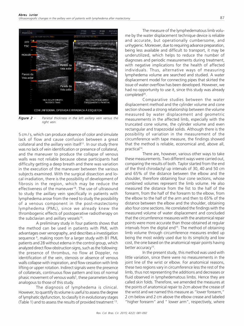
Abreu JuniorAbreu JuniorAbreu JuniorAbreu JuniorAbreu JuniorUltrasonografic changes in the axillary vein of patients with lymphedema after mastectomy 87
Rev. Col. Bras. Cir. 2015; 42(2): 081-092
5 cm / s, which can produce absence of color and simulatelack of flow and cause confusion between a greatcollateral and the axillary vein itself11. In our study therewas no lack of vein identification or presence of collateral,and the maneuver to produce the collapse of venouswalls was not reliable because obese participants haddifficulty getting a deep breath and there was variationin the execution of the maneuver between the varioussubjects examined. With the surgical dissection and lo-cal irradiation, there is the possibility of development offibrosis in the region, which may be reduce theeffectiveness of the maneuver18. The use of ultrasoundto study the axillary vein specifically in patients withlymphedema arose from the need to study the possibilityof a venous component in the post-mastectomylymphedema (PML), since we already knew thethrombogenic effects of postoperative radiotherapy onthe subclavian and axillary vessels19.
A preliminary study in four patients shows thatthe method can be used in patients with PML withadvantages over venography, and describes a investigationsequence 6, making room for a larger study with 81 PMLpatients and 28 without edema in the control group, whichanalyzed direct flow obstruction signs, such as the following:the presence of thrombus, no flow at Doppler, noidentification of the vein, stenosis or absence of venouswalls collapse with inspiration, and flow cessation with limblifting or upper rotation. Indirect signals were the presenceof collaterals, continuous flow pattern and loss of normalphasic movement of venous walls5, these parameters beinganalogous to those of this study.
The diagnosis of lymphedema is clinical.However, to quantify its volume is useful to assess the degreeof lymphatic dysfunction, to classify it in evolutionary stages(Table 1) and to assess the results of provided treatment 13.
The measure of the lymphedematous limb volu-me by the water displacement technique device is reliableand accurate, but operationally cumbersome, andunhygienic. Moreover, due to requiring advance preparation,being less available and difficult to transport, it may beunderutilized, which helps to reduce the number ofdiagnoses and periodic measurements during treatment,with negative implications for the health of affectedindividuals. Thus, alternative ways of measuringlymphedema volume are searched and studied. A waterdisplacement model for connecting pipes that skirted theissue of water overflow has been developed. However, wehad no opportunity to use it, since this study was alreadycompleted20.
Comparative studies between the waterdisplacement method and the cylinder volume and conesection showed a strong relationship between the volumemeasured by water displacement and geometricmeasurements in the affected limb, especially with thetruncated cone volume, the cylinder volume and evenrectangular and trapezoidal solids. Although there is thepossibility of variation in the measurement of thecircumference with tape measure, the findings showedthat the method is reliable, economical and, above all,practical14.
There are, however, various other ways to takethese measurements. Two different ways were carried out,comparing the results of both. Taylor started from the endof the third chirodactyl up intervals of 30, 40 and 50 cmand 65% of the distance between the elbow and theshoulder, therefore obtaining four cone sections, whosecombined volumes represent the limb volume. He alsomeasured the distance from the fist to the half of theforearm, from the half of the forearm to the elbow, fromthe elbow to the half of the arm and then to 65% of thedistance between the elbow and the shoulder, obtainingalso four cone sections. He correlated the findings with themeasured volume of water displacement and concludedthat the circumference measures with the anatomical repairpoints were more accurate than those obtained at regularintervals from the digital end15. The method of obtaininglimb volume through circumference measures ended upbeing the most widely used due to its simplicity and lowcost, the one based on the anatomical repair points havingbetter accuracy15.
In the present study, this method was used withlittle variation, since there were no measurements in thejoint line of the wrist or elbow. For anatomical reasons,these two regions vary in circumference less the rest of thelimb, thus not representing the additions and decreases orfluid observed in lymphedematous limbs. Hence they arecalled skin folds. Therefore, we amended the measures atthe points of anatomical repair to 2cm above the crease ofthe wrist and we named this measure as “lower forearm,”2 cm below and 2 cm above the elbow crease and labeled“higher forearm” and “ lower arm”, respectively, where
Figure 2 Figure 2 Figure 2 Figure 2 Figure 2 - Parietal thickness in the left axillary vein versus theright vein.
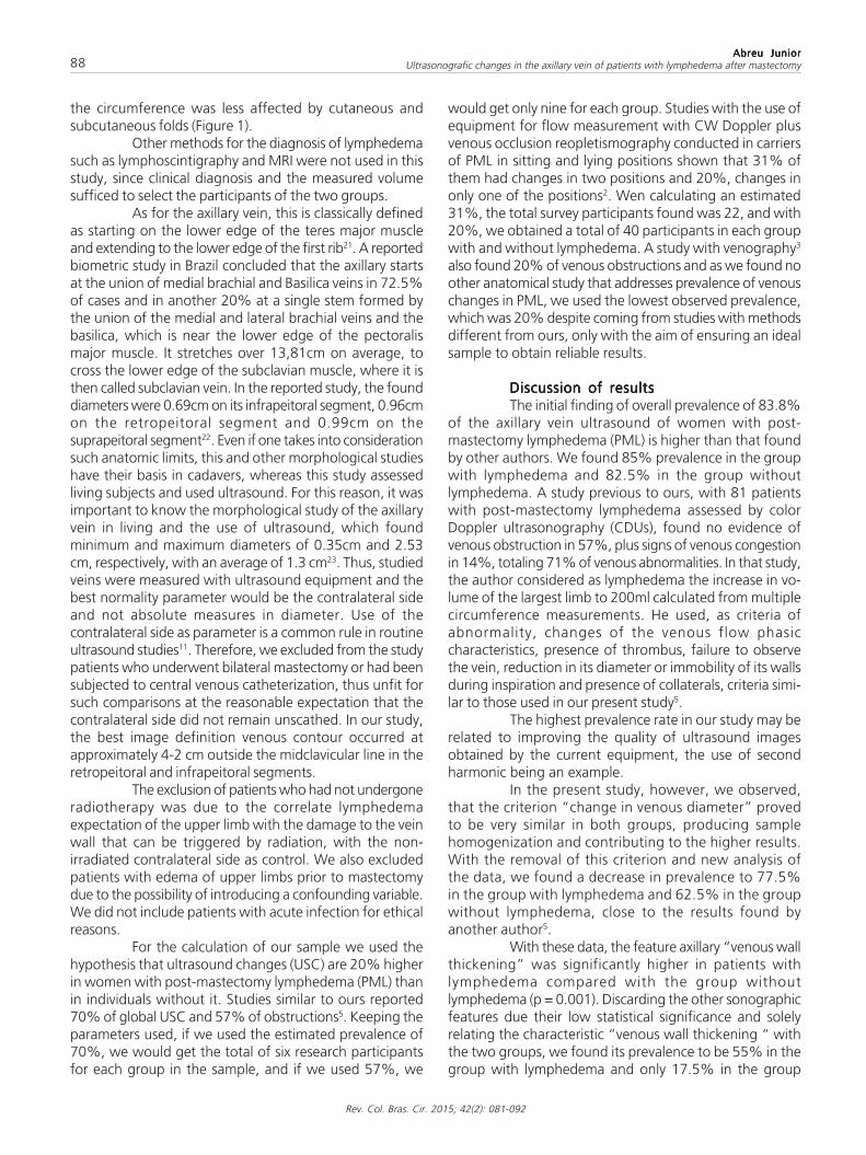
88
Rev. Col. Bras. Cir. 2015; 42(2): 081-092
Abreu JuniorAbreu JuniorAbreu JuniorAbreu JuniorAbreu JuniorUltrasonografic changes in the axillary vein of patients with lymphedema after mastectomy
the circumference was less affected by cutaneous andsubcutaneous folds (Figure 1).
Other methods for the diagnosis of lymphedemasuch as lymphoscintigraphy and MRI were not used in thisstudy, since clinical diagnosis and the measured volumesufficed to select the participants of the two groups.
As for the axillary vein, this is classically definedas starting on the lower edge of the teres major muscleand extending to the lower edge of the first rib21. A reportedbiometric study in Brazil concluded that the axillary startsat the union of medial brachial and Basilica veins in 72.5%of cases and in another 20% at a single stem formed bythe union of the medial and lateral brachial veins and thebasilica, which is near the lower edge of the pectoralismajor muscle. It stretches over 13,81cm on average, tocross the lower edge of the subclavian muscle, where it isthen called subclavian vein. In the reported study, the founddiameters were 0.69cm on its infrapeitoral segment, 0.96cmon the retropeitoral segment and 0.99cm on thesuprapeitoral segment22. Even if one takes into considerationsuch anatomic limits, this and other morphological studieshave their basis in cadavers, whereas this study assessedliving subjects and used ultrasound. For this reason, it wasimportant to know the morphological study of the axillaryvein in living and the use of ultrasound, which foundminimum and maximum diameters of 0.35cm and 2.53cm, respectively, with an average of 1.3 cm23. Thus, studiedveins were measured with ultrasound equipment and thebest normality parameter would be the contralateral sideand not absolute measures in diameter. Use of thecontralateral side as parameter is a common rule in routineultrasound studies11. Therefore, we excluded from the studypatients who underwent bilateral mastectomy or had beensubjected to central venous catheterization, thus unfit forsuch comparisons at the reasonable expectation that thecontralateral side did not remain unscathed. In our study,the best image definition venous contour occurred atapproximately 4-2 cm outside the midclavicular line in theretropeitoral and infrapeitoral segments.
The exclusion of patients who had not undergoneradiotherapy was due to the correlate lymphedemaexpectation of the upper limb with the damage to the veinwall that can be triggered by radiation, with the non-irradiated contralateral side as control. We also excludedpatients with edema of upper limbs prior to mastectomydue to the possibility of introducing a confounding variable.We did not include patients with acute infection for ethicalreasons.
For the calculation of our sample we used thehypothesis that ultrasound changes (USC) are 20% higherin women with post-mastectomy lymphedema (PML) thanin individuals without it. Studies similar to ours reported70% of global USC and 57% of obstructions5. Keeping theparameters used, if we used the estimated prevalence of70%, we would get the total of six research participantsfor each group in the sample, and if we used 57%, we
would get only nine for each group. Studies with the use ofequipment for flow measurement with CW Doppler plusvenous occlusion reopletismography conducted in carriersof PML in sitting and lying positions shown that 31% ofthem had changes in two positions and 20%, changes inonly one of the positions2. Wen calculating an estimated31%, the total survey participants found was 22, and with20%, we obtained a total of 40 participants in each groupwith and without lymphedema. A study with venography3
also found 20% of venous obstructions and as we found noother anatomical study that addresses prevalence of venouschanges in PML, we used the lowest observed prevalence,which was 20% despite coming from studies with methodsdifferent from ours, only with the aim of ensuring an idealsample to obtain reliable results.
Discussion of resultsDiscussion of resultsDiscussion of resultsDiscussion of resultsDiscussion of resultsThe initial finding of overall prevalence of 83.8%
of the axillary vein ultrasound of women with post-mastectomy lymphedema (PML) is higher than that foundby other authors. We found 85% prevalence in the groupwith lymphedema and 82.5% in the group withoutlymphedema. A study previous to ours, with 81 patientswith post-mastectomy lymphedema assessed by colorDoppler ultrasonography (CDUs), found no evidence ofvenous obstruction in 57%, plus signs of venous congestionin 14%, totaling 71% of venous abnormalities. In that study,the author considered as lymphedema the increase in vo-lume of the largest limb to 200ml calculated from multiplecircumference measurements. He used, as criteria ofabnormality, changes of the venous flow phasiccharacteristics, presence of thrombus, failure to observethe vein, reduction in its diameter or immobility of its wallsduring inspiration and presence of collaterals, criteria simi-lar to those used in our present study5.
The highest prevalence rate in our study may berelated to improving the quality of ultrasound imagesobtained by the current equipment, the use of secondharmonic being an example.
In the present study, however, we observed,that the criterion “change in venous diameter” provedto be very similar in both groups, producing samplehomogenization and contributing to the higher results.With the removal of this criterion and new analysis ofthe data, we found a decrease in prevalence to 77.5%in the group with lymphedema and 62.5% in the groupwithout lymphedema, close to the results found byanother author5.
With these data, the feature axillary “venous wallthickening” was significantly higher in patients withlymphedema compared with the group withoutlymphedema (p = 0.001). Discarding the other sonographicfeatures due their low statistical significance and solelyrelating the characteristic “venous wall thickening “ withthe two groups, we found its prevalence to be 55% in thegroup with lymphedema and only 17.5% in the group

Abreu JuniorAbreu JuniorAbreu JuniorAbreu JuniorAbreu JuniorUltrasonografic changes in the axillary vein of patients with lymphedema after mastectomy 89
Rev. Col. Bras. Cir. 2015; 42(2): 081-092
without it (PR = 3.14; 95% CI: 1.51 to 6.51). The cause ofthis thickening is not clear, and we can attribute it to theaxillary vein thrombosis or to factors external to it such assurgery and radiotherapy.
The similarity of prevalence in both groups ofother sonographic features, namely, change of diameter,presence of intraluminal images, compressibility, floweasiness and collapsing walls at inspiration, gave greaterstatistical value the unique feature that proved dominantin a given group, the one with lymphedema.
In reviewing the other sonographic features, wefound that the flow was phasic in all sample cases, notbeing able to separate interest groups. In addition, thecollapsing findings of the axillary vein similar to inspirationin both groups, and even slightly higher in patients withlymphedema, the venous diameter similar in both groups,and also the finding of just one intraluminal image in thewhole sample, made us consider the possibility that theoccurrence of thrombosis of the axillary vein can beinfrequent, non-occlusive or quick resolution, or that theresulting impairment of venous function is of minorsignificance.
Furthermore, we observed that venouscompression maneuver for venous thrombus was not asreliable, as some veins showed initially incompressible,though with normal flow demonstrable by Doppler. In mostcases, venous occlusion obtained with the maneuverrequired more compression than on the contralateral controlside. The compression caused by the equipment transducerpromoted the movement of all the tissue block surroundingthe vein, keeping it with its normal diameter or with smalldeformation, precisely in an area where sometimes thetransducer’s own weight is sufficient to occlude the vein. Inpatients submitted to total mastectomy, there is anatomicalchange in the chest wall at this location, which, associatedwith postoperative fibrosis, contributes to alter from convexityand typical softness to concavity and reduction of elasticityand thickening of the overlying skin. Subsequentradiotherapy may contribute to exacerbate this situationand this set of tissue changes makes the compressivemaneuver difficult24.
The maneuver to obtain venous collapsing atinspiration proved to be very dependent on the examinedindividual and appears to be hampered in overweight statesand in the supine position. Moreover, pleuropulmonarylesions are common in this type of patients and we did notinvestigate their presence in our sample; their participationin the reported maneuvers is not known.
If we consider the set of signals that we deemedto be ultrasound changes for this work, we find that theiroccurrence is statistically similar in both groups, with andwithout lymphedema, except for the vessel wall thickening.We can also conclude that ultrasound changes are notassociated with post-mastectomy lymphedema. It is possiblethat the adopted set of signals is not appropriate to separatethe affected from the non-affected as for their local venous
circulatory function within the universe of patients exposedto mastectomy plus radiotherapy. However, it is alsoexpected that the venous disease, usually correlated withultrasound changes, is not an independent factor in thegenesis of post-mastectomy lymphedema, but one amongstother contributory factors. We do not know the weight ofthis participation. Other factors related to the clinical andepidemiological aspects correlated with the presence ofpost-mastectomy lymphedema.
The groups with and without lymphedema weresimilar in many aspects, such as age, surgical technique,operated side, and number of radiotherapy andchemotherapy sessions, helping to give confidence in thecorrelation of the findings related to the risk of USCoccurrence in some of the groups.
The age group between 47 and 57 yearsdisplayed increased risk of USC occurrence, of 3.82 times,moving far away from the risk seen in younger age groupsand maintaining superiority, but closer to the age groupabove, a finding that suggests USC increased prevalencewith increasing age. As the postoperative time did not conferincreased risk, age appears to be an independent riskfactor25.
Total mastectomy showed 3.17-fold risk ofincreased USC prevalence compared with partialmastectomy, which is understandable due to the increasedremoval of affected lymph nodes and greater surgicalaggression, with sequelae to the corresponding side, suchas local fibrosis and limb mobility reduction, regardless ofthe operated side9. The risk of increased USC prevalencewas also 9.33 times higher when the number of radiotherapysessions ranged between 20 and 25. This is possibly due toall research subjects in our study underwent both therapeuticmethods, and the association between PML and radiationis reported in the literature when associated with previoussurgery, there being little evidence that it contributes toPML alone9,25. This factor is well accepted in the literatureas capable of aggravating local fibrosis, which can reducethe ability of spontaneous lymphatic compensation,contributing to the emergence or worsening of the post-mastectomy lymphedema 24-26.
There were differences between the groups inthe aspect lymphedema stadium, which was expected, sincein the group without lymphedema would be the stage 0participants, thus inevitably differing from the other group,where would be individuals with all the other developmentalstages.
The aspect BMI also showed differences betweengroups, with overweight subjectes predominating in the onewithout lymphedema, while in the group with lymphedemathere was balance between overweight and obeseindividuals, suggesting, therefore, that with the BMIevolution from overweight to obesity, there is migration tothe group with lymphedema. We also observed increasedUSC prevalence. This correlation between high BMI andlymphedema has been reported by other authors25-27. We

90
Rev. Col. Bras. Cir. 2015; 42(2): 081-092
Abreu JuniorAbreu JuniorAbreu JuniorAbreu JuniorAbreu JuniorUltrasonografic changes in the axillary vein of patients with lymphedema after mastectomy
have not found participants with BMI compatible with ide-al weight.
The association between increased USCprevalence and number of chemotherapy sessions was 3.31times from as early as the fifth session on. There is doubtabout the association between chemotherapy and PML,with acceptance, however, of its participation inpostoperative painful morbidity 28.
During participants examination there were nolimitations of movements that caused any restrictions, andwe observed a wide freedom of movement of the upperlimbs, whose amplitude seemed normal.
It was noteworthy the absence of venouscollateral circulation in cases of recorded axillary veinthrombosis, which is described in studies involving axillaryvein thrombosis due to effort, after catheterization, claviclefracture, all without radiotherapy. Radiation therapy mayhave a role in it24.
We recognize some limitations in this study. Thelack of information on occupation, income and educationdid not allow correlating the prevalence of ultrasoundchanges and the presence of lymphedema withenvironmental risk.
Implications for clinical practiceImplications for clinical practiceImplications for clinical practiceImplications for clinical practiceImplications for clinical practiceThis study allowed ultrasound changes to be
detected in the axillary vein, showing its involvement withupper limb post-mastectomy lymphedema.
Since ultrasound alterations are easily found inpost-thrombotic sequelae, this possibility should be aired inthe postoperative period, and measures implemented,including prevention of venous thrombosis and early upperlimb motor rehabilitation. Another aspect is that there mustbe caution with the axillary vein during surgery, pluspostoperative care, in order to minimize the resulting scar.
The results of this study point to the need toreconsider some ultrasonographic signs found in the axillaryvein when one wants to correlate them with the presence ofpost-mastectomy lymphedema. Changing in the diameter,presence of intraluminal images, compressibility as sign ofthrombosis in the region and the absence of venous wallscollapse seen at ultrasound as signs of post-thrombotic venousdisease showed no reliability in our sample. As the venouswall thickening correlated strongly with the group with post-mastectomy lymphedema, this sonographic characteristicseemed reliable, rendering possible the correlation betweenthe findings and high-risk patients, identifying thosecandidates for more often clinical follow-up.
Weight gain, with the research subject in theobesity range, correlated with the presence of PML. Effortsshould be made in a multidisciplinary way for weightreduction when treating patients with post-mastectomylymphedema.
The performance of total mastectomy is still areality and this technique is related to increased risk ofPML. There was increased USC prevalence in these cases
and effective measures of early diagnosis and treatmentshould contribute to the decline of such rates. Mastectomy,when held on the right side, does not seem to be apredisposing factor for lymphedema, although most peopleare right-handed and may put the dominant member tothe effort, and consequently to the risk.
Implications for researchImplications for researchImplications for researchImplications for researchImplications for researchMany questions remain open on the subject
venous disease and post-mastectomy lymphedema andspecifically in its ultrasound assessment.
The presence of vessel wall thickening in 55%of cases with PML compared with only 17.5% in non-carrierscorrelates vein injury with post-mastectomy lymphedema.Nonetheless, we do not know if such injury precedes orfollows the onset of lymphedema. Nor do we knowwhether such injury is the sequel of venous thrombosis orresulting from radiotherapy. Studies lack before and aftermastectomy and before and after radiotherapy, with short,medium and long-term follow-up, to establish the naturalhistory of venous repercussions and their relationship withlymphedema in patients undergoing mastectomy andradiotherapy for breast cancer.
We do not know if the venous walls collapse atinspiration can be influenced by the presence ofpleuropulmonary injury secondary to radiotherapy andfurther research is necessary in this area.
The possibility of spontaneous postoperativelymphatic compensation to ensure the lymphatic outflowin the venous system is well established in the literature inliving29 and dead30, which, of course, may be compromisedby tissue aggression subsequent to surgery, such as infectionsand radiation.
The continuous study of the axillary vein and re-gional tissue changes in PML carriers by usingcomplementary venous angiotomography or new ultrasoundelastography techniques and magnetic resonance imagingcan be important to select patients with the disease, sincethe ultrasound alone fails to diagnose it7. Elastography hasemerged as an auxiliary tool for ultrasound diagnosis.Elastograms are images of tissue stiffness and may becolored, in shades of gray, or a combination of the two.Recent advances in elastography include quantification usingvoltage ratios, the acoustic radiation image, power boost,speed and wave cutting estimate. The first and mostcommon elastography application is for the diagnosis ofbreast lesions and characterization of the focal masses suchas liver cirrhosis of thyroid nodules, evaluation of lymphnodes for metastatic disease, detection of prostate cancer,among others. Promising applications include study ofatheroma, assessment of venous and arterial wall andevaluation of characteristics and age of thrombi31.Studieswith ultrasound elastography are conducted to selectsuitable locations for the employment of lymphatic-venousanastomoses32 and determination of the age of thethrombus33, while other studies with magnetic resonance

Abreu JuniorAbreu JuniorAbreu JuniorAbreu JuniorAbreu JuniorUltrasonografic changes in the axillary vein of patients with lymphedema after mastectomy 91
Rev. Col. Bras. Cir. 2015; 42(2): 081-092
elastography, investigate the tissue consequences of diseaseprocesses, such as inflammation and fibrosis34. the use ofthese new technologies in a prospective study before andafter mastectomy can help to better understand the factorsthat influence the installation of PML.
More studies are needed to assess the possibilityof treatment of axillary venous hypertension with venousdecompression by section of the pectoralis major18 or thesuitability for venous flow unblocking or correction of stenosisand placement of a stent7 if the injuries are appreciable.However, this study is not conclusive in this direction.
Finally, other questions must be answered on thetheme post-mastectomy lymphedema and, among those
that were addressed in this study, we highlight some, suchas the need to deepen the study of the forms of limb volu-me measurement to lymphedema definition and staging.There is no standard way of measuring a lymphedematouslimb and therefore to search correlation between themeasured volume obtained by water displacement and thevarious forms of circumference measurement, which couldhelp to clarify this question.
The prevalence of ultrasound changes wassignificantly higher in patients undergoing mastectomy andwith lymphedema compared with those withoutlymphedema. These findings correlate with increased riskof developing post-mastectomy lymphedema.
R E S U M OR E S U M OR E S U M OR E S U M OR E S U M O
Objetivo:Objetivo:Objetivo:Objetivo:Objetivo: verificar se existe prevalência de alterações ultrassonográficas (AUS) na veia axilar de pacientes portadoras e nãoportadoras de linfedema do membro superior pós-mastectomia. Métodos:Métodos:Métodos:Métodos:Métodos: uma amostra de 80 mulheres, alocadas em dois gruposiguais, com e sem linfedema foi estudada com ultrassonografia modo B, Doppler colorido e pulsado. A variável primária AUS foidefinida como: alteração do diâmetro venoso, espessamento parietal, imagens intraluminares, compressibilidade, colapso parietal àinspiração e característica do fluxo venoso axilar no lado operado. Como variáveis secundárias: estádio do linfedema, técnicaoperatória, número de sessões de radio e quimioterapia, volume do membro, peso e idade. As diferenças entre as proporções nosgrupos foram verificadas com o teste qui-quadrado de Pearson e/ou exato de Fisher. Para variáveis contínuas usamos o teste deMann-Whitney. Para estimar a magnitude das associações utilizou-se como medida de frequência a prevalência de AUS em ambosos grupos e como medida de associação, a razão de prevalência (RP) obtida em função do risco relativo (RR) e estimada por meio doteste de homogeneidade de Mantel-Haenszel. Adotou-se o nível de significância estatístico de 5% (pd”0,05). Resultados:Resultados:Resultados:Resultados:Resultados: somenteo critério “espessamento parietal” se relacionou fortemente com o grupo com linfedema (p=0,001). A prevalência de AUS foi 55%no grupo com linfedema e 17,5% no grupo sem linfedema, com diferença de prevalências de 37,5%. Conclusão:Conclusão:Conclusão:Conclusão:Conclusão: a prevalência deAUS foi maior nas pacientes submetidas à mastectomia e com linfedema do que naquelas sem linfedema.
Descritores: Descritores: Descritores: Descritores: Descritores: Neoplasias da mama. Veia Axilar. Linfedema. Ultrassonografia.
REFERENCESREFERENCESREFERENCESREFERENCESREFERENCES
1. Megret G. Iatrogenic venous pathology and post-radiosurgeryswollen arm. Apropos of 100 cases. Phlebologie. 1985;38(4):609-19.
2. Gruffaz J. Venous component in lymphedema in the upper extremityafter radio-surgical therapy of cancer of the breast. Phlebologie.1986;39(3):517-25.
3. McIvor J, O’Connell D. The investigation of postmastectomyoedema of the arm by lymphography and venography. Clin Radiol.1978;29(4):457-62.
4. Tarquini A, Arru A, Puliga A, Armeni S, Piga A, Malloci A. Role ofvenous injury in the origin of post-mastectomy lymphedema ofthe arm. Results of 150 measurements of the venous pressure ofthe homo- and counter-lateral arm in mastectomy. Minerva Chir.1987;42(12):1053-9.
5. Svensson WE, Mortimer PS, Tohno E, Cosgrove DO. Colour Dopplerdemonstrates venous flow abnormalities in breast cancer patientswith chronic arm swelling. Eur J Cancer. 1994;30A(5):657-60.
6. Svensson WE, Mortimer PS, Tohno E, Cosgrove DO, Badger C, alMurrani B. The use of colour Doppler to define venous abnormalitiesin the swollen arm following therapy for breast carcinoma. ClinRadiol. 1991;44(4):249-52.
7. Szuba A, Razavi M, Rockson S. Diagnosis and treatment ofconcomitant venous obstruction in patients with secondarylymphedema. J Vasc Interv Radiol. 2002;13(8):799-803.
8. Pain SJ, Vowler S, Purushotham AD. Axillary vein abnormalitiescontribute to development of lymphoedema after surgery forbreast cancer. Br J Surg. 2005;92(3):311-5.
9. Erickson VS, Pearson ML, Ganz PA, Adams J, Kahn KL. Arm edemain breast cancer patients. J Natl Cancer Inst. 2001;93(2):96-111.
10. Gooding GA, Woodruff A. Color Doppler imaging in the subclavian-axillary region and upper extremity. Clin Imaging. 1994;18(3):165-72.
11. Nazarian GK, Foshager MC. Color Doppler sonography of thethoracic inlet veins. Radiographics. 1995;15(6):1357-71.
12. Patel MC, Berman LH, Moss HA, McPherson SJ. Subclavian andinternal jugular veins at Doppler US: abnormal cardiac pulsatilityand respiratory phasicity as a predictor of complete centralocclusion. Radiology. 1999;211(2):579-83.
13. International Society of Lymphology. The diagnosis and treatmentof peripheral lymphedema. 2009 Concensus Document of theInternational Society of Lymphology. Lymphology. 2009;42(2):51-60.
14. Sander AP, Hajer NM, Hemenway K, Miller AC. Upper-extremityvolume measurements in women with lymphedema: a comparisonof measurements obtained via water displacement withgeometrically determined volume. Phys Ther. 2002;82(12):1201-12.
15. Taylor R. Jayasinghe UW, Koelmeyer L, Ung O, Boyages J. Reliabilityand validity of arm volume measurements for assessment oflymphedema. Phys Ther. 2006;86(2):205-14.
Rodrigues JuniorRodrigues JuniorRodrigues JuniorRodrigues JuniorRodrigues JuniorOpen abdomen management: single institution experience 93
Rev. Col. Bras. Cir. 2015; 42(2): 093-096
Original ArticleOriginal ArticleOriginal ArticleOriginal ArticleOriginal Article
Open abdomen management: single institution experienceOpen abdomen management: single institution experienceOpen abdomen management: single institution experienceOpen abdomen management: single institution experienceOpen abdomen management: single institution experience
Abdômen aberto: experiência em uma única instituiçãoAbdômen aberto: experiência em uma única instituiçãoAbdômen aberto: experiência em uma única instituiçãoAbdômen aberto: experiência em uma única instituiçãoAbdômen aberto: experiência em uma única instituição
ADILSON COSTA RODRIGUES JUNIOR1; FERNANDO DA COSTA FERREIRA NOVO1; RAFAEL DE CASTRO SANTANA AROUCA1; FRANCISCO DE SALLES
COLLET E SILVA, TCBC-SP1; EDNA FRASSON DE SOUZA MONTERO,TCBC-SP2; EDIVALDO MASSAZO UTIYAMA, TCBC-SP2
A B S T R A C TA B S T R A C TA B S T R A C TA B S T R A C TA B S T R A C T
Objective:Objective:Objective:Objective:Objective: to evaluate the outcome of abdominal wall integrity of both techniques. Methods:Methods:Methods:Methods:Methods: a retrospective study was carried
out at the Hospital das Clínicas, Faculdade de Medicina, Universidade de São Paulo, identifying the patients undergoing temporary
abdominal closure (TAC) from January 2005 to December 2011. Data were collected through the review of clinical charts. Inclusion
criteria were indication of TAC and survival to definitive abdominal closure. In the post-operative period only a group of three
surgeons followed all patients and performed the reoperations. Results:Results:Results:Results:Results:Twenty eightpatients were included. The difference in
primary closure rates and mean time for fascial closure did not reach statistical significance (p=0.98 and p=0.23, respectively).
Conclusion:Conclusion:Conclusion:Conclusion:Conclusion: VAC and Bogota Bag do not differ significantly regarding the outcome of abdominal wall integrity, due to the
monitoring of a specific team and the adoption of progressive closure
Key words: Key words: Key words: Key words: Key words: Peritonitis. Trauma. Sepsis. Negative-Pressure Wound Therapy. Abdominal Wound Closure Techniques. Intra-
Abdominal Hypertension.
1. General and Trauma Surgery, Hospital das Clínicas, Medical School at the University of São Paulo, São Paulo, Brazil; 2. Surgery Departmentof Medical School the University São Paulo. São Paulo, Brazil.
INTRODUCTIONINTRODUCTIONINTRODUCTIONINTRODUCTIONINTRODUCTION
Most of the time, at the end performing a laparotomy,the abdomen is closed with primary closure of
aponeurosis. However, in some cases, the surgeon is forcedto leave the open abdomen tactic that is associated with amortality rate of 30%1-3.
The open abdomen technique (AA) is a surgicalstrategy used in patients with related life-threatening intra-abdominal hemorrhage, prevention or treatment of intra-abdominal hypertension and treatment of intra-abdominalsepsis. It is a temporary measure to prioritize the control ofbleeding, correction of metabolic disorders and hypothermia,and facilitate access to the abdominal cavity4.
In the 40s, leaving open abdomen after alaparotomy was considered technical failure. However, inthe 70, the disseminated abdominal infections representeda major challenge, with mortality rates ranging from 30%to 80%5. Therefore, at that time, Hay et al.6 andSteinberg7proposed to maintain the open abdomen to treatsevere peritonitis. In the same period, Champaultet al.8
advocated reoperations scheduled for cleaning the abdo-minal cavity.
Since 1981, the Department of General Surgeryand Trauma Hospital of the Faculty of Medicine, Universi-dade de São Paulo (HCFMUSP), it was established that theopen abdomen (AA) with planned reoperations would be
an option for the treatment of intra-infections abdominalwith systemic repercussions, whose infectious focus couldnot be removed in one operation9. In the 90’s was designedthe Damage Control (DC). First described in 1993 byRotondo et al. as an alternative to final laparotomy inpatients with exsanguinantes bleeding related to lesionsof large vessels and multiple lesions intra-abdominalviscera10. It was later shown to initiate damage controlearly, before the patient’s clinical conditions deteriorateto the extreme [massive blood loss, severe injury (ISS>25), hypothermia (<34°C), acidosis (pH <7.25) and bloodcoagulation (APTT> 19sec)], reduces mortality11.Furthermore, keep the cavity open abdominal exposespatients to the risk of perforation of hollow viscera andincreases the risk of developing complex hernias.Temporary abdominal closure techniques (FAT) with sutureor skin closure Backaus calipers reduce thesecomplications, but increase the risk of abdominalcompartment syndrome (ACS). After recognition ofmorbidity and mortality attributed to SCA, have beendeveloped various methods to avoid this complication12.
The ideal technique for the FAT was defined asone that contains the abdominal viscera, limitcontamination, prevent the loss of abdominal fluid, avoidadhesions, allow easy access to the abdominal cavity, avoiddamage and the retraction of the abdominal wall and standin the way ACS13.
DOI: 10.1590/0100-69912015002005

94
Rev. Col. Bras. Cir. 2015; 42(2): 093-096
Rodrigues JuniorRodrigues JuniorRodrigues JuniorRodrigues JuniorRodrigues JuniorOpen abdomen management: single institution experience
The application of the Bogota Stock Exchange(BB) has become the most popular and effective method oftemporary abdominal closure. It is still used in many hospitalsin developing countries because of its low cost and easyhandling. It was created by Oswaldo Borráez in 1984 andthe name “Bogota bag” was created by Mattox, during avisit to a hospital in Bogota - Colombia, in 199714,15.
A decade ago, the negative pressure applicationconcept was introduced by Barker et al. as a new means oftemporary abdominal closure16. After the introduction ofthis vacuum closure technique, a more comprehensivemethod to manage negative pressure therapy to an openabdominal wound was developed: the closing assistedvacuum, the English “Vacuum Assisted Closure” (VAC).This technique enabled by the possibility of FAT drainperitoneal fluid, to minimize visceral edema, fascial applygreater tension in the abdominal wall and promoting thedefinitive abdominal closure in patients with open abdomen,one month after the initial laparotomy17,18.
Today, despite advances in AA use with thedevelopment of vacuum therapy, the Bogota StockExchange (BB) is still widely used. In our institution andthroughout Brazil, both techniques are used and the study’sgoal is to evaluate the morbidity and the result of the integrityof the abdominal wall after the use of both techniques.
METHODSMETHODSMETHODSMETHODSMETHODS
A retrospective study was conducted atHCFMUSP, identifying patients undergoing temporary ab-dominal closure (FAT) between January 2005 andDecember 2011. Data were collected through review ofmedical records. Data were compared in patientsundergoing treatment with assisted closing the vacuum andthe Bogota bag.
Inclusion criteria were temporary abdominalclosure indication survival and the definitive abdominalclosure. FAT indications included: abdominal trauma, severeabdominal sepsis and ACS. Data collected included age,FAT indication, the number of procedures in the operatingroom and the primary fascial closure rate. In thepostoperative period, one group of three surgeons followedall patients and performed all reoperations. As soon aspossible, the aponeurosis of the edges were subjected toprogressive approach with careful not to cause abdominalhypertension.
The demographics of the two study groups (BBand VAC) were compared using the chi-square test ofPearson or Fisher’s exact test for categorical variables andthe Student t test for continuous variables.
RESULTSRESULTSRESULTSRESULTSRESULTS
During the study period, 59 patients require somekind of temporary abdominal closure (FAT), however, only29 patients survived the final abdominal closure (52.5%mortality). One patient was excluded (subject to closureBackaus forceps). Thus, 28 patients were included, and,after two years of follow-up, none of them developed ab-dominal hernia or intestinal fistulas.
There was no statistical difference between thestudy groups with respect to age (p> 0.05) and a significantdifference regarding indications for temporary abdominalclosure (p <0.05) (Table 1).
The primary closure rates were similar in bothgroups (p = 0.98). The average time (days) for fascial closurewas 10.8 days for the BB group and 7.52 days in the groupVAC (Table 2).
DISCUSSIONDISCUSSIONDISCUSSIONDISCUSSIONDISCUSSION
In this study, there was no stat ist ica ldifference between the closing assisted vacuum andthe Bogota bag (VAC and BB) when analyzed thenumber of operations, the primary closure, and theaverage time of closing. However, a previous study19
showed better results when using VAC BB was comparedon the primary closure (50 to 70 %% and 88% for BBVAC). The best approach to achieve the definitive ab-dominal closure in patients with open abdomen remainscontroversial. To improve the fascial closure rate, theexcess volume resuscitation should be avoided, thewater balance should be carefully implemented, notonly on admission, but also throughout the course oftreatment with open abdomen20. The high rate ofprimary closure found in our patients, 80% for BB and96% for the VAC, no statistical difference was due tothe above mentioned guidelines and monitoring carriedout by the same team of surgeons in all reoperations.This may also explain the same results of other variablesanalyzed with use of the BB or VAC.
Table 1 -Table 1 -Table 1 -Table 1 -Table 1 - Epidemiological data.
Bogotá (n=10)Bogotá (n=10)Bogotá (n=10)Bogotá (n=10)Bogotá (n=10) VAC (n=17)VAC (n=17)VAC (n=17)VAC (n=17)VAC (n=17) ppppp
Average age (years ± SD) 39.5 ± 14.8 33.17 ± 16.6 0.17
Trauma 6 (60%) 6 (35.6%) 0.012
Non-trauma 4 (40%) 11 (64.7%) 0.01

Rodrigues JuniorRodrigues JuniorRodrigues JuniorRodrigues JuniorRodrigues JuniorOpen abdomen management: single institution experience 95
Rev. Col. Bras. Cir. 2015; 42(2): 093-096
A major obstacle to the abdominal closure is theretraction of the rectus abdominal muscles, which shouldbe avoided at all costs. While the patient is open abdomen,the fascial edges are placed under tension by interruptedsuture with nonabsorbable suture caliber. This strategyavoids the fascial retraction and facilitates the gradualapproximation of aponeurotic edges in each reoperationuntil definitive abdominal closure21. These techniques alsoapplied in our patients, reflect the high rate of abdominalclosure, and reduced closing average time found that study,even when BB is the chosen method.
Another factor is the indication of the methodused to implement the FAT. Some studies have shown theefficiency of the use of VAC in wounds that were not causedby trauma22,23, results similar to those found in our study.Differences were found in the temporary abdominal closureindications: the Bogota bag was most appropriate in cases
involving trauma, while VAC was more indicated in casesof abdominal sepsis, abdominal compartment syndromeand mesenteric ischemia.
The absence of abdominal hernias and intestinalfistulas in patients undergoing FAT in our service is due tothe technique used and the care adopted and applied bythe same surgical team.
In our experience, the closing assisted vacuum(VAC) and the Bogota bag do not differ significantly withrespect to the result of the integrity of the abdominal wall,although there is a tendency in our institution to use BBpreferably in trauma and VAC in cases “no trauma”. Themonitoring of a specific team and the progressive closureof adoption are the factors that can explain the absenceof difference between the groups and the high closingrate of the abdominal wall for both VAC and for the Bogotabag.
Table 2 -Table 2 -Table 2 -Table 2 -Table 2 - Results.
Bogotá (n=10)Bogotá (n=10)Bogotá (n=10)Bogotá (n=10)Bogotá (n=10) VAC (n=17)VAC (n=17)VAC (n=17)VAC (n=17)VAC (n=17) ppppp
Average operations 2.4 (1-8) 2.05 (1-3) 0.3
Primary closure 8 (80%) 16 (94.1%) 0.98
Closure average (days ± SD) 10.8 ± 14.46 7.52 ± 9.03 0.23
R E S U M OR E S U M OR E S U M OR E S U M OR E S U M O
Objetivo:Objetivo:Objetivo:Objetivo:Objetivo: avaliar o resultado da integridade da parede abdominal após utilização do fechamento assistido a vácuo e da bolsa de
Bogotá. Métodos:Métodos:Métodos:Métodos:Métodos: um estudo retrospectivo foi realizado no Hospital das Clínicas da Faculdade de Medicina da Universidade de São
Paulo (HCFMUSP), identificando os pacientes submetidos à técnica de fechamento abdominal temporário (FAT) entre janeiro de
2005 e dezembro de 2011. Os dados foram coletados por meio de revisão de prontuários. Os critérios de inclusão foram indicação
de FAT e sobrevivência até o fechamento definitivo da parede abdominal. No período pós-operatório, apenas um grupo de três
cirurgiões, seguiu todos os pacientes e realizou as reoperações. Além disso, independente da técnica de FAT utilizada, foi aplicada a
tática de fechamento fascial progressivo durante as reoperações. Resultados:Resultados:Resultados:Resultados:Resultados: Vinte e oito pacientes foram incluídos. Não houve
diferença estatística nas taxas de fechamento primário e tempo médio de fechamento fascial. Conclusão:Conclusão:Conclusão:Conclusão:Conclusão: O fechamento assistido
a vácuo e a bolsa de Bogotá não diferem significativamente em relação ao resultado da integridade da parede abdominal após as
reoperações. Isso se deve ao acompanhamento de uma equipe específica e a adoção de técnica de fechamento fascial progressivo.
Descritores: Descritores: Descritores: Descritores: Descritores: Peritonite. Trauma. Sepse. Tratamento de Ferimentos com Pressão Negativa. Técnicas de Fechamento de Ferimentos
Abdominais. Síndrome Compartimental Abdominal.
REFERENCESREFERENCESREFERENCESREFERENCESREFERENCES
1. Keremati M, Srivastava A, Sakabu S, Rumbolo P, Smock M, PollackJ, et al. The Wittmann Patch is a temporary abdominal closuredevice after descompressive celiotomy for abdominal compartmentsyndrome following burn. Burns. 2008;34(4):493-7.
2. Kushimoto S, Miyauchi M, Yokota H, Kawai M. Damage controlsurgery and open abdominal management: recent advances andour approach. J Nippon Med Sch. 2009;76(6):280-90.
3. Perez A, Hilvano S. Abdominal zippers for temporary abdominalclosure in planned relaparotomies for peripancreatic sepsis:experience in a developing country. J Hepatobiliary Pancreat Surg.2001;8(5):449-52.
4. Schecter WP, Ivatury RR, Rotondo MF, Hirshberg A. Open abdomenafter trauma and abdominal sepsis: a strategy for management.J Am Coll Surg. 2006;203(3):390-6.
5. Farthmann EH, Schoffel U. Principles and limitations of operativemanagement of intraabdominal infections. World J Surg.1990;14(2):210-7.
6. Hay J, Duchatelle P, Elman A, Flamant Y, Maillard JN. Les ventreslaissés ouvert. Chirurgie. 1979;105(6):508-10.
7. Steinberg D. On leaving the peritoneal cavity open in acutegeneralized suppurative peritonitis. Am J Surg. 1979;137(2):216-20.

96
Rev. Col. Bras. Cir. 2015; 42(2): 093-096
Rodrigues JuniorRodrigues JuniorRodrigues JuniorRodrigues JuniorRodrigues JuniorOpen abdomen management: single institution experience
8. Champault G, Magnier M, Psalmon F, Patel JC. L’eviscerationcontrolée dans le traitement des peritonites graves. Chirurgie.1979;105(9):866-9.
9. Utiyama EM, Pflug ARM, Damous SHB, Rodrigues Jr AC, MonteroEFS, Birolini CAV. Temporary abdominal closure with zipper-meshdevice for management of intra-abdominal sepsis. Rev Col BrasCir. 2015;42(1). In press.
10. Rotondo MF, Schwab CW, McGonigal MD, Phillips GR 3rd,Fruchterman TM, Kauder DR, et al. ‘Damage control’: an approachfor improved survival in exsanguinating penetrating abdominalinjury. J Trauma. 1993;35(3):375-82; discussion 382-3.
11. Hirshberg A, Mattox KL. Planned reoperation for severe trauma.Ann Surg. 1995;222(1):3-8.
12. Offner PJ, de Souza AL, Moore EE, Biffl WL, Franciose RJ, JohnsonJL, et al. Avoidance of abdominal compartment syndrome indamage-control laparotomy after trauma. Arch Surg.2001;136(6):676-81.
13. Stone HH, Strom PR, Mullins RJ. Management of the majorcoagulopathy with onset during laparotomy. Ann Surg.1983;197(5):532-5.
14. Borráez OA. Abdomen Abierto: la herida más desafiante. RevColomb Cir. 2008;23(4):204-9.
15. Ghimenton F, Thomson SR, Muckart DJ, Burrows R. Abdominalcontent containment: practicalities and outcome. Br J Surg.2000;87(1):106-9.
16. Barker DE, Kaufman HJ, Smith LA, Ciraulo DL, Richart CL, BurnsRP. Vacuum pack technique of temporary abdominal closure: a 7-year experience with 112 patients. J Trauma. 2000;48(2):201´-6;discussion 206-7.
17. Miller PR, Thompson JT, Faler BJ, Meredith JW, Chang MC. Latefascial closure in lieu of ventral hernia: the next step in openabdomen management. J Trauma. 2002;53(5):843-9.
18. Boele van Hensbroek P, Wind J, Dijkgraaf MG, Busch OR, GoslingsJC. Temporary closure of the open abdomen: a systematic reviewon delayed primary fascial closure in patients with an openabdomen. World J Surg. 2009;33(2):199-207.
19. Miller PR, Meredith JW, Johnson JC, Chang MC. Prospectiveevaluation of vacuum-assisted fascial closure after open abdomen:planned ventral hernia rate is substantially reduced. Ann Surg.2004;239(5):608-14; discussion 614-6.
20. Goussous N, Kim BD, Jenkins DH, Zielinski MD. Factors affectingprimary fascial closure of the open abdomen in the nontraumapatient. Surgery. 2012;152(4):777-83; discussion 783-4.
21. Cothren CC, Moore EE, Johnson JL, Moore JB, Burch JM. Onehundred percent fascial approximation with sequential abdominalclosure of the open abdomen. Am J Surg. 2006;192(2):238-42.
22. Perez D, Wildi S, Demartines N, Bramkamp M, Koehler C, ClavienPA. Prospective evaluation of vacuum-assisted closure in abdomi-nal compartment syndrome and severe abdominal sepsis. J AmColl Surg. 2007;205(4):586-92.
23. Adkins AL, Robbins J, Villalba M, Bendick P, Shanley CJ. Openabdomen management of intra-abdominal sepsis. Am Surg.2004;70(2):137-40; discussion 140.
Received on 06/05/2014Accepted for publication 30/06/2014Conflict of interest: none.Source of funding: none.
Address for correspondence:Address for correspondence:Address for correspondence:Address for correspondence:Address for correspondence:Edivaldo MassazoUtiyamaE-mail: [email protected]

92
Rev. Col. Bras. Cir. 2015; 42(2): 081-092
Abreu JuniorAbreu JuniorAbreu JuniorAbreu JuniorAbreu JuniorUltrasonografic changes in the axillary vein of patients with lymphedema after mastectomy
16. Grassi CJ, Polak JF. Axillary and subclavian venous thrombosis:follow-up evaluation with color Doppler flow US and venography.Radiology. 1990;175(3):651-4.
17. Baarslag HJ, van Beek EJ, Koopman MM, Reekers JA. Prospectivestudy of color duplex ultrasography compared with contrastvenography in patients suspected of having deep venous thrombosisof the upper extremities. Ann Intern Med. 2002;136(12):865-72.Erratum in: Ann Intern Med. 2003;138(5):438.
18. Segerström K, Bjerle P, Henriksson TG, Nyström A. Decompressionof the axillary vein for oedema of the arm caused by axillarydissection and irradiation. Scand J Plast Reconstr Surg Hand Surg.1991;25(3):245-8.
19. Wilson CB, Lambert HE, Scott RD. Subclavian and axillary veinthrombosis following radiotherapy for carcinoma of the breast.Clin Radiol. 1987;38(1):95-6.
20. Carvalho RM, Perez MCJ, Miranda Jr F. Volúmetro com câmarascomunicantes: apresentação de um protótipo de dimensões re-duzidas desenvolvido para a avaliação objetiva do volume de mão.J vasc bras. 2010;9(2):76-80.
21. Warwick R, Williams PL. Gray Anatomia. 35a Ed. Rio de Janeiro:Guanabara Koogan; 1979. Capítulo 6, Angiologia; p. 558-706.
22. Rivello T, Messière B. Estudo biométrico de veia axilar em sereshumanos. Rev Angiol Cir Vasc. 2002;11(2):80-4.
23. Galloway S, Bodenham A. Ultrasound imaging of the axillary vein—anatomical basis for central venous access. Br J Anaesth.2003;90(5):589-95.
24. Kocak Z, Overgaard J. Risk factors of arm lymphedema in breastcancer patients. Acta Oncol. 2000;39(3):389-92.
25. Bentzen SM, Dische S. Morbidity related to axillary irradiation inthe treatment of breast cancer. Acta Oncol. 2000;39(3):337-47.
26. Rezende LF, Pedras FV, Ramos CD, Gurgel MSC. Avaliação dascompensações linfáticas no pós-operatório de câncer de mamacom dissecção axilar através da linfocintilografia. J vascbras. 2008;7(4):370-5.
27. Freitas Júnior R, Ribeiro LFJ, Taia L, Kajita D, Fernandes MV, QueirozGS. Linfedema em pacientes submetidas à mastectomia radicalmodificada. Rev Bras Ginecol Obstet. 2001;23(4):205-8.
28. Hack TF, Cohen L, Katz J, Robson LS, Goss P. Physical andpsychological morbidity after axillary lymph node dissection forbreast cancer. J Clin Oncol. 1999;17(1):143-9.
29. Földi E, Földi M, Clodius L. The lymphedema chaos: a lancet. AnnPlast Surg. 1989;22(6):505-15.
30. Suami H, Pan WR, Taylor GI. Changes in the lymph structure of theupper limb after axillary dissection: radiographic and anatomicalstudy in a human cadaver. Plast Reconstr Surg. 2007;120(4):982-91.
31. Garra BS. Elastography: current status, future prospects, andmaking it work for you. Ultrasound Q. 2011;27(3):177-86.
32. Mihara M, Hayashi Y, Murai N, Moriguchi H, Iida T, Hara H, et al.Regional diagnosis of lymphoedema and selection of sites forlymphaticovenular anastomosis using elastography. Clin Radiol.2011;66(8):715-9. Erratum in: Clin Radiol. 2012;67(11):1128.
33. Karpiouk AB, Aglyamov SR, Mallidi S, Shah J, Scott WG, Rubin JM,et al. Combined ultrasound and photoacoustic imaging to detectand stage deep vein thrombosis: phantom and ex vivo studies. JBiomed Opt. 2008;13(5):054061.
34. Mariappan YK, Glaser KJ, Ehman RL. Magnetic resonanceelastography: a review. Clin Anat. 2010;23(5):497-511.
Received on 01/07/2014Accepted for publication 15/08/2014Conflict of interest: none.Source of funding: none.
Address for correspondence:Address for correspondence:Address for correspondence:Address for correspondence:Address for correspondence:Gilberto Ferreira de Abreu JuniorE-mail: [email protected]

Becha raBecha raBecha raBecha raBecha raMontreal classification of patient operated for Crohn’s disease and identification of surgical recurrence predictors 97
Rev. Col. Bras. Cir. 2015; 42(2): 097-104
Original ArticleOriginal ArticleOriginal ArticleOriginal ArticleOriginal Article
Montreal classification of patient operated for Crohn’s diseaseMontreal classification of patient operated for Crohn’s diseaseMontreal classification of patient operated for Crohn’s diseaseMontreal classification of patient operated for Crohn’s diseaseMontreal classification of patient operated for Crohn’s diseaseand identification of surgical recurrence predictorsand identification of surgical recurrence predictorsand identification of surgical recurrence predictorsand identification of surgical recurrence predictorsand identification of surgical recurrence predictors
Caracterização de pacientes operados por doença de Crohn pelaCaracterização de pacientes operados por doença de Crohn pelaCaracterização de pacientes operados por doença de Crohn pelaCaracterização de pacientes operados por doença de Crohn pelaCaracterização de pacientes operados por doença de Crohn pelaclassificação de Montreal e identificação de fatores preditores de suaclassificação de Montreal e identificação de fatores preditores de suaclassificação de Montreal e identificação de fatores preditores de suaclassificação de Montreal e identificação de fatores preditores de suaclassificação de Montreal e identificação de fatores preditores de suarecorrência cirúrgicarecorrência cirúrgicarecorrência cirúrgicarecorrência cirúrgicarecorrência cirúrgica
CRISTIANE DE SOUZA BECHARA1; ANTONIO LACERDA FILHO, TCBC-MG2; MARIA DE LOURDES ABREU FERRARI3; DÉBORAH ALMEIDA ROQUETTE
ANDRADE4; MAGDA MARIA PROFETA DA LUZ2; RODRIGO GOMES DA SILVA2
A B S T R A C TA B S T R A C TA B S T R A C TA B S T R A C TA B S T R A C T
ObjectiveObjectiveObjectiveObjectiveObjective: to evaluate Crohn’s disease recurrence and its possible predictors in patients undergoing surgical treatment. MethodsMethodsMethodsMethodsMethods:
We conducted a retrospective study with Crohn’s disease (CD) patients undergoing surgical treatment between January 1992 and
January 2012, and regularly monitored at the Bowel Clinic of the Hospital das Clínicas of the UFMG. ResultsResultsResultsResultsResults: we evaluated 125
patients, 50.4% female, with a mean age of 46.12 years, the majority (63.2%) diagnosed between 17 and 40 years of age. The
ileum was involved in 58.4%, whereas stenotic behavior was observed in 44.8%, and penetrating, in 45.6%. We observed perianal
disease in 26.4% of cases. The follow-up average was 152.40 months. Surgical relapse occurred in 29.6%, with a median time of
68 months from the first operation. ConclusionConclusionConclusionConclusionConclusion: The ileocolic location, penetrating behavior and perianal involvement (L3B3p) were
associated with increased risk of surgical recurrence.
Key words:Key words:Key words:Key words:Key words: Crohn’s Disease. Classification. Recurrence. Risk factors.
1. Coloproctology Service, Clinics Hospital, Federal Universityof Minas gerais – UFMG, Belo Horizonte, Minas Gerais State – MG, Brazil; 2.Department of the Surgery, Faculty of Medicine, UFMG; 3. Department of Internal Medicine, Faculty of Medicine, UFMG; 4. Facultyof Medicine,UFMG.
INTRODUCTIONINTRODUCTIONINTRODUCTIONINTRODUCTIONINTRODUCTION
C rohn’s disease (CD) is a chronic transmuralinflammatory disease that can affect any segment of
the digestive tract associated with intestinal manifestationsand other immunological alterations. Its clinical presentationdepends on the location and include diarrhea, abdominalpain, weight loss, fever, mucorreia or hematochezia. Clinicalbehavior comprises alternating periods of exacerbation andremission. The typical presentation is the involvement ofdiscontinuous segments of the gastrointestinal tract. Theinflammatory process can lead to the development ofcomplications such as stenosis, abscesses and fistulas1.
Among the many classifications proposed for CD,the Montreal2, introduced in 2005, is the most used andaims to standardize the characterization of the disease,using reproducible clinical and epidemiologicalcharacteristics. Such categorization is desirable so that onecan correlate particular disease phenotypes with possibleclinical outcomes and prognosis, to select the besttherapeutic strategy and the most appropriate follow-upfor each patient.
Despite the increasing success with the clinicaltreatment of CD, about 75% of patients require surgical
intervention within 20 years of onset. Surgery, however, isnot curative, postoperative recurrence being common3. Thisrecurrence may be defined as clinical, endoscopic,histological, radiological or surgical. Often the disease isasymptomatic until the intestinal inflammation is severe,corresponding to the development of complications requiringa new operation4. Little is known specifically on surgicalrecurrence, there being few studies published in this respect.
The aim of this study was to characterize a cohortof patients with Crohn’s disease treated at a tertiary referralcenter for intestinal inflammatory diseases, based on theMontreal classification, and to evaluate surgical recurrenceand its possible predictors.
METHODSMETHODSMETHODSMETHODSMETHODS
We retrospectively analyzed data from 137patients diagnosed with Crohn’s disease (CD) who werefollowed-up at the Bowel Intestine Clinic of the Hospitaldas Clínicas, UFMG, and underwent surgical treatmentbetween January 1992 and January 2012. Twelve wereexcluded due to loss of essential data for the preparationof the study, 125 patients operated remaining for evaluation.
DOI: 10.1590/0100-69912015002006

98
Rev. Col. Bras. Cir. 2015; 42(2): 097-104
Becha raBecha raBecha raBecha raBecha raMontreal classification of patient operated for Crohn’s disease and identification of surgical recurrence predictors
Data were collected from medical records and recorded ina specific protocol.
This study was approved by the Ethics in ResearchCommittee of the Universidade Federal de Minas Gerais(CEP-UFMG - opinion ETIC 0156.0.203.000-11).
The variables studied were age, gender, clinicalpresentation according to the Montreal classification, timeof disease evolution, smoking (smoking at diagnosis,regardless of the number of cigarettes smoked and thehabit interruption after treatment start), clinical treatmentperformed before the first operation, progression of thedisease until operation, surgical indication, type of surgery,postoperative complications, medications for maintenanceof remission after operation, surgical recurrence, numberof operations, type of new operation and date ofperformance.
Patients for whom the diagnosis of CD wasperioperatively established, without previous medicaltreatment, were considered as receiving urgent surgicaltreatment.
Montreal classificationMontreal classificationMontreal classificationMontreal classificationMontreal classificationThis classification subdivides CD according to
three main phenotypic characteristics: age at diagnosis (A),topographic location (L) and clinical behavior (B)2.
The age parameter takes account of the timewhen the diagnosis was definitely established. There arethree possibilities: £16 years (A1); between 17 and 40 years(A2); and > 40 years (A3)2.
Location is defined as the point of maximumanatomical extent of the disease at any time. There arefour possibilities: the terminal ileum (L1, disease limited tothe lower third of the small intestine, with or without theinvolvement of the cecum); colon (L2, anywhere betweenthe cecum and the rectum, without involvement of theupper digestive tract); ileocolic (L3, terminal ileum diseaseand anywhere between the ascending colon and therectum); and the upper gastrointestinal tract (L4, anylocation proximal to the terminal ileum, except the mouth).If there is concomitant proximal (L4) and distal (L1 to L3)involvement, the L4 category should be added as a modifier;if the distal condition is not found, L4 remains exclusivelyconsidered. This creates seven location possibilities: L1, L2,L3, L4, L4 + L1, L2+ L4 and L3 + L42.
Clinical behavior is classified as non-stenotic andnon-penetrating (B1), stenosing (B2) and penetrating (B3).Non-penetrating, non-stenotic disease is defined as thepresence of inflammation, without evidence of stenosis, orfistula. Stenotic disease is defined by the occurrence ofluminal narrowing. Penetrating disease is defined by theoccurrence of abdominal fistulas or inflammatory massesand / or abscess in any period of the disease. When there isperianal disease, this should be separately indicated by amarker (p), which must be added to the categories B1, B2and B3. Thus, there are six possibilities: B1, B2, B3, B1p,B2p and B3p2.
Statistical analysisStatistical analysisStatistical analysisStatistical analysisStatistical analysisData descriptions were based on frequency tables
and graphs. For quantitative variables, we computed theaverage and standard deviation when they presented nor-mal distribution, and median (interquartile range) when thedistribution was not normal, after application of the Shapiro-Wilk test.
Initially we analyzed the association between thestudy variables and the number of operations (1 and e”2)and subsequently we evaluated the association of thevariables with the type of operation (elective and emergency)through the asymptotic or accurate Pearson chi-square tests.For the analysis of quantitative variables we used theStudent t or Mann-Whitney tests, depending on thedistribution’ the normality or not. The level of significancewas 5% (p <0.05).
We performed the multivariate analysis of theassociation between the number of operations and the studyvariables by the logistic regression model adjustment. Themodel was adequate when the p value was less than 0.05.
RESULTSRESULTSRESULTSRESULTSRESULTS
There were similar gender distribution, with aslight predominance of females (63 patients - 50.4%) andthe mean age was 46.12 ± 12.2 years. Forty-eight patientswere smokers (38.4%).
Six patients (4.8%) were diagnosed younger thanage 16 (A1), 79 (63.2%) between 17 and 40 years (A2)and 40 (32%), over 40 (A3).
In 73 (58.4%) patients the terminal ileum wasaffected (L1); in 22 (17.6%), the colon (L2) and 29 (23.2%)had ileocolic disease (L3). The upper gastrointestinal tract(L4) was committed in six patients, one in isolation (0.8%),four (3.2%) in association with involvement of the terminalileum (L1 + L4) and in one patient (0.8%), in combinationwith ileocolic involvement (L1 + L3).
Twelve patients (9.6%) had non-stenotic / non-penetrating disease (B1); 56 (44.8%) stenotic disease (B2)and 57 (45.6%) had penetrating disease (B3); 33 patients(26.4%) had associated perianal disease: five (4%) non-stenotic and non-penetrating (B1P), five (4%) stenotic (B2P)and 23 (18.4%) penetrating (B3p).
Fifty-four patients (43.2%) received preoperativeclinical treatment (Table 1), the association of another typeof medicine being common. Seventy-one (56.8%) patientsunderwent surgical treatment, usually of urgency, withoutprior medical treatment nor confirmed diagnosis.
The mean follow-up of patients was 152.4months. The first surgery was performed on average 29months after diagnosis. The most frequent surgicalindications were acute inflammatory abdomen and ilealobstruction (Table 2).
The main surgical decisions during the firstoperation were ileocolectomy in 49 (39.2%) patients and

Becha raBecha raBecha raBecha raBecha raMontreal classification of patient operated for Crohn’s disease and identification of surgical recurrence predictors 99
Rev. Col. Bras. Cir. 2015; 42(2): 097-104
bowel resection in 46 (36.8%). The laparoscopic approachwas applied in three patients. Anoperineal interventionswere performed in 17 patients: simple fistulotomy, abscessdrainage, placement of seton and excision of plicomas werethe most common procedures (Figure 1).
Postoperative medication was used in 98 patients(78.4%), some patients receiving more than one drug;azathioprine was used in 61 (48.8%), mesalazine in 27(21.6%), sulfasalazine in 25 (20%), infliximab in 16(12.8%), methotrexate in two patients (1.6%) andadalimumab in one (0.8%) (Table 3).
Postoperative complications occurred in 23patients (18.4%). Anastomotic leaks occurred in 15individuals (12%), superficial surgical site infection in six(4.8%), pneumonia in one patient (0.8%) and deep veinthrombosis in one (0.8 %), which evolved to death due topulmonary thromboembolism.
Surgical recurrence occurred in 37 patients(29.6%), on average 68 months after the first intervention,ranging from three to 204 months. In 27 patients (57.5%)recurrence ensued in the pre-anastomotic region, and in 20patients (42.5%) it occurred in other places: proximal smallintestine in nine (7.2%), terminal ileum in five (4%), perianalregion in five patients (4%), colon in one (0.8%) and rectumin one patient (0.8%). Patients with symptoms recurrencerequired two or more operations. Fourteen patients (11.2%)underwent three operations, the second recurrencehappening, on average, 47 months after the first. Fourpatients (3.2%) required four operations, 72 months beingthe average time of the third recurrence (Figure 2).
The ileocolic location (L3), the penetratingbehavior (B3) and presence of perianal disease (B3p), aswell as the time of disease progression, were significantlyassociated with surgical recurrence (Tables 3 and 4).
When the emergency operations were comparedwith the elective ones, we observed that there was nodifference in relation to surgical recurrence. Regarding theMontreal classification, we observed that patients youngerthan age 16 and aged between 17 and 40 years at diagnosisunderwent elective surgery more often. On the other hand,patients aged over 40 years underwent emergency surgerymore frequently (Table 5).
Also regarding the type of operation, the mainindication for surgery in the group undergoing emergencyprocedures was inflammatory acute abdomen in 30 patients(42.9%), followed by intestinal obstruction in 27 (38.6%).In the group of patients undergoing elective operations,the main indication was intestinal obstruction in 17 patients(30.9%), followed by perianal sepsis in 11 (20%) and clinicalintractability in ten (18.2%) (Table 5).
DISCUSSIONDISCUSSIONDISCUSSIONDISCUSSIONDISCUSSION
The Montreal classification has been increasinglyused in patients with Crohn’s disease (CD) due to its
diagnostic and therapeutic implications. However, despiteits great utility, it is still little used in Brazil, especially whenit comes to evaluating potential predictors of recurrence ina population of surgical patients. Its use in a cohort ofpatients with CD treated surgically, evaluated and monitoredon a university reference center for treatment ofinflammatory bowel disease can contribute to a greaterunderstanding of the disease presentation and its response
Table 1 -Table 1 -Table 1 -Table 1 -Table 1 - Preoperative clinical treatment (n = 54).
Previous DrugPrevious DrugPrevious DrugPrevious DrugPrevious Drug n (%)n (%)n (%)n (%)n (%)
Sulfasalazine 25 (20)Mesalazine 14(11.2)Corticosteroids 42(33.6)Azathioprine 27(21.6)Mercatopurina 1 (0.8)Methotrexate 5 (4)Infliximab 6 (4.8)
Table 2 -Table 2 -Table 2 -Table 2 -Table 2 - Main surgical indications (n=125).
Ind icat ionInd icat ionInd icat ionInd icat ionInd icat ion n (%)n (%)n (%)n (%)n (%)
Acute Abdomen 36 (28.8)Ileal Obstruction 35 (28)Perianal Disease 17 (13.6)Ileal Perforation 10 (8)Clinical Intractability 10 (8)Jejunal Obstruction 8 (6.4)Enterocutaneous Fistula 4 (3.2)Enterovesical Fistula 3 (2.4)Enteroenteric Fistula 2 (1.6)Colonic Perforation 2 (1.6)Colonic Obstruction 1 (0.8)Enterouterine Fistula 1 (0.8)Refractory Bleeding 1 (0.8)
Figure 1 -Figure 1 -Figure 1 -Figure 1 -Figure 1 - Conduct adopted in the first operation (n=125).

100
Rev. Col. Bras. Cir. 2015; 42(2): 097-104
Becha raBecha raBecha raBecha raBecha raMontreal classification of patient operated for Crohn’s disease and identification of surgical recurrence predictors
to operative treatment. In the Brazilian literature, only asingle study aimed to assess the factors of CD surgicalrecurrence5. In that study, Albuquerque et al. observed thatpenetrating clinical presentation and the presence of intes-tinal perforation are predictors of recurrence in surgicalpatients5. However, that article does not apply the Montre-al classification and its prognostic implications.
Moreover, one may wonder whether CD wouldnot be less frequently suspected in patients over 40 yearsof age, since inflammatory bowel diseases are consideredto traditionally affect younger patients.
It is of paramount importance to study theconcurrent involvement of the upper gastrointestinal and
Table 3 -Table 3 -Table 3 -Table 3 -Table 3 - Analysis of association between the number of operations in patients with Crohn’s disease and the study variables (n= 125).
Number of SurgeriesNumber of SurgeriesNumber of SurgeriesNumber of SurgeriesNumber of Surgeries
Variables n (%)Variables n (%)Variables n (%)Variables n (%)Variables n (%) 11111 >>>>> 22222 Tota lTota lTota lTota lTota l p-va luep-va luep-va luep-va luep-va luen=88 (70,4%)n=88 (70,4%)n=88 (70,4%)n=88 (70,4%)n=88 (70,4%) n=37 (29,6%)n=37 (29,6%)n=37 (29,6%)n=37 (29,6%)n=37 (29,6%) n=125n=125n=125n=125n=125
Age of diagnosisA1 3 (3.4) 3 (8.1) 6 (4.8) 0.319A2 59 (67.0) 20 (50.1) 79 (63.2)A3 26 (29.5) 14 (37.8) 40 (32.0)LocationL1 57 (64.8) 16 (43.2) 73 (58.4) 0.039L2 15 (17.0) 7 (18.9) 22 (17.6)L3 16 (18.2) 13 (35.1) 29 (23.2)L4 0 1 (2.7) 1 (0.8)BehaviorB1 8 (9.1) 4 (10.8) 12 (9.6) 0.001B2 49 (55.7) 7 (18.9) 12 (9.6)B3 31 (35.2) 26 (70.3) 57 (45.6)Perianal DiseaseB1p 4 (4.5) 1 (2.7) 5 (4.0) 0.033B2p 5 (5.7) 0 5 (4.0)B3p 11 (12.5) 12 (32.4) 23 (18.4)SmokingYes 33 (37.5) 15 (40.5) 48 (38.4) 0.319No 55 (62.5) 22 (59.5) 40 (32.0)Previous medicationSulfasalazine 17 (19.3) 8 (21.6) 25 (20) 0.769Mesalamine 13 (14.8) 1 (2.7) 14 (11.2) 0.063Corticoids 32 (36.4) 10 (27.0) 42 (33.6) 0.313Azathioprine 23 (26.1) 4 (10.8) 27 (21.6) 0.057Mercaptopurine 1 (1.1) 0 1 (0.8) 1.000Methotrexate 5 (5.7) 0 1 (0.8) 0.320Infliximab 6 (6.8) 0 6 (4.8) 0.178Postoperative medicationAzathioprine 43 (48.9) 18 (48.6) 61 (48.8) 0.982Infliximab 12 (13.6) 4 (10.8) 16 (12.8) 0.776Other 44 (50.0) 15 (40.5) 50 (47.2) 0.334
other locations, a fact that was not contemplated by otherclassifications, with possible underestimation of the diseaseincidence in such topography. In this study, the involvementof the upper gastrointestinal (GIT) tract occurred in sixpatients (4.8%), 4% concurrent to other locations. This dataare similar to the published by Thin et al., in that only 1%of the study population had an isolated upper GIT conditionand 3.3% had concomitant involvement, L1, L2 or L37. Ithas also been reported that the risk of recurrence is higherwhen the disease occurs in the terminal ileum and ileocolicregions 8.9. In this cohort, patients with L3 location had highersurgical recurrence, which had already been confirmed byother authors3

Becha raBecha raBecha raBecha raBecha raMontreal classification of patient operated for Crohn’s disease and identification of surgical recurrence predictors 101
Rev. Col. Bras. Cir. 2015; 42(2): 097-104
It is well established that the penetratingbehavioral features more aggressive clinical course and morerecurrence index4, as well as the presence of perianaldisease, often associated with colonic disease, which hasalso been considered an important marker of severedisease10. In this study, it was observed that B3 behaviorpatients had more surgical recurrence, which confirms thehigher aggressiveness of this kind of CD behavior. However,despite the perianal disease is more associated with CDpenetrating form, even grouped together with this form inthe old Vienna classification11, enteric fistula does not occurin up to 80% of patients with perianal disease2, which isprobably relatedto the genetic profile of the disease.
With regard to surgical treatment, ileocolectomyand enterectomy were the most common procedures, whichconforms to the CD topographical location in the studypopulation (L1 in 58.4% of patients). In this cohort, fewpatients underwent plasty of stenosis, always Heine-typeMickulicz, in segmental stenoses12. This is because moststenoses were long, resulting in the realization ofenterectomies or the need for more extensive plasties, typeJaboulay or Michelassi12, which are not used in our service.Moreover, as most of the patients underwent emergencysurgery, resection allowed the confirmation of CD by
histological analysis of the specimen in patients withoutprior diagnosis.
In this cohort, 12% of patients had anastomoticleak and 4.8% had superficial surgical site infection. Thesecomplication rates are high, since the reports of septic ab-dominal complication rates in the postoperative period ofintestinal resection for CD range from 8% to 16%. Theserates are higher than those recorded in patients undergoingbowel resection by other benign diseases13. It must bepointed out that this study was conducted in a referencecenter, at a hospital in tertiary complexity and thereforemany of the operated patients are severely ill, withcompromised nutritional status and in use of medicationsthat lead to anastomotic dehiscence risk.
The obtained surgical recurrence rate of 29.6%,with a mean 68-month time onset, corresponding to 5.6years, is consistent with the rates reported in the literature,which vary according to the definition of recurrence used:endoscopic, surgical or clinical14. Patients undergoingendoscopic ileocolectomy present relapse in 90% of casesin the absence of treatment, while clinical recurrencereaches 20-25% per year15. In what concerns surgicalrecurrence, reoperation rates range from, 11 to 32% infive years, 20-44% in ten and 46 to 65% in 20 years16.
Among the Montreal classification features, thefactors most related to surgical recurrence in this study werethe ileocolic location (L3) and the penetrating behavior (B3).In the national literature, in a paper published byAlbuquerque et al., who also evaluated postoperativerecurrence, penetrating behavior was also associated withhigher recurrence rates5.
We also observed that the duration of the diseasewas more associated with recurrence, which conforms toCD’s natural history, a chronic, progressive and almostinvariably evolving into complications that culminate withsurgical treatment. The long follow-up of this cohort in auniversity academic ambulatory-care character, averaging152.4 months, accounting for more than 12 years, makespossible the proper assessment of a chronic disease withhigh morbidity but relative low mortality.
As for the analysis of other surgical recurrencepredictors, smoking is widely deemed a negative prognosticfactor, being recognized both as a risk factor for the onset
Table 4 -Table 4 -Table 4 -Table 4 -Table 4 - Multivariate analysis between the number of operations, the time of evolution and disease behavior.
Amount of operationsAmount of operationsAmount of operationsAmount of operationsAmount of operations
Var iablesVar iablesVar iablesVar iablesVar iables 11111 >>>>> 22222 Tota lTota lTota lTota lTota l O RO RO RO RO R IC95% ORIC95% ORIC95% ORIC95% ORIC95% OR p-va luep-va luep-va luep-va luep-va lueN=88N=88N=88N=88N=88 n=37n=37n=37n=37n=37 n=125n=125n=125n=125n=125
Time evolution 133.8 ± 97.2 179.4 ± 81.1 147.3 ± 94.8 1.01 1.00 ; 1.01 0.031BehaviorB1 8 (66.7) 4 (33.3) 12 1B2 49 (87.5) 7 (12.5) 56 0.10 0.02 ; 0.54 0.686B3 31 (54.4) 26 (45.6) 57 1.34 0.33 ; 5.48 0.007
Figure 2 -Figure 2 -Figure 2 -Figure 2 -Figure 2 - Surgical recurrence time in patients operated forCrohn’s disease (n=37).

102
Rev. Col. Bras. Cir. 2015; 42(2): 097-104
Becha raBecha raBecha raBecha raBecha raMontreal classification of patient operated for Crohn’s disease and identification of surgical recurrence predictors
of the disease, and for recurrence after surgical treatment3.5.
Nonetheless, this was not confirmed in this study (Table 4).We did not aim to evaluate clinical treatment,
nor the association between the type of drug treatmentwith the need for surgical treatment. It is noteworthy thatin this study most patients (56.4%) underwent surgicaltreatment without previous clinical treatment, ie, hadsuspected and confirmed diagnosis peri- and postoperatively.Importantly, patients belong to a reference clinic, whichoften receives individuals who were operated in variousother services, often undiagnosed. One cannot say whetherthese patients had a more acute disease course or stoppedseeking medical assistance at the start of symptoms, losingthe opportunity to establish the diagnosis without the needfor surgical intervention. Many may have been treated inemergency rooms, where the initial condition of acuteinflammation of the ileocecal region was interpreted as
inflammatory acute abdomen, with strong suspicion ofacute appendicitis.
The relationship between appendicitis,appendectomy and Crohn’s disease is not well established.Many patients who present with acute ileitis can besubmitted to appendectomy for suspected acuteappendicitis, when the true diagnosis is Crohn’s disease,which can be diagnosed in the perioperative period or afew months later. However, it is not yet confirmed ifappendectomy is a risk factor for CD or previousappendectomy changes the natural history of the CD17.
We did not to evaluate postoperative prophylacticmedication, as many of these patients were operated onother services, sometimes staying long periods without anymedication, being referred only when they becamesymptomatic again. In any case, there was no relationshipbetween preoperative clinical treatment and greater surgical
Table 5 -Table 5 -Table 5 -Table 5 -Table 5 - Association between the type of operation (elective or emergency) in patients with Crohn’s disease and the studyvariables (n=125).
Types of surgeryTypes of surgeryTypes of surgeryTypes of surgeryTypes of surgery
Var iablesVar iablesVar iablesVar iablesVar iables Urgency (n=70)Urgency (n=70)Urgency (n=70)Urgency (n=70)Urgency (n=70) Elective (n=55)Elective (n=55)Elective (n=55)Elective (n=55)Elective (n=55) Total (n=125)Total (n=125)Total (n=125)Total (n=125)Total (n=125) p-va luep-va luep-va luep-va luep-va lue
Number of surgeries1 46 (65.7) 42 (76.4) 88 (70.4) 0.195>2 24 (34.3) 13 (23.6) 37 (29.6)Age at diagnosisA1 1 (1.4) 5 (9.1) 6 (4.8) 0.04A2 42 (60) 37 (67.3) 79 (63.2)A3 27 (38.6) 13 (23.6) 40 (32)LocationL1 43 (61.4) 30 (54.5) 73 (58.4) 0.084L2 8 (11.4) 14 (25.5) 22 (17.6)L3 19 (27.1) 10 (18.2) 29 (23.2)L4 0 (0) 1 (1.8) 1 (0.8)BehaviorB1 6 (8.6) 6 (10.9) 12 (9.6) 0.902B2 32 (45.7) 24 (46.3) 56 (44.8)B3 32 (45.7) 25 (45.5) 57 (45.6)Surgical IndicationInflammatory acute abdomen 30 (40.2) 6 (10.9) 36 (28.8) <0.0001Jejunal Obstruction 4 (5.7) 4 (7.3) 8 (6.4) 1.000Ileal obstruction 23 (32.9) 12 (21.8) 35 (28.0) 0.172Colonic pseudo-obstruction 0 (0) 1 (1.8) 1 (0.8) 0.440Refractory bleeding 1 (1.4) 0 (0) 1 (0.8) 1.000Enterocutaneous fistula 1 (1.4) 3 (5.5) 4 (3.2) 0.319Enteroenteric fistula 0 (0) 2 (3.6) 2 (1.6) 0.192Enterovesical fistula 1 (1.4) 2 (3.6) 2 (2.4) 0.582Enterouterine fistula 0 (0) 1 (1.8) 1 (0.8) 0.331Ileal perforation 7 (10.0) 3 (5.5) 10 (8.0) 0.510Colon perforation 0 (0) 2 (3.6) 2 (1.6) 0.192Clinical intractability 0 (0) 10 (18.2) 10 (8.0) <0.0001Perianal sepsis 6 (8.6) 11 (20.0) 17 (13.6) 0.064
Becha raBecha raBecha raBecha raBecha raMontreal classification of patient operated for Crohn’s disease and identification of surgical recurrence predictors 103
Rev. Col. Bras. Cir. 2015; 42(2): 097-104
recurrence rate, nor regarding the use of prophylacticpostoperative medication (Table 4).
In a study published by Latellaet al.18, whoevaluated CD’s clinical course in patients undergoing surgicaltreatment for acute abdomen and had their diagnosisrevealed at surgery, and observed that the risk of thesepatients being submitted to reintervention due to surgicalrecurrence was lower. The authors also found that patientswith involvement of the terminal ileum and penetrating orstenotic behavior were at greater risk of being subjected toexploratory laparotomy without prior diagnosis. In anotherstudy19, in which they compared two groups who underwentearly surgery (without medical treatment) and late surgery(along the course of the disease due to complications),patients who underwent early surgery had a longer clinicalremission 19. However, the reoperation rate was the same.In this cohort, there was no statistically significant differencebetween the two groups of patients regarding surgicalrecurrence. The most common indication for surgery in theemergency surgery group was laparotomy for suspectedinflammatory acute abdomen, followed by bowelobstruction, whereas in patients undergoing elective surgery,the main indication was intestinal obstruction, followed byclinical intractability.
The perianal disease affects about 33% ofpatients with CD in ten years of evolution and has highmorbidity due mainly to pain and drainage in the area,
which can lead to complications such as dermatitis,itching, among others 10. We observed perianalinvolvement in 26.4% of patients. In a recent study,Eglinton et al. reported prevalence of symptomaticperianal disease in 26.6% of patients with DC10, muchlike the rate found in our cohort. In this, the most frequenttype of injury was the perianal fistula, followed byabscess, fissure, plicoma and stenosis. The goal oftreatment of perianal disease should be aimed to improvethe quality of life and is determined by the complexity ofinvolvement. Simple abscesses may be drained, alwaysas close to the anal verge as possible. Simple fistulascan be treated by fistulotomy, as occurred in 12 patientsin this study. The seton placement is indicated by thepresence of complex fistula, to drain the perianal sepsisand to prevent the sphincter section, which can lead tofecal incontinence20. This technique was used in sixpatients. It is known that the association of seton withimmunobiological therapy provides better results in fistulashealing than each type of therapy alone20.
In conclusion, surgical recurrence of Crohn’sdisease affects a significant portion of patients (29.6%),usually more than five years after the first intervention. Thepenetrating behavior, especially in the ileocolic topography,associated with perianal involvement most often lead torecurrence after surgical treatment, confirming theprognostic importance of the Montreal classification.
R E S U M OR E S U M OR E S U M OR E S U M OR E S U M O
Objetivo:Objetivo:Objetivo:Objetivo:Objetivo: avaliar a recorrência da doença de Cröhn e seus possíveis fatores preditores em pacientes submetidos ao tratamentocirúrgico. Métodos:Métodos:Métodos:Métodos:Métodos: estudo retrospectivo de pacientes com doença de Crohn (DC) submetidos a tratamento cirúrgico entre janeirode 1992 e janeiro de 2012, em acompanhamento regular no Ambulatório de Intestino Clínico do Hospital das Clínicas da UFMG.
Resultados:Resultados:Resultados:Resultados:Resultados: foram avaliados 125 pacientes, sendo 50,4% do sexo feminino, com média de idade de 46,12 anos, a maioria (63,2%)com diagnóstico entre 17 e 40 anos de idade. O íleo terminal foi envolvido em 58,4%, sendo que o comportamento estenosante foiobservado em 44,8% e o penetrante em 45,6% dos pacientes. Doença perianal foi observada em 26,4% dos casos. A média detempo de acompanhamento foi 152,40 meses. Recorrência cirúrgica foi observada em 29,6%, com um tempo médio de 68 mesesaté a segunda operação. Conclusão:Conclusão:Conclusão:Conclusão:Conclusão: a localização ileocólica, o comportamento penetrante e o acometimento perianal (L3B3p)estão associados ao maior risco de recorrência cirúrgica.
Descritores:Descritores:Descritores:Descritores:Descritores: Doença de Crohn. Classificação. Recorrência. Fatores de Risco.
REFERENCESREFERENCESREFERENCESREFERENCESREFERENCES
1. Freeman HJ. Long-term natural history of Crohn’s disease. WorldJ Gastroenterol. 2009;15(11):1315-8.
2. Silverberg MS, Satsangi J, Ahmad T, Arnott ID, Bernstein CN,Brant SR, et al. Toward an integrated clinical, molecular andserological classification of inflammatory bowel disease: report ofa Working Party of the 2005 Montreal World Congress ofGastroenterology. Can J Gastroenterol. 2005;19 Suppl A:5-36A.
3. Yamamoto T. Factors affecting recurrence after surgery forCrohn’s disease. World J Gastroenterol. 2005;11(26):3971-9.
4. Swoger JM, Regueiro M. Evaluation for postoperative recurrenceof Crohn disease. Gastroenterol Clin North Am. 2012;41(2):303-14.
5. Albuquerque IC, Carvalho RB, Lopes JM, Pincinato AL, MonteiroEP, Formiga GJS. Doença de Crohn: fatores de risco para recidivano pós-operatório. Rev bras colo-proctol. 2008;28(1):36-9.
6. Freeman HJ. Application of the Montreal classification for Crohn’sdisease to a single clinician database of 1015 patients. Can JGastroenterol. 2007;21(6):363-6.
7. Magro F, Portela F, Lago P, Ramos de Deus J, Vieira A, Peixe P, etal. Crohn’s disease in a southern European country: Montrealclassification and clinical activity. Inflamm Bowel Dis.2009;15(9):1343-50.
8. Walfish A, Sachar D. Phenotype classification in IBD: Is there animpact on therapy? Inflamm Bowel Dis. 2007;13(12):1573-5.
9. Oostenbrug LE, van Dullemen HM, te Meerman GJ, Jansen PL,Kleibeuker JH. Clinical outcome of Crohn’s disease according to

104
Rev. Col. Bras. Cir. 2015; 42(2): 097-104
Becha raBecha raBecha raBecha raBecha raMontreal classification of patient operated for Crohn’s disease and identification of surgical recurrence predictors
the Vienna classification: disease location is a useful predictor ofdisease course. Eur J Gastroenterol Hepatol. 2006;18(3):255-61.
10. Eglinton TW, Barclay ML, Gearry RB, Frizelle FA. The spectrum ofperianal Crohn’s disease in a population-based cohort. Dis ColonRectum. 2012;55(7):773-7.
11. Gasche C, Scholmerich J, Brynskov J, D’Haens G, Hanauer SB,Irvine EJ, et al. A simple classification of Crohn’s disease: report ofthe Working Party for the World Congresses of Gastroenterology,Vienna 1998. Inflamm Bowel Dis. 2000;6(1):8-15.
12. Campbell L, Ambe R, Weaver J, Marcus SM, Cagir B. Comparisonof conventional and nonconventional strictureplasties in Crohn’sdisease: a systematic review and meta-analysis. Dis Colon Rectum.2012;55(6):714-26.
13. Hendren SK, Morris AM. Evaluating patients undergoing colorectalsurgery to estimate and minimize morbidity and mortality. SurgClin North Am. 2013;93(1):1-20.
14. Swoger JM, Regueiro M. Preventive therapy in postoperativeCrohn’s disease. Curr Opin Gastroenterol. 2010;26(4):337-43.
15. Van Assche G, Dignass A, Reinisch W, van der Woude CJ, Sturm A,De Vos M, et al. The second European evidence-based Consensuson the diagnosis and management of Crohn’s disease: Specialsituations. J Crohns Colitis. 2010;4(1):63-101.
16. Borowiec AM, Fedorak RN. Predicting, treating and preventingpostoperative recurrence of Crohn’s disease: the state of thefield. Can J Gastroenterol. 2011;25(3):140-6.
17. Radford-Smith GL. What is the importance of appendectomy inthe natural history of IBD? Inflamm Bowel Dis. 2008;14 Suppl2:S72-4.
18. Latella G, Cocco A, Angelucci E, Viscido A, Bacci S, Necozione S, etal. Clinical course of Crohn’s disease first diagnosed at surgery foracute abdomen. Dig Liver Dis. 2009;41(4):269-76.
19. Fasci Spurio F, Aratari A, Margagnoni G, Doddato MT, Papi C.Early treatment in Crohn’s disease: do we have enough evidenceto reverse the therapeutic pyramid? J Gastrointestin Liver Dis.2012;21(1):67-73.
20. Taxonera C, Schwartz DA, García-Olmo D. Emerging treatmentsfor complex perianal fistula in Crohn’s disease. World JGastroenterol. 2009;15(34):4263-72.
Received on 10/07/2014Accepted for publication 10/08/2014Conflict of interest: none.Source of funding: parcial funding of the Conselho Nacional de Desen-volvimento Científico e Tecnológico (CNPq).
Address for correspondence:Address for correspondence:Address for correspondence:Address for correspondence:Address for correspondence:Antonio Lacerda FilhoE-mail: [email protected]
106
Rev. Col. Bras. Cir. 2015; 42(2): 106-110
Gove iaGove iaGove iaGove iaGove iaProfile of hip arthroplasty patients in a teaching hospitalOriginal ArticleOriginal ArticleOriginal ArticleOriginal ArticleOriginal Article
Profile of hip arthroplasty patients in a teaching hospitalProfile of hip arthroplasty patients in a teaching hospitalProfile of hip arthroplasty patients in a teaching hospitalProfile of hip arthroplasty patients in a teaching hospitalProfile of hip arthroplasty patients in a teaching hospital
Perfil dos pacientes submetidos à artroplastia do quadril em hospital dePerfil dos pacientes submetidos à artroplastia do quadril em hospital dePerfil dos pacientes submetidos à artroplastia do quadril em hospital dePerfil dos pacientes submetidos à artroplastia do quadril em hospital dePerfil dos pacientes submetidos à artroplastia do quadril em hospital deensinoensinoensinoensinoensino
VANIA REGINA GOVEIA1; ISABEL YOVANA QUISPE MENDOZA1; BRÁULIO ROBERTO GONÇALVES MARINHO COUTO2; JOSE ANTONIO GUIMARÃES
FERREIRA3; EDSON BARRETO PAIVA4; GILBERTO LIMA GUIMARÃES1; MARIA APARECIDA RESENDE STOIANOFF3
A B S T R A C TA B S T R A C TA B S T R A C TA B S T R A C TA B S T R A C T
ObjectiveObjectiveObjectiveObjectiveObjective: to characterize the epidemiological profile of patients undergoing hip replacement, primary or revisional. MethodsMethodsMethodsMethodsMethods: we
conducted a retrospective, descriptive study, including hip arthroplasties performed from January 2009 to June 2012 in a Belo
Horizonte teaching hospital, Minas Gerais State – MG, Brazil. Data were analyzed using descriptive statistics. ResultsResultsResultsResultsResults: orthopedic
procedures represented 45% of the operations at the hospital in the period, 1.4% hip arthroplasties. There were 125 hip replacements,
85 total, 27 partial and 13 reviews. Among the patients, 40% were male and 60% were female. Age ranged between 20 and 102
years, mean and median of 73 and 76 years, respectively. The most frequent diagnosis (82%) was femoral neck fracture by low-
energy trauma caused by falling form standing position. In 13 revision operations, 12 required removal of the prosthesis. The
infectious complication led to revision in 54% of the time, followed by dislocation (15%), peri-prosthetic fracture (15%) and aseptic
loosening (15%). The infection etiologic agent was identified in 43% of occasions. The average length of the prosthesis to a revision
operation was eight months. ConclusionConclusionConclusionConclusionConclusion: patients undergoing hip arthroplasty are elderly, with femoral neck fracture caused by
falling form standing position, affecting more women. The incidence of hip prosthesis loosening was 10%. The main cause of the
infection was loosening. The incidence of revisional hip arthroplasty was 10% and the incidence of hospital mortality in patients
undergoing hip arthroplasty was 7.2%.
Key wordsKey wordsKey wordsKey wordsKey words: Orthopedics. Hip Arthroplasty. Reoperation. Surgical Wound Infection.
1. Nursing School, Universidade Federal de Minas Gerais (EE / UFMG); 2. Institute of Engineering and Technology, Belo Horizonte University Center(EIT / UNIBH); 3. Institute of Biological Sciences, Universidade Federal de Minas Gerais (ICB / UFMG); 4. Risoleta Tolentino Neves OrthopedicsHospital, Universidade Federal de Minas Gerais (HRTN / UFMG).
INTRODUCTIONINTRODUCTIONINTRODUCTIONINTRODUCTIONINTRODUCTION
Currently, the use of biomaterials in orthopedic implantsis well known. This application has the prospect of
becoming more and more intense when considering thehigh life expectancy of the population in recent decades.Among the obstacles faced in the implementation of theseprostheses, there is the rejection of the body to the materialsused and infections resulting from surgical procedures.Biocompatibility studies that assess the tolerance of the bodyto the presence of a foreign body and its toxicity has beenmade1.
Hip arthroplasty represents an advance in surgicalprocedures of the modern era and, after five decades sincethe first total hip replacement surgery, the scientific literaturedata prove the success, between 90 and 95% for 10 to 15years, of an operation that relieves pain and correctsdeformities2.3.
The failure of implants can occur by mechanicalor biological reasons. The mechanical ones includesexcessive use of the prosthesis, its displacement ormisalignment, physical stress and peri-prosthetic bone
fracture. The biological failure, on its turn, basically includesinflammation, which can occur as an infectious responseor a consequence of the presence of particles in the site. Ingeneral and in practice, any of these events requiressurgical revision. Infectious complication causesapproximately 1.5% of prosthesis loosening. However, thenon-infectious complication is the main cause of surgicalrevision, for the so-called aseptic prosthesis loosening. Inthe United States approximately 500,000 arthroplastiestake place a year and over 40,000 reviews for asepticloosening are carried out3-5.
Epidemiological studies evaluating the surgicalprocedures and the performance of implants are important,especially to identify postoperative complications, as wellas their causes. The Brazilian Society of Orthopedics andTraumatology (SBOT), similar to what occurs in manycountries, proposed a national arthroplasty registry in 2007,when a pilot project was initiated in some Brazilian cities.The partnership with the National Agency of SanitarySurveillance (ANVISA) was established and soon thenational registry will be extended to all healthestablishments6.
DOI: 10.1590/0100-69912015002007

Gove iaGove iaGove iaGove iaGove iaProfile of hip arthroplasty patients in a teaching hospital 107
Rev. Col. Bras. Cir. 2015; 42(2): 106-110
Considering the magnitude of the loosening,whether septic or aseptic, of hip joints with regard toincreased morbidity, care costs of such complications andalso the absence of national data on the subject, it isnecessary to produce knowledge that will assess currentcare practices. Also noteworthy is the contribution to nursingassisting patients in the perioperative period and processingsurgical materials.
This study aims to characterize theepidemiological profile of patients undergoing hiparthroplasty.
METHODSMETHODSMETHODSMETHODSMETHODS
We conducted a retrospective study, withdescriptive approach, for the analysis of hip arthroplastiesperformed in a public teaching hospital from January 2009to June 2012. The hospital performs an average of 500operations per month, 45% orthopedic and of these, 1.4%hip arthroplasties. The team of orthopedic surgery consistsof 24 surgeons, six of them hip experts, operating weeklyand leading all arthroplasties in the institution during thestudy period.
We used a specific instrument for collecting datathrough query to the electronic medical records of allpatients undergoing hip replacement, primary or revisional,from January 2009 to June 2012. The exclusion criteria were:electronic records unavailable or incomplete filling. Theinterest collected variables were: gender; age; dates ofhospitalization, surgical procedure and outcome (discharge,transfer or death); admission diagnosis, presence of fracture,its cause; the type of surgical treatment (partial, total orrevisional arthroplasty); surgeon; type of prosthesis; in thecase of surgical revision: time of prosthesis and cause ofthe review; postoperative complications; and clinical care.We created a database and the variables of interest wereanalyzed at first, by using descriptive statistics. All incidencerates were calculated using point estimate and 95%confidence intervals7. We applied the Garden classification8
to determine the types of fractureThe research project was approved by the Ethics
in Research Committee of the Federal University of MinasGerais – UFMG, Opinion No ETIC 0300.0.203.000-10 andits conduction was authorized by the Teaching, Researchand Extension Nucleus of the Risoleta Tolentino NevesHospital.
RESULTSRESULTSRESULTSRESULTSRESULTS
During the period from January 2009 to June 201219,233 operations were carried out in the hospital, 8,701orthopedic, corresponding to 45% of the procedures.Among these, 125 were hip replacements, whichcorresponds to 1.4% of the orthopedic operations.
Among patients undergoing hip arthroplasty, 50were male (40%) and 75 (60%) female. Patient age rangedbetween 20 and 102 years, mean and median of 73 and76 years, respectively. The sample of patients had astandard deviation of 14.6 years and 20% coefficient ofvariation, which indicates an intermediate variability (Table1).
Most patients had femoral neck fracture asadmission diagnosis, its main cause being the low-energytrauma by falling from standing position Regarding the typeof operation, the majority was total hip replacement (68%).As for the type of prosthesis, in the 27 partial arthroplastiesthe femoral prosthesis was cemented. Among the 85 totalarthroplasties with acetabular and femoral prosthesis, bothwere cemented on 18 occasions, both were non-cementedon 56 occasions, only the femoral one was cemented in sixand only the acetabular was cemented on five. Twoorthopedic surgeons were responsible for the absolutemajority of the procedures (77%).
Of the 102 patients with hip fracture diagnosis,53 had the Garden classification defined: two Garden typeII fractures, complete fracture without deviation (4%), 17Garden type III, partially varus deviation (32%) and 34Garden IV, completely deviation, with displacement of thefemoral head (64%). The two patients with fracturesclassified as Garden II had symptomatic coxarthrosis priorto the fracture.
Of the 13 surgical revisions, infectious cause wasobserved in 54%, requiring prosthesis removal. The surgicalsite infection was classified as deep, whose etiologic agentwas identified in only 43% of patients (Table 1). Sevenother patients underwent reoperation, on three occasionsto the repositioning of the prosthesis due to dislocation andfour times for surgical cleaning due to superficial infection.In one, culture examination was performed, and theetiologic agent was Staphylococcus aureus.
Also in the 13 reviews the average time ofprosthesis to the revision operation was eight months,ranging from 10 days to 48 months. Among the revisions,38% occurred within 45 days after arthroplasty. Thefrequency of prosthesis loosening was observed in 12patients, with an incidence of 9.6% (95% CI = 4.4% to14.8%). The incidence of prosthetic loosening of infectiouscause was 5.6% (95% CI = 1.6% to 9.6%). Of the 125patients studied, nine died during hospitalization, with ahospital mortality of 7.2% (95% CI = 2.7% to 11.7%).
DISCUSSIONDISCUSSIONDISCUSSIONDISCUSSIONDISCUSSION
Hip arthroplasty is considered a successful surgicalprocedure for treating diseases that manifest in a moreadvanced stage of life. In this study, only 12.8% of patientswere younger than 60 years, the majority (63%) consistingof individuals aged between 70 and 90 years. The elderlypopulation has increased both in developed and developing

108
Rev. Col. Bras. Cir. 2015; 42(2): 106-110
Gove iaGove iaGove iaGove iaGove iaProfile of hip arthroplasty patients in a teaching hospital
countries. In Brazil, data from the the Brazilian Institute ofGeography and Statistics (IBGE) show 23.5 million peopleover 60 years of age, that is, 12.1% of the population9.
Femoral neck fracture in the elderly affects menand women. In this study, the vast majority of patientsundergoing hip arthroplasty had femoral neck fracture and60% were women. A study carried out in an orthopedichospital in the state of Mato Grosso identified the incidenceof 11% of elderly patients with hip fracture, 63% of themwomen. Most underwent fracture surgical correction10.Another study, conducted in São Paulo, identified thepredominance of women among elderly hip fracturevictims11.
Researchers have conducted investigations tosearch beyond the incidence and prevalence of falls amongthe elderly, institutionalized or not, and assess their causesand predisposing factors. A study in Goiania withinstitutionalized elderly found a prevalence of 38% in falls12.
In Belo Horizonte, among the elderly enrolled in a healthcenter, the prevalence of falls was 59%, and half fell fromstanding position13. Fall from standing position was also themost common mechanism of injury among elderly patientsfrom the emergency department of the São Paulo HolyHome14. In the present study the main cause of hipreplacement surgery was femoral neck fracture caused byfalling from a standing position, in 92% of cases.
Infectious and non-infectious complications cancause loosening of the hip prosthesis. In this study, theinfection was identified as the leading cause of surgicalintervention in 54% of revisional cases, resulting in prosthesisremoval. The literature brings different results, with theinfectious complication accounting for 8% of the revisionalcases in a retrospective US study that evaluated more thana thousand records of patients undergoing hip arthroplastysurgical revision. Aseptic loosening is the cause of 51% ofrevisions; however, infection has become a major cause of
Table 1 -Table 1 -Table 1 -Table 1 -Table 1 - Characteristics of patients undergoing hip arthroplasty between the years of 2009 and 2012.
Var iableVar iableVar iableVar iableVar iable Categor iesCategor iesCategor iesCategor iesCategor ies FrequencyFrequencyFrequencyFrequencyFrequency PercentagePercentagePercentagePercentagePercentage
Age < 60 anos 16 12,8%60 – 69 21 16,8%70 – 79 40 32,0%80 – 89 39 31,2%90 – 99 8 6,4%>100 1 0,8%
Admission diagnosis Femoral neck fracture 102 81,6%Secondary osteoarthritis 8 6,4%Infection 7 5,6%Peri-prosthetic fracture 2 1,6%Prosthesis dislocation 2 1,6%Osteonecrosis 2 1,6%Prosthesis aseptic loosening 2 1,6%
Etiology of femoral Low-energy trauma 94 92%neck fractures (n = 102) High-energy trauma 6 6%
Other 2 2%Surgery type Total hip arthroplasty 85 68%
Partial hip arthroplasty 27 22%Revisional arthroplasty 13 10%
Reason for reoperation (n = 7) Superficial infection 4 57%Prosthesis dislocation 3 43%
Reason for revisional Infection 7 54%operation (n=13) Peri-prosthetic fracture 2 15%
Prosthesis dislocation 2 15%Prosthesis aseptic loosening 2 15%
Prosthesis removed Yes 12 92%after review (n=13) No 1 8%Identification of the Yes 3 43%etiologic agent (n=7) No 4 57%Etiological agent (n=3) Enterococcus faecalis 1 33%
Escherichia coli, Proteus mirabilis 1 33% Staphylococcus aureus 1 33%

Gove iaGove iaGove iaGove iaGove iaProfile of hip arthroplasty patients in a teaching hospital 109
Rev. Col. Bras. Cir. 2015; 42(2): 106-110
prosthesis loss after the surgical intervention for 30% ofpatients15. Other studies showed similar results16.17.
Understanding the interaction betweenmicroorganisms, prosthesis and host is necessary to establishthe best approach for both diagnosis and treatment ofinfections associated with the implants. Although infectionin hip arthroplasties are not so frequent, when it occurs ithas devastating effects, with high morbidity and significantcost. Although the skin colonizing microorganisms areassociated with implants infection, their diagnosis is complexand has been studied by many researchers. Conventionalculture methods must be combined with other laboratorytests, since studies show negative culture results in 20% ofcases of prostheses infection. In addition, it is estimatedthat over 10% present false positive results16-19.
In this study, microbiological investigation wasperformed in less than half of the cases of infection bystandard clinical methods. Identification of gram-positivebacteria was consistent with the literature, which describesthe gram-positive cocci as responsible for approximately65% of infections in orthopedic prostheses18.
Infections in implants caused by virulentmicroorganisms, such as S. aureus and gram-negative rods,typically manifest themselves as acute infection in the firstthree months after surgery17,18. In this study, almost half ofsurgical revisions occurred up to three months after hipreplacement, and infection was the main cause. It is worthmentioning that reoperations due to infection for surgicalcleaning were not effective, requiring surgical revisionapproach for prosthesis replacement.
Considering the recent developments with regardto the diagnostic investigation of infection associated withorthopedic prostheses, and also to prevention and treatment,despite the hospital has a team of orthopedic surgeons, twoheldd the vast majority of procedures, a fact that enablesthe extensive discussion and review of techniques andprocedures related to the perioperative period. Peopleinvolved in infection control, perioperative nursing andsurgeons must work together to discuss best practices to beimplemented, as well as reviewing those being applied.
Although this study answered the questionspreviously established, it has some limitations. The first isthe retrospective data collection, from the electronic medicalrecords, which has a variability related to forgetting inrecording important details about the patient in theperioperative period. The second limitation is related tothe creation of a heterogeneous group that included totaland partial arthroplasties, making it impossible to drawmeaningful conclusions, only allowing to know the realityof the scenario in question. The partial hip arthroplasty isindicated for more severe patients with worse clinical state,low life expectancy and low functional demand whencompared with total arthroplasty. The third limitation is theinclusion of revisions of those undergoing primaryarthroplasty in other services.
The concern with improving the quality of careleads us to reflect on strategies to minimize risks andenhance security of the elderly patients undergoingarthroplasty. The results suggest a careful evaluation toassess the risk of falls in hospitalized elderly as a measureto prevent fracture and complications arising from it. Animportant aspect to be investigated is the adhesion ofassistance professionals to infection prevention protocols inarthroplasties, that being the most important complicationidentified in this study. Considering the aseptic loosening,they have been associated with the presence of endotoxinsand its origin may be due to both the excessive use of theprosthesis and the presence of gram negative bacteria.Therefore, investigating the presence of bacteria in surgicalinstruments that are reprocessed using drinking waterbecomes essential in order to reset guidelines forreprocessing.
Patients undergoing hip arthroplasty in the studyhospital are elderly, with femoral neck fracture caused byfalling from standing position, affecting more women thanmen. The incidence of hip prosthesis loosening was 10%,and infection, 5.6%. The main cause of loosening wasinfection. The incidence of hip arthroplasty surgical revisionwas 10%. The hospital mortality in patients undergoinghip arthroplasty was 7.2%.
R E S U M OR E S U M OR E S U M OR E S U M OR E S U M O
ObjetivoObjetivoObjetivoObjetivoObjetivo: caracterizar perfil epidemiológico de pacientes submetidos à artroplastia do quadril, primária ou de revisão. MétodosMétodosMétodosMétodosMétodos:estudo retrospectivo, descritivo, incluindo artroplastias do quadril realizadas no período de janeiro/2009 a junho/2012 em hospital deensino de Belo Horizonte, MG. Os dados foram analisados por estatística descritiva. ResultadosResultadosResultadosResultadosResultados: as operações ortopédicascorrespondem a 45% das realizadas no Hospital e 1,4% à artroplastias do quadril. No período, foram realizadas 125 artroplastias doquadril, sendo 85 totais, 27 parciais e 13 revisões. Dentre os pacientes, 40% pertenciam ao sexo masculino e 60%, ao feminino. Aidade variou entre 20 e 102 anos, com média e mediana de 73 e 76 anos. O diagnóstico mais frequente (82%) foi fratura de colode fêmur por trauma de baixa energia causado por queda da própria altura. Em 13 operações de revisão, 12 necessitaram aremoção da prótese. A complicação infecciosa motivou a revisão em 54% das ocasiões, seguida de luxação (15%), fratura periprótese(15%) e soltura asséptica (15%). O agente etiológico de infecção foi identificado em 43% das ocasiões. O tempo médio de próteseaté a operação de revisão foi oito meses. ConclusãoConclusãoConclusãoConclusãoConclusão: os pacientes submetidos à artroplastia do quadril são idosos, com fratura decolo fêmur, causada por queda da própria altura, que acometeu mais as mulheres. A incidência de soltura de prótese do quadril foi10%. A principal causa de soltura foi a infecção. A incidência de revisão cirúrgica de artroplastia do quadril foi 10% e a incidência demortalidade hospitalar nos pacientes submetidos à artroplastia do quadril foi 7,2%.
DescritoresDescritoresDescritoresDescritoresDescritores: Ortopedia. Artroplastia de Quadril. Reoperação. Infecção da Ferida Operatória.

110
Rev. Col. Bras. Cir. 2015; 42(2): 106-110
Gove iaGove iaGove iaGove iaGove iaProfile of hip arthroplasty patients in a teaching hospital
REFERENCESREFERENCESREFERENCESREFERENCESREFERENCES
1. Mello GMR. Efeito de elementos betagênicos na estabilidade defases e propriedades de ligas de titânio para implantes ortopédi-cos [tese]. Campinas/SP: Universidade Estadual de Campinas, Fa-culdade de Engenharia Mecânica; 2004.
2. Harris WH. The first 50 years of total hip arthroplasty: lessonslearned. Clin Orthop Relat Res. 2009;467(1):28-31.
3. Revell PA. The combined role of wear particles, macrophages andlymphocytes in the loosening of total joint prostheses. J R SocInterface. 2008;5(28):1263-78.
4. Campoccia D, Montanaro L, Arciola CR. The significance of infectionrelated to orthopedic devices and issues of antibiotic resistance.Biomaterials. 2006;27(11):2331-9.
5. Greenfield EM, Bi Y, Ragab AA, Goldberg VM, Nalepka JL, SeaboldJM. Does endotoxin contribute to aseptic loosening of orthopedicimplants? J Biomed Mater Res B Appl Biomater. 2005;72(1):179-85.
6. Sociedade Brasileira de Ortopedia e Traumatologia. Radar SBOT:Comissão RNA – novos avanços. Jornal da SBOT 2012; 104:7.Acessado em: 28 jan 2013. Disponível em: http://www.por ta l sbot .o rg .b r /pub l i c /documents / jo rna l sbot /jornalsbot104_71.pdf
7. Altman DG. Practical statistics for medical research. London:Chapman and Hall; 1991.
8. Garden RS. The structure and function of the proximal end of thefemur. J Bone Joint Surg Br. 1961;43B(3):576-89.
9. Brasil. Secretaria de Direitos Humanos. Secretaria Nacional dePromoção Defesa dos Direitos Humanos. Dados sobre envelheci-mento no Brasil. Acessado em: 05 set 2013. Disponível em: http://www.sdh.gov.br/assuntos/pessoa-idosa/dados-estatisticos/DadossobreoenvelhecimentonoBrasil.pdf
10. Ferreira AC, Almeida DRC, Carrijo FM, Tomazelli R, Romão DF.Incidência e caracterização de idosos na clínica ortopédica porfratura de fêmur, Cáceres MT. Rev Eletr Gestão e Saúde. 2013;4(2):53-67.
11. Cunha PTS, Artifon AN, Lima DP, Marques WV, Rahal MA, RibeiroRR, et al. Fratura de quadril em idosos: tempo de abordagemcirúrgica e sua associação quanto a delirium e infecção. Actaortop bras. 2008;16(3):173-6.
12. Barbosa AM, Oliveira CL. Prevalência de quedas, fatores de riscoe nível de atividade física em idosos institucionalizados. RBCEH.2012; 9(1):57-70.
13. Chianca TCM, Andrade CR, Albuquerque J, Wenceslau LCC, Ta-deu LFR, Macieira TGR, et al. Prevalência de quedas em idosos emum centro de saúde de Belo Horizonte. Rev bras enferm.2013;66(2):234-40.
14. Parreira JG, Farrath S, Soldá SC, Perlingeiro JAG, Assef JC. Análisecomparativa das características do trauma entre idosos com ida-de superior e inferior a 80 anos. Rev Col Bras Cir. 2013;40(4):269-74.
15. Jafari SM, Coyle C, Mortavazi J, Sharkey PF, Parvizi J. Revision hiparthroplasty: infection is the most common cause of failure. ClinOrthop Relat Res. 2010;468(8):2046-51.
16. Parvizi J, Suh DH, Jafari SM, Mullan A, Purtill JJ. Aseptic looseningof total hip arthroplasty: infection always should be ruled out. ClinOrthop Relat Res. 2011;469(5):1401-5.
17. Trampuz A, Widmer AF. Infections associated with orthopedicimplants. Curr Opin Infect Dis. 2006;19(4):349-56.
18. Del Pozo JL, Patel R. Clinical practice. Infection associated withprosthetic joints. N Engl J Med. 2009;361(8):787-94.
19. Berbari E, Mabry T, Tsaras G, Spangehl M, Erwin PJ, Murad MH,et al. Inflammatory blood laboratory levels as markers of prostheticjoint infection: a systematic review and meta-analysis. J Bone JointSurg Am. 2010;92(11):2102-9.
Received on 02/04/2014Accepted for publication 20/06/2014Conflict of interest: none.Source of funding: CNPq, protocol nº 473384/2010-8
Address for correspondence:Address for correspondence:Address for correspondence:Address for correspondence:Address for correspondence:Vania Regina GoveiaE-mail: [email protected]
Ba r ro sBa r ro sBa r ro sBa r ro sBa r ro sEfficacy of varicose vein surgery with preservation of the great safenous vein 111
Rev. Col. Bras. Cir. 2015; 42(2): 111-115
Original ArticleOriginal ArticleOriginal ArticleOriginal ArticleOriginal Article
Efficacy of varicose vein surgery with preservation of the greatEfficacy of varicose vein surgery with preservation of the greatEfficacy of varicose vein surgery with preservation of the greatEfficacy of varicose vein surgery with preservation of the greatEfficacy of varicose vein surgery with preservation of the greatsafenous veinsafenous veinsafenous veinsafenous veinsafenous vein
Eficácia do tratamento cirúrgico das varizes com preservação de veia safenaEficácia do tratamento cirúrgico das varizes com preservação de veia safenaEficácia do tratamento cirúrgico das varizes com preservação de veia safenaEficácia do tratamento cirúrgico das varizes com preservação de veia safenaEficácia do tratamento cirúrgico das varizes com preservação de veia safenainternainternainternainternainterna
BERNARDO CUNHA SENRA BARROS1; ANTONIO LUIZ DE ARAUJO, TCBC-RJ2; CARLOS EDUARDO VIRGINI MAGALHÃES1; RAIMUNDO LUIZ SENRA
BARROS3; STENIO KARLOS ALVIM FIORELLI2; RAPHAELLA FERREIRA GATTS1
A B S T R A C TA B S T R A C TA B S T R A C TA B S T R A C TA B S T R A C T
ObjectiveObjectiveObjectiveObjectiveObjective: To evaluate the efficacy of surgical treatment of varicose veins with preservation of the great saphenous vein.
MethodsMethodsMethodsMethodsMethods: We conducted a prospective study of 15 female patients between 25 and 55 years of age with clinical, etiologic,
anatomic and pathophysiologic (CEAP) classification 2, 3 and 4. The patients underwent surgical treatment of primary varicose veins
with great saphenous vein (GSV) preservation. Doppler ultrasonography exams were carried out in the first and third months
postoperatively. The form of clinical severity of venous disease, Venous Clinical Severity Score (VCSS) was completed before and
after surgery. We excluded patients with history of deep vein thrombosis, smoking or postoperatively use of elastic stockings or
phlebotonics. ResultsResultsResultsResultsResults: All patients had improved VCSS (p <0.001) and reduction in the diameter of the great saphenous vein (p
<0.001). There was a relationship between VCSS and the GSV caliber, as well as with preoperative CEAP. There was improvement
in CEAP class in nine patients when compared with the preoperative period (p <0.001). ConclusionConclusionConclusionConclusionConclusion: The varicose vein surgery with
preservation of the great saphenous vein had beneficial effects to the GSV itself, with decreasing caliber, and to the symptoms when
the vein had maximum caliber of 7.5 mm, correlating directly with the CEAP. The decrease in GSV caliber, even without complete
abolition of reflux, leads to clinical improvement by decreasing the reflux volume.
Key wordsKey wordsKey wordsKey wordsKey words: Venous insufficiency. Saphenous vein. Varicose veins. Saphenous vein/ultrasonography. Saphenous vein/surgery.
1. Hospital Universitário Pedro Ernesto, Universidade Estadual do Rio de Janeiro (UERJ), Rio de Janeiro, Brazil; 2. Hospital Universitário Gaffrée eGuinle, Universidade Federal do Estado do Rio de Janeiro (UNIRIO), Rio de Janeiro, Brazil; 3. Hospital Municipal Miguel Couto, Rio de Janeiro, Brazil.
INTRODUCTIONINTRODUCTIONINTRODUCTIONINTRODUCTIONINTRODUCTION
Chronic venous insufficiency (CVI) is defined as anabnormality in the functioning of the venous system
due to venous hypertension caused by venous reflux,obstruction of venous flow, or by the combination of thesetwo factors1. CVI can affect the superficial venous system,the deep venous system, or both. Furthermore, the venousdysfunction may be the result of a congenital or acquireddisorder2-4.
Reflux-induced venous hypertension can becaused by breakdown or damage to the venous valves;more recently, it is believed that the lesion and weaknessof the vessel wall play a more important role in causingreflux and even in the genesis of valve disease1,5 -7.
The prevalence of chronic venous insufficiencyin the population increases with age. In Europe, 5 to 15%of adults between 30 and 70 years of age have this disease,and 1% of them have varicose ulcers. In the United States,about seven million people have chronic venousinsufficiency, which accounts for 70 to 90% of all lowerextremity ulcers8-10. The prevalence of primary venousdisease can reach 20% of the population11. Maffei et al.12
conducted an epidemiological study on venous alterationsof the lower limbs in the population of Botucatu / SP andfound an estimated prevalence of 35.5% of varicose veinsand 1.5% of severe forms of CVI, with active or healedulcers. With the aging of the world population, thisprevalence tends to increase.
Being a chronic, progressive and relapsingcharacter disease, CVI is still far from being understoodand treated properly, although several theories and methodsmay be employed with relative immediate success, butwithout proven long-term results13.
Histological results of the saphenous vein wallwith marked valvular insufficiency in patients with alteredlipid profile show clear resulting subintimal thickening ofintense reflux, hypertension and inflammatory reaction si-milar to atherosclerosis13.
Several methods have been used to assess thedegree of CVI, such as the Venous Clinic Severity Score(VCSS), air plethysmography and echo-color-Doppler14-16,but none of them proved reliable stratification when relatingclinical, etiologic, anatomic and pathophysiologic (CEAP)classification, nor has been successfully used to demonstrateefficacy of treatment methods.
DOI: 10.1590/0100-69912015002008

112
Rev. Col. Bras. Cir. 2015; 42(2): 111-115
Ba r ro sBa r ro sBa r ro sBa r ro sBa r ro sEfficacy of varicose vein surgery with preservation of the great safenous vein
The authors aimed to evaluate the importanceof surgical treatment of lower limb varicose veins withpreservation of the great saphenous vein in clinicalregression of chronic venous disease.
METHODSMETHODSMETHODSMETHODSMETHODS
We evaluated a total of 22 lower limbs of 15female patients with chronic venous insufficiency (CVI),according to the clinical, etiologic, anatomic andpathophysiologic classification. The patients underwent apreoperative protocol for surgical treatment of primaryvaricose veins of the lower limbs with preservation of thegreater saphenous vein17. The VCSS form was completedbefore and after surgery to assess venous disease clinicalseverity.
The inclusion criteria adopted were: 1) femalepatient; 2) aged between 25 and 55 years; 3) diagnosis ofchronic venous insufficiency (CEAP classification – clinical,etiologic, anatomic and pathophysiologic – between 2 and4); 4) surgical treatment; 5) insufficiency in at least one ofthe saphenous veins; and 6) Maximum 7.5 mm diameterof the great saphenous vein. Exclusion criteria were: 1)history of lower limbs deep vein thrombosis); 2) smoking;3) use of compression socks or phlebotonic medicationpostoperatively; 4) postoperative complications such as deepvenous thrombosis or infection; 5) maximum diameter ofthe greater saphenous vein greater than 7.5 mm; and 6)prior saphenectomy.
Patients were treated at the Vascular Surgeryclinic, Hospital Universitário Gaffree e Guinle, HospitalUniversitário Pedro Ernesto and VASCLAR Clinic. Theselection was made from the history and physicalexamination of each patient (CEAP classification and VCSS).All patients underwent examination with eco-color-Dopplerto identify and classify the degrees of venous insufficiency,completing the CEAP classification.
Postoperatively, patients were followed withqueries made at one week, one month and three monthsafter surgery. Each evaluation consisted of clinical history,physical examination and venous echo-color-Doppler examof the lower limbs. The comparative analysis was performedbetween the results of the three tests applied during thevisits.
The varicose paths were marked in standingposition. Patients received spinal anesthesia and sedationand underwent surgery in the supine and Trendelenburgpositions. The varicose paths were resected after ligationof insufficient veins directly and indirectly communicatingto the great saphenous vein, which was preserved.
The main indicators in the evaluation of the greatsaphenous vein by Doppler ultrasonography were: thediameter of the vein in the groin, thigh and leg; the presenceof insufficiency and the location of the insufficiency. Theseindicators were evaluated preoperatively and
postoperatively, and subsequently compared.The following parameters were evaluated: VCSS,
diameter and insufficiency of the great saphenous vein(GSV), CEAP classification between the pre- andpostoperative periods, as well as differences in individualswithin the CEAP classification. For the statistical analysiswe used the Student’s t test (p <0.05).
RESULTSRESULTSRESULTSRESULTSRESULTS
We operated, between August 2011 and August2012, 15 female patients between 25 and 55 years of age,bearers of varicose veins. All patients (Figure 1) improvedclinical criteria (VCSS) postoperatively (p <0.001) and therewas decrease in the diameter of the great saphenous vein(p = 0.002) (Figure 2).
We observed that there was no postoperativeimprovement of the CEAP class in 11 patients, one patientshowing improvement of CEAP classification in one of thelimbs (p <0.001) (Table 1). There was restoration of theflow of the great saphenous vein in seven operated legs,with statistical significance (p = 0.001) (Table 2). Therewas a directly proportional relationship between the VCSSand the great saphenous vein diameter, and the preoperativeclinical, etiologic, anatomic and pathophysiologicclassification (Table 3).
Figure 2 Figure 2 Figure 2 Figure 2 Figure 2 - Diameter of the great saphenous vein.
Figure 1 -Figure 1 -Figure 1 -Figure 1 -Figure 1 - Pre- and postoperative VCCS (Venous Clinic SeveretyScore) values.
Patients
Proximal Saphenous Vein
Preop
Post 1
Post 3
Dia
met
er

Ba r ro sBa r ro sBa r ro sBa r ro sBa r ro sEfficacy of varicose vein surgery with preservation of the great safenous vein 113
Rev. Col. Bras. Cir. 2015; 42(2): 111-115
DISCUSSIONDISCUSSIONDISCUSSIONDISCUSSIONDISCUSSION
Radical saphenectomy plays a role in thetreatment of chronic venous disease, but with increasinglylimited indication, given its importance as a vascularsubstitute for various beds of the circulatory system.Associated with this, Pittaluga et al.17 showed that thegreat saphenous vein, when presenting with a diameterof approximately 7.5 mm, shall, after the withdrawal ofcollaterals and the ligation of the perforating, decrease indiameter, with improvement or complete abolition of
reflux, associated with improvement of clinical parameters,confirming the effectiveness of the proposed method.Nonetheless, it might be questioned that the diameter ofthe saphenous vein may have no direct correlation or bethe only significant predictor18 associated with thesymptoms.
Resection of varicose veins with preservation ofthe saphenous vein and venous reservoir resection17 restoresvenous flow, resulting in the beneficial effects to thesaphenous vein, reducing signs and symptoms, and thereforethe degree of venous disease of the patients thus operated.
Table 1Table 1Table 1Table 1Table 1 – Classification according to CEAP and VCSS criteria.
Pat ientsPat ientsPat ientsPat ientsPat ients L imbL imbL imbL imbL imb AgeAgeAgeAgeAge Pre RLLPre RLLPre RLLPre RLLPre RLL Post RLLPost RLLPost RLLPost RLLPost RLL Pre LLLPre LLLPre LLLPre LLLPre LLL Post LLLPost LLLPost LLLPost LLLPost LLL P reP reP reP reP re Pos tPos tPos tPos tPos t
s tud ieds tud ieds tud ieds tud ieds tud ied CEAPCEAPCEAPCEAPCEAP CEAPCEAPCEAPCEAPCEAP CEAPCEAPCEAPCEAPCEAP CEAPCEAPCEAPCEAPCEAP VCSSVCSSVCSSVCSSVCSS VCSSVCSSVCSSVCSSVCSS
TMLT RLL/ LLL 51 3 2 3 1 8 2
TLS RLL 47 3 2 - - 5 0
TPA RLL 35 2 1 - - 2 0
MS RLL / LLL 48 3 2 3 2 6 1
MJNS LLL 55 - - 2 2 3 0
MCAN RLL / LLL 53 2 2 2 2 4 0
LSS LLL 28 - - 2 1 5 1
LN RLL 29 2 1 - - 2 0
DFFP RLL / LLL 50 3 2 3 2 5 0
ASI RLL / LLL 40 3 2 2 2 6 1
RML RLL / LLL 48 2 1 3 2 6 1
MCA RLL / LLL 49 3 2 4 2 6 2
MAG MIE 34 - - 1 1 2 0
EPM MIE 34 - - 2 1 9 2
COC MIE 47 - - 4 4 8 2
CEAP – clinical, etiologic, anatomic and pathophysiologic; RLL - right lower limb; LLL – left lower limb; VCSS – Venous Clinic Severety Score.
Table 2Table 2Table 2Table 2Table 2 – Restoration of flow in the greater saphenous vein (GSV).
Pat ientsPat ientsPat ientsPat ientsPat ients RLL PreoperativeRLL PreoperativeRLL PreoperativeRLL PreoperativeRLL Preoperative RLL PostoperativeRLL PostoperativeRLL PostoperativeRLL PostoperativeRLL Postoperative LLLPreoperativeLLLPreoperativeLLLPreoperativeLLLPreoperativeLLLPreoperative LLLPostoperativeLLLPostoperativeLLLPostoperativeLLLPostoperativeLLLPostoperative
ref luxref luxref luxref luxref lux ref luxref luxref luxref luxref lux ref luxref luxref luxref luxref lux ref luxref luxref luxref luxref lux
TMLT P P P A
TLS P P
TPA P A
MS P P P A
MJNS P P
MCAN P P P P
LSS P A
LN P A
DFFP P P A A
ASI P A A A
RML A A P P
MCA P P P P
MAG P P
EPM P P
COC P A
Legends: RLL - right lower limb; LLL – left lower limb.P- presenT; A- absent

114
Rev. Col. Bras. Cir. 2015; 42(2): 111-115
Ba r ro sBa r ro sBa r ro sBa r ro sBa r ro sEfficacy of varicose vein surgery with preservation of the great safenous vein
The decrease in diameter of the great saphenousvein without completely abolishing reflux can lead to clinicalimprovement by decreasing the volume of reflux18-20, sincethis is in direct proportion to its diameter.
Recently, less invasive methods with laser andradio frequency and even dense foam opened new horizonsin the treatment of chronic venous disease with less invasivetechniques, an outpatient basis and results comparable toconventional surgery21.
Prospective studies with longer follow-up areneeded to define the method to be used in cases of dilationand greater saphenous vein insufficiency and in cases ofvenous symptoms recurrence in patients with chronic venousinsufficiency.
Table 3 Table 3 Table 3 Table 3 Table 3 - Preoperative CEAP, VCSS and saphenous vein diameter.
Patients /Patients /Patients /Patients /Patients / Pre RRLPre RRLPre RRLPre RRLPre RRL Pre LLLPre LLLPre LLLPre LLLPre LLL P reP reP reP reP re SVDSVDSVDSVDSVD SVDSVDSVDSVDSVD SVDSVDSVDSVDSVD SVDSVDSVDSVDSVD SVDSVDSVDSVDSVD SVDSVDSVDSVDSVD
ParameterParameterParameterParameterParameter CEAPCEAPCEAPCEAPCEAP CEAPCEAPCEAPCEAPCEAP VCSSVCSSVCSSVCSSVCSS right archright archright archright archright arch right thighright thighright thighright thighright thigh right legright legright legright legright leg left archleft archleft archleft archleft arch left thighleft thighleft thighleft thighleft thigh left legleft legleft legleft legleft leg
TMLT 3 3 8 6 4.2 5.4 3
TLS 3 2 5 6.9 4.7 3.5
TPA 2 2 2 3.5 3.2 3.7
MS 3 3 6 6.7 5 4 5 3.5 3.5
MJNS 2 2 3 5 4.5 2.5
MCAN 2 2 4 5.5 3.7 3 4.5 2.5 2.5
LSS 2 2 5 4.5 2.5 2
LN 2 2 2 4 3.5 2.5
DFFP 3 3 5 6 4.5 4.5 4 2 2.2
ASI 3 2 6 6.3 4.7 4 2.3
RML 2 3 6 4.4 3 4.9 3.1
MCA 3 4 6 6 5.5 5 5 5 4
MAG 2 1 2 6 5 3 3
EPM 4 2 9 7.2 3.6
COC 4 4 8 5.5 4.5 4
CEAP: clinical, etiologic, anatomic and pathophysiologic; RLL: right lower limb; LLL: left lower limb; VCSS: Venous Clinic Severety Score; SVD:saphenous vein diameter.
The long-term follow-up of these patients mayhelp elucidate the most effective method in the treatmentof lower limb varicose veins, its success and recurrencerates, bringing a new, less invasive proposal, with fewercomorbidities, to the therapeutic arsenal of lower limbsvaricose disease.
In the authors’ view, the preservation of the greatsaphenous vein, even if dilated and insufficient, is possible.It provides improvement of symptoms and also preservesthe best autologous vascular conduit replacement. The agingpopulation and the emergence of new instruments mayinfluence the choice of radical treatment of the greatsaphenous vein, but not modify the surgical outcome inthe short term.
R E S U M OR E S U M OR E S U M OR E S U M OR E S U M O
ObjetivoObjetivoObjetivoObjetivoObjetivo: avaliar a eficácia do tratamento cirúrgico das varizes de membros inferiores com preservação da veia safena magna.
MétodosMétodosMétodosMétodosMétodos: estudo prospectivo realizado em 15 pacientes do sexo feminino entre 25 e 55 anos com a classificação clínica,
etiológica, anatômica e patofisiológica (CEAP) 2, 3 e 4. Os pacientes foram submetidos ao tratamento cirúrgico das varizes
primárias dos membros inferiores com preservação da veia safena magna (VSM). Foram realizados exames com eco-Doppler no
primeiro e terceiro meses de pós-operatório. O formulário da gravidade clinica da doença venosa, Venous Clinical Severity Score
(VCSS) foi preenchido no pré e pós-operatório para graduá-la. Foram excluídos pacientes com historia de trombose venosa
profunda, tabagismo, uso de meia elástica ou flebotômicos no pós-operatório. Resultados: Resultados: Resultados: Resultados: Resultados: todos os pacientes obtiveram
melhora do VCSS (P<0,001) e redução do calibre da veia safena magna (P<0,001). Houve relação do VCSS com o calibre da VSM,
assim como, com o CEAP no pré-operatório. Houve melhora da classe CEAP em nove pacientes comparado com o pré-operatório
(P<0,001). ConclusãoConclusãoConclusãoConclusãoConclusão: a operação de varizes com preservação da veia safena magna teve efeito benéfico à própria VSM, com
a diminuição de calibre, e à sintomatologia quando a veia apresentava calibre máximo de 7,5mm, correlacionando-se diretamen-
te com a CEAP. A diminuição do calibre da VSM mesmo sem abolição total do refluxo leva a melhora clinica por diminuição do
volume de refluxo.
Descritores:Descritores:Descritores:Descritores:Descritores: Insuficiência venosa. Varizes. Veia safena. Veia safena/ultrassografia. Veia safena/cirurgia.

Ba r ro sBa r ro sBa r ro sBa r ro sBa r ro sEfficacy of varicose vein surgery with preservation of the great safenous vein 115
Rev. Col. Bras. Cir. 2015; 42(2): 111-115
REFERENCESREFERENCESREFERENCESREFERENCESREFERENCES
1. Labropoulos N, Giannoukas AD, Delis K, Mansour MA, Kang SS,Nicolaides AN, et al. Where does venous reflux starts? J Vasc Surg.1997;26(5):736-42.
2. Porter JM, Moneta LG. Reporting standards in venous disease: anupdate. International Consensus Committee on Chronic VenousDisease. J Vasc Surg. 1995;21(4):635-45.
3. Labropoulos N, Leon L, Kwon S, Tassiopoulos A, Gonzalez-Fajardo JA, Kang SS, et al. Study of the venous reflux progression.J Vasc Surg. 2005;41(2):291-5.
4. Fiebig A, Krusche P, Wolf A, Krawczak M, Timm B, Nikolaus S, etal. Heritability of chronic venous disease. Hum Genet.2010;127(6):669-74.
5. Tran NT, Meissner MH. The epidemiology, pathophysiology, andnatural history of chronic venous disease. Semin Vasc Surg.2002;15(1):5-12.
6. Raffetto JD, Khalil RA. Mechanisms of varicose vein formation:valve dysfunction and wall dilation. Phlebology. 2008;23(2):85-98.
7. Sansilvestri-Morel P, Fioretti F, Rupin A, Senni K, Fabiani JN, GodeauG, et al. Comparison of extracellular matrix in skin and saphenousveins from patients with varicose veins: does the skin reflect venousmatrix changes? Clin Sci. 2007;112(4):229-39.
8. Brand FN, Dannenberg AL, Abbott RD, Kannel WB. Theepidemiology of varicose veins: the Framingham Study. Am J PrevMed. 1988;4(2):96-101.
9. Heit JA, Rooke TW, Silverstein MD, Mohr DN, Lohse CM, PettersonTM, et al. Trends in the incidence of venous stasis syndrome andvenous ulcer: a 25-year population-based study. J Vasc Surg.2001;33(5):1022-7.
10. Caggiati A, Rosi C, Heyn R, Franceschini M, Acconcia MC. Age-relates variations of varicose veins anatomy. J Vasc Surg.2006;44(6):1291-5.
11. Meissner MH, Gloviczki P, Bergan J, Kistner RL, Morrison N, PannierF, et al. Primary chronic venous disorders. J Vasc Surg. 2007;46Suppl S:54S-67S.
12. Maffei FH, Magaldi C, Pinho SZ, Lastoria S, Pinho W, Yoshida WB,et al. Varicose veins and chronic venous insufficiency in Brazil:prevalence among 1755 inhabitants of a country town. Int JEpidemiol. 1986;15(2):210-7.
13. Araújo AL, Ferreira RCA, Oliveira CAB. Semelhança morfológicaentre lesões vasculares: varizes dos membros inferiores eaterosclerose. Rev Angiol Cir Vasc. 2007;6(1):29-33.
14. Engelhorn CA, Beffa CV, Bochi G Pullig RC, Cassou MF, Cunha SS.A pletismografia a ar avalia a gravidade da insuficiência venosacrônica. J vasc bras. 2004;3(4):311-6.
15. Nishibe T, Kudo F, Miyazaki K, Kondo Y, Nishibe M, Dardik A.Relationship between air-pletismographic venous function andclinical severity in primary varicose veins. Int Angiol. 2006;25(4):352-5.
16. Pittaluga P, Chastanet S, Réa B, Barbe R, Guex JJ, Locret T.Corrélation entre l’âge, les signes et symptômes de l’insuffisanceveineuse superficielle et les résultats de l’exploration écho-Doppler.Phlébologie. 2006;59(2):149-56.
17. Pittaluga P, Réa B, Barbe R. Méthode ASVAL (Ablation Sélectivede Varices sous Anethésie Locale): principes et résultats préliminaires.Phlébologie. 2005;58(2):175-81.
18. Gibson K, Meissner M, Wright D. Great saphenous vein diameterdoes not correlate with worsening quality of life scores in patientswith great saphenous vein incompetence. J Vasc Surg.2012;56(6):1634-41.
19. Christopoulos D, Nicolaides AN, Szendro G. Venous reflux:quantification and correlation with the clinical severity of chronicvenous disease. Br J Surg. 1988;75(4):352-6.
20. Joh JH, Park HC. The cutoff value of saphenous vein diameter topredict reflux. J Korean Surg Soc. 2013;85(4):169-74.
21. Nesbitt C, Eifell RK, Coyne P, Badri H, Bhattacharya V, Stansby G.Endovenous ablation (radiofrequency and laser) and foamsclerotherapy versus conventional surgery for great saphenousvein varices. Cochrane Database Syst Rev. 2011;5(10)CD005624.
Received on 15/03/2014Accepted for publication 25/05/2014Conflict of interest: none.Source of funding: none.
Address for correspondence:Address for correspondence:Address for correspondence:Address for correspondence:Address for correspondence:Bernardo Cunha Senra BarrosE-mail: [email protected]
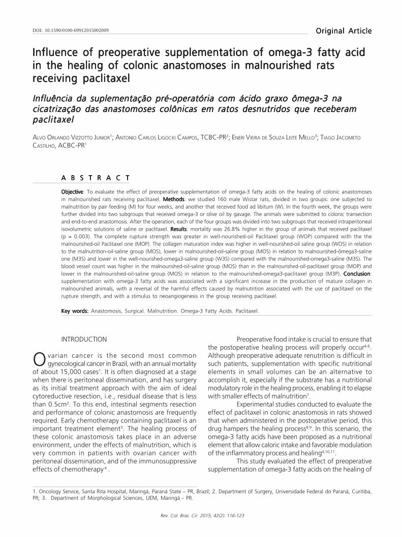
116
Rev. Col. Bras. Cir. 2015; 42(2): 116-123
Vizzoto JuniorVizzoto JuniorVizzoto JuniorVizzoto JuniorVizzoto JuniorInfluence of preoperative supplementation of omega-3 fatty acid in the healing of colonic anastomoses in malnourished rats receiving paclitaxelOriginal ArticleOriginal ArticleOriginal ArticleOriginal ArticleOriginal Article
Influence of preoperative supplementation of omega-3 fatty acidInfluence of preoperative supplementation of omega-3 fatty acidInfluence of preoperative supplementation of omega-3 fatty acidInfluence of preoperative supplementation of omega-3 fatty acidInfluence of preoperative supplementation of omega-3 fatty acidin the healing of colonic anastomoses in malnourished ratsin the healing of colonic anastomoses in malnourished ratsin the healing of colonic anastomoses in malnourished ratsin the healing of colonic anastomoses in malnourished ratsin the healing of colonic anastomoses in malnourished ratsreceiving paclitaxelreceiving paclitaxelreceiving paclitaxelreceiving paclitaxelreceiving paclitaxel
Influência da suplementação pré-operatória com ácido graxo ômega-3 naInfluência da suplementação pré-operatória com ácido graxo ômega-3 naInfluência da suplementação pré-operatória com ácido graxo ômega-3 naInfluência da suplementação pré-operatória com ácido graxo ômega-3 naInfluência da suplementação pré-operatória com ácido graxo ômega-3 nacicatrização das anastomoses colônicas em ratos desnutridos que receberamcicatrização das anastomoses colônicas em ratos desnutridos que receberamcicatrização das anastomoses colônicas em ratos desnutridos que receberamcicatrização das anastomoses colônicas em ratos desnutridos que receberamcicatrização das anastomoses colônicas em ratos desnutridos que receberampaclitaxelpaclitaxelpaclitaxelpaclitaxelpaclitaxel
ALVO ORLANDO VIZZOTTO JUNIOR1; ANTONIO CARLOS LIGOCKI CAMPOS, TCBC-PR2; ENERI VIEIRA DE SOUZA LEITE MELLO3; TIAGO JACOMETO
CASTILHO, ACBC-PR1
A B S T R A C TA B S T R A C TA B S T R A C TA B S T R A C TA B S T R A C T
ObjectiveObjectiveObjectiveObjectiveObjective: To evaluate the effect of preoperative supplementation of omega-3 fatty acids on the healing of colonic anastomoses
in malnourished rats receiving paclitaxel. MethodsMethodsMethodsMethodsMethods: we studied 160 male Wistar rats, divided in two groups: one subjected to
malnutrition by pair feeding (M) for four weeks, and another that received food ad libitum (W). In the fourth week, the groups were
further divided into two subgroups that received omega-3 or olive oil by gavage. The animals were submitted to colonic transection
and end-to-end anastomosis. After the operation, each of the four groups was divided into two subgroups that received intraperitoneal
isovolumetric solutions of saline or paclitaxel. ResultsResultsResultsResultsResults: mortality was 26.8% higher in the group of animals that received paclitaxel
(p = 0.003). The complete rupture strength was greater in well-nourished-oil Paclitaxel group (WOP) compared with the the
malnourished-oil Paclitaxel one (MOP). The collagen maturation index was higher in well-nourished-oil saline group (WOS) in relation
to the malnutrition-oil-saline group (MOS), lower in malnourished-oil-saline group (MOS) in relation to malnourished-ômega3-saline
one (M3S) and lower in the well-nourished-omega3-saline group (W3S) compared with the malnourished-omega3-saline (M3S). The
blood vessel count was higher in the malnourished-oil-saline group (MOS) than in the malnourished-oil-paclitaxel group (MOP) and
lower in the malnourished-oil-saline group (MOS) in relation to the malnourished-omega3-paclitaxel group (M3P). ConclusionConclusionConclusionConclusionConclusion:
supplementation with omega-3 fatty acids was associated with a significant increase in the production of mature collagen in
malnourished animals, with a reversal of the harmful effects caused by malnutrition associated with the use of paclitaxel on the
rupture strength, and with a stimulus to neoangiogenesis in the group receiving paclitaxel.
Key words: Key words: Key words: Key words: Key words: Anastomosis, Surgical. Malnutrition. Omega-3 Fatty Acids. Paclitaxel.
1. Oncology Service, Santa Rita Hospital, Maringá, Paraná State – PR, Brazil; 2. Department of Surgery, Universidade Federal do Paraná, Curitiba,PR; 3. Department of Morphological Sciences, UEM, Maringá - PR.
INTRODUCTION
O varian cancer is the second most commongynecological cancer in Brazil, with an annual mortality
of about 15,000 cases1. It is often diagnosed at a stagewhen there is peritoneal dissemination, and has surgeryas its initial treatment approach with the aim of idealcytoreductive resection, i.e., residual disease that is lessthan 0.5cm2. To this end, intestinal segments resectionand performance of colonic anastomosis are frequentlyrequired. Early chemotherapy containing paclitaxel is animportant treatment element3. The healing process ofthese colonic anastomosis takes place in an adverseenvironment, under the effects of malnutrition, which isvery common in patients with ovarian cancer withperitoneal dissemination, and of the immunosuppressiveeffects of chemotherapy 4 .
Preoperative food intake is crucial to ensure thatthe postoperative healing process will properly occur4-6.Although preoperative adequate renutrition is difficult insuch patients, supplementation with specific nutritionalelements in small volumes can be an alternative toaccomplish it, especially if the substrate has a nutritionalmodulatory role in the healing process, enabling it to elapsewith smaller effects of malnutrition7.
Experimental studies conducted to evaluate theeffect of paclitaxel in colonic anastomosis in rats showedthat when administered in the postoperative period, thisdrug hampers the healing process8.9. In this scenario, theomega-3 fatty acids have been proposed as a nutritionalelement that allow caloric intake and favorable modulationof the inflammatory process and healing4,10,11.
This study evaluated the effect of preoperativesupplementation of omega-3 fatty acids on the healing of
DOI: 10.1590/0100-69912015002009

Vizzoto JuniorVizzoto JuniorVizzoto JuniorVizzoto JuniorVizzoto JuniorInfluence of preoperative supplementation of omega-3 fatty acid in the healing of colonic anastomoses in malnourished rats receiving paclitaxel 117
Rev. Col. Bras. Cir. 2015; 42(2): 116-123
colonic anastomoses in malnourished rats receivingintraperitoneal paclitaxel.
METHODSMETHODSMETHODSMETHODSMETHODS
Experimental design and administered solutionsWe used 160 male Wistar rats (Rattus norvegicus
albinos, Roentia Mammalia) with body weight ranging from200 to 330 grams (288.76 ± 4,45g) from the vivarium ofthe State University of Maringá. The animals were housedin individual boxes and divided into two main groups: W -well-nourished rats (n = 80) who received food and waterad libitum; M - malnourished rats (n = 80) who receivedhalf the amount of the diet that their well-nourished pairshad received the previous day (malnutrition process, pairfeeding method). Each group was divided into foursubgroups with 20 animals each, according to lipidsupplementation and the administration of saline orpaclitaxel.
The lipid solutions were extra-virgin olive oilemulsion and omega-3 fatty acids consisting ofeicosapentaenoic acid (EPA) and docosahexaenoic acid(DHA) in a ratio of 80:20 and concentration of 20mg / ml.The administration comprised a single daily dose of 100mg/ kg via orogastric gavage without anesthesia.
The drug used was paclitaxel at a dose of 3.5mg / kg diluted in isotonic saline solution at a concentrationof 0.35 mg / ml. The administration of chemotherapy or0.9% isotonic saline solution was carried out withintraperitoneal injection, immediately after the surgicalprocedure.
The well-nourished rats (W) received chow adlibitum the four weeks prior to the procedure and weredivided into four subgroups, being supplemented with:WOS – olive oil and saline; W3S – omega 3 and saline;WOP - olive oil and paclitaxel; and W3P – omega 3 andpaclitaxel. The malnourished rats (M) were divided intofour subgroups and were supplemented with: MOS –olive oil and saline; M3S – omega 3 and saline; MOP –olive oil and paclitaxel; and M3P - omega-3 andpaclitaxel.
Surgical procedure and evolutionSurgical procedure and evolutionSurgical procedure and evolutionSurgical procedure and evolutionSurgical procedure and evolutionThe surgical procedure, conducted under gene-
ral anesthesia with a combination of 10 mg / kg xylazineand 50 mg / kg ketamine, consisted of two colontransections (colostomies), one at the colon proximalsegment, 4.0 cm from the ileocecal valve, and one at thedistal colon segment, 3.0 cm above the peritoneal reflectionof the rectum. The reconstructions were performed in totalsingle plane end-to-end anastomosis with interrupted sutureusing eight stitches of 6-0 monofilament, synthetic,nonabsorbable suture (Ethicon?). After anesthetic recoveryall animals received food and water ad libitum until thetime of death.
We observed and weighed the animals daily, andthose which died underwent necropsy to evaluate thepresence of fistula or peritonitis. Euthanasia, with a lethaldose of ketamine and xylazine association, was taken onthe fifth day after surgery. After death, the animalsunderwent celiotomy for evaluation of the following:presence of wound infection, adhesions (by the Knigthlyindex)12 and anastomotic complications (dehiscence orstenosis). Then we excised the proximal and distal colonsegments containing the anastomoses. The proximalsegments, about 2.0 cm long, were named proximalspecimen (PS) and used for pathological andimmunohistochemical study. The distal segments, 4 cmlong, were named distal specimen (DS) and subjected totensile test.
Tension trialTension trialTension trialTension trialTension trialThe distal specimen was subjected to tensile test,
using a computerized mechanical testing machine (EMIC®).Tension was applied with a 50 mm / minute speed. We useda 1Kg load cell with a sensitivity of 50 grams for a 4500 gfstrength limit, until complete anastomotic rupture. Thevariables evaluated were the maximum tensile strength(MTS) and the complete rupture strength (CRS), measuredin Newtons (N), and the maximum tension (MT), measuredin N / cm2.
Histological evaluationHistological evaluationHistological evaluationHistological evaluationHistological evaluationThe proximal specimen was fixed in buffered 10%
formalin and sent for histological processing and evaluationby hematoxylin-eosin (HE), picrosirius-red (PSR) andimmunohistochemistry.
HE staining was used to assess the degree ofinflammatory reaction (poly and monomorfonuclearinfiltrate), interstitial edema, vascular congestion,granulation tissue and fibrosis. The data were classified assevere, moderate, mild or absent, and transformed intoquantitative variables by assignment of indexes to thehistological findings, as follows: absent, index 0; discrete,index 1; moderate, index 2; and severe, index 3.
The staining with F3BA picrosirius-red was madeto identify the mature and immature collagen fibers bymicroscopy technique with polarized light and computerizedmorphometric analysis. Each section was assessed by opticalmicroscopy at 200x magnification using a polarized lightsource (C-SP simple polarizer). We analyzed two fields,proximal and distal to the anastomotic line, containing theentire thickness of the colonic segment. The images werecaptured by a Nikon DS-FI1C camera and transmitted viathe D-DA simple system analyzer, to the G205HV® colorLCD monitor, frozen and scanned. We then performedimage analysis using the Image Pro-plus™ software. In theRGB (“Red, Green, Blue”) system, we quantified areas ofmature collagen fibers – collagen type I (shades of red,yellow and orange) – and immature ones – type III collagen(green tones). We computed the between the area

118
Rev. Col. Bras. Cir. 2015; 42(2): 116-123
Vizzoto JuniorVizzoto JuniorVizzoto JuniorVizzoto JuniorVizzoto JuniorInfluence of preoperative supplementation of omega-3 fatty acid in the healing of colonic anastomoses in malnourished rats receiving paclitaxel
percentages of collagen type I and collagen type III,determined as collagen maturation index (CMI).
Immunohistochemical evaluationImmunohistochemical evaluationImmunohistochemical evaluationImmunohistochemical evaluationImmunohistochemical evaluationBy immunohistochemical method, we evaluated
CD31 (Monoclonal, JC70 code, Cell Marque, Moleenstraat,NL), calponin (Monoclonal, code CALP, Cell Marque,Moleenstraat, NL) and Ki-67 (Monoclonal, SP6 code, CellMarque, Moleenstraat , NL), on separate slides. We usedthese markers to evaluate neoangiogenesis, research formyofibroblasts, and for cells in the replication phase (otherthan G0) present in the healing process area. We carriedout antigen retrieval by exposing the materials to heat andpressure. Development of the slides was made byDiaminobenzidine (DAB) brown chromogen; the detectiontechnique, by HRP-polymer (free biotin). We used positiveand negative controls to validate the reactions. Weexamined the slides under 200x magnification to countvessels and 400x to count myofibroblasts and proliferatingcells. The images were made using the pictures OPT 5000Power Opticam® capture system, of 5.0 megapixels,connected to a computer with image analysis software (VMS3.5 Measuring Software HPower®). We performedquantitative analysis of the number of vessels, fibroblastsand proliferating cells in the area of the anastomosis, aswell as 10mm proximal and 10mm distal to it, in fourrandomly selected different fields.
Statistical analysisStatistical analysisStatistical analysisStatistical analysisStatistical analysisWe carried out the statistical analysis according
to the nature of the data. We used parametric tests forquantitative variables with normal (Gaussian) distribution.For comparison between all groups, was applied the analysisof variance (ANOVA), and for the pair-wise comparison ofgroups, we used the Student’s t test. For quantitativevariables without normal (Gaussian) distribution, we usednon-parametric analysis. For comparison between all groups,we applied the Kruskal-Wallis test, and for pair-wise analysis,the Mann-Whitney test (Wilcoxon test for independentgroups). We evaluated dichotomous variables, such as afatal outcome, presence of wound infection andanastomotic complications (dehiscence or stenosis) by thechi-square test and the Cochran test. The level ofsignificance was p <0.05.
RESULTSRESULTSRESULTSRESULTSRESULTS
Of the initial 160 animals experiment on, 117survived until the day of euthanasia, with an overall mortalityrate of 26.8%. Of the 43 deaths, 42 were for anastomoticcomplications, 15 in the animals receiving saline and 28 ingroups receiving paclitaxel, significantly higher in the latter(p = 0.03).
The average weight of the animals was 294.8 ±1,67g. There was no difference between the groups average
weight on the first day (D1) of the experiment (p = 0.8037).The animals of well-nourished groups had an average weightgain of 23.1% between D1 and D28. The animals of themalnourished groups had an average weight loss of 16.6%between D1 and D28. Between the day of the operationand the euthanasia there was a mean weight loss of 18%in the well-nourished group and 10% in the malnourishedgroup (Figure 1).
On the operation day (Figure 2), the averageweight of the MOS group (236.5 ± 4.2 g) was significantlylower (p <0.05) than the average M3S group weight (253.8± 4.1g).
Anastomotic stenosis occurred in 14 rats of thewell-nourished group and in 31 of the malnourished group,being significantly higher in this group (p = 0.007).
Full tensile strength (Figure 3) was significantlyhigher in the WOP group compared with the MOP group(1.28 ± 0.17 vs. 0.77 ± 0.07, p <0.05). There was nodifference between the groups in terms of maximum tension(p = 0.2119) and for the maximum tensile strength (p =0.3638).
The mean intensity of the inflammatory infiltrate(Figure 4) was significantly lower in the MOS groupcompared with the M3S group (1.61 ± 0.78 vs. 2.50 ±0.63, p <0.05).
Regarding the collagen maturation index (CMI)(Figure 5), it was significantly higher in the WOS groupthan in the MOS one (1.45 ± 0.4 vs. 0.35 ± 0.08, p<0.05),significantly lower in the W3S group than in M3S one (2.08± 0.35 vs. 2.82 ± 0.33, p<0.05), and significantly lower inthe MOS group than in groups M3S (0.35 ± 0.08 vs. 2.82 ±
Figure 1 Figure 1 Figure 1 Figure 1 Figure 1 - Evolution of animal weight.X-axis: days of the experiment - D1: Day 1; D7: 7th day; D14: 14th
day; DOP- day of operation; and DEUT: day of mice death(euthanasia).Y-axis: Weight (grams). WOS: well-nourished-oil-saline; W3S: well-nourished-omega-3-saline; WOP: well-nourished-oil-paclitaxel; W3P:well-nourished-omega-3-paclitaxel; MOS: malnourished-oil-saline;M3S: malnourished-omega-3-saline; MOP: malnourished-oil-paclitaxel;M3P: : : : : malnourished-omega-3-paclitaxel.

Vizzoto JuniorVizzoto JuniorVizzoto JuniorVizzoto JuniorVizzoto JuniorInfluence of preoperative supplementation of omega-3 fatty acid in the healing of colonic anastomoses in malnourished rats receiving paclitaxel 119
Rev. Col. Bras. Cir. 2015; 42(2): 116-123
0.33, p<0.05) and MOP (0.35 ± 0.08 vs. 1.58 ± 0.15,p<0.05).
The average blood vessels count (Figure 6) wassignificantly higher in the M3S group compared with theMOP group (12.64 ± 1.7 vs. 6.45 ± 0.28, p <0.05) andsignificantly lower in the MOP group in relation to the M3Pgroup (6.45 ± 0.28 vs. 8.91 ± 0.88, p <0.05).
DISCUSSIONDISCUSSIONDISCUSSIONDISCUSSIONDISCUSSION
The overall incidence of deaths in this study was26.5%, consistent with other studies using similarmethodologies. Trubian observed 24.5% of deaths whenassessing malnutrition with toxemia induction13. Themortality rate was higher in the groups receiving paclitaxelcompared with the groups that received saline, most dueto anastomotic postoperative complications.
The immunosuppressive action of chemotherapydrugs such as paclitaxel is one of the possible factorsassociated with higher death rates observed in the groupsreceiving this drug 2,14. Another possible explanation for thishigher death rate is because such drugs act by inhibitingcell proliferation by different mechanisms of action. As suchproliferation is essential in the healing process, they playan important attenuation of the fibroplasia process8,9,15. Theinhibitory action on fibroplasia results in higher anastomoticdehiscence rate, causing fistula and fecal peritonitis 15,which is consistent with what was observed at the animals‘necropsy.
The average starting weight of the animals in allgroups was homogeneous. The induction of protein-energymalnutrition, by restriction to 50% of the standard diet,promoted progressive weight loss during the 28 days. Theanimals in well-nourished groups had an average weightgain of 23.1% between D1 and D28, while the ones inmalnourished group had a mean weight loss of 16.6%between D1 and D28. The average weight of themalnourished group was lower compared with the well-nourished ones at the end of 28 days, showing the efficiencyof the food restriction procedure on obtaining malnutrition.Similar results were found in other studies13,16.
Relatively few studies have evaluated the roleof supplementation of omega-3 fatty acids in post-anastomosis recovery of the digestive tract, most of themclinical studies. Clinical trials such as the ones of Farreraset al., in 200517, Torrinhas, 20134, and Aoyama et al., in201318, evaluated the effect of omega-3 fatty acidssupplementation in patients undergoing digestive tractanastomosis. Only in the latter, the weight was evaluatedas a nutritional recovery indicator, and the omega-3 fattyacids had an important role for nutritional recovery andweight gain in postoperative patients undergoinggastroenteric anastomosis for stomach cancer18.
We also observed that in the operation day, afterone week of olive oil or omega-3 fatty acids administration,
Figure 3-Figure 3-Figure 3-Figure 3-Figure 3- Complete Rupture Strength (CRS) in groups. Y-axis:force in Newtons. X axis: groups; WOS: well-nourished-oil-saline; W3S: well-nourished-omega-3-saline; WOP:well-nourished-oil-paclitaxel; W3P: well-nourished-omega-3-paclitaxel; MOS: malnourished-oil-saline;M3S: malnourished-omega-3-saline; MOP:malnourished-oil-paclitaxel; M3P: : : : : malnourished-omega-3-paclitaxel. There was no significantdifference between groups (p = 0.3638).
Figure 2 Figure 2 Figure 2 Figure 2 Figure 2 - Mean weights in malnourished groups on the day ofoperation. Y-axis: weight in grams; X-axis: groups:MOS - malnourished-oil-saline; M3S - malnourished-omega-3-saline; MOP - malnourished-oil-paclitaxel;M3P - malnourished-omega-3-paclitaxel; e - Averageof MOS group weight is smaller than the averageweight of M3S group (p <0.05).
the MOS group mean weight was greater than the M3Sgroup. This finding, consistent with the literature, suggeststhat supplementation with omega-3, besides favorablymodulating the anastomotic healing process, also contributesas an important nutritional energy intake factor in therecovery from undernourishment18.
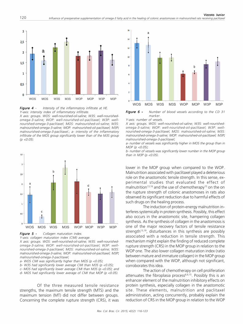
120
Rev. Col. Bras. Cir. 2015; 42(2): 116-123
Vizzoto JuniorVizzoto JuniorVizzoto JuniorVizzoto JuniorVizzoto JuniorInfluence of preoperative supplementation of omega-3 fatty acid in the healing of colonic anastomoses in malnourished rats receiving paclitaxel
lower in the MOP group when compared to the WOP.Malnutrition associated with paclitaxel played a deleteriousrole on the anastomotic tensile strength. In this sense, ex-perimental studies that evaluated the effect ofmalnutrition13,16 and the use of chemotherapy15 on the onthe rupture strength of colonic anastomoses in rats alsoobserved its significant reduction due to harmful effects ofsuch drugs on the healing process.
The induction of protein-energy malnutrition in-terferes systemically in protein synthesis. Possibly, this effectalso occurs in the anastomotic site, hampering collagensynthesis. As the synthesis of collagen in the anastomosis isone of the major recovery factors of tensile resistancestrength13,19, disturbances in this synthesis are possiblyassociated with a reduction in tensile strength. Thismechanism might explain the finding of reduced completerupture strength (CRS) in the MOP group in relation to theWOP one. The also lower collagen maturation index (ratiobetween mature and immature collagen) in the MOP groupwhen compared with the WOP, although not significant,corroborates this idea.
The action of chemotherapy on cell proliferationattenuates the fibroplasia process8,9,15. Possibly this is anenhancer element of the malnutrition inhibitory effects onprotein synthesis, especially collagen in the anastomoticsite. These elements, malnutrition and paclitaxeladministration, acting concurrently, probably explain thereduction of CRS in the MOP group in relation to the WOP
Figure 6 -Figure 6 -Figure 6 -Figure 6 -Figure 6 - Number of blood vessels according to the CD 31marker.
Y-axis: number of vessels.X axis: groups. WOS: well-nourished-oil-saline; W3S: well-nourished-omega-3-saline; WOP: well-nourished-oil-paclitaxel; W3P: well-nourished-omega-3-paclitaxel; MOS: malnourished-oil-saline; M3S:malnourished-omega-3-saline; MOP: malnourished-oil-paclitaxel; M3P:::::malnourished-omega-3-paclitaxel;a- number of vessels was significantly higher in MOS the group than inMOP (p <0.05);b- number of vessels was significantly lower number in the MOP groupthan in M3P (p <0.05).
Figure 5 -Figure 5 -Figure 5 -Figure 5 -Figure 5 - Collagen maturation index.Y-axis: collagen maturation index (CMI) average.X axis: groups. WOS: well-nourished-oil-saline; W3S: well-nourished-omega-3-saline; WOP: well-nourished-oil-paclitaxel; W3P: well-nourished-omega-3-paclitaxel; MOS: malnourished-oil-saline; M3S:malnourished-omega-3-saline; MOP: malnourished-oil-paclitaxel; M3P:::::malnourished-omega-3-paclitaxel;a- WOS CMI was significantly higher than MOS (p <0.05);b- W3S had significantly lower average CMI than M3S (p <0.05);c- MOS had significantly lower average CMI than M3S (p <0.05); andd- MOS had significantly lower average of CMI that MOP (p <0.05).
Figure 4 Figure 4 Figure 4 Figure 4 Figure 4 - Intensity of the inflammatory infiltrate at HE.Y-axis: intensity index of inflammatory infiltrate.X axis: groups. WOS: well-nourished-oil-saline; W3S: well-nourished-omega-3-saline; WOP: well-nourished-oil-paclitaxel; W3P: well-nourished-omega-3-paclitaxel; MOS: malnourished-oil-saline; M3S:malnourished-omega-3-saline; MOP: malnourished-oil-paclitaxel; M3P:::::malnourished-omega-3-paclitaxel.; a- intensity of the inflammatoryinfiltrate of the MOS group significantly lower than of the M3S group(p <0.05).
Of the three measured tensile resistancestrengths, the maximum tensile strength (MTS) and themaximum tension (MT) did not differ between groups.Concerning the complete rupture strength (CRS), it was

Vizzoto JuniorVizzoto JuniorVizzoto JuniorVizzoto JuniorVizzoto JuniorInfluence of preoperative supplementation of omega-3 fatty acid in the healing of colonic anastomoses in malnourished rats receiving paclitaxel 121
Rev. Col. Bras. Cir. 2015; 42(2): 116-123
group, as this reduction was not observed in the group ofanimals submitted only to malnutrition without paclitaxeladministration (MOS) in relation to the WOS group.
On the other hand, there was no significantdifference in CRP between the M3P and W3P groups. Thisfinding suggests that nutritional supplementation withomega-3 fatty acids can reverse the deleterious effectscaused by the association of malnutrition and paclitaxeladministration in colonic anastomosis CRS, for thesignificant reduction of this force, observed in the MOPgroup in relation to the WOP one, was not observed whenthe M3P group was compared with the W3P group. Thisfinding is consistent with the ones of Ekçi et al.20, who alsoobserved a significant increase in the colonic anastomosisrupture pressure of rats receiving dietary supplementationwith omega-3, although used in combination with ascorbicacid. The favorable modulation of omega-3 on collagensynthesis in the anastomotic site is one possible explanationfor the observed recovery of the colonic anastomosis rupturestrength.
We also noted an increase in cell activity in theanastomotic site, considered as inflammatory infiltrate, inthe M3S group compared with the M3S one. This findingreinforces the idea that omega-3 has immunomodulatingeffects in inflammation and healing, by stimulating cellularactivity, and corroborates the findings of McDaniel et al.21,who observed an inflammatory infiltrate increase of humanhealing skin wounds, as well as a significantly higher levelof interleukin 1â (IL-1 â) in the inflammatory lesion fluidin the group that received the administration of EPA andDHA. This finding may seem conflicting with that proposedin the literature, that the omega-3 fatty acids have lesspro-inflammatory activity by replacing arachidonic acid(omega-6 fatty acid) in the cell membrane and increase,through its metabolism, the production of eicosanoids oflower inflammatory potential, such as leukotriene B
5 and
prostaglandin E3
10,22. Authors speculate that this increasedcellular activity of the anastomotic environment is due tospecific cell types, such as macrophages, fibroblasts, andmyofibroblasts. It is possible that at the anastomotic siteeicosanoids derived from the omega-3 fatty acids, althoughdisplay an inhibitory effect on the inflammatory processin general, can stimulate the proliferation of specific celltypes. The largest production of mature collagen (CMI) inthe M3S group compared with the M3S one contributesindirectly to this idea, as this increased production iscorrelated with increased activity of cells producingcollagen (fibroblasts and myofibroblasts) in the anastomoticenvironment.
The evaluation of collagen densitometry showeda significant reduction of mature collagen (type I) in relationto the immature one (type III), when assessed by thecollagen maturation index (CMI), in the MOS groupcompared with the WOS group. This finding suggests thatmalnutrition is an important factor reducing the productionof mature collagen in the healing anastomosis. Trubian also
observed a reduction of CMI in groups of malnourishedrats compared with the group of well-nourished ones13.
The recovery of the tensile strength is morerelated to type I collagen than with type III one. Thedeposition and remodeling of type I collagen fibers is themain factor for recovery of anastomotic resistance19.
The M3S group showed an important gain ofmature collagen in relation to the immature one whencompared to W3S group, as the CMI of the former wasgreater than the one of the latter. This finding suggeststhat the administration of omega-3 in the week before thesurgical intervention stimulates the production of maturecollagen in malnourished animals. The CMI was alsosignificantly higher in the M3S group compared with theMOS one. This finding supports the idea that preoperativeadministration of omega-3 can reverse the deleteriouseffects of malnutrition in the production of mature collagenin the healing anastomotic environment.
The migration and proliferation of fibroblasts andmyofibroblasts in the healing environment, andconsequently the production of collagen, is mediated bythe action of inflammatory prostanoids (prostaglandins andleukotrienes) produced by inflammatory cells. Therefore,the higher CMI in the M3S group in relation to the MOS ispossibly correlated with the higher level of cellular activityobserved in the M3S group compared with the MOS group.
Another finding of this study that favors theconcept of immunomodulation produced by omega-3 wasthe observation made in the number of blood vessels(neoangiogenesis) in the anastomotic healing environment.In immunohistochemistry, CD31 is mainly used todemonstrate the presence of endothelial cells in histologictissue sections. This allows evaluating the degree of tissueangiogenesis23. We observed a decrease in the number ofblood vessels in the MOP group in relation to the MOSgroup, suggesting that the administration of paclitaxel,which is a drug with antiangiogenic action24, is the causativefactor of such reduction, agreeing with another study8 thatshowed significant reduction in neovascularization of colonicanastomoses of rats receiving paclitaxel. Supplementationwith omega-3 in the M3P group was associated withrecovery of the number of vessels, since this number washigher in this group compared with the MOP group. Thisfinding suggests that the inhibition that paclitaxel causeson neoangiogenesis of the colonic anastomosis healingprocess can be reversed with the addition of omega-3preoperatively. It is speculated that the eicosanoids producedby the metabolism of omega-3 fatty acids, such asprostaglandin E
3 and leukotriene B
5, stimulate the migration
and proliferation of endothelial cells precursors in theanastomotic environment.
Myofibroblasts are (predominantly) type Icollagen-producing cells, and its quantification can also beused as a healing evaluation method13. Fibroblasts start toappear in the wound around the third day after surgery,proliferate in response to growth factors and begin to
122
Rev. Col. Bras. Cir. 2015; 42(2): 116-123
Vizzoto JuniorVizzoto JuniorVizzoto JuniorVizzoto JuniorVizzoto JuniorInfluence of preoperative supplementation of omega-3 fatty acid in the healing of colonic anastomoses in malnourished rats receiving paclitaxel
R E S U M OR E S U M OR E S U M OR E S U M OR E S U M O
Objetivo: Objetivo: Objetivo: Objetivo: Objetivo: avaliar o efeito da suplementação pré-operatória dos ácidos graxos ômega-3 sobre a cicatrização das anastomosescolônicas em ratos desnutridos que receberam paclitaxel. Métodos:Métodos:Métodos:Métodos:Métodos: foram estudados 160 ratos Wistar, distribuídos em dois grupos:um submetido à desnutrição pelo pair feeding (D) durante quatro semanas, e outro que recebeu ração ad libitum (N). Na quartasemana, os grupos foram subdivididos em dois subgrupos que receberam, por gavagem, ácido graxo ômega-3 ou azeite de oliva. Osanimais foram submetidos à transecção colônica e anastomose término-terminal. Após a operação, foram distribuídos em doissubgrupos que receberam soluções isovolumétricas de salina ou paclitaxel, intraperitonial. Resultados:Resultados:Resultados:Resultados:Resultados: A mortalidade foi 26,8%,maior no grupo de animais que recebeu paclitaxel (p=0,003). A força de ruptura completa foi maior no grupo nutrido-azeite-paclitaxel (NAP) em relação ao grupo desnutrido-azeite-paclitaxel (DAP). O índice de maturação de colágeno foi maior no gruponutrido-azeite-salina (NAS) em relação ao grupo desnutrido-azeite-salina (DAS), menor no grupo desnutrido-azeite-salina (DAS) emrelação ao grupo desnutrido-ômega3-salina (DOS) e menor no grupo nutrido-ômega3-salina (NOS) em relação ao desnutrido-ômega3-salina (DOS). A contagem do número de vasos sanguíneos foi maior no grupo desnutrido-azeite-salina (DAS) em relação aogrupo desnutrido-azeite-paclitaxel (DAP) e menor no grupo desnutrido-azeite-salina (DAS) em relação ao grupo desnutrido-ômega3-paclitaxel (DOP). Conclusão:Conclusão:Conclusão:Conclusão:Conclusão: a suplementação com ácidos graxos ômega-3 associou-se ao aumento significativo na produção decolágeno maduro nos animais desnutridos, à reversão do efeito deletério causado pela desnutrição em associação ao uso dopaclitaxel, sobre a força de ruptura, e ao estímulo da neoangiogênese no grupo que recebeu paclitaxel.
DescritoresDescritoresDescritoresDescritoresDescritores: Anastomose Cirúrgica. Desnutrição. Acido Graxo Ômega-3. Paclitaxel.....
REFERENCESREFERENCESREFERENCESREFERENCESREFERENCES
1. Siegel R, Ward E, Brawley O, Jemal A. Cancer statistics, 2011: theimpact of eliminating socioeconomic and racial disparities onpremature cancer deaths. CA Cancer J Clin. 2011;61(4):212-36.
produce collagen25. We observed a significant reduction inthe average number of myofibroblasts in the M3S groupcompared with the W3S one. This finding suggests thatmalnutrition is a deleterious effect to the myofibroblastsproduction. Trubian also found a significant reduction inthe number of myofibroblasts in colonic anastomosis inmalnourished rats13.
Proliferating cells are the very hallmark ofanastomotic healing. The Ki-67 antigen is present on allactive phases of the cell cycle (G1, S, G2 and mitosis), butis absent on resting cells (G0)26. In this study, there was nodifference between groups in the number of proliferatingcells when assessed by Ki67. Given the previously reportedfindings and hypotheses developed, one would expect tofind more proliferating cells in the groups receiving omega-3 fatty acids, particularly in subgroups that did not receivechemotherapy, when compared with groups receiving oliveoil, but we did not. We could not establish objective reasonsfor this. One suggested possibility is that proliferating celltypes are different between the groups olive oil and omega-3 fatty acids. In the latter, with healing mediated byeicosanoids of lower inflammatory potential (prostaglandinE
3 and leukotriene B
5) derived from the metabolism of
omega-3 fatty acids, cell types would be composed ofmyofibroblasts, fibroblasts, endothelial cells and theirprecursors; in the former group (olive oil), with healingmediated by prostanoid of more inflammatory potential,other cell types would prevail, like polymorphonuclear andmononuclear ones.
In agreement with the literature, the datapresented here suggest that the use of omega-3 wasassociated with modulation of the inflammatory process,with stimuli to collagen production and to its quality in theanastomotic environment19. As already mentioned, theamount of mature collagen in relation to the immature, asmeasured by the CMI, was significantly higher in the M3Sgroup compared with the MOS. Also in the M3S group,cellular activity represented by the inflammatory infiltratein the anastomosis was higher than in the M3S group. Thesetwo concomitantly assessed findings support the idea thatpreoperative omega-3 administration in malnourishedanimals contributes positively to the healing processmodulation of colonic anastomoses in rats, corroboratingdata from another work20, which pointed to the beneficialaction of fatty acids.
The supplementation of omega-3 was also animportant energy intake nutritional factor, as it attenuatedweight loss in the M3S group compared with the MOS.This supplementation also played an important role in therecovery of the deleterious effects of paclitaxel onneoangiogenesis, since the number of blood vessels in theM3P group was higher than in the MOP one.
The exact mechanisms by which omega-3 mayhave acted to produce this study‘s findings still need to beclarified. Omega-3 fatty acids have immunomodulatoryactivities, still under study, on the production of cytokines,activation of T lymphocytes and angiogenesis27, which, whenunderstood, can contribute to a better data comprehension.
2. du Bois A, Quinn M, Thigpen T, Vermorken J, Avall-Lundqvist E,Bookman M, et al. 2004 consensus statements on the managementof ovarian cancer: final document of the 3rd International GynecologicCancer Intergroup Ovarian Cancer Consensus Conference (GCIGOCCC 2004). Ann Oncol. 2005;16 Suppl 8:viii7-viii12.

Vizzoto JuniorVizzoto JuniorVizzoto JuniorVizzoto JuniorVizzoto JuniorInfluence of preoperative supplementation of omega-3 fatty acid in the healing of colonic anastomoses in malnourished rats receiving paclitaxel 123
Rev. Col. Bras. Cir. 2015; 42(2): 116-123
3. Trimbos JB, Parmar M, Vergote I, Guthrie D, Bolis G, Colombo N,et al. International Collaborative Ovarian Neoplasm trial 1 andAdjuvant ChemoTherapy In Ovarian Neoplasm trial: two parallelrandomized phase III trials of adjuvant chemotherapy in patientswith early-stage ovarian carcinoma. J Natl Cancer Inst.2003;95(2):105-12.
4. Campos ACL. Tratado de nutrição e metabolismo em Cirurgia. Riode Janeiro: Rubrio; 2013.
5. Braga M, Gianotti L, Nespoli L, Radaelli G, Di Carlo V. Nutritionalapproach in malnourished surgical patients. A prospectiverandomized study. Arch Surg. 2002;137(2):174-80.
6. Kavalukas SL, Barbul A. Nutrition and wound healing: an update.Plast Reconstr Surg. 2011;127 Suppl 1:38S-43S.
7. Balogun N, Forbes A, Windswendter M, Lanceley A. Noninvasivenutritional management of ovarian cancer patients: beyond in-testinal obstruction. Int J Gynecol Cancer. 2012;22(6):1089-95.
8. Yüceyar S, Ertürk S, Temiz M, Doðusoy G, Gümüstas K, OzgüroðluM, et al. Role of the antiangiogenetic drug paclitaxel on healing ofintestinal anastomosis: an experimental study. Tech Coloproctol.2005;9(3):201-5.
9. Bozkurtoglu H, Bozkurt S, Celik F, Kaya MA, Zemheri E. Effect ofthe intraperitoneal paclitaxel on the healing of colonic anastomosis.Int Surg. 2005;90(1):12-7.
10. Calder PC. Immunomodulation by omega-3 fatty acids.Prostaglandins Leukot Essent Fatty Acids. 2007;77(5-6):327-35.
11. Waitzberg DL, Torrinhas RS, Jacintho TM. New parenteral lipidemulsions for clinical use. JPEN J Parenter Enteral Nutr.2006;30(4):351-67.
12. Knightly JJ, Agostino D, Cliffton EE. The effect of fibrinolysin andheparin on the formation of peritoneal adhesions.Surgery.1962;52:250-8.
13. Trubian PS. Nutrição perioperatória e cicatrização de anastomosesem ratos desnutridos e toxêmicos: tensiometria, densitometria docolágeno e quantificação de angiogênese e miofibroblasto [tese].Curitiba: Universidade Federal do Paraná, Ciências da Saúde;2008.
14. von Gruenigen VE, Huang HQ, Gil KM, Frasure HE, Armstrong DK.Wenzel LB. The association between quality of life domains andoverall survival in ovarian cancer patients during adjuvantchemotherapy: a Gynecologic Oncology Group Study. GynecolOncol. 2012;124(3):379-82.
15. Vizzotto Júnior AO, Noronha L, Scheffel DLH, Campos ACL. Influ-ência da cisplatina administrada no pré e no pós-operatório sobrecicartrização de anastomoses colônicas em ratos. J Bras PatolMed Lab. 2003;39(2):143-9.
16. Gonçalves CG, Groth AK, Ferreira M, Matias JE, Coelho JC, Cam-pos AC. Influence of preoperative feeding on the healing of colonicanastomosis in malnourished rats. JPEN J Parenter Enteral Nutr.2009;33(1):83-9.
17. Farreras N, Artigas V, Cardona D, Rius X, Trias M, Gonzáles JA.Effect of early postoperative enteral immunonutrition on woundhealing in patients undergoing surgery for gastric cancer. ClinNutr. 2005;24(1):55-65.
18. Aoyama T, Hayashi T, Fujikawa H, Ogata T, Cho H, Wada H, et al.Effect of enteral nutrition enriched with eicosapentaenoic acid onbody weight loss and compliance with S-1 adjuvant chemotherapyafter gastric cancer surgery. Gan To Kagaku Ryoho.2013;40(12):2289-91.
19. Doillon CJ, Dunn MG, Berg RA, Silver FH. Collagen deposition duringwound repair. Scan Electron Microsc. 1985;(Pt 2):897-903.
20. Ekçi B, Karabicak I, Atukeren P, Altinlio E, Tomaoglu K, Tasci I. Theeffect of omega-3 fatty acid and ascorbic acid on healing ofischemic colon anastomoses. Ann Ital Chirur. 2011;82(6):475-9.
21. McDaniel JC, Belury M, Ahijevych K, Blakely W. omega-3 fattyacids effects on wound healing. Wound Repair Regen.2008;16(3):337-45.
22. Cockbain AJ, Toogood GJ, Hull MA. Omega-3 polyunsaturatedfatty acids for the treatment and prevention of colorectal cancer.Gut. 2012;61(1):135-49.
23. Jackson DE. The unfolding tale of PECAM-1. FEBS Lett. 2003;540(1-3):7-14.
24. Klein LE, Freeze BS, Smith AB 3rd, Horwitz SB. The microtubulestabilizing agent discodermolide is a potent inducer of acceleratedcell senescence. Cell Cycle. 2005;4(3):501-7.
25. Kiyama T, Witte MB, Thornton FJ, Barbul A. The route of nutritionsupport affects the early phase of wound healing. JPEN J ParenterEnteral Nutr. 1998;22(5):276-9.
26. Bullwinkel J, Baron-Lühr B, Lüdemann A, Wohlenberg C, Gerdes J,Scholzen T. Ki-67 protein is associated with ribosomal RNAtranscription in quiescent and proliferating cells. J Cell Physiol.2006;206(3):624-35.
27. Segura JG. Extração e caracterização de óleos de resíduos depeixe de água doce [dissertação]. Pirassununga/SP: Universidadede São Paulo, Faculdade de Zootecnia e Engenharia de Alimen-tos; 2012.
Received on 10/07/2014Accepted for publication 15/08/2014Conflict of interest: none.Source of funding: none.
Address for correspondence:Address for correspondence:Address for correspondence:Address for correspondence:Address for correspondence:Alvo Orlando Vizzotto JuniorE-mail: [email protected]

S a n t o sS a n t o sS a n t o sS a n t o sS a n t o sPulmonar recruitment in acute respiratory distress syndrome. What is the best strategy? 125
Rev. Col. Bras. Cir. 2015; 42(2): 125-129
Rev iewRev iewRev iewRev iewRev iew
Pulmonar recruitment in acute respiratory distress syndrome.Pulmonar recruitment in acute respiratory distress syndrome.Pulmonar recruitment in acute respiratory distress syndrome.Pulmonar recruitment in acute respiratory distress syndrome.Pulmonar recruitment in acute respiratory distress syndrome.What is the best strategy?What is the best strategy?What is the best strategy?What is the best strategy?What is the best strategy?
Recrutamento pulmonar na síndrome do desconforto respiratório agudo. Qual aRecrutamento pulmonar na síndrome do desconforto respiratório agudo. Qual aRecrutamento pulmonar na síndrome do desconforto respiratório agudo. Qual aRecrutamento pulmonar na síndrome do desconforto respiratório agudo. Qual aRecrutamento pulmonar na síndrome do desconforto respiratório agudo. Qual amelhor estratégia?melhor estratégia?melhor estratégia?melhor estratégia?melhor estratégia?
CÍNTIA LOURENÇO SANTOS1; CYNTHIA DOS SANTOS SAMARY2; PEDRO LAURINDO FIORIO JÚNIOR3; BRUNA LOURENÇO SANTOS4;ALBERTO SCHANAIDER, TCBC-RJ1
A B S T R A C TA B S T R A C TA B S T R A C TA B S T R A C TA B S T R A C T
Supporting patients with acute respiratory distress syndrome (ARDS), using a protective mechanical ventilation strategy
characterized by low tidal volume and limitation of positive end-expiratory pressure (PEEP) is a standard practice in the
intensive care unit. However, these strategies can promote lung de-recruitment, leading to the cyclic closing and reopening of
collapsed alveoli and small airways. Recruitment maneuvers (RM) can be used to augment other methods, like positive end-
expiratory pressure and positioning, to improve aerated lung volume. Clinical practice varies widely, and the optimal method
and patient selection for recruitment maneuvers have not been determined, considerable uncertainty remaining regarding
the appropriateness of RM. This review aims to discuss recent findings about the available types of RM, and compare the
effectiveness, indications and adverse effects among them, as well as their impact on morbidity and mortality in ARDS patients.
Recent developments include experimental and clinical evidence that a stepwise extended recruitment maneuver may cause
an improvement in aerated lung volume and decrease the biological impact seen with the traditionally used sustained inflation,
with less adverse effects. Prone positioning can reduce mortality in severe ARDS patients and may be an useful adjunct to
recruitment maneuvers and advanced ventilatory strategies, such noisy ventilation and BIVENT, which have been useful in
providing lung recruitment.
Key words:Key words:Key words:Key words:Key words: Valsava Maneuver. Positive-Pressure Respiration. Respiratory Distress Syndrome, Adult. Respiration, Artificial. Prone
Position.
1. Experimental Surgery Center, Faculty of Medicine, Universidade Federal do Rio de Janeiro, Rio de Janeiro, Rio de Janeiro State – RJ, Brazil; 2.Pulmonary Research Laboratory, Carlos Chagas Filho Institute of Biophysics, Universidade Federal do Rio de Janeiro; 3. Universidade Federal deLavras, Lavras, Minas Gerais State – MG, Brazil; 4. Universidade Estacio de Sá, Rio de Janeiro; 5. Department of Surgery, Faculty of Medicine,Universidade Federal do Rio de Janeiro.
INTRODUCTIONINTRODUCTIONINTRODUCTIONINTRODUCTIONINTRODUCTION
The acute respiratory distress syndrome (ARDS) is aworldwide public health problem, occurring even today,
with high rates of mortality. Despite the many strategiesproposed so far, the only isolated therapy that effectivelychanged the prognosis of patients, with a significantreduction in morbidity and mortality rates, was theprotective ventilatory strategy, characterized by the use oflow tidal volume (4-8 ml / kg)1.2. However, this strategycan facilitate alveolar de-recruitment and promote the cyclicopening and closing of alveoli, which is considered one ofthe mechanisms of promotion and exacerbation of lunginjury3. In this context, various strategies, ranging fromventilation modes to specific maneuvers, have beenproposed to minimize alveolar collapse and promote a morehomogeneous ventilation distribution. The use of recruitmentmaneuvers (RM) aims to reopen collapsed alveolar, units
based on a transient increase in transpulmonary pressure(P
TP) during mechanical ventilation4,5.
Nevertheless, the RM can also exacerbate thedamage to pulmonary epithelial6 and endothelial7 cells,increasing the alveolar-capillary permeability, which canexacerbate the syndrome8.
The objective of this study is to discuss the mainstrategies used to promote alveolar recruitment in patientswith ARDS, as well as its benefits, indications and limitations.Finally, it aims to apply the concepts to clinical practice, inpatients with ARDS.
METHODSMETHODSMETHODSMETHODSMETHODS
We conducted a wide survey in the database of“National Library of Medicine” / Pubmed using the followingkey words and descriptors, alone or in combination:
DOI: 10.1590/0100-69912015002010

126
Rev. Col. Bras. Cir. 2015; 42(2): 125-129
S a n t o sS a n t o sS a n t o sS a n t o sS a n t o sPulmonar recruitment in acute respiratory distress syndrome. What is the best strategy?
“Mechanical ventilation”, “Acute respiratory DistressSyndrome”, “Recruitment maneuvers” “Prone positioning,”“Noisy ventilation”, “positive end expiratory pressure.” Weselected the most relevant articles, as well as the classicworks on mechanical ventilation in acute respiratory distresssyndrome. We included in the selection clinical and experi-mental original articles, multicenter studies and meta-analyzes. We tried to make a critical analysis of the currentdata available regarding the use of recruitment maneuversin ARDS, as well as its benefits, indications and limitations.
Lung recruitmentLung recruitmentLung recruitmentLung recruitmentLung recruitmentThe recruitment aims to promote reopening of
collapsed alveoli. Therefore, there can be used severalmechanical ventilation strategies, patient positioning andspecific recruitment maneuvers or association of one or moreof these mechanisms.
Prone positioningProne positioningProne positioningProne positioningProne positioningThe prone position is a relatively simple and safe
method for improved oxygenation, which can be regardedas a recruitment maneuver itself, improving gas exchange,promoting alveolar recruitment, without providing areas ofhyperinflation2,9.
When patients are placed in the prone position,the chest wall compliance decreases, and the P
TP is
redistributed from dorsal to ventral and, as a consequence,there is a recruitment of pulmonary dorsal regions, whichdirectly reflects the improvement in patient’s oxygenation10-
12. By promoting a more balanced ventilation associatedwith recruitment, the prone position also results in a betterdistribution of blood flow13, preventing its inappropriateredirection from hyperinflated areas to the collapsed onesin response to increased average airway pressure andpositive end-expiratory pressure (PEEP)14,15 .
In addition to direct effects, studies show thatthe ventilation in prone protects from, or at least slows thedevelopment of, injuries associated with mechanicalventilation16. Therefore, to provide a more homogeneousdistribution of the P
TP gradient, there is a redirection of
ventilation, making it more uniform17, which helps toestablish and maintain lung recruitment in response toPEEP18, as well as reducing alveolar hyperinflation19.
The benefits of the prone position to ARDSpatients during mechanical ventilation have been provenby several studies. However, the reduction of the mortalityrate was proven recently in the PROSEVA study, publishedin 201320. The multicenter data established that this positionis strongly indicated in patients with severe ARDS20, which,according to the latest definition of Berlin (2012), includespatients with a PaO
2 / FiO
2 ratio of less than 100mmHg21.
Moreover, these data clearly show that there is no advantageof this positioning in relation to increased survival in mildARDS (PaO
2 / FiO
2 from 200 to 300 mmHg)20.
Regarding moderate ARDS, the data are stillcontroversial. By making a more detailed analysis of recently
published large studies20,22,23, it can be suggested that pronepositioning should be considered for patients with PaO
2 /
FiO2 below 150mmHg, when they are under a PEEP than
higher 5cmH2O and FiO
2 the greater than 0.6.
Another point to be considered is at what ARDSstage should the patient be put in the prone position. Despitepromoting effective improvement in oxygenation after manydays of the onset of the syndrome, the data on survivalsuggest that the best response is related to the earlypositioning of the patient in prone20. This fact can beexplained by factors for which the prone position has itsmost evident benefit, such as edema, reversible alveolarcollapse areas and absence of major structural lung changes.In addition, prone applied early is more effective at reducingthe risk of ventilator-associated lung injury (VALI) whencompared with more advanced stages, since in these thedamage has already been established20.23.
Importantly, due to prone positioning promotingsignificant improvement in oxygenation, it eventuallyreduces the need for other ventilatory interventions thatmay be iatrogenic. Moreover, it may allow reducing thefraction of inspired oxygen (FiO
2) and airway pressure, also
reducing the need for fluid infusion and, thus, the risk ofadditional injury to the mechanically stressed membranesand of cardiac overload.
Ventilation strategiesVentilation strategiesVentilation strategiesVentilation strategiesVentilation strategiesSome non-traditional ventilation modes have
recently been suggested to promote lung recruitment. Thebiphasic positive airway pressure (BIVENT) mode allowsventilation with two levels of CPAP (Continuous PositiveAirway Pressure) - high pressure (Phigh) and low pressure(Plow) and, when associated with PSV (Pressure SupportVentilation), results in increase of the mean airway pressureand hence, increased P
TP24. Thus, it facilitates the opening
of the previously collapsed airways by means of the installedpressure gradient. In addition, through the help ofspontaneous breathing with diaphragmatic contraction,there is an increased ventilation of the lower posterior areasof the lungs, minimizing the airway pressure24,25.
The “variable ventilation”, characterized bychanges in tidal volume and respiratory rate, cycle by cycle,simulates the breath of normal individuals. Experimentalstudies show that it leads to improved oxygenation andrespiratory mechanics and the reduction of diffuse alveolardamage26-28. By generating different levels of volume withinbiological parameters, it reaches a critical opening pressureof the collapsed airways, followed by the opening theremaining airways with less opening pressure, leading toimprovement in gas exchange and reduction of alveolarcollapse29,30.
Since these structures have different timeconstants in different regions of the lung, the mechanicalventilation with different pressure patterns and inspiratorytimes may be useful to recruit and stabilize the lungs whencompared to regular respiratory patterns.

S a n t o sS a n t o sS a n t o sS a n t o sS a n t o sPulmonar recruitment in acute respiratory distress syndrome. What is the best strategy? 127
Rev. Col. Bras. Cir. 2015; 42(2): 125-129
Recruitment maneuversRecruitment maneuversRecruitment maneuversRecruitment maneuversRecruitment maneuversThe most used RM is sustained inflation,
characterized by an abrupt increase in airway pressure (40cm H
2O) over a specific time (within 60 seconds)31. The
sustained inflation is effective in reducing pulmonaryatelectasis32, improving oxygenation33 and respiratorymechanics33, preventing alveolar de-recruitment.
However, this maneuver requires high inspiratoryflow and, when applied to an inhomogeneous lungparenchyma, may bring deleterious effects, predisposingto alveolar deformation during pulmonary distress,contributing to VALI, with bacterial34 and cytokinestranslocation into the systemic circulation35. Other studieshave shown the benefit of this maneuver has limitedduration, associated with high hemodynamic instability,increased risk of barotrauma / volutrauma36, increasedintracranial pressure37, and reduced clearance of alveolarfluid8, resulting in poor oxygenation and38 serious clinicalconsequences36.
A recruitment maneuver considered “morephysiological” is setting longer breath cycles duringmechanical ventilation with a constant tidal volume,mimicking the sigh observed during normal breathing inhealthy individuals. It can be obtained from a sequence ofindependent or consecutive breaths to reach a high plateaupressure in a volume- or pressure-controlled ventilation modeor by the periodic PEEP increase for a few cycles4. The sighcounterbalances the alveolar collapse tendency duringventilation with low tidal volumes, thus improving therespiratory function in patients with acute respiratory distresssyndrome (ARDS), both in the controlled (PCV)4 and in thesupport (PSV)39 ventilation modes. In the latter, experimen-tal models suggest that the sigh reduces alveolar collapseand helps protect the lung from VALI40.
Gradual recruitment (step) maneuvers have beenquite effective when applied to heterogeneous lungparenchyma, with different time constants for small airwaysopening, thus promoting lower biological impact whencompared with the abrupt pressure increase 41. The stepcan be obtained through a slow and gradual increase ofPEEP or also by increasing inspiratory driving pressure up toa threshold pressure, in general 40cm H
2O. Furthermore, in
step there are smaller hemodynamic effects, since theaverage pressure achieved during this maneuver is lower41.
FINAL CONSIDERATIONSFINAL CONSIDERATIONSFINAL CONSIDERATIONSFINAL CONSIDERATIONSFINAL CONSIDERATIONS
Which patients have a better response torecruitment maneuvers?
Recruitment maneuvers (RM) are not withoutrisks. To reduce the number of patients unnecessarilyexposed can prevent potential complications. Importantly,to date no multicenter study demonstrated the superiorityof RM, associated with protective strategy, in terms ofsurvival. Thus, their use should always be cautious and somepoints should be observed. The earlier or exudative phaseof acute respiratory distress syndrome (ARDS), the betterthe chance of RM success compared with a later or fibroticphase42. Patients with extrapulmonary etiology of ARDShave better response to recruitment41.43. Therefore, thosewith diffuse changes on imaging studies have better chanceof RM success than those with focal changes3. Patients withsevere ARDS respond better to RM19 and the high respiratorysystem elastance is associated with better response torecruitment in clinical trials19. On the other hand, whenthere is low thoracic wall elastance, the response to RMwill be worse42.
R E S U M OR E S U M OR E S U M OR E S U M OR E S U M O
O suporte a pacientes com a Síndrome do Desconforto Respiratório Agudo (SDRA), realizado com baixos volumes correntes e limiteda pressão positiva ao final da expiração (PEEP), é o padrão ouro no tratamento de pacientes internados em Unidades de TerapiaIntensiva. No entanto, essas estratégias podem promover o desrecrutamento pulmonar levando ao fechamento e reabertura cíclicosde alvéolos colapsados e de pequenas vias aéreas. As manobras de recrutamento (MR) podem ser usadas em conjunto a outrosmétodos, como a PEEP e posicionamento dos pacientes, para promover melhora no volume pulmonar aerado. Diversos métodos sãoutilizados na prática clínica, mas o mais adequado e a seleção de qual paciente se beneficiaria de MR ainda não estão estabelecidos.Além disso, ainda permanecem consideráveis incertezas em relação a adequação da MR. Esta revisão objetiva discutir as últimasdescobertas acerca das MR existentes e compará-las no que tange a suas eficácias, indicações e complicações. Descobertasrecentes incluem evidencias clínicas e experimentais que a manobra de recrutamento em “STEP” pode promover uma melhora dovolume pulmonar aerado e reduzir o impacto biológico observado na insulflação sustentada tradicionalmente usada. O posicionamentoem prona pode reduzir a mortalidade em pacientes com SDRA grave e ser um coadjuvante nas manobras de recrutamento eestratégias ventilatórias avançadas como a ventilação variável e o BIVENT tem se mostrado úteis em proporcionar recrutamentopulmonar.
Descritores:Descritores:Descritores:Descritores:Descritores: Manobras de Valsava. Respiração com Pressão Positiva, Síndrome do Desconforto Respiratório do Adulto. RespiraçãoArtificial. Decúbito Ventral.

128
Rev. Col. Bras. Cir. 2015; 42(2): 125-129
S a n t o sS a n t o sS a n t o sS a n t o sS a n t o sPulmonar recruitment in acute respiratory distress syndrome. What is the best strategy?
REFERENCESREFERENCESREFERENCESREFERENCESREFERENCES
1. Amato MB, Barbas CS, Medeiros DM, Magaldi RB, Schettino GP,Lorenzi-Filho G,et al. Effect of a protective-ventilation strategy on mortality in the acute respiratory distresssyndrome. N Engl J Med. 1998;338(6):347-54.
2. Tonelli AR, Zein J, Adams J, Ioannidis JP. Effects of interventions onsurvival in acute respiratory distress syndrome: an umbrella reviewof 159 published randomized trials and 29 meta-analyses.IntensiveCare Med. 2014;40(6):769-87.
3. Pelosi P, Goldner M, McKibben A, Adams A, Eccher G, Caironi P,et al. Recruitment and derecruitment during acute respiratoryfailure: an experimental study. Am J Respir Crit Care Med.2001;164(1):122-30.
4. Pelosi P, Cadringher P, Bottino N, Panigada M, Carrieri F, Riva E, etal.Sigh in acute respiratory distress syndrome. Am J Respir CritCare Med. 1999;159(3):872-80.
5. Tremblay LN, Slutsky AS. Ventilator-induced lung injury: from thebench to the bedside. Intensive Care Med. 2006;32(1):24-33.
6. Silva PL, Moraes L, Santos RS, Samary C, Ornellas DS, Maron-Gutierrez T, et al. Impact of pressure profile and duration ofrecruitment maneuvers on morphofunctional and biochemicalvariables in experimental lung injury. Crit Care Med.2011;39(5):1074-81.
7. Silva PL, Cruz FF, Fujisaki LC, Oliveira GP, Samary CS, Ornellas DS, etal. Hypervolemia induces and potentiates lung damage afterrecruitment maneuver in a model of sepsis-induced acute lunginjury. Crit Care 2010;14(3):R114.
8. Constantin JM, Cayot-Constantin S, Roszyk L, Futier E, SapinV, Dastugue B, et al. Response to recruitment maneuver influencesnet alveolar fluid clearance in acute respiratory distress syndrome.Anesthesiology. 2007;106(5):944-51.
9. Cornejo RA, Díaz JC, Tobar EA, Bruhn AR, Ramos CA, GonzálezRA, et al. Effects of prone positioning on lung protection in patientswith acute respiratory distress syndrome. Am J Respir Crit CareMed. 2013;188(4):440-8.
10. Gattinoni L, Pelosi P, Vitale G, Pesenti A, D’Andrea L, MascheroniD. Body position changes redistribute lung computed-tomographicdensity in patients with acute respiratory failure. Anesthesiology.1991;74(1):15-23.
11. Gattinoni L, Pesenti A, Carlesso E. Body position changesredistribute lung cumputed-tomographic density in patients withacute respiratory failure: impact and clinical fallout though thefollowing 20 years. Intensive Care Med. 2013;39(11):1909-15.
12. Wilson JG, Matthay MA. Mechanical ventilationin acute hypoxemic respiratory failure: a review of new strategiesfor the practicing hospitalist.J Hosp Med. 2014;9(7):469-75.
13. Petersson J, Ax M, Frey J, Sánchez-Crespo A, Lindahl SG, Mure M.Positive end-expiratory pressure redistributes regionalblood flow and ventilation differently in supineand prone humans.Anesthesiology. 2010;113(6):1361-9.
14. Lamm WJ, Graham MM, Albert RK. Mechanism by which theprone position improves oxygenation in acute lung injury. Am JRespir Crit Care Med, 1994;150(1):184-93.
15. Richter T, Bellani G, Scott Harris R, Vidal Melo MF, WinklerT, Venegas JG, et al. Effect of prone position on regional shunt,aeration, and perfusion in experimental acute lung injury. Am JRespir Crit Care Med. 2005;172(4):480-7.
16. Slutsky AS, Ranieri VM. Ventilator-induced lung injury. N Engl JMed. 2013;369(22):2126-36. Erratum in: N Engl J Med.2014;370(17):1668-9.
17. Mentzelopoulos SD, Roussos C, Zakynthinos SG. Prone positionreduces lung stress and strain in severe acute respiratory distresssyndrome. Eur Respir J. 2005;25(3):534-44.
18. Rival G, Patry C, Floret N, Navellou JC, Belle E, Capellier G. Proneposition and recruitment manoeuvre: the combined effect improvesoxygenation. Crit Care. 2011;15(3):R125.
19. Galiatsou E, Kostanti E, Svarna E, Kitsakos A, KoulourasV, Efremidis SC, et al. Prone position augments recruitment and
prevents alveolar overinflation in acute lung injury. Am J RespirCrit Care Med. 2006;174(2):187-97.
20. Guérin C, Reignier J, Richard JC, Beuret P, Gacouin A, Boulain T, etal.Prone positioning in severe acute respiratory distress syndrome.N Engl J Med. 2013;368(23):2159-68.
21. ARDS Definition Task Force, Ranieri VM, Rubenfeld GD, ThompsonBT, Ferguson ND, Caldwell E, et al. Acute respiratory distresssyndrome: the BerlinDefinition. JAMA. 2012;307(23):2526-33.
22. Sud S, Friedrich JO, Taccone P, Polli F, Adhikari NK, Latini R, et al.Prone ventilation reduces mortality in patients with acuterespiratory failure and severe hypoxemia: systematic review andmeta-analysis. Intensive Care Med. 2010;36(4):585-99.
23. Lee JM, Bae W, Lee YJ, Cho YJ. The efficacy and safetyof prone positional ventilationin acute respiratory distress syndrome: updated study-level meta-analysis of 11 randomized controlled trials. Crit CareMed. 2014;42(5):1252-62.
24. Saddy F, Moraes L, Santos CL, Oliveira GP, Cruz FF, Morales MM, et al. Biphasic positive airway pressure minimizes biological impacton lung tissue in mild acute lung injury independent of etiology.Crit Care. 2013;17(5):R228.
25. Saddy F, Oliveira GP, Garcia CS, Nardelli LM, Rzezinski AF, OrnellasDS, et al. Assisted ventilation modes reduce the expression oflung inflammatory and fibrogenic mediators in a model of mildacute lung injury. Intensive Care Med. 2010;36(8):1417-26.
26. Funk DJ, Graham MR, Girling LG, Thliveris JA, McManusBM, Walker EK, et al. A comparison of biologically variableventilation to recruitment manoeuvres in a porcine model of acutelung injury. Respir Res. 2004;5:22.
27. Spieth PM, Carvalho AR, Pelosi P, Hoehn C, Meissner C, KasperM, et al. Variable tidal volumes improve lung protective ventilationstrategies in experimental lung injury. Am J Respir Crit CareMed. 2009;179(8):684-93.
28. Bellardine CL, Hoffman AM, Tsai L, Ingenito EP, Arold SP, LutchenKR, et al. Comparison of variable and conventional ventilation in asheep saline lavage lung injury model. Crit CareMed. 2006;34(2):439-45.
29. Suki B, Barabási AL, Hantos Z, Peták F, Stanley HE. Avalanchesand power-law behaviour in lung inflation. Nature.1994;368(6472):615-8.
30. Mutch WA, Harms S, Ruth Graham M, Kowalski SE, Girling LG,Lefevre GR. Biologically variable or naturally noisy mechanicalventilation recruits atelectatic lung. Am J Respir Crit CareMed. 2000;162(1):319-23.
31. Fan E, Wilcox ME, Brower RG. Recruitment maneuvers for acutelung injury: a systematic review. Am J Respir Crit Care Med.2008;178(11):1156-63.
32. Farias LL, Faffe DS, Xisto DG, Santana MC, Lassance R, ProtaLF, et al. Positive end-expiratory pressure prevents lung mechanicalstress caused by recruitment/derecruitment. J Appl Physiol(1985). 2005;98(1):53-61.
33. Lapinsky SE, Aubin M, Mehta S, Boiteau P, Slutsky AS. Safety andefficacy of a sustained inflation for alveolar recruitment in adultswith respiratory failure. Intensive Care Med. 1999;25(11):1297-301.
34. Cakar N, Akinci O, Tugrul S, Ozcan PE, Esen F, Eraksoy H, et al.Recruitment maneuver: does it promote bacterial translocation?Crit Care Med. 2002;30(9):2103-6.
35. Halbertsma FJ, Vaneker M, Pickkers P, Neeleman C, Scheffer GJ,Hoeven van der JG. A single recruitment maneuver in ventilatedcritically ill children can translocate pulmonary cytokines into thecirculation. J Crit Care. 2010;25(1):10-5.
36. Meade MO, Cook DJ, Griffith LE, Hand LE, Lapinsky SE, StewartTE, et al. A study of the physiologic responses to a lung recruitmentmaneuver in acute lung injury and acute respiratory distresssyndrome. Respir Care. 2008;53(11):1441-9.
37. Bein T, Kuhr LP, Bele S, Ploner F, Keyl C, Taeger K. Lung recruitmentmaneuver in patients with cerebral injury: effects on intracranialpressure and cerebral metabolism. Intensive Care Med.2002;28(5):554-8.

S a n t o sS a n t o sS a n t o sS a n t o sS a n t o sPulmonar recruitment in acute respiratory distress syndrome. What is the best strategy? 129
Rev. Col. Bras. Cir. 2015; 42(2): 125-129
38. Musch G, Harris RS, Vidal Melo MF, O’Neill KR, Layfield JD, WinklerT, et al. Mechanism by which a sustained inflation can worsenoxygenation in acute lung injury. Anesthesiology. 2004;100(5):323-30.
39. Patroniti N, Foti G, Cortinovis B, Maggioni E, Bigatello LM, CeredaM, et al. Sigh improves gas exchange and lung volume in patientswith acute respiratory distress syndrome undergoing pressuresupport ventilation. Anesthesiology. 2002;96(4):788-94.
40. Moraes L, Santos CL, Santos RS, Cruz FF, Saddy F, Morales MM,et al. Effects of sigh during pressure control and pressuresupport ventilation in pulmonary and extrapulmonary mild acutelung injury. Crit Care. 2014;18(4):474.
41. Silva PL, Moraes L, Santos RS, Samary C, Ramos MB, Santos CL, etal. Recruitment maneuvers modulate epithelial and endothelialcell response according to acute lung injury etiology. Crit CareMed. 2013;41(10):e256-65.
42. Grasso S, Mascia L, Del Turco M, Malacarne P, Giunta F, BrochardL, et al. Effects of recruiting maneuvers in patients with acuterespiratory distress syndrome ventilated with protective ventilatorystrategy. Anesthesiology. 2002;96(4):795-802.
43. Foti G, Cereda M, Sparacino ME, De Marchi L, Villa F, Pesenti A.Effects of periodic lung recruitment maneuvers on gas exchangeand respiratory mechanics in mechanically ventilated acuterespiratory distress syndrome (ARDS) patients. Intensive Care Med.2000;26(5):501-7.
Received on 20/03/2014Accepted for publication 20/05/2014Conflict of interest: none.Source of funding: none.
Address for correspondence:Address for correspondence:Address for correspondence:Address for correspondence:Address for correspondence:Cíntia Lourenço SantosE-mail:[email protected]
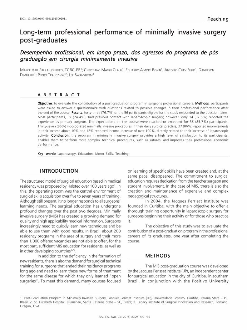
130
Rev. Col. Bras. Cir. 2015; 42(2): 130-135
Loure i roLoure i roLoure i roLoure i roLoure i roLong-term professional performance of minimally invasive surgery post- graduatesTeachingTeachingTeachingTeachingTeaching
Long-term professional performance of minimally invasive surgeryLong-term professional performance of minimally invasive surgeryLong-term professional performance of minimally invasive surgeryLong-term professional performance of minimally invasive surgeryLong-term professional performance of minimally invasive surgerypost-graduatespost-graduatespost-graduatespost-graduatespost-graduates
Desempenho profissional, em longo prazo, dos egressos do programa de pósDesempenho profissional, em longo prazo, dos egressos do programa de pósDesempenho profissional, em longo prazo, dos egressos do programa de pósDesempenho profissional, em longo prazo, dos egressos do programa de pósDesempenho profissional, em longo prazo, dos egressos do programa de pósgraduaçåo em cirurgia minimamente invasivagraduaçåo em cirurgia minimamente invasivagraduaçåo em cirurgia minimamente invasivagraduaçåo em cirurgia minimamente invasivagraduaçåo em cirurgia minimamente invasiva
MARCELO DE PAULA LOUREIRO, TCBC-PR1; CHRISTIANO MAGGI CLAUS1; EDUARDO AIMORÉ BONIN1; ANTONIO CURY FILHO1; DANIELSON
DIMBARRE1; PEDRO TRAUCZINSKI2; LEE SWANSTROM3
A B S T R A C TA B S T R A C TA B S T R A C TA B S T R A C TA B S T R A C T
ObjectiveObjectiveObjectiveObjectiveObjective: to evaluate the contribution of a post-graduation program in surgeons professional careers. MethodsMethodsMethodsMethodsMethods: participants
were asked to answer a questionnaire with questions related to possible changes in their professional performance after
the end of the course. ResultsResultsResultsResultsResults: forty-three (76.7%) of the 56 participants eligible for the study responded to the questionnaires.
Most participants, 32 (74.4%), had previous contact with laparoscopic surgery; however, only 14 (32.5%) reported the
experience as primary surgeon. The expectations on the course were reached or exceeded for 36 (83.7%) participants.
Thirty-seven (86%) incorporated minimally invasive procedures in their daily surgical practice, 37 (86%) reported improvements
in their income above 10% and 12% reported income increase of over 100%, directly related to their increase of laparoscopic
activity. ConclusionConclusionConclusionConclusionConclusion: the program in minimally invasive surgery provides a high level of satisfaction to its participants,
enables them to perform more complex technical procedures, such as sutures, and improves their professional economic
performance.
Key words:Key words:Key words:Key words:Key words: Laparoscopy. Education. Motor Skills. Teaching.
1. Post-Graduation Program in Minimally Invasive Surgery, Jacques Perissat Institute (IJP), Universidade Positivo, Curitiba, Paraná State – PR,Brazil; 2. St. Elizabeth Hospital, Blumenau, Santa Catarina State – SC, Brazil; 3. Legacy Institute of Surgical Innovation and Research, Portland,Oregon, USA.
INTRODUCTION INTRODUCTION INTRODUCTION INTRODUCTION INTRODUCTION
The structured model of surgical education based in medicalresidency was proposed by Halsted over 100 years ago1. Inthis, the operating room was the central environment ofsurgical skills acquisition over five to seven years of training.Although still present, it no longer responds to all surgeons’learning needs. The surgical education has undergoneprofound changes over the past two decades. Minimallyinvasive surgery (MIS) has created a growing demand forquality and high applicability medical information. Surgeonsincreasingly need to quickly learn new techniques and beable to use them with good results. In Brazil, about 200residency programs in the area of surgery and their morethan 1,000 offered vacancies are not able to offer, for themost part, sufficient MIS education for residents, as well asin other developing countries1- 5.
In addition to the deficiency in the formation ofnew residents, there is also the demand for surgical technicaltraining for surgeons that ended their residency programslong ago and need to learn these new forms of treatmentfor the same disease for which they only learned “opensurgeries”. To meet this demand, many courses focused
on learning of specific skills have been created and, at thesame pace, disappeared. The commitment to surgicaleducation requires dedication from the teacher surgeon andstudent involvement. In the case of MIS, there is also thecreation and maintenance of expensive and complexpedagogical structures.
In 2004, the Jacques Perrisat Institute wasfounded in Curitiba, with the main objective to offer athorough training opportunity in laparoscopic surgery forsurgeons beginning their activity or for those who practiceit.
The objective of this study was to evaluate thecontribution of a post-graduation program in the professionalcareers of its graduates, one year after completing thecourse.
METHODSMETHODSMETHODSMETHODSMETHODS
The MIS post-graduation course was developedby the Jacques Perissat Institute (IJP), an independent centerfor surgical education in the city of Curitiba, in southernBrazil, in conjunction with the Positivo University
DOI: 10.1590/0100-69912015002011
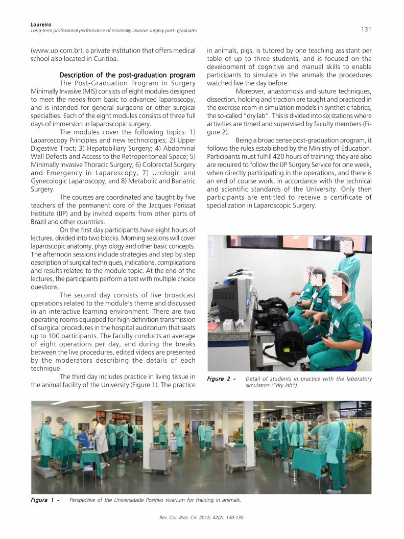
Loure i roLoure i roLoure i roLoure i roLoure i roLong-term professional performance of minimally invasive surgery post- graduates 131
Rev. Col. Bras. Cir. 2015; 42(2): 130-135
(www.up.com.br), a private institution that offers medicalschool also located in Curitiba.
Description of the post-graduation programDescription of the post-graduation programDescription of the post-graduation programDescription of the post-graduation programDescription of the post-graduation programThe Post-Graduation Program in Surgery
Minimally Invasive (MIS) consists of eight modules designedto meet the needs from basic to advanced laparoscopy,and is intended for general surgeons or other surgicalspecialties. Each of the eight modules consists of three fulldays of immersion in laparoscopic surgery.
The modules cover the following topics: 1)Laparoscopy Principles and new technologies; 2) UpperDigestive Tract; 3) Hepatobiliary Surgery; 4) AbdominalWall Defects and Access to the Retroperitoneal Space; 5)Minimally Invasive Thoracic Surgery; 6) Colorectal Surgeryand Emergency in Laparoscopy; 7) Urologic andGynecologic Laparoscopy; and 8) Metabolic and BariatricSurgery.
The courses are coordinated and taught by fiveteachers of the permanent core of the Jacques PerissatInstitute (IJP) and by invited experts from other parts ofBrazil and other countries.
On the first day participants have eight hours oflectures, divided into two blocks. Morning sessions will coverlaparoscopic anatomy, physiology and other basic concepts.The afternoon sessions include strategies and step by stepdescription of surgical techniques, indications, complicationsand results related to the module topic. At the end of thelectures, the participants perform a test with multiple choicequestions.
The second day consists of live broadcastoperations related to the module’s theme and discussedin an interactive learning environment. There are twooperating rooms equipped for high definition transmissionof surgical procedures in the hospital auditorium that seatsup to 100 participants. The faculty conducts an averageof eight operations per day, and during the breaksbetween the live procedures, edited videos are presentedby the moderators describing the details of eachtechnique.
The third day includes practice in living tissue inthe animal facility of the University (Figure 1). The practice
in animals, pigs, is tutored by one teaching assistant pertable of up to three students, and is focused on thedevelopment of cognitive and manual skills to enableparticipants to simulate in the animals the procedureswatched live the day before.
Moreover, anastomosis and suture techniques,dissection, holding and traction are taught and practiced inthe exercise room in simulation models in synthetic fabrics,the so-called “dry lab”. This is divided into six stations whereactivities are timed and supervised by faculty members (Fi-gure 2).
Being a broad sense post-graduation program, itfollows the rules established by the Ministry of Education.Participants must fulfill 420 hours of training; they are alsoare required to follow the IJP Surgery Service for one week,when directly participating in the operations, and there isan end of course work, in accordance with the technicaland scientific standards of the University. Only thenparticipants are entitled to receive a certificate ofspecialization in Laparoscopic Surgery.
Figura 1 -Figura 1 -Figura 1 -Figura 1 -Figura 1 - Perspective of the Universidade Positivo vivarium for training in animals.
Figure 2 -Figure 2 -Figure 2 -Figure 2 -Figure 2 - Detail of students in practice with the laboratorysimulators (“dry lab”).

132
Rev. Col. Bras. Cir. 2015; 42(2): 130-135
Loure i roLoure i roLoure i roLoure i roLoure i roLong-term professional performance of minimally invasive surgery post- graduates
Study designStudy designStudy designStudy designStudy designAll participants in the MIS Post-Graduation
Program of the Jacques Perissat Institute between 2005and 2009 were invited to participate.
We collected basic demographic data at the timeof enrollment in the course. Age, gender, level of surgicaltraining, current practice profile, and previous experiencein MIS were evaluated. After completing the program, allparticipants received a structured questionnaire.
To better assess the impact of such training inclinical practice, only those who had completed the courseat least one year before were considered to participate.Questionnaires were sent by letter or email and focusedon the assessment of the expertise acquired after thecourse, comparing with pre-course expectations. Inaddition, we assessed the professional and economicimpact of the course in their lives. All data are presentedas descriptive statistics.
RESULTSRESULTSRESULTSRESULTSRESULTS
Profile of MIS Post-Graduation participantsProfile of MIS Post-Graduation participantsProfile of MIS Post-Graduation participantsProfile of MIS Post-Graduation participantsProfile of MIS Post-Graduation participantsand previous experienceand previous experienceand previous experienceand previous experienceand previous experience.
Forty-three (76.7%) of the 56 participants in theprogram eligible for the study responded to questionnairesand had their answers matched and compared with theones provided at the beginning of the course. There were38 men and five women. They had completed the post-graduation program in an average of 2.18 years (1-5 years).Twenty-four (56%) were between 25 and 40 years of age.Nineteen (44%) started the program less than five yearsafter completion of residency training, while 10 (23.2%)had completed residency more than 20 years before.Fourteen participants (32.5%) were from cities with lessthan 200,000 inhabitants, while the majority (67.5%) wasfrom larger cities.
Thirty-two participants (74.4%) had previouscontact with laparoscopic surgery and 14 (32.5%) reportedexperience in the performed procedures as the primarysurgeon. Prior to the course, seven of these participantsoperated only less complex laparoscopic procedures, suchas cholecystectomies, while the other seven performedintermediate or advanced complex procedures, such asfundoplication or colectomy (Figures 3 and 4).
Motivations and expectations regardingMotivations and expectations regardingMotivations and expectations regardingMotivations and expectations regardingMotivations and expectations regardingthe Post-Graduationthe Post-Graduationthe Post-Graduationthe Post-Graduationthe Post-Graduation
The main motivation of students to start thecourse were learning and the development of newtechniques for 16 of them (37.2%) and the interest in thelong-term course format. Eight students (18.6%)participated in the program to keep their knowledge up todate.
Thirty-two (74.4%) were defined as “activeparticipants” throughout the program. Three (7%) confessed
little involvement. The expectations regarding the purposeof the course were fully achieved for 36 (83.7%) them.Seven (16.2%) had partially met expectations. Of these,five (71.4%) were older than 40 years.
Professional and Economic ImpactProfessional and Economic ImpactProfessional and Economic ImpactProfessional and Economic ImpactProfessional and Economic ImpactThirty-seven participants (86%) incorporated
minimally invasive procedures in their daily surgical practice.Only six (13.9%) of the evaluated students mentioned notincorporating minimally invasive procedures to their routines.The main barrier to the adoption of MIS, both basic andadvanced, was the lack of investment in infrastructure /equipment by hospital where they worked.
Considering the participant’s self-perception oftheir technical skills, 28 (65.1%) felt fully able to performlaparoscopic suturing; 10 (23.2%) felt suitable, but slow;and five (11.6%) still felt unsafe to perform laparoscopicsuturing. Three of these five (60%) were over 40 years of
Figure 3 -Figure 3 -Figure 3 -Figure 3 -Figure 3 - Previous experience in laparoscopy before startingthe MIS course.
Figure 4 -Figure 4 -Figure 4 -Figure 4 -Figure 4 - Laparoscopic procedures performed before startingthe MIS course.
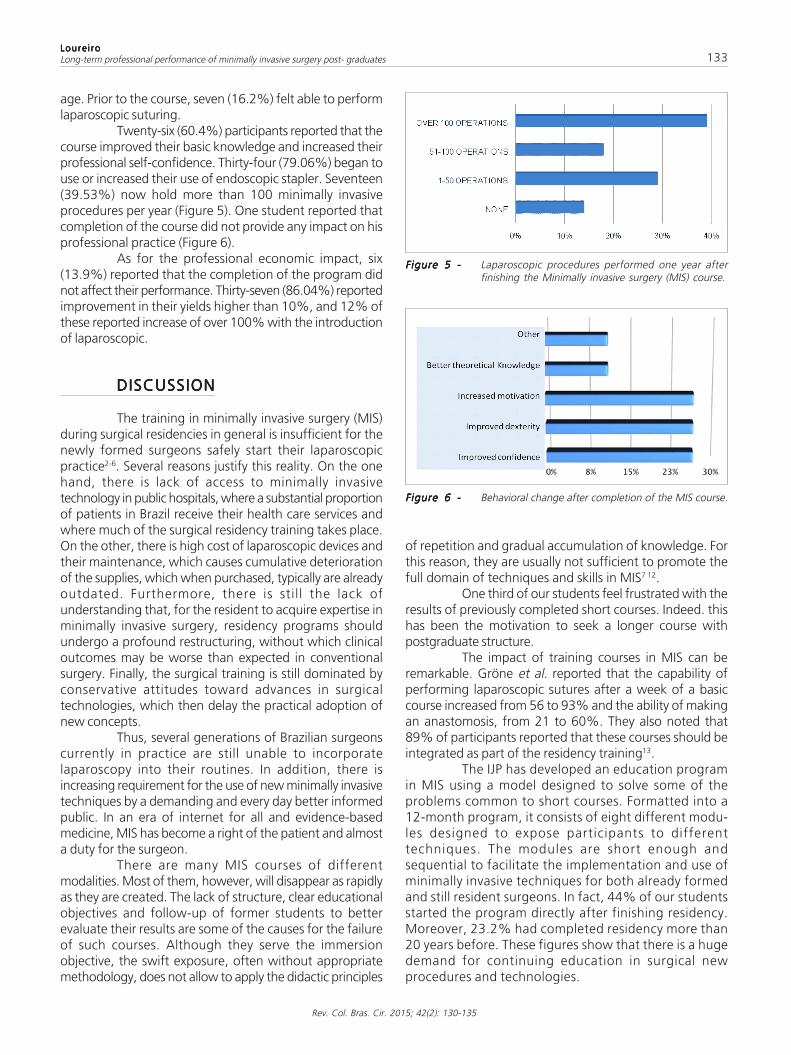
Loure i roLoure i roLoure i roLoure i roLoure i roLong-term professional performance of minimally invasive surgery post- graduates 133
Rev. Col. Bras. Cir. 2015; 42(2): 130-135
age. Prior to the course, seven (16.2%) felt able to performlaparoscopic suturing.
Twenty-six (60.4%) participants reported that thecourse improved their basic knowledge and increased theirprofessional self-confidence. Thirty-four (79.06%) began touse or increased their use of endoscopic stapler. Seventeen(39.53%) now hold more than 100 minimally invasiveprocedures per year (Figure 5). One student reported thatcompletion of the course did not provide any impact on hisprofessional practice (Figure 6).
As for the professional economic impact, six(13.9%) reported that the completion of the program didnot affect their performance. Thirty-seven (86.04%) reportedimprovement in their yields higher than 10%, and 12% ofthese reported increase of over 100% with the introductionof laparoscopic.
DISCUSSIONDISCUSSIONDISCUSSIONDISCUSSIONDISCUSSION
The training in minimally invasive surgery (MIS)during surgical residencies in general is insufficient for thenewly formed surgeons safely start their laparoscopicpractice2-6. Several reasons justify this reality. On the onehand, there is lack of access to minimally invasivetechnology in public hospitals, where a substantial proportionof patients in Brazil receive their health care services andwhere much of the surgical residency training takes place.On the other, there is high cost of laparoscopic devices andtheir maintenance, which causes cumulative deteriorationof the supplies, which when purchased, typically are alreadyoutdated. Furthermore, there is still the lack ofunderstanding that, for the resident to acquire expertise inminimally invasive surgery, residency programs shouldundergo a profound restructuring, without which clinicaloutcomes may be worse than expected in conventionalsurgery. Finally, the surgical training is still dominated byconservative attitudes toward advances in surgicaltechnologies, which then delay the practical adoption ofnew concepts.
Thus, several generations of Brazilian surgeonscurrently in practice are still unable to incorporatelaparoscopy into their routines. In addition, there isincreasing requirement for the use of new minimally invasivetechniques by a demanding and every day better informedpublic. In an era of internet for all and evidence-basedmedicine, MIS has become a right of the patient and almosta duty for the surgeon.
There are many MIS courses of differentmodalities. Most of them, however, will disappear as rapidlyas they are created. The lack of structure, clear educationalobjectives and follow-up of former students to betterevaluate their results are some of the causes for the failureof such courses. Although they serve the immersionobjective, the swift exposure, often without appropriatemethodology, does not allow to apply the didactic principles
of repetition and gradual accumulation of knowledge. Forthis reason, they are usually not sufficient to promote thefull domain of techniques and skills in MIS7 12.
One third of our students feel frustrated with theresults of previously completed short courses. Indeed. thishas been the motivation to seek a longer course withpostgraduate structure.
The impact of training courses in MIS can beremarkable. Gröne et al. reported that the capability ofperforming laparoscopic sutures after a week of a basiccourse increased from 56 to 93% and the ability of makingan anastomosis, from 21 to 60%. They also noted that89% of participants reported that these courses should beintegrated as part of the residency training13.
The IJP has developed an education programin MIS using a model designed to solve some of theproblems common to short courses. Formatted into a12-month program, it consists of eight different modu-les designed to expose participants to differenttechniques. The modules are short enough andsequential to facilitate the implementation and use ofminimally invasive techniques for both already formedand still resident surgeons. In fact, 44% of our studentsstarted the program directly after finishing residency.Moreover, 23.2% had completed residency more than20 years before. These figures show that there is a hugedemand for continuing education in surgical newprocedures and technologies.
Figure 5 -Figure 5 -Figure 5 -Figure 5 -Figure 5 - Laparoscopic procedures performed one year afterfinishing the Minimally invasive surgery (MIS) course.
Figure 6 -Figure 6 -Figure 6 -Figure 6 -Figure 6 - Behavioral change after completion of the MIS course.
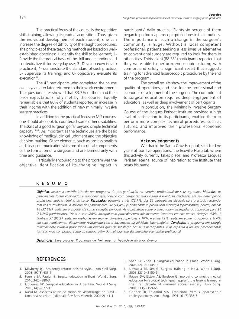
134
Rev. Col. Bras. Cir. 2015; 42(2): 130-135
Loure i roLoure i roLoure i roLoure i roLoure i roLong-term professional performance of minimally invasive surgery post- graduates
R E S U M OR E S U M OR E S U M OR E S U M OR E S U M O
ObjetivoObjetivoObjetivoObjetivoObjetivo: avaliar a contribuição de um programa de pós-graduação na carreira profissional de seus egressos. MétodosMétodosMétodosMétodosMétodos: osparticipantes foram convidados a responder questionário com perguntas relacionadas a eventuais mudanças em seu desempenhoprofissional após o término do curso. Resultados:Resultados:Resultados:Resultados:Resultados: quarenta e três (76,7%) dos 56 participantes elegíveis para o estudo responde-ram aos questionários. A maioria dos participantes, 32 (74,4%) já tinha contato prévio com a cirurgia laparoscópica, porém, apenas14 (32,5%) relataram a experiência como cirurgião principal. As expectativas sobre o curso foram alcançadas ou superadas para 36(83,7%) participantes. Trinta e sete (86%) incorporaram procedimentos minimamente invasivos em sua prática cirúrgica diária. Etambém 37 (86%) relataram melhorias em seus rendimentos superiores a 10%, e ainda 12% relataram aumento superior a 100%em seus rendimentos, diretamente relacionado com o incremento da atividade laparoscópica. Conclusão:Conclusão:Conclusão:Conclusão:Conclusão: o programa em cirurgiaminimamente invasiva proporciona um elevado grau de satisfação aos seus participantes, e os capacita a realizar procedimentostécnicos mais complexos, como as suturas, além de melhorar seu desempenho economico profissional.
Descritores:Descritores:Descritores:Descritores:Descritores: Laparoscopia. Programas de Treinamento. Habilidade Motora. Ensino.
The practical focus of the course is the repetitiveskills training, allowing its gradual acquisition. Thus, giventhe individual development of each student, one canincrease the degree of difficulty of the taught procedures.The principles of these teaching methods are based on well-established doctrines: 1. Identify the skill to be learned; 2-Provide the theoretical basis of the skill understanding andcontextualize it for everyday use; 3- Develop exercises topractice it; 4- demonstrate the standard of such practice;5- Supervise its training; and 6- objectively evaluate itsexecution14.
The 43 participants who completed the courseover a year later later returned to their work environment.The questionnaires showed that 83.7% of them had theirprior expectations fully met by the course. Equallyremarkable is that 86% of students reported an increase intheir income with the addition of new minimally invasivesurgery practices.
In addition to the practical focus on MIS courses,one should also look to counteract some other disabilities.The skills of a good surgeon go far beyond simple technicalcapacity15-17. As important as the techniques are the basicknowledge of medical, clinical judgment and the objectivedecision-making. Other elements, such as professionalismand clear communication skills are also critical componentsof the formation of a surgeon and are learned only withtime and guidance.
Particularly encouraging to the program was theobjective identification of its changing impact in
participants’ daily practice. Eighty-six percent of thembegan to perform laparoscopic procedures in their routines.The importance of such a change in the surgeon’scommunity is huge. Without a local competentprofessional, patients seeking a less invasive alternativeto conventional surgery are required to look for them inother cities. Thirty-eight (88.3%) participants reported thatthey were able to perform endoscopic suturing withcomfort and safety, a significant result that suggeststraining for advanced laparoscopic procedures by the endof the program.
The overall results show the improvement of thequality of operations, and also for the professional andeconomic development of the surgeon. The commitmentto surgical education requires deep engagement ofeducators, as well as deep involvement of participants.
In conclusion, the Minimally Invasive SurgeryCourse of the Jacques Perissat Institute provided a highlevel of satisfaction to its participants, enabled them toperform more complex technical procedures, such assutures, and improved their professional economicperformance.
AcknowledgementsAcknowledgementsAcknowledgementsAcknowledgementsAcknowledgementsWe thank the Santa Cruz Hospital, seat for five
years of our live operations; the Ecoville Hospital, wherethis activity currently takes place; and Professor JacquesPerissat, eternal source of inspiration to the Institute thatbears his name.
REFERENCESREFERENCESREFERENCESREFERENCESREFERENCES
1. Mayberry JC. Residency reform Halsted-style. J Am Coll Surg.2003;197(3):433-5.
2. Ferreira EA, Rasslan S. Surgical education in Brazil. World J Surg.2010;34(5):880-3.
3. Gutiérrez VP. Surgical education in Argentina. World J Surg.2010;34(5):877-9.
4. Nácul M. Aspectos atuais do ensino da videocirurgia no Brasil –Uma análise crítica [editorial]. Rev Bras Videocir. 2004;2(1):1-4.
5. Shen BY, Zhan Q. Surgical education in China. World J Surg.2008;32(10):2145-9.
6. Udwadia TE, Sen G. Surgical training in India. World J Surg.2008;32(10):2150-5.
7. Rogers DA, Elstein AS, Bordage G. Improving continuing medicaleducation for surgical techniques: applying the lessons learned inthe first decade of minimal access surgery. Ann Surg.2001;233(2):159-66.
8. Gadacz TR, Talamini MA. Traditional versus laparoscopiccholecystectomy. Am J Surg. 1991;161(3):336-8.

Loure i roLoure i roLoure i roLoure i roLoure i roLong-term professional performance of minimally invasive surgery post- graduates 135
Rev. Col. Bras. Cir. 2015; 42(2): 130-135
9. Zucker KA. Training issues [editorial]. Surg Laparosc Endosc.1992;2:187.
10. Rock JA, Warshaw JR. The history and future of operativelaparoscopy. Am J Obstet Gynecol. 1994;170(1 Pt 1):7-11.
11. Gates EA. New surgical procedures: can our patients benefit whilewe learn? Am J Obstet Gynecol. 1997;176(6):1293-8; discussion1298-9.
12. Reznick R. Let’s not forget that CME has an “E”. Foc Surg Ed.1999;17:1-2.
13. Gröne J, Ritz JP, Buhr HJ, Lauscher JC. Sustainability of skill coursesfor general and visceral surgery—evaluation of the long-termeffect. Langenbecks Arch Surg. 2010;395(3):277-83.
14. Starr RA, Wagstaff NV. Implementing a surgical skills trainingprogram. Obstet Gynecol Clin North Am. 2006;33(2):247-58, viii.
15. Patil NG, Cheng SW, Wong J. Surgical competence. World J Surg.2003;27(8):943-7.
16. Cuschieri A, Francis N, Crosby J, Hanna GB. What do mastersurgeons think of surgical competence and revalidation? Am JSurg. 2001;182(2):110-6.
17. Satava RM, Gallagher AG, Pellegrini CA. Surgical competenceand surgical proficiency: definitions, taxonomy, and metrics. J AmColl Surg. 2003;196(6):933-7.
Received on 15/04/2014Accepted for publication 20/05/2014Conflict of interest: none.Source of funding: none.
Address for correspondence:Address for correspondence:Address for correspondence:Address for correspondence:Address for correspondence:Marcelo de Paula LoureiroE-mail: [email protected]