Subject- BOTANY Course name- Biology & Diversity of Algae ...
39
Course name- Biology & Diversity of Algae, Bryophytes & Pteridophytes Paper code BOT – 502 Block IV Pteridophytes Unit 18: Telome theory & Stelar Evolution Dr. Prabha Dhondiyal Department of Botany Uttarakhand Open University Haldwani Subject- BOTANY
Transcript of Subject- BOTANY Course name- Biology & Diversity of Algae ...
Course name- Biology & Diversity of Algae,
Bryophytes & Pteridophytes
Paper code BOT – 502
Block IV Pteridophytes
Unit 18: Telome theory & Stelar Evolution
Dr. Prabha Dhondiyal
Department of Botany
Uttarakhand Open University
Haldwani
Subject- BOTANY

Contents
•Introduction
•Meaning of Telome
•Origin of Telomes
•Evolution of Higher plants by Telome Theory
•Merits
•Demerits
•Stelar Evolution

Introduction
The discovery of a group of earliest known land plants with simple
organization of the sporophyte (rootless, dichotomously branched,
single terminal sporangium, protostele vascular cylinder) from the
upper Silurian and middle Devonian deposits has been of great
importance in the understanding of the structure and phylogeny of
vascular plants.
A number of theories on land plant evolution exists of which the
Telome theory of Walter Zimmermann (1930, and later elaborated
on 1952) is the most comprehensive.

This theory is based on fossil record and synthesizes the
major steps in the evolution of vascular plants.
According to this theory, all vascular plants evolved either
directly or indirectly from a simple leafless Rhynia type
ancestral form made up of sterile and fertile axes (the
telomes).
Evolutionary modification of its parts produce more
advanced vascular plants with roots, stems, leaves, protected
sporangia and more complex vascular systems.
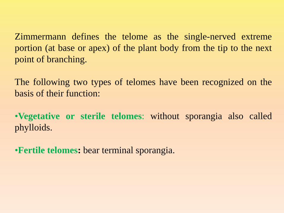
Zimmermann defines the telome as the single-nerved extreme
portion (at base or apex) of the plant body from the tip to the next
point of branching.
The following two types of telomes have been recognized on the
basis of their function:
•Vegetative or sterile telomes: without sporangia also called
phylloids.
•Fertile telomes: bear terminal sporangia.

The telome grow and divides dichotomously, the new
segments become new telomes and older segment below
are mesomes.
Following evolutionary development telomes may be
grouped together in various ways to form more complex
bodies Telome Truss or Syntelome.
(i) Phylloid Truss- Only sterile telomes
(ii) Fertile Truss- Only Fertile telomes

According to the Telome theory the early land plants
originated from the green algae which lived in tidal zone of
the Cambrian and Silurian sea coasts.
The plant body of those algal ancestors was undifferentiated
branched thallus (primitive telome).
Origin of Telomes and Primitive land plants

According to Zimmermann these primitive telomes were formed
from the unicellular stage by the following five elementary
processes:
(i) Interconnection of cells
(ii) Differentiation of meristem
(iii) Rotation of cell axis
(iv) Shifting of chief phases in alternation of generation
(v) Differentiation of various permanent tissues

The Primitive Land Plant
The telome theory visualizes the Psilophytales of the upper Silurian
and lower and middle Devonian deposits (Zosterophyllum, Rhynia,
Horneophyton and Psilophyton etc) as representing the sporophyte
of the ancient vascular plants.
The sporophyte was relatively undifferentiated (no distinction
between leaf and stem) and consisted of single-veined (protostele)
telomes which may be sterile and fertile.
The aerial portion developed stomata and the basal portion, hairs or
rhizoids. The fertile telome produced terminal sporangia.

Fig.1.1 Hypothetical diagramme of a primitive land plant
Evolution of Higher Land Plants
From the primitive syntelome of the early land plants the
sporophytes of higher land plant evolved by certain organogenetic
processes called “elementary processes” each following its own
trends. Zimmerman suggested that the these elementary processes
were responsible for the development of higher vascular plants
from the early vascular cryptogams.

Overtopping:
• One of the two dichotomizing
branches becomes stronger and erect
becoming the axis while other
remained overtopped as short lateral
branch.
•It leads to the formation of an axis
with lateral appendages, the leaves,
e.g. open-veined pinnately compound
type of fern leaf.
• Overtopping mesomes form the
rachis and the overtopped mesomes
constitute the leaflets.
Process of Telome Theory

Planation:
•Rearrangement of telomes and
mesomes from a three dimensional
pattern to a single plane.
•Branching in more than one plane
(cruciate dichotomy) is replaced by a
dichotomy in a single plane (fan
shaped dichotomy).
•By this process an organ of radial
symmetry gives rise to one of
bilateral symmetry.
•Plantation concerns mainly the
evolution of the leaf.

Syngenesis (fusion or webbing):
• Fusion of the telome or telome trusses by the development of
connecting tissue (as in the foot of duck) is called syngenesis or
webbing.
•Telomes and mesomes connect by the formation of
parenchymatous tissue between them (parenchymatous webbing)
sometimes accompanied by the fusion of their stele.

Syngenesis is a very important elementary process because it
explains the origin and evolution of both the leaf and stele of the
stem. It leads to the formation of:
(i) Foliar appendages with open dichotomous venation. In this
case the sterile telomes (Phylloids) become united only by the
development of parenchymatous webbing
(ii) Pinnately veined leaf: Parenchymatous webbing was
accompanied by over-topping.
(iii) Leaf with reticulate venation: if fusion of steles or vascular
bundles also occurred.
(iv) Parenchymatous webbing led to the polystelic condition (in
an open form) as in many species of Selaginella.

Reduction:
•Activity of terminal meristem of each telome suppressed
resulting into much shorter branches
• It involved the transformation of a syntelome into a single
needle-like leaf (conifers).
•According to Zimmermann the microphyllous leaves of
Lycopsida and Sphenopsida were evolved by the reduction of
telome trusses.

Curvation:
The fertile Telomes become curved or bend downwards. Wilson
(1953) recognized two separate sub-processes
•Recurvation: When telomes bent down inwards, it is called
Recurvation. During this process, the fertile telomes
(sporangiophores) were reflexed and sporangia became inverted.
•Incurvation: This process accounts for the shifting of sporangia
from terminal position to the ventral surface of the leaf in ferns
Fig1.3. Steps in the process of IncurvationFig 1.2. Steps in the process of Recurvation

Merits
•It provides an interpretation of origin and evolution of sporophytes
of land plants.
•Structure of the sporophytes of the most primitive known land
plants is defined.
•It interprets the morphological features of the lower vascular plant
such as the nature of the aerial portion of the plant body of the
family Ophioglossaceae and coenopterid ferns.
•This theory emphasises on the fact that the plant body is an axis
with a descending portion, the root and an aerial portion, the shoot
whose appendages are modified parts of the stem.
•According to Eames, though the theory is built upon structure in
the lowest known vascular plants, higher plant can also be
interpreted in this way.
•It also connects the fossil and living plants by their phylogenetical
relations.
Demerits
•According to Thomas (1950), the telome theory does not explain
the whorled or spiral arrangement of sporangia, which is observed
in some ancient and primitive plants.
•Application of the telome theory to the origin of Lycopsida has
been greatly criticised. Andrews (1960) supports this theory to
some extent so far as Sphenopsida and Pteropsida are concerned,
but for Lycopsida, he is not in agreement with Zimmermann.
•According to Bower (1946), this theory does not explain how a
telome-like characterized body has been developed. It has been
taken for granted by Zimmermann (1930) that a telome type body
is ‘ready-made’.
Stewart (1964) also criticised the telome theory because it does not
explain the derivation of the dictyostelic condition.
Many other plants of greater complexity, than Rhynia fossils have
been discovered e., Zosterophyllum, Baragnnathia (Leclercq,
1954) Lyon and Heuber (1964), Heuber and Banks (1967) and
Lyon (1964) observed lateral sporangia on short vascularised
stalks in Psilophyton and Asteroxylon instead of usual terminal
sporangia.
This theory has received little attention by angiosperm
morphologists. Its application to stamens, venation pattern of
leaves and carpels have been critised from time to time.

The stele (=Greek word meaning pillar or column) is defined as a
central vascular cylinder, with or without pith. Endodermis is the
boundary between cortex and stele. The central cylinder or core of
vascular tissue, consisting of xylem, phloem, pericycle and
sometimes mudullary rays and pith, is technically called the stele.
The concept of the stele as the fundamental unit of vascular system
was put forward by Van Tieghem and Douliot (1886) who
proposed and developed Stelar theory.
The stele of stem was connected with that of leaf by a vascular
connection known as leaf trace.
STELAR EVOLUTION
Types of stele in pteridophytes
The stele in pteridophytes can be differentiated into two major
groups.
1. Protostele: It is the most simplest and primitive type of stele. In
protostele, the vascular bundle is a concentric solid mass and the
central core of xylem is surrounded by a layer of phloem and
finally surrounded by a layer of pericycle. The protostele exists
in following forms:
a) Haplostele: This is the most primitive type of stele. In this the
xylem forms a solid, smooth and spherical central core which is
surrounded by a continuous concentric layer of phloem e.g.
Lygodium, Selaginella.

b) Actinostele: This is the modification of the haplostele and
somewhat more advanced in having the central xylem core with
radiating ribs and phloem is not concentric but present in
between the radiating ribs of xylem e.g. Psilotum. The
protoxylem is present at the tips of radiating arms.

c) Plectostele: In the stem of some species of Lycopodium the
solid core of xylem gets broken in a number of plate-like lobes,
more or less lying parallel to one another. The phloem alternates
with xylem plates e.g. Lycopodium volubile. This specialized
form of protostele is termed as plectostele.
d) Mixed protostele: In this type, masses of xylem and phloem
are uniformly distributed. Scattered groups of xylem are
embedded in the ground tissue of phloem e.g. Lycopodium
cernnum.

e) Protostele with mixed pith: In the centre there is parenchyma
cells associated alongwith the tracheids e.g. Lepidodendron. It
may be derived from the transformation of the tracheids into
parenchyma and may be first step in formation of pith. Such type
of protostele is termed as mixed-protostele.
Stelar System: A. Plectostele B. Mixed protostele C. Mixed protostele with pith

2. Siphonostele: Medullated protostele is called siphonostele.
It is characteristic of Filicophyta. This is the modification of
protostele. During the development the siphonostele the central
core of xylem is replaced by parenchymatous cells so that
definite pith surrounded by xylem appears in the centre.
On the basis of branch and leaf gaps Jeffrey (1910),
distinguished two types of siphonosteles, cladosiphonic
siphonosteles (leaf gaps not found) and phyllosiphonic
siphonosteles (leaf and branch gaps are present).

Origin of pith in siphonostele
There is a general acceptance that siphonostele is evolved from a
protostele. Two theories have been proposed to explain the origin
of pith.
Intrastelar origin of pith: According to this view, pith has
originated as a result of transformation of tracheary elements of
the central xylem core into parenchyma. Thus, the pith is totally
intra –stelar. This view is supported by Boodle (1901), Gwynne–
Vaughani (1908), Bower (1911). E.g., Osmunda regalis,
Botrychium ternatum where tracheids are scattered throughout the
pith (mixed pith).
Extrastelar origin of pith: This hypothesis was supported By
Jeffrey (1902, 1910 and 1917). According to this view, protostele
has transformed into siphonostele due to migration of cortical cells
into stelar axis. Openings such as leaf and branch gaps, probably
provided passage for invasion of parenchyma.

This view derives support from amphiphloic siphonostele that has
two endodermal layers; one delimits the stele from the cortex and
the other from the pith. Since endodermis is a structure peculiar to
cortex, the pith is considered as cortical in origin.
Stelar seytem: A. Polycyclic solenostele, B. Polycyclic dictyostele, C. Polystele

Jeffrey (1898) classified Siphonostele into following two types,
on the basis of position of phloem.
(a) Ectophloic siphonostele: The pith is surrounded by
concentric xylem cylinder and next to xylem the concentric
phloem cylinder. It means the phloem is restricted only on the
external sides of the xylem. (e.g. Osmunda and Schizea). The
pith is central in position. The phloem is externally surrounded
by pericycle and endodermis.
(b) Amphiphloic siphonostele: The pith is surrounded by the
vascular tissue. The concentric inner phloem cylinder surrounds
the central pith. Next is the concentric xylem cylinder which is
immediately surrounded by outer phloem cylinder. It means
phloem is present both sides of xylem. (e.g., Marsilea rhizome).


Other modification of Siphonostele
3. Solenostele
If the siphonostele perphorated at the place or places to the
origin of the leaf trace, such a condition is known as
Solenostele.
Ectophloic solenostele : This type of solenostele derived from
the ectophloic siphonostele. So, here the phloem is present
only side of xylem.
Amphiphloic solenostele : This type of solenostele derived
from the amphiphloic siphonostele. So in this case the phloem
is present on both the sides of the xylem.


4. Dictyostele
In the more advanced siphonosteles of Pteropsida, the
successive gaps may overlap each other.
Brebner (1902) called the siphonosteles with overlapping gaps
as dictyosteles. So, in this case the solenostele broken into a
network of separate vascular strands, due to crowded leaf gaps.
In such cases each separate vascular strand is known as
meristele. Each meristele is of protostelic type. The dictyostele
is a ring of many meristeles.

5. Eustele
According to Brebner (1902), there is one more modification
of the siphonostele known as eustele.
Here the stele is split into distinct collateral vascular bundles.
So the vascular system consists of a ring of collateral or
bicollateral vascular bundles situated on the periphery of the pith.
In such steles, the inter-fascicular areas and the leaf gaps are
not distinguished from each other very clearly. The example of
this type is Equisetum.

6. Atactostele
In atactostele the vascular strands are scattered. It occurs into
monocotyledons. George Brebner (1902) coined the term
atactostele (Greek atact-without order) for vein arrangement
seen in transverse view which has been described later as
“scattered” by Berg (1997).
A. Eustele, B. Atactostele

7. Polycylic Stele
When the vascular tissue present in the form of two or more
concentric cylinders, such a stele is called polycyclic stele. A
typical polycyclic stele possesses two or more concentric rings of
vascular tissue. This may be a solenostele or a dictyostele. Two
concentric rings of vascular tissue are found in Pteridium
aquilinum and three in Matonia pectinata.
8. Polystele
Some times more than one stele are present in the axis of some
pteridophytes. Such a condition is called Polystelics. Such stele
shows polystelic condition. Certain species of Selaginella have
polystelic condition.

Polystelic (distelic) condition in Selaginella Stem

Evolution of the stelar system
It is now generally clear that the simplest type of stele is
protostele.
It is fundamental type for the vascular plants in general and the
pteridophyta in general and all the other types of stele have been
derived from it in the course of evolutionary specialization. As far
as known all pteridophytes in the initial (sporeling) stage start
with protostelic stem.
When the shoot grows accompanying an increase in size, there is
internal differentiation of the stele. The first step is appearance of
parenchyma, scattered in the xylem core and finally a central
parenchymatous medulla or pith is developed in the protostele.
Such type of stele is called Siphonostele.

The method by which medullation came about in a protostele and
formed a siphonostele is a debated question and explained by two
hypotheses viz. Extra-stelar and Intra-stelar origin of pith.
In its simplest form the siphonostele has no leaf gaps e.g.
Selaginella and known as cladosiphonic siphonostele in contrast
with phyllosiphonic condition with leaf gaps.
Siphonosteles which are perforated by scattered leaf gaps are
known as Solenostele and a siphonostele with more overlapping
gaps is known as dissected siphonostele or dictyostele.
The final elaboration of the stellar organization in pteridophytes
consists in the development of a number of separate steles. Such
a stele is known as Polycyclostele. Another modification of
siphonostele is the Eustele in which the vascular system consists
of collateral vascular bundles.

Different types of Steles arranged in evolutionary sequence