Subependymoma of the Lateral Ventricles: Report of Two Cases · Subependymoma is a rare benign...
5
Subependymoma of the Lateral Ventricles: Report of Two Cases Lateral Ventrikiil Subependimomasl: iki Vaka Sunumu BiLGE BiLGiC;, NESiMi BUYUKBABANi, SEMRA VURAL, ~iC;EK BAYINDIR Istanbul University, Istanbul Medical Faculty, Department of Pathology Abstract: Sub ependymoma is a rare benign tumor of the ventricular system. It was described as a distinct entity by Scheinker in 1945. The fourth ventricle is the most common location. Many cases are asymptomatic and discovered at autopsy. Symptomatic cases often present with hydrocephalus.We present two cases of subependymoma located in lateral ventricles. Case 1 was a 69-year-old male with non-Hodgkin's large B- cell lymphoma. He died following seizures and respiratory depression while he was receiving chemotherapy. At autopsy brain oedema and a small nodular mass located in the right lateral ventricular wall were discovered. Case 2 was a 38-year-old female presenting with headache. MR revealed a mass, approximately 2.5cm in diameter in the right lateral ventricle extending to the third ventricle. Both tumors were diagnosed as subependymoma. Microscopically, a fibrillary background, cell clusters with small uniform nuclei and rnicrocysts were the main histopathological characteristics. Microcysts contained slightly basophilic material stained with alcian blue pH2.5 and toluidine blue pH? This material was interpreted as extracellular mucin. Tumoral infiltration was positive for GFAP and S-100' protein. Ki-6? (MIB-l) proliferation index was lower than 1%. P53 was negative in both cases. Key Words: Subependymoma, autopsy, lateral ventricle, mucin, p53 126 Ozet: Subependimom, ventrikiiler sistemin nadir goriilen benign bir tiimoriidiir. Scheinker tarafmdan, 1945 yJlmda ayn bir antite olarak tammlanml~hr. En slk yerle~im yeri 4. ventrikUldiir. Vakalann <;ogu asemptomatiktir ve otopside tesadiifen bulunurlar. Semptom veren vakalar, <;ogunlukla hidrosefali ile kar~lmlza <;lkarlar. Bu <;ab~mada, lateral ventrikiil yerle~imli 2 subependimom vakasl sunulmaktadu. ilk vaka 69 ya~mda, B hiicreli non-Hodgkin lenfomab erkek hastadlr. Kemoterapi altmda, konvUlziyon ve solunum yetmezligi ile kaybedilrni~tir. Otopside beyin odemi ve sag ventrikiil duvannda kii<;iik nodUler lezyon saptanml~tu. ikinci vaka 38 ya~mda kadmdlr ve ba~agnsl ~ikayeti ile klinige baFurmu~tur. Radyolojik incelemede MR'da sag ventrikiilde, ii<;iincii ventrikiile uzamm gosteren, 2.5 em <;apmda kitle gorUlmii~tiir. Mikroskopik olarak her iki vakada da benzer morfoloji saptanml~tlr: fibriler zemin, kii<;iikve uniform <;ekirdekli hiicrelerin olu~turdugu hiicre gruplan ve rnikrokistler. Mikrokistlerin i<;indeki materyel, alcian blue pH2.5 ve toluidine blue pH? ile pozitif boyanarak miisin olarak yorumlanml~tlr. Tiimorde GFAP ve S-100 protein pozitiftir. Proliferasyon indeksi Ki-6?(MIB-l) % l'den az ve p53 negatiftir. Anahtar Kelimeler: Subependimom, otopsi, lateral ventrikiil, miisin, p53.
Transcript of Subependymoma of the Lateral Ventricles: Report of Two Cases · Subependymoma is a rare benign...

Subependymoma of the Lateral Ventricles:Report of Two Cases
Lateral Ventrikiil Subependimomasl: iki Vaka Sunumu
BiLGE BiLGiC;, NESiMi BUYUKBABANi, SEMRA VURAL, ~iC;EK BAYINDIR
Istanbul University, Istanbul Medical Faculty, Department of Pathology
Abstract: Subependymoma is a rare benign tumor of theventricular system. It was described as a distinct entityby Scheinker in 1945. The fourth ventricle is the mostcommon location. Many cases are asymptomatic anddiscovered at autopsy. Symptomatic cases often presentwith hydrocephalus.We present two cases ofsubependymoma located in lateral ventricles.Case 1 was a 69-year-old male with non-Hodgkin's largeB- cell lymphoma. He died following seizures andrespiratory depression while he was receivingchemotherapy. At autopsy brain oedema and a smallnodular mass located in the right lateral ventricular wallwere discovered.
Case 2 was a 38-year-old female presenting withheadache. MR revealed a mass, approximately 2.5cm indiameter in the right lateral ventricle extending to thethird ventricle. Both tumors were diagnosed assubependymoma.Microscopically, a fibrillary background, cell clusterswith small uniform nuclei and rnicrocysts were the mainhistopathological characteristics. Microcysts containedslightly basophilic material stained with alcian bluepH2.5 and toluidine blue pH? This material wasinterpreted as extracellular mucin.Tumoral infiltration was positive for GFAP and S-100'protein. Ki-6? (MIB-l) proliferation index was lower than1%. P53 was negative in both cases.
Key Words: Subependymoma, autopsy, lateral ventricle,mucin, p53
126
Ozet: Subependimom, ventrikiiler sistemin nadirgoriilen benign bir tiimoriidiir. Scheinker tarafmdan,1945 yJlmda ayn bir antite olarak tammlanml~hr. En slkyerle~im yeri 4. ventrikUldiir. Vakalann <;oguasemptomatiktir ve otopside tesadiifen bulunurlar.Semptom veren vakalar, <;ogunlukla hidrosefali ilekar~lmlza <;lkarlar.Bu <;ab~mada, lateral ventrikiil yerle~imli 2subependimom vakasl sunulmaktadu. ilk vaka 69ya~mda, B hiicreli non-Hodgkin lenfomab erkekhastadlr. Kemoterapi altmda, konvUlziyon ve solunumyetmezligi ile kaybedilrni~tir. Otopside beyin odemi vesag ventrikiil duvannda kii<;iik nodUler lezyonsaptanml~tu. ikinci vaka 38 ya~mda kadmdlr veba~agnsl ~ikayeti ile klinige baFurmu~tur. Radyolojikincelemede MR'da sag ventrikiilde, ii<;iincii ventrikiileuzamm gosteren, 2.5 em <;apmda kitle gorUlmii~tiir.Mikroskopik olarak her iki vakada da benzer morfolojisaptanml~tlr: fibriler zemin, kii<;iikve uniform <;ekirdeklihiicrelerin olu~turdugu hiicre gruplan ve rnikrokistler.Mikrokistlerin i<;indeki materyel, alcian blue pH2.5 vetoluidine blue pH? ile pozitif boyanarak miisin olarakyorumlanml~tlr. Tiimorde GFAP ve S-100 proteinpozitiftir. Proliferasyon indeksi Ki-6?(MIB-l) % l'den azve p53 negatiftir.
Anahtar Kelimeler: Subependimom, otopsi, lateralventrikiil, miisin, p53.

Turkish Neurosurgery 13: 126-130, 2003
INTRODUCTION
Subependymoma is a rare benign glial tumorof the ventricular system.
The fourth ventricle is the most common
location. They are often found incidentally atautopsy in elderly patients. Symptomatic casesoccur primarily in middle-aged patients and oftenpresent with hidrocephalus.
Prognosis is good even with subtotal resection(1, 23).
We present a symptomatic case ofsubependymoma and an incidental case found atautopsy.
CASE 1:
A 69-year-old male was admitted to hospitalin January 2001 with complaints
of weight loss and fatigue. Following physicalexamination and laboratory investigation, cervicallymph node and trephine bone marrow biopsieswere performed. Both material showed neoplasticinfiltration. Histopathological diagnosis was alarge B-cell non-Hodgkin's lymphoma rich in Tcells. The bone marrow was infiltrated by the sametumor. The patient received a regimen of
CHOP chemotherapy and his status began toimprove clinically. Three weeks after the firstchemotherapy he developed focal seizures andsoon after a respiratory depression. Followingintubation and anticonvulsive therapyspontaneous breathing was achieved. A few dayslater the convulsive status reappeared and thepatient died. In this period MR and CT revealed noinfiltration or hemorrhage.
To explain his neurological status a braindissection was done. The brain was fixed in
10%formalin for 4 weeks. The fixed brain weighed1500g.It was cut in
1cm thick coronal slices. Macroscopically, nohemorrhage or mass lesion was detected. The brain
Bilgif: Subepelldymoma of the Lateral Velltrides: Report of Two Cases
was heavy and oedematous. A small nodularlesion, 0.8 cm in diameter was observed in the
anterior horn of the right lateral ventricle (Fig 1).
The tumor was attached to the ventricular
wall, well circumscribed with no infiltration of the
brain parenchyme.
CASE 2:
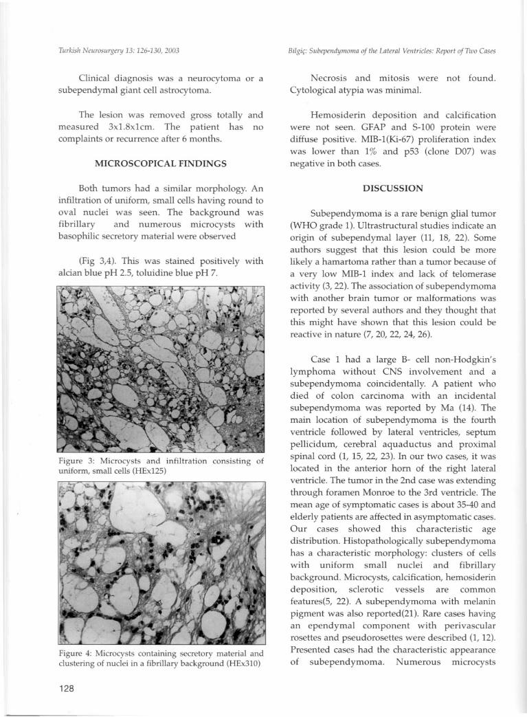
The patient was a 38-year-old female. Shecomplained of headache for 2 months and MRexamination revealed a mass in the right anteriorhorn of the lateral ventricle which was extending tothe 3rd ventricle (Fig 2).
Figure 1: A nodular mass attached to the ventricular wallin case 1 .
Figure 2: Radiological appearance of the tumor in case 2
127
Turkish Neurosurgery 13: 126-130, 2003
Clinical diagnosis was a neurocytoma or asubependymal giant cell astrocytoma.
The lesion was removed gross totally andmeasured 3x1.8xlcm. The patient has nocomplaints or recurrence after 6 months.
MICROSCOPICAL FINDINGS
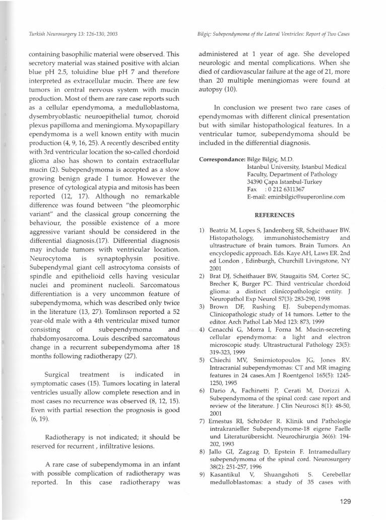
Both tumors had a similar morphology. Aninfiltration of uniform, small cells having round tooval nuclei was seen. The background wasfibrillary and numerous microcysts withbasophilic secretory material were observed
(Fig 3,4). This was stained positively withalcian blue pH 2.5, toluidine blue pH 7.
Figure 3: Microcysts and infiltration consisting ofuniform, small cells (HEx125)
Figure 4: Microcysts containing secretory material andclustering of nuclei in a fibrillary background (HEx310)
128
BilgiC SubepelldYl11011laof tire Lateral Ventricles: Report of Two Cases
Necrosis and mitosis were not found.
Cytological atypia was minimal.
Hemosiderin deposition and calcificationwere not seen. GFAP and S-100 protein werediffuse positive. MIB-l(Ki-67) proliferation indexwas lower than 1% and p53 (clone D07) wasnegative in both cases.
DISCUSSION
Subependymoma is a rare benign glial tumor(WHO grade 1). Ultrastructural studies indicate anorigin of subependymal layer (11, 18, 22). Someauthors suggest that this lesion could be morelikely a hamartoma rather than a tumor because ofa very low MIB-l index and lack of telomeraseactivity (3, 22). The association of subependymomawith another brain tumor or malformations was
reported by several authors and they thought thatthis might have shown that this lesion could bereactive in nature (7, 20, 22, 24, 26).
Case 1 had a large B- cell non-Hodgkin'slymphoma without CNS involvement and asubependymoma coincidentally. A patient whodied of colon carcinoma with an incidental
subependymoma was reported by Ma (14). Themain location of subependymoma is the fourthventricle followed by lateral ventricles, septumpellicidum, cerebral aquaductus and proximalspinal cord (1, 15, 22, 23). In our two cases, it waslocated in the anterior horn of the right lateralventricle. The tumor in the 2nd case was extendingthrough foramen Monroe to the 3rd ventricle. Themean age of symptomatic cases is about 35-40 andelderly patients are affected in asymptomatic cases.Our cases showed this characteristic agedistribution. Histopathologically subependymomahas a characteristic morphology: clusters of cellswith uniform small nuclei and fibrillarybackground. Microcysts, calcification, hemosiderindeposition, sclerotic vessels are commonfeatures(5, 22). A subependymoma with melaninpigment was also reported(21). Rare cases havingan ependymal component with perivascularrosettes and pseudorosettes were described (I, 12).Presented cases had the characteristic appearanceof subependymoma. Numerous microcysts
Turkish Neurosurgery 13: 126-130, 2003
containing basophilic material were observed. Thissecretory material was stained positive with alcianblue pH 2.5, toluidine blue pH 7 and thereforeinterpreted as extracellular mucin. There are fewtumors in central nervous system with mucinproduction. Most of them are rare case reports suchas a cellular ependymoma, a medulloblastoma,dysembryoblastic neuroepithelial tumor, choroidplexus papilloma and meningioma. Myxopapillaryependymoma is a well known entity with mucinproduction (4, 9, 16, 25). A recently described entitywith 3rd ventricular location the so-called chordoid
glioma also has shown to contain extracellularmucin (2). Subependymoma is accepted as a slowgrowing benign grade I tumor. However thepresence of cytological atypia and mitosis has beenreported (12, 17). Although no remarkabledifference was found between "the pleomorphicvariant" and the classical group concerning thebehaviour, the possible existence of a moreaggressive variant should be considered in thedifferential diagnosis.(17). Differential diagnosismay include tumors with ventricular location.Neurocytoma is synaptophysin positive.Subependymal giant cell astrocytoma consists ofspindle and epithelioid cells having vesicularnuclei and prominent nucleoli. Sarcomatousdifferentiation is a very uncommon feature ofsubependymoma, which was described only twicein the literature (13, 27). ;r:'omlinson reported a 52year-old male with a 4th ventricular mixed tumorconsisting of subependymoma andrhabdomyosarcoma. Louis described sarcomatouschange in a recurrent subependymoma after 18months following radiotherapy (27).
Surgical treatment is indicated insymptomatic cases (15). Tumors locating in lateralventricles usually allow complete resection and inmost cases no recurrence was observed (8, 12, 15).
Even with partial resection the prognosis is good(6, 19).
Radiotherapy is not indicated; it should bereserved for recurrent, infiltrative lesions.
A rare case of subependymoma in an infantwith possible complication of radiotherapy wasreported. In this case radiotherapy was
Bilgi~: 5ubependymoma of the Lateral Ventricles: Report of Two Cases
administered at 1 year of age. She developedneurologic and mental complications. When shedied of cardiovascular failure at the age of 21, morethan 20 multiple meningiomas were found atautopsy (10).
In conclusion we present two rare cases ofependymomas with different clinical presentationbut with similar histopathological features. In aventricular tumor, subependymoma should beincluded in the differential diagnosis.
Correspondance: Bilge BilgiC;,MD.Istanbul University, Istanbul MedicalFaculty, Department of Pathology34390 ~apa Istanbul-TurkeyFax: 0 212 6311367
E-mail: [email protected]
REFERENCES
1) Beatriz M, Lopes S, Jandenberg SR, Scheithauer BW.Histopathology, immunohistochemistry andultrastructure of brain tumors. Brain Tumors. An
encyclopedic approach. Eds. Kaye AH, Laws ER. 2nded London, Edinburgh, Churchill Livingstone, NY2001
2) Brat DJ, Scheithauer BW, Staugaitis SM, Cortez SC,Brecher K, Burger Pc. Third ventricular chordoidglioma: a distinct clinicopathologic entity. JNeuropathol Exp Neuro157(3): 283-290, 1998
3) Brown DF, Rushing EJ. Subependymomas.Clinicopathologic study of 14 tumors. Letter to theeditor. Arch Pathol Lab Med 123: 873, 1999
4) Cenacchi G, Morra I, Forna M. Mucin-secretingcellular ependymoma: a light and electronmicroscopic study. Ultrastructural Pathology 23(5):319-323, 1999
5) Chiechi MY, Smirniotopoulos JG, Jones RY.Intracranial sub ependymomas: CT and MR imagingfeatures in 24 cases.Am J Roentgenol 165(5): 12451250, 1995
6) Dario A, Fachinetti P, Cerati M, Dorizzi A.Subependymoma of the spinal cord: case report andreview of the literature. J Clin Neurosci 8(1): 48-50,2001
7) Ernestus RI, Schroder R. Klinik und Pathologieintrakranieller Subependymome-18 eigene Faelleund Literaturtibersicht. Neurochirurgia 36(6): 194202, 1993
8) Jallo GI, Zagzag D, Epstein F. Intramedullarysubependymoma of the spinal cord. Neurosurgery38(2): 251-257,1996
9) Kasantikul V, Shuangshoti S. Cerebellarmedulloblastomas: a study of 35 cases with
129

Turkish Neurosurgery 13: 126-130, 2003
particular reference to cellular differentiation. SurgNeuroI26(6): 532-541, 1986
10) Kurihara M, Kumagai K, Watanabe M, Yagishita S.An autopsy case of a sub ependymoma in infancyand multipl meningiomas in adulthood: problems ofusing radiation therapy for brain tumors in infancy.No To Hattatsu 27(3): 246-250, 1995
11) Lach B, Russell N, Benoit B. Atypicalsubependymoma of the spinal cord: ultrastructuralimmunohistochemical studies. Neurosurgery 27(2):319-325,1990
12) Lombardi 0, Scheithauer BW, Meyer FB, Forbes GS,Shaw EG, Gibney OJ, Katzmann JA. Symtomaticsubependymoma: a clinicopathological and flowcytometric study. J Neurosurg 75(4): 583-588, 1991.
13) Louis ON, Hedley-Whyte ET, Martuza RL.Sarcomatous proliferation of the vasculature in asubependymoma: a follow-up study of sarcomatousdifferentiation. Acta Neuropathol 80(5): 573-574,1990
14) Ma TK, Ang LC, Mamelak M, Kish SJ, Young B,Lewis AJ. Narcolepsy secondary to fourthventricular subependymoma. Can J Neurol Sci 23(1):59-62,1996
15) Maiuri F, Gangemi M, Iaconetta G, Signorelli F, DelBasso De Caro M. Symtomatic subependymomas ofthe lateral ventricles. Report of eight cases. ClinNeurol Neurosurg 99(1): 17-22, 1997
16) Maruyama R, Koga K, Nakahara T, Kishida K,Nabeshima K. Cerebral myxopapillaryependymoma. Hum Pathol 23(8): 960-962, 1992
17) Matsumura A, Ahyai A, Hori A, Schaake T.Intracerebral subependymomas. Clinical andneuropathological analyses with special reference tothe possible existence of a lei's benign variant. ActaNeurochir(Wien) 96(1-2): 15-25, 1089
18) Moss TH. Observations on the nature of
130
Bilgi~: Subepel1dYl1lol1la of the Lateral Ventricles: Report of Two Cases
subependymoma: an electron microscopic study.Neuropathol App Neurobioll0(l): 63-75, 1984
19) Nishio S, Morioka T, Mihara F, Fukui M.Subependymoma of the lateral ventricles. NeurosurgRev 23(2): 98-103, 2000
20) Piatt JH Jr, D'agostiono A. The Chiari IImalformation: lesions discovered within the fourth
ventricle. Pediatr Neurosurg 30(2): 79-85, 199921) Rosenblum MK, Erlandson RA, Aleksic SN,
Budzilovich GN. Melanotic ependymoma andsubependymoma. Am J Surg Pathol 14(8): 729-736,1990
22) Ryken TC, Robinson RA, Van Gilder Jc. Familialoccurence of subependymoma. J Neurosurgery 80:1108-1111,1994
23) Scheithauer BW, Burger Pc. Atlas of TumorPathology. Tumors of Central Nervous System. Eds.Rosai J, Sobin LH . Armed Forces InstItute ofPathology, Washington DC, 1994
24) Singh M, Corboy JR, Stears JC, Kleinschmidt-DeMasters BK. Diffuse leptomeningeal gliomatosisassociated with multifocal CNS infarcts. Surg Neurol50(4): 356-362, 1998
25) Specht CS, Smith TW, De Ginolami U, Price JM.Myxopapillary ependymoma of the filum terminale.A light and electron microscopic study. Cancer 58(2):310-317, 1986
26) Tolnay M, Kaim A, Probst A, Ulrich J.Subependymoma of the third ventricle after partialresection of a craniopharyngioma and repeatedpostoperative irradiation. Clin Neuropathol 15(2):63-66,1996
27) Tomlinson FH, Scheithauer BW, Kelly PJ, Forbes GS.Subependymoma with rhabdomyosarcomatousdifferentiation: report of a case and literature review.Neurosurgery 28(5): 761-768, 1991