Subacute Encephalopathy with Transhemispheric Transition ...

www.tnpj.com The Nurse Practitioner • March 2011 31
G was a 42-year-old man in optimal health who worked full-time and was an active weight lifter, hunter, and fi sherman. RG presented to the internist
with complaints of malaise, shortness of breath, and low-grade fever for weeks. Upon exam, RG had “scattered crackles,” a temperature of 100.4º F, slight shortness of breath on exertion, and diaphoresis. Otherwise, the physical exam was normal.
The records indicated normal S1S
2 heart sounds with no men-
tion of murmur, rub, or gallop. Without any kind of signifi cant medical history noted, RG was sent home with a differential diagnosis of bronchitis and viral syndrome and was prescribed an oral antibiotic and inhaler. A week later, family members brought RG to the ED due to the inability to walk from bilateral lower extremity edema.
By Kristen Luttenberger, RN, MSN, CCRN, APN; and Mary DiNapoli, RN, MSN, APN-c
R
Subacute Bacterial Endocarditis:
making the diagnosis
Abstract: The presentation of endocarditis varies from patient to patient, making it a
diffi cult infection to diagnose correctly. While some patients will develop symptoms acutely
over days, it may take weeks or months for symptoms to develop as in the case of subacute
bacterial endocarditis.
Key words: Bacterial endocarditis, subacute bacterial endocarditis, prosthetic valve endocarditis
Illus
tratio
n by
Kev
in A
. Som
ervi
lle/P
hoto
take
©
Copyright © 2011 Lippincott Williams & Wilkins. Unauthorized reproduction of this article is prohibited.
32 The Nurse Practitioner • Vol. 36, No. 3 www.tnpj.com
Subacute bacterial endocarditis: making the diagnosis
Within the next few hours in the ED, RG became confused. The labs returned with a blood urea nitrogen of 45 mg/dL, cre-atinine of 3.3 mg/dL, aspartate aminotransferase-321 units/L, alanine aminotransferase-408 units/L, total bilirubin-2.4 mg/dL, and negative blood cultures masked by the weeklong therapy of oral antibiotics. RG developed multisystem organ failure from septic shock involving his kidneys, liver, and heart. A case of subacute bacterial endocarditis (SBE) turned acute, and a previously unknown bicuspid aortic valve from birth was the cause.
Although this case ended well with an aortic valve re-placement, mitral valve repair, and a relatively full recovery, the lesson for NPs is that the vague symptoms of SBE can be easily misdiagnosed leading to catastrophic results.
■ Defi nitionsThe history of endocarditis dates as far back 1723 where Lazaire Riviere fi rst described gross autopsy fi ndings of the disease.1 Then it was the Gulstonian lectures of 1885 where Sir William Osler (hence “Osler nodes”) drew a distinction between “simple” and “malignant” forms of the disease. The “simple” form of endocarditis correlates to what now has become SBE and the “malignant” form is now characterized as an acute onset and fulminant course referred to as acute infective endocarditis (IE) or acute bacterial endocarditis (ABE).2 It is worthy to note that many studies of diagnosis and treatment of endocarditis do not differentiate between acute and subacute disease process, and many principles of diagnosis and management are identical.3 Some physi-cian researchers also argue that the distinction between subacute and acute is not useful for clinical management and is arbitrary. The opinion is that the focus of diagnosis and clinical relevance should be on the causative organism involved (such as Staphylococcus aureus bacterial endocar-ditis).4,5 Some types of endocarditis are also not caused by bacteria (that is, Haemophilus parainfl uenzae, fungi, culture negative), so using the word “bacteria” in diagnosis may be misleading.
In the literature, clear-cut definitions of SBE versus ABE have been published by Burke A. Cunha, MD, Chief, Infectious Disease Division, Winthrop-University Hospital, Mineola, N.Y.6-8 In Dr. Cunha’s work, he characterizes SBE by fever less than 102º F, a heart murmur, a continuous bacteremia with possible aseptic emboli phenomenon, pri-marily caused by viridian streptococci from the oral cavity, an avirulent organism causing endocarditis in patients with preexisting valvular heart disease, and a variety of peripheral manifestations. The majority of organisms responsible for SBE are avirulent/noninvasive pathogens of viridans strep-tococci, which include S. sanguis, S. intermedius, S. sanguinis, S. anginosus, S. salivarius, S. mutans, and others.6-8 SBE may
pursue a protracted course of weeks to months, cures may be produced with antibiotics alone, and a later mortality ensues (up to a year) if not treated.9
ABE is characterized by more dramatic symptoms such as a continuous bacteremia, temperatures spiking over 102º F, frequent septic embolic complications (metastatic focus), an acute and virulent organism attacking both damaged and normal heart valves (more often normal), and a new murmur with valvular destruction.6,8 Due to the dangers of septic embolic phenomena, myocardial abscesses, valve destruction, sepsis, and diffi culty curing with antibiotics, ABE is usually treated as a medical emergency requiring car-diac valvular replacement. If untreated, an earlier mortality ensues.8,9 (See Clinical differences between SBE and ABE).
Nosocomial endocarditis (NE) is considered a hospital-acquired form of ABE. It has similar characteristics in that it is acute, temperatures over 102º F, and affects normal heart valves. NE is usually related to an intracardiac device placed in the hospital setting such as temporary central venous cath-eters, pulmonary artery catheters, and pacemakers. The im-portance of the skin prep for the introduction of percutaneous central devices cannot be emphasized enough because MSSA (methicillin-sensitive S. aureus) and MRSA (methicillin-resis-tant S. aureus) are the usual pathogens of cause.6 Sherwood et al. compared outcomes of MSSA endocarditis with MRSA endocarditis and found no signifi cant difference.10
Similarly, there is prosthetic valve endocarditis (PVE), which involves an infection of a prosthetic cardiac valve or relating structure. In this situation patients would have bacteremia and cardiac vegetation on the prosthetic structure. PVE is frequently caused by coagulase-negative staphylococci (CoNS), which is a normal skin fl ora.8,11 The importance of skin prep must be emphasized, along with the practitioner’s ability to recognize the possibility of en-docarditis in prosthetic valve patients with positive blood cultures of CoNS. CoNS may not just be a skin contaminant in these patients.
Nonbacterial thrombotic endocarditis (NBTE) is char-acterized with small sterile thrombi on the leafl ets of the cardiac valves that do not elicit any infl ammatory reaction but can become systemic emboli producing infarcts. NBTE is usually seen in debilitated patients in a hypercoagulable state, such as cancer or sepsis, and has been referred to as ma-rantic endocarditis in the past. Finally there is Libman-Sacks endocarditis, which is occasionally encountered in systemic lupus erythematosus (SLE). These vegetations are sterile and may be encountered with a valvulitis in this disorder.9
■ EpidemiologyEndocarditis accounts for approximately 1 case per 1000 U.S. hospital admissions, with a range of 0.16 to 5.4 cases
Copyright © 2011 Lippincott Williams & Wilkins. Unauthorized reproduction of this article is prohibited.

Subacute bacterial endocarditis: making the diagnosis
www.tnpj.com The Nurse Practitioner • March 2011 33
per 1,000 admissions.4 The American Heart As-sociation (AHA) places the annual incidence of IE in the United States at 10,000 to 20,000 new cases.12 Although the primary causes of endo-carditis have varied, the incidence of the disease has remained relatively the same over the past 30 years. However, this epidemiology may possibly vary due to changes in the diagnostic criteria of endocarditis and changes in defi nitions over time.4
Endocarditis is more common in men and the incidence increases with age due to the cur-rent, low incidence of acute rheumatic heart disease.13,14 It is also more commonly involved with the mitral valve only (40%), followed by the aortic valve only (36%), and followed by multi-valvular disease.15-17
Rheumatic heart disease was the most com-mon cause of endocarditis in the preantibiotic era. Nowadays about 75% of patients who de-velop endocarditis have some type of underlying structural heart disease (that is, congenital or calcifi cation). These include mitral regurgita-tion; aortic valve disease, including stenosis and regurgitation; and congenital heart disease.17-19 Prosthetic valves (1% to 4% 1st year), prior his-tory of endocarditis, invasive medical procedures, drug use, healthcare-associated infections, soci-etal hazards as in body piercings, and some sys-temic medical conditions (that is, HIV, end-stage kidney disease, diabetes mellitus) are also among risk factors.4,17 The majority of the time a right-sided heart valve involvement is a good indica-tion of I.V. drug abuse (tricuspid valve-78% of cases, mitral-24%, and aortic-8%).4 S. aureus has been identifi ed as the most common pathogen worldwide in endocarditis,20,21 and embolic risk has been found to be the highest among the fi rst days following the initiation of antibiotics and in cases where the vegetation is very large and mobile ( greater than 15 mm).22
■ PathophysiologyEndocarditis is the infl ammation of the part of the heart that lines the cardiac chambers called the endothelial layer and the endocardium, which is the origin of the large vessels in direct prox-imity to the heart. This inflammation can be attributable to infectious or noninfectious causes and mainly refers to the inflammation of the heart valves themselves. The heart valves consist
Clinical differences between SBE and ABE
Symptoms SBE ABE
Anorexia + –
Myalgias/arthralgias +/– +
Fatigue + –
Dyspnea/cough – +
Pleuritic chest pain/ hemoptysis – +
Lumbar back pain + +
Weight loss +/– –
Headache +/– +
Mental status changes +/– +
Acute confusional states – +
Unexplained stroke + –
Sudden unilateral blindness + –
Left upper quadrant pain Splenic abscess
Splenic abscess
Signs
Fever (° F) <102 >102
New heart murmur – +/–
Splenomegaly + –
Petechiae + +
Osler nodes + –
Janeway lesions – +
Splinter hemorrhages + +/–
Roth spots + –
Heart failure – +
Laboratory tests
Anemia + –
Marked leukocytosis – +
Hematuria, proteinuria, RBC casts + –
Microscopic hematuria + +/–
Elevated ESR (mm/hour) >50 <50
Elevated RF titer + –
Elevated VDRL titer + –
CSF Aseptic meningitis
profi le
Purulent meningitis
profi le
Heart block (ECG) – +
CXR (septic pulmonary emboli) – +
Brain CT/MRI Usually nega-tive, possible microembolic
stroke
Possible cere-britis micro-abscesses or hemorrhage
purulent meningitis
Legend: + commonly present +/– uncommonly present – rarely, if ever, present
CSF = cerebrospinal fl uid; MRI: magnetic resonance imaging; RBC = red blood cell.
Adapted from: Cunha BA, D’Elia AA, Pawar N, Schoch P. Viridans streptococcal (Strepto-coccus intermedius) mitral valve subacute bacterial endocarditis (SBE) in a patient with mitral valve prolapse after a dental procedure: the importance of antibiotic prophylaxis. Heart Lung. 2010;39(1):64-72.
Copyright © 2011 Lippincott Williams & Wilkins. Unauthorized reproduction of this article is prohibited.

34 The Nurse Practitioner • Vol. 36, No. 3 www.tnpj.com
Subacute bacterial endocarditis: making the diagnosis
of three normal membranous layers called the fi brosa, the spongiosa, and the laminosa, which are all covered on both sides by endothelial cells. There are also different types of these endothelial cells called microvascular endothelial cells, arteriolar endothelial cells, aortic endothelial cells, and ve-nous endothelial cells. The function of these endothelial cells includes regulating vascular tone, permeability, coagulation, fi brinolysis, and an infl ammatory response to defend against an invading microorganism. When these cells are invaded by an organism, endothelial cell injury and dysfunction results with alterations in vascular permeability, leukocyte and platelet activation and adhesion, and homeostasis. The entire infl ammatory cytokine cascade begins similar to sep-sis.23 Specifi cally to SBE, Presterl et al. found the decreased response of patient monocytes to the pathogens may con-tribute to the low-grade infl ammatory response and to the course of streptococcal endocarditis.24 The destructive na-ture of endocarditis is a result of pathogens exerting a brisk infl ammatory response. Monocyte attraction, ulceration, tissue destruction, and fi brotic scarring eventually affect valve function and myocardial hemodynamics. In addition to this immune response, the type of pathogen itself largely impacts the course and severity of endocarditis.23
Many researchers view the hallmark of endocarditis as the presence of friable, bulky, potentially destructive vegeta-tions containing fi brin, infl ammatory cells, and bacteria or other organisms on the heart valves.9 Vegetations develop due to the previously mentioned infl ammatory activation of the endothelium, which disrupts the endothelial cell bar-rier in many ways. When viewed under a microscope, white blood cells that the body uses to fi ght infection are uncom-mon. This fi nding explains why antibiotics are needed for many weeks to kill the infecting organism (see Bacterial
endocarditis). Although not completely understood, the absence of white blood cells may likely relate to the dense nature of the vegetation tissue where the bacteria causing endocarditis is buried in a nongrowing state deep in the veg-etation.25 Specifi cally the vegetations of SBE are associated with less valvular destruction and have granulation tissue indicative of healing at their bases. There lie many dangers in regard to the vegetation. They can erode into the underlying myocardium and produce a ring abscess. Or, they can em-bolize causing infection at the sites where the emboli lodge, stroke, or a variety of other dangerous organ infarctions.9,23
Even in these modern times of medicine, there is still very little known about the specifi c role of the endothe-lium in the pathogenesis of endocarditis and induction of endocardial infl ammation. Studies continue to analyze the ability of bacteria to attach to the endothelial cells by means of cell-envelope glycopolymer found on certain specifi c bacteria such as teichoic acid, lipoteichoic acid, and fi bronectin.23 Banks et al. identifi ed two bacterial proteins (involving S. Sanguis endocarditis) as being responsible for the majority of the cytokine induction. They hypothesized that these proteins, or the receptors to which they bind, may be therapeutic targets and may allow the development of adjunctive therapies to prevent endocardial damage during the treatment of endocarditis.26
■ PresentationHistoryThe presentation of endocarditis varies from patient to pa-tient, making it diffi cult sometimes to diagnose correctly as in the previous case study. While some patients will develop symptoms acutely over days, others’ symptoms develop over weeks or months as in SBE. Most patients complain of fever and nonspecifi c constitutional symptoms, such as fatigue, malaise, and weight loss, along with 50% complaining of frank arthritis to diffuse myalgias.27 It is important to note that patients who report muscular skeletal symptoms that have been misdiagnosed as rheumatic disorders may be inap-propriately treated with steroids.8 These patients may actually have SBE. With the use of corticosteroids for nonspecifi c symptoms, SBE can be easily masked and therefore the diag-nosis of endocarditis can be delayed. Other chief complaints may include dyspnea, headache, backache, chills, chest pain, and anorexia.1
The presentation of ABE varies from SBE in that they are resulting from either embolic or intracardiac suppura-tive complications. ABE’s onset is abrupt, and causes rapid progressive destruction of the infected valve. The infected valve is destroyed by the bacteria that rapidly multiply within the friable vegetations. The complications that de-velop include severe dyspnea and fatigue resulting from
Bacterial endocarditis
Source: Rubin R, Strayer D, eds. Rubin’s Pathology: Clinicopathologic Foundations of Medicine. 5th ed. Philadelphia, PA: Wolters Kluwer/ Lippincott Williams & Wilkins. 2008:462.
Copyright © 2011 Lippincott Williams & Wilkins. Unauthorized reproduction of this article is prohibited.

Subacute bacterial endocarditis: making the diagnosis
www.tnpj.com The Nurse Practitioner • March 2011 35
heart failure (HF), neuropsychiatric complications that result from central nervous system (CNS) involvement, and overall signs of a septic picture.1
In the review of symptoms, questions should include any history of abrupt onset or a protracted course of fl ulike symp-toms. Because it is well known that bacteremia can result from various invasive procedures, the patients should be questioned on any history of periodontal disease and/or dental work, preexisting heart anomalies, or history of gastroinstinal and urinary tract procedures. The potential for invasive procedures to produce a bacteremia varies greatly; however, healthcare-associated bacteremias (mostly associated with intravascular lines) have more than doubled in the last few years.1
■ Physical examDue to the sometimes vague symptoms of SBE, an extremely thorough physical exam needs to be performed. Fever is present in nearly 50% of patients in most studies; however, elderly patients and patients with renal failure or heart fail-ure may be less likely to have a febrile response.27 Embolic phenomena may be present, such as petechiae, splinter hemorrhages, and Osler nodes, all of which are seen on the skin of the hands and feet, as well as the conjunctiva and palate.5 Osler nodes have been tradi-tionally reported as painful red-purple raised cutaneous nodules in association with SBE, which may exemplify more pathogenesis of an immunologic phe-nomenon. Although some crossover is hypothesized among the two types of skin lesions, the painless Janeway lesions are believed to be more associated with ABE, and be more a product of the septic microemboli.28 Eighty-fi ve percent of patients have a new, audible murmur, either systolic or diastolic, and any evidence of heart failure is a late sign. Splenomegaly is also a fi nding that is commonly present on physical exam in SBE. Finally, a neurologic exam should be included for any signs of major vessel embolism, visual fi eld defects, or altered mental status, and also to serve as a baseline for any changes in the future. Purulent meningitis may be observed in patients with ABE compared with the aseptic type observed in SBE.1
■ DiagnosisThe diagnosis of SBE is usually through the gold standard of positive blood cultures, along with presenting clinical symptoms. In the diagnosis of this disease, the most widely accepted clinical criteria is the Duke criteria, which have an estimated 76% to 100% sensitivity, and 88% to 100% speci-fi city, with a negative predictive value of at least 92%.27 This criterion relies on blood culture and echocardiogram data
to make a defi nitive diagnosis. (See Modifi ed Duke criteria for the diagnosis of IE).
Lab testsThe criterion standard test for diagnosing SBE is the doc-umentation of a continuous bacteremia based on blood culture results. The growth of Gram-positive bacteria is the most common pathogens causing endocarditis. Staphy-lococcal endocarditis is the most severe form of the disease, occurring in 3 to 4 cases per 10,000 hospital admissions, and in 10% to 25% of endocarditis cases.29 Streptococcus viridans is also one of the more common organisms causing SBE, and most commonly occurs in the setting of preexisting valvular disease. Exceptions to positive blood cultures are observed on patients with PVE and right-sided IE.1 False-negative culture results are also seen with prior use of antibiotics. In diagnosing SBE, three to fi ve sets of blood cultures are needed for confi rmation of diagnosis. Other lab work should include complete blood cell count with differential to evalu-
ate anemia, possible leukocytosis with a left-sided shift, along with elevated erythrocyte sedimentation rate (ESR), RF titer, and Venereal Disease Research Laboratories (VDRL) titers. Hematuria may also be present in urinalysis.
CXRA chest X-ray with an anterior/posterior and lateral view may be abnormal, with consolidation, atelectasis, pleural effusions, or evidence of HF. Septic pulmonary emboli may be present in ABE.
Imaging studiesPrimarily performed in the diagnostic workup of SBE, a 2-D transthoracic echocardiogram (TTE) can detect 85% of vegetations.1 Transesophageal echocardiograms (TEE) are indicated when the patient has a mechanical prosthetic valve or to visualize any myocardial abscesses. Immunos-cintigraphy with Tc-Fab-1 fragments in combination with TEE improves diagnostic accuracy with TTE/TEE in patients with SBE.30 ECGs are done to detect whether there is any conduction delays that may be present. Multislice computed tomography (CT) scans of the chest for valvular images and also of the head (for those who show CNS symptoms) are also helpful in the diagnostic workup.
Embolic phenomena may be present,
such as petechiae, splinter hemorrhages,
and Osler nodes.
Copyright © 2011 Lippincott Williams & Wilkins. Unauthorized reproduction of this article is prohibited.

36 The Nurse Practitioner • Vol. 36, No. 3 www.tnpj.com
Subacute bacterial endocarditis: making the diagnosis
■ ComplicationsThe mortality in non-I.V. drug users with acute/subacute endocarditis with S. aureus is approximately 40%.31 Valve re-placement may be required, especially when the aortic valve is involved in ABE.32 Complications occur when vegetations embolize to blood vessels and organs causing mycotic an-eurysms, infarcts, abscesses, and arteritis.33 Cerebral arteries and the spleen are the most frequent sites of embolization
in left-sided SBE, whereas pulmonary embolism is frequent in right-sided and pacemaker lead SBE.22 These embolic events are a frequent and sometimes life-threatening com-plication of SBE, and are associated with higher mortality and morbidity. Sepsis and multisystem organ failure may also result in severe cases of ABE that have either gone too far without ample treatment (late diagnosis) or are not responding to present treatment.
■ TreatmentThe primary goals of therapy for IE are to eliminate the infectious agent and address the complications, if any, of the valvular in-fection. In heart failure, which results from insuffi ciency of the valve affected, medical management is the treatment of choice. If the disease is progressive, despite achieving a cure with antibiotic therapy, valve surgery is the end choice of treatment.
Traditionally, antibiotic therapy has been the standard of care and the main choice of treatment for SBE. In SBE, treatment may be safely delayed until results of the cultures and sensitivities are available, if the patient is clinically stable without evidence of HF or end-organ complications. I.V. administra-tion of antibiotics for 4 to 6 weeks has been the preferred, traditional route because of the more reliable therapeutic levels that are achieved. Definitive treatment with anti-biotics is guided by the responsible patho-gen that has been isolated from the culture specimens. When the correct organism or pathogen has been clearly identifi ed, the cul-ture sensitive treatment regimen can begin. The standard therapy for endocarditis that is caused by enterococci includes penicillin or ampicillin plus gentamicin. When the en-docarditis is caused by S. aureus, the choice of antibiotics is nafcillin plus gentamicin or vancomycin or daptomycin plus gentamicin. Another alternative is cefazolin plus genta-micin. These types of combination therapies are the most appropriate empirical coverage for patients with native valve endocarditis (NVE). If MRSA is suspected and/or con-fi rmed, the drugs of choice are vancomycin and gentamicin, as well as with patients who are penicillin allergic. In those cases where patients have a PVE, the treatment of choice is vancomycin, gentamicin, and rifampin.1,5
Modifi ed Duke criteria for the diagnosis of IE
MAJOR CRITERIA
Blood culture positive
• Typical organism (x-hemolytic streptococcus, S. bovis, HACEK organ-isms [Haemophilus species, Actinobacillus actinomycetemcomitans, Cardiobacterium hominis, Eikenella corrodens, and Kingella species]., or community-acquired S. aureus or enterococcus without a primary focus ) from two separate blood cultures
• Persistent bacteremia with any organism (two positive cultures >12 hours apart or three positive cultures or a majority of four or more cultures >1 hour apart
• Bacteremia with S. aureus, regardless of whether the bacteremia was nosocomially acquired or whether a removable focus of infection is found
Evidence of endocardial involvement
• Echocardiographic fi ndings: mobile mass attached to valve or valve apparatus, or abscess, or new partial dehiscence of prosthetic valve
• New valvar regurgitation
Serology
• Single positive blood culture for Coxiella burnetii or antiphase 1 immunoglobulin G antibody titer >1:800
MINOR CRITERIA
• Predisposing condition: I.V. drug use or predisposing cardiac condition• Temperature >38° C• Vascular phenomena: arterial embolism, septic pulmonary emboli,
mycotic aneurysm, intracranial hemorrhage, conjunctival hemorrhages, Janeway lesions
• Immunologic phenomena: glomerulonephritis, Osler nodes, Roth spots, rheumatoid factor
• Echocardiogram fi ndings consistent with endocarditis but not meeting major criteria
• Microbiologic evidence: positive blood cultures not meeting major criteria or serologic evidence of active infection consistent with endocarditis
Defi nite Infective Endocarditis
Possible Infective Endocarditis
Rejected Infective Endocarditis
• Pathologically proven infective endocarditis
• Clinical criteria meeting either 2 major criteria, or 1 major and three minor criteria, or 5 minor criteria
• Findings that fall short of defi nite infective endocarditis but not rejected
• Firm alternative diagnosis• Resolution of infective
endocarditis syndrome with antibiotic therapy of 4 days or less
• No pathologic evidence of infective endocarditis at surgery or autopsy with antiobiotic therapy of 4 days or less
Adapted with permission from Li JS, Sexton DJ, Mick N, et al. Proposed modifi cations to the Duke criteria for the diagnosis of infective endocarditis. Clin Infect Dis. 2000; 30(4):633-638.
Copyright © 2011 Lippincott Williams & Wilkins. Unauthorized reproduction of this article is prohibited.
Subacute bacterial endocarditis: making the diagnosis
www.tnpj.com The Nurse Practitioner • March 2011 37
Patients should have blood cultures taken after 3 to 4 days of treatment to document the eradication of the bac-teremia and are especially important if persistent fever or other signs develop that suggest failing treatment. Failure to eradicate the bacteremia or fever lasting longer than 10 days should prompt a search for superlative complications (for example, abscesses or metastatic infections).1
Patients with IE/SBE may eventually require surgery. In patients who have a NVE, the primary indicator for surgery is heart failure. This is especially indicated when during antibiotic therapy, or after completion of antibiotics, there is a second relapse of symptoms. Patients with paravalvular abscess or persistent hypermobile vegetations should be treated with surgery,1 especially in those with history of embolization and fevers after 7 days of antibiotic therapy. Mitral valve repair using Carpentier techniques in patients with active endocarditis offers very good long-term re-sults with a low rate of recurrence or reoperation.34 Early surgery for NVE is associated with an in-hospital mortality benefi t compared with medical therapy alone.35 If early surgical intervention is necessary then antibiotic therapy is still required for 4 to 8 weeks after the procedure.
Finally, in an attempt to decrease length of stay and prolonged parenteral therapy, I.V.–to-oral switch programs have been used suc-cessfully to treat ABE/SBE caused by a variety of organisms.36 Most patients in need of long-term I.V. antibiotics require a peripherally inserted central catheter line that necessitates additional patient education and monitoring.
■ PrognosisWhen IE/SBE is left untreated, the end result is usually fatal. However, with aggressive medical and possibly surgical treat-ment, the outcomes for patients are dramatically different. For the appropriately managed NVE patient the cure rates are as follows: S. viridans and S. bovis infection-98%, en-terococci and S. aureus infection who abuse I.V. drugs-90%, community-acquired S. aureus infection in individuals who do not abuse I.V. drugs-60% to 70%, infection with aerobic Gram-negative organisms-40% to 60%, and infections with fungal organisms-less than 50%. For appropriately managed PVE, the cure rates are 10% to 15% lower for all previously mentioned organisms in NVE, surgery is required more frequently, and approximately 60% of early CoNS PVE cases and 70% of late CoNS PVE cases are curable.1
■ PreventionIn 1997, the AHA guidelines recommended prophylactic antibiotics for the prevention of SBE for cardiac condi-
tions with a high to moderate risk of SBE. These condi-tions include prosthetic heart valves, history of bacterial endocarditis, complex cyanotic congenial heart disease, congenital malformations, acquired valvular heart disease, mitral valve prolapsed with regurgitation, and hypertrophic cardiomyopathy. These cardiac conditions all required pro-phylactic antibiotics depending on type of procedure being performed.12,37
In April 2007, the AHA revised the guidelines about the role of antimicrobials in the prevention of IE.12 These new guidelines were based on in vitro studies, clinical experi-ence, and experimental animal model, rather than random-ized controlled clinical trials. They recommended a more narrowed range in cardiac conditions and medical/dental procedures when antibiotic prophylaxis is indicated. The AHA concluded that antibiotic prophylaxis may prevent only a small number of SBE cases for those patients who undergo invasive procedures and also that there is a greater
risk in the adverse reactions of the antibiotic rather than the proven benefi t from prophylaxis. The recommendation is to use antibiotic prophylaxis for those individuals only at high risk for SBE. These include individuals with pros-thetic heart valves, a history of infectious endocarditis, an unrepaired congenital heart disease, a repaired congenital heart defect with prosthetic material or a device, any re-paired congenital heart defect with residual defects near the prosthetic device, or cardiac valvulopathy in a trans-planted heart.12,38 The AHA also created a wallet card that specifi cally details the dosage/antibiotics for those patients at risk for endocarditis.
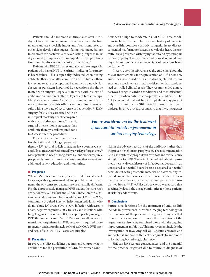
■ ConclusionsFuture considerations for the treatment of endocarditis include improvements in cardiac imaging technology for the diagnosis of the presence of vegetation. Agents that prevent the formation or promote the dissolution of the vegetation are also being examined, along with the ongoing improvement in antibiotics. This improvement includes the investigation of involving cell wall-specifi c enzymes and antibacterial antibodies that act as adjuncts to antibiotics in facilitating bacteriologic clearance.4
SBE can have serious consequences, and the potential for malpractice litigation due to failure to diagnose or
Future considerations for the treatment
of endocarditis include improvements in
cardiac imaging technology.
Copyright © 2011 Lippincott Williams & Wilkins. Unauthorized reproduction of this article is prohibited.
38 The Nurse Practitioner • Vol. 36, No. 3 www.tnpj.com
Subacute bacterial endocarditis: making the diagnosis
delayed diagnosis accompanied by a poor outcome for the patient. There have been documented case studies of failure of the current AHA recommendations even when they are applied appropriately.7 Therefore, NPs must follow the revised 2007 AHA guidelines, keeping in mind that they may need modifi cation in particular circumstances based on the individual clinical setting and risk of developing SBE. Overall, the key to maximizing treatment success is knowl-edge of this disease process and early diagnosis. SBE is complex, and the clinical team must have an understanding of the epidemiology, microbiology, and natural history of endocarditis, as well as a grasp of the AHA guiding prin-ciples of prophylaxis, diagnosis, and medical and surgical management.
REFERENCES 1. Brusch JL. Infective endocarditis; 2009 (updated August 23, 2009).
http://emedicine.medscape.com.
2. Osler W. Gustonian lectures on malignant endocarditis. Br Med J. 1885;1(1262):467-470, 522-526, 577-579.
3. McDonald JR. Acute infective endocarditis. Infect Dis Clin North Am. 2009;23(3):643-664.
4. Fowler VG, Bayer AS. Infective endocarditis. In: Goldman L, Ausiello DA, eds. Cecil Medicine. 23rd ed. Philadelphia, PA: Saunders; 2008:537-548.
5. Medline. Infective endocarditis. Arch Intern Med. 2010 Jan 25 commentary; 2010 (updated January 27th, 2010). http://medline.com.
6. Cunha BA, Eisenstein LA, Hamid NS. Pacemaker-induced Staphylococcus aureus mitral valve acute bacterial endocarditis complicated by persistent bacteremia from a coronary stent: cure with prolonged/high-dose daptomycin without toxicity. Heart Lung. 2006;35(3):207-211.
7. Cunha BA, D’Elia AA, Pawar N, Schoch P. Viridans streptococcal (Streptococcus intermedius) mitral valve subacute bacterial endocarditis (SBE) in a patient with mitral valve prolapse after a dental procedure: the importance of antibiotic prophylaxis. Heart Lung. 2010;39(1):64-72.
8. Turnier L, Nausheen S, Cunha BA. Fatal Streptococcus viridians (S. oralis) aortic prosthetic valve endocarditis (PVE) with paravalvular abscesses related to steroids. Heart Lung. 2009;38(2):167-171.
9. Schoen FJ. Infective endocarditis. In: Cotran RS, Kumar V, Collins T, eds. Pathologic Basis of Disease. 8th ed. Philadelphia, PA: W.B. Saunders Company; 2009:566-569.
10. Sherwood M, Smith D, Crisel R, Veledar E, Lerakis S. Staphylococcus aureus endocarditis: The Grady Memorial Hospital Experience. Am J Med Sci. 2006;331(2):84-87.
11. Cunha BA, Esrick MD, LaRusso M. Staphylococcus hominis native mitral valve bacterial endocarditis (SBE) in a patient with hypertrophic obstructive cardiomyopathy. Heart Lung. 2007;36(5):380-382.
12. Wilson W, Taubert KA, Gewitz M, et al. Prevention of infective endocarditis: guidelines from The American Heart Association: a guideline from The American Heart Association Rheumatic Fever, Endocarditis, and Kawasaki Disease Committee, Council on Cardiovascular Disease in the Young and the Council on Clinical Cardiology, Council on Cardiovascular Surgery and Anesthesia, and the Quality of Care and Outcomes Research Interdisciplinary Working Group. Circulation. 2007;116(15):1736-1754.
13. Cabell CH, Fowler VG Jr, Engemann JJ et al. Endocarditis in the elderly: incidence, surgery, and survival in 16,921 patients over 12 years. Circulation. 2002;106(19):547.
14. Fowler VG, Scheld WM, Bayer AS. Endocarditis and intravascular infections. In: Mandell GL, Bennett JE, Dolin R, eds. Mandell, Douglas and Bennett’s Principles and Practice of Infectious Diseases. 6th ed. Philadelphia, PA: Churchill Livingstone; 2005:975-1021.
15. Miro JM, Anguera I, Cabell CH, et al. Staphylococcus aureus native valve infective endocarditis: report of 566 episodes from the International Collaboration on Endocarditis Merged Database. Clin Infect Dis. 2005;41(4):507-514.
16. McDonald JR, Olaison L, Anderson DJ, et al. Enterococcal endocarditis: 107 cases from the International Collaboration on Endocarditis Merged Database. Am J Med. 2005;118(7):759-766.
17. McDonald JR. Acute infective endocarditis. Infect Dis Clin North Am. 2009;23(3):643-664.
18. Michel PL, Acar J. Native cardiac disease predisposing to infective endocarditis. Eur Heart J. 1995;16(suppl B):2-9.
19. Weinberger I, Rotenberg Z, Zacharovitch D, Fuchs J, Davidson E, Agmon J. Native valve infective endocarditis in the 1970s versus 1980s: underlying cardiac lesions and infecting organisms. Clin Cardiol. 1990;13(2):94-98.
20. Murdoch DR, Corey GR, Hoen B, et al. Clinical presentation, etiology, and outcomes of infective endocarditis in the 21st century: the International Collaboration on Endocarditis-Prospective Cohort Study. Arch Intern Med. 2009;169(5):463-473.
21. Fowler VG Jr, Miro JM, Hoen B, et al. Staphylococcus aureus endocarditis: a consequence of medical progress. JAMA. 2005;293(24):3012-3021.
22. Habib G. Embolic risk in subacute bacterial endocarditis: determinants and role of transesophageal echocardiography. Curr Card Rep. 2003;5:129-136.
23. Presterl E, Rokita E, Graninger W, Hirschi AM. Dysregulation of monocyte oxidative burst in streptococcal endocarditis. Eur J Clin Invest. 2001;31(10):902-906.
24. Chorianopoulos E, Bea F, Katus HA, Frey N. The role of endothelial cell biology in endocarditis. Cell Tissue Res. 2009;335(1):153-163.
25. Cabell CH, Abrutyn E, Karchmer AW. Cardiology patient page. Bacterial endocarditis: the disease, treatment, and prevention. Circulation. 2003;107(20):e185-e187.
26. Banks J, Poole S, Nair SP, et al. Streptococcus sanguis secretes CD14-binding proteins that stimulate cytokine synthesis: a clue to the pathogenesis of infective (bacterial) endocarditis? Microb Pathog. 2002;32(3):105-116.
27. Fowler VG, Scheld WM, Bayer AS. Endocarditis and intravascular infections. In: Mandell GL, Bennett JE, Dolin R, eds. Mandell, Douglas and Bennett’s Principle and Practice of Infectious Diseases. 6th ed. Philadelphia, PA: Churchill Livingstone; 2005:975-1021.
28. Gunson TH, Oliver GF. Osler’s nodes and Janeway lesions. Australas J Dermatol. 2007;48:251-255.
29. Graninger W, Presterl E, Wenisch C, Schwameis E, Breyer S, Vukovich T. Management of serious staphylococcal infections in the outpatient setting. Drugs. 1997;54(suppl 6):21-28.
30. Gratz S, Raddatz D, Hagenah G, Behr T, Behe M, Becker W. 99mTc-labelled antigranulocyte monoclonal antibody FAB( fragments versus echocardiography in the diagnosis of subacute infective endocarditis. Int J Cardiol. 2000;75(1):75-84.
31. Cunha BA, Gill MV, Lazar JM. Acute infective endocarditis. Diagnostic and therapeutic approach. Infect Dis Clin North Am. 1996;10(4):811-834.
32. Dreyfus G, Serraf A, Jebara VA, et al. Valve repair in acute endocarditis. Ann Thorac Surg. 1990;49(5):760-711.
33. Magilligan DJ Jr, Quinn EL, eds. Endocarditis, Medical and Surgical Management. New York: Marcel Dekker; 1986:207-264.
34. Zegdi R, Debieche M, Latremouille C, et al. Long-term results of mitral valve repair in active endocarditis. Circulation. 2005;111(19);2532-2536.
35. Lalani T, Cabell C, Benjamin DK, et al. Analysis of the impact of early surgery on in-hospital mortality of native valve endocarditis: use of propensity score and instrumental variable methods to adjust for treatment-selection bias. Circulation. 2010;121(8):1005-1013.
36. Garcia Rodriguez JF, Mesias Prego JA, Dominguez Gomez D. Treatment of endocarditis due to penicillin-susceptible streptococci with a 2-week course of ceftriaxone followed by oral amoxicillin. Eur J Clin Microbiol Infect Dis. 1992;11(10):952-953.
37. Dajani AS, Disno AL, Chung KJ, et al. Prevention of bacterial endocarditis. Recommendations by the American Heart Association. JAMA. 1990;264(22):2919-2922.
38. Van der Meer JT, Van Wijk W, Thompson J, Vandenbroucke JP, Valkenburg HA, Michel MF. Effi cacy of antibiotic prophylaxis for prevention of native-valve endocarditis. Lancet. 1992;339(8786):135-139.
Kristen Luttenberger is a CCU Unit Educator and Mary DiNapoli is an acute adult nurse practitioner at Morristown Memorial Hospital, Morristown, NJ.
DOI-10.1097/01.NPR.0000393971.15598.0c
Copyright © 2011 Lippincott Williams & Wilkins. Unauthorized reproduction of this article is prohibited.