Sub-acute toxicity and effect of Hwangryunhaedok-tang on ...
16
http://dx.doi.org/10.13048/jkm.17014 15 Sub-acute toxicity and effect of Hwangryunhaedok-tang on human drug-metabolizing enzymes Seong Eun Jin 1 , Mee-Young Lee 1 , Chang-Seob Seo 1 , Hyeun-Kyoo Shin 1 , Jae-Woo Cho 2 , Hyekyung Ha 1,* 1 K-herb Research Center, Korea Institute of Oriental Medicine 2 Pathology Research Center, Korea Institute of Toxicology Original Article ⋅Received:6 April 2017 ⋅Revised:29 May 2017 ⋅Accepted:29 May 2017 ⋅Correspondence to:Hyekyung Ha K-herb Research Center, Korea Institute of Oriental Medicine 1672 Yuseong-daero, Yuseong-gu, Daejeon 34054, Republic of Korea Tel:+82-42-868-9513, Fax:+82-42-864-2120, E-mail:[email protected] Objectives: Hwangryunhaedok-tang (HHT; Huanglianjiedu-tang, Orengedoku-to), a traditional herbal formula, is used for treating inflammation, hypertension, gastritis, liver dysfunction, cerebrovascular diseases, dermatitis and dementia. The objective of this study was to assess the sub-acute toxicity of HHT in Sprague-Dawley (SD) rats, and its effect on the activities of human microsomal cytochrome P450s (CYP450s) and UDP-glucuronosyltransferases (UGTs). Methods: Male and female SD rats were orally administered HHT once daily at doses of 0, 500, 1000 and 2000 mg/kg for 4 weeks. We analyzed mortality, clinical observations, body weight, food consumption, organ weights, urinalysis, hematology, serum biochemistry, and histopathology. The activities of major human CYP450s (CYP1A2, CYP3A4, CYP2B6, CYP2C9, CYP2C19, CYP2D6, and CYP2E1) and UGTs (UGT1A1, UGT1A4, and UGT2B7) were assessed using in vitro fluorescence- and luminescence-based enzyme assays, respectively. Results: No toxicologically significant changes related to the repeated administration of HHT were observed in both male and female SD rats. The no observed adverse effect level (NOAEL) value was more than 2000 mg/kg/day for both sexes. HHT inhibited the activities of human microsomal CYP1A2, CYP2C19, CYP2D6, and CYP2E1, whereas it weakly inhibited the activities of CYP2B6, CYP2C9, CYP3A4, and UGT1A1. In addition, HHT negligibly inhibited the activities of human microsomal UGT1A4 and UGT2B7 with IC 50 values in excess of 1000 μg/mL. Conclusions: Our findings indicate that HHT may be safe for repeated administration up to 4 weeks. In addition, these findings provide information on the safety and effectiveness of HHT when co-administered with conventional drugs. Key Words : Hwangryunhaedok-tang, sub-acute toxicity, cytochrome P450, UDP-glucuronosyltransferase, herb-drug interactions Introduction Herbal medicines have been used for the treatment of a multitude of diseases, and are often used in conjunction with conventional drugs. Some herbal medicines have shown significant side effects even though they comprise natural products. However, there is limited scientific evidence to validate the safety of majority of the herbal medicines 1) . In particular, the co-administration of herbal formulas with conventional drugs raises even deeper safety concerns because herbal formulas comprise various herbal medicines and constituents. Herbal medicines and their constituents can alter the activity or expression of drug-metabolizing enzymes, which can lead to changes in the efficacy J Korean Med. 2017;38(2):15-30 http://dx.doi.org/10.13048/jkm.17014 pISSN 1010-0695 • eISSN 2288-3339
Transcript of Sub-acute toxicity and effect of Hwangryunhaedok-tang on ...
http://dx.doi.org/10.13048/jkm.17014 15
Sub-acute toxicity and effect of Hwangryunhaedok-tang on human drug-metabolizing enzymes
Seong Eun Jin1, Mee-Young Lee1, Chang-Seob Seo1, Hyeun-Kyoo Shin1, Jae-Woo Cho2, Hyekyung Ha1,*
1K-herb Research Center, Korea Institute of Oriental Medicine2Pathology Research Center, Korea Institute of Toxicology
Original Article
⋅Received:6 April 2017 ⋅Revised:29 May 2017 ⋅Accepted:29 May 2017
⋅Correspondence to:Hyekyung Ha
K-herb Research Center, Korea Institute of Oriental Medicine1672 Yuseong-daero, Yuseong-gu, Daejeon 34054, Republic of Korea
Tel:+82-42-868-9513, Fax:+82-42-864-2120, E-mail:[email protected]
Objectives: Hwangryunhaedok-tang (HHT; Huanglianjiedu-tang, Orengedoku-to), a traditional herbal formula, is used for treating inflammation, hypertension, gastritis, liver dysfunction, cerebrovascular diseases, dermatitis and dementia. The objective of this study was to assess the sub-acute toxicity of HHT in Sprague-Dawley (SD) rats, and its effect on the activities of human microsomal cytochrome P450s (CYP450s) and UDP-glucuronosyltransferases (UGTs). Methods: Male and female SD rats were orally administered HHT once daily at doses of 0, 500, 1000 and 2000 mg/kg for 4 weeks. We analyzed mortality, clinical observations, body weight, food consumption, organ weights, urinalysis, hematology, serum biochemistry, and histopathology. The activities of major human CYP450s (CYP1A2, CYP3A4, CYP2B6, CYP2C9, CYP2C19, CYP2D6, and CYP2E1) and UGTs (UGT1A1, UGT1A4, and UGT2B7) were assessed using in vitro fluorescence- and luminescence-based enzyme assays, respectively. Results: No toxicologically significant changes related to the repeated administration of HHT were observed in both male and female SD rats. The no observed adverse effect level (NOAEL) value was more than 2000 mg/kg/day for both sexes. HHT inhibited the activities of human microsomal CYP1A2, CYP2C19, CYP2D6, and CYP2E1, whereas it weakly inhibited the activities of CYP2B6, CYP2C9, CYP3A4, and UGT1A1. In addition, HHT negligibly inhibited the activities of human microsomal UGT1A4 and UGT2B7 with IC50 values in excess of 1000 μg/mL. Conclusions: Our findings indicate that HHT may be safe for repeated administration up to 4 weeks. In addition, these findings provide information on the safety and effectiveness of HHT when co-administered with conventional drugs.
Key Words : Hwangryunhaedok-tang, sub-acute toxicity, cytochrome P450, UDP-glucuronosyltransferase, herb-drug interactions
Introduction
Herbal medicines have been used for the
treatment of a multitude of diseases, and are often
used in conjunction with conventional drugs. Some
herbal medicines have shown significant side effects
even though they comprise natural products.
However, there is limited scientific evidence to
validate the safety of majority of the herbal
medicines1). In particular, the co-administration of
herbal formulas with conventional drugs raises even
deeper safety concerns because herbal formulas
comprise various herbal medicines and constituents.
Herbal medicines and their constituents can alter
the activity or expression of drug-metabolizing
enzymes, which can lead to changes in the efficacy
J Korean Med. 2017;38(2):15-30http://dx.doi.org/10.13048/jkm.17014
pISSN 1010-0695 • eISSN 2288-3339

Journal of Korean Medicine 2017;38(2)
http://dx.doi.org/10.13048/jkm.1701416
(150)
and toxicity of conventional drugs. Therefore,
information on the potential of herbal medicines to
inhibit or induce the activity or expression of
drug-metabolizing enzymes when co-administered
with conventional drugs is important.
In general, studies on the safety of herbal
medicines, including herbal formulas, should include
at least in vitro and in vivo genotoxicity assays,
long-term rodent carcinogenicity tests, reproductive
and developmental toxicity studies, and investigation
of their effects on drug-metabolizing enzymes2).
Drug metabolism is divided into phase Ⅰ and
phase Ⅱ transformation reactions. Cytochrome P450
(CYP450), a class of phase Ⅰ enzymes, exists as a
superfamily of heme containing enzymes that catalyze
the oxidative metabolism of various endogenous and
xenobiotic substrates3). Approximately 73% of all
known drugs are metabolized by hepatic CYP4504).
In particular, CYP1A2, CYP2C9, CYP2C19, CYP2D6,
and CYP3A4 are responsible for approximately 80%
of the known oxidative drug reactions, and they are
highly subjected to inhibition owing to their broad
specificity for structurally diverse substrates4).
UDP-glucuronosyltransferases (UGT), a class of
phase Ⅱ enzymes, catalyze the conjugation of
glucuronic acid to exogenous substances and
endogenous components5). The human UGTs are
generally categorized into UGT1A, UGT2A, and
UGT2B subfamilies6).
Hwangryunhaedok-tang (HHT; Huanglianjiedu-tang,
Orengedoku-to) is a traditional herbal formula
comprising Coptidis Rhizoma, Scutellariae Radix,
Phellodendri Cortex, and Gardeniae Fructus7). HHT
has been used for treating inflammation, hypertension,
gastritis, liver dysfunction, cerebrovascular diseases,
dermatitis, and dementia8,9). In our previously study,
the sub-chronic toxicity of HHT in Sprague-Dawley
(SD) rats was reported. Under our experimental
conditions, the no observed adverse effect level
(NOAEL) of HHT for rats of either sex was 750
mg/kg/day10). However, other studies on the toxic
and influence of HHT on drug-metabolizing
enzymes are required to evaluate the safety of HHT.
In this study, we evaluated the sub-acute toxicity
of HHT in SD rats, and the effects of HHT on the
activities of major human CYP450s (CYP1A2,
CYP2B6, CYP2C9, CYP2C19, CYP2D6, CYP2E1,
and CYP3A4) and UGTs (UGT1A1, UGT1A4, and
UGT2B7) drug metabolizing enzymes.
Materials and methods
1. Chemicals and materials
Vivid®CYP450 Screening Kits (Vivid® CYP1A2
Blue, Vivid® CYP2B6 Blue, Vivid®CYP2C9 Blue,
Vivid® CYP2C19 Blue, Vivid® CYP2D6 Blue,
Vivid® CYP2E1 Blue, and Vivid® CYP3A4 Green)
were purchased from Invitrogen Co. (Camarillo,
CA, USA). These kits use 7-ethoxy-methyloxy-3
-cyanocoumarin (EOMCC) as a substrate for
CYP1A2, CYP2D6, CYP2C19, and CYP2E1.
Additionally, di(benzyloxymethoxy)fluorescein (DBOMF)
was used as a substrate for CYP3A4, and 7
-benzyloxy-4-trifluoromethylcoumarin (BOMCC) was
used as a substrate for CYP2B6 and CYP2C9.
UGT-Glo™ UGT1A1 and UGT2B7 Screening
Systems were purchased from Promega (Madison,
WI, USA). The recombinant human UGT1A4
enzyme was purchased from Corning, Inc. Life
Sciences (Tewksbury, MA, USA). α-Naphthoflavone,
ketoconazole, miconazole, sulfaphenazole, quinidine,
sodium diethyldithiocarbamate trihydrate, diclofenac,
and lopinavir were obtained from Sigma Chemical
Co. (St. Louis, MO, USA). All other chemicals
were of analytical grade.
2. Preparation of HHT
Preparation of HHT was performed as previously
reported11). In brief, we prepared the HHT extract in
our laboratory. The four medicinal herbs that
constitute HHT, Coptis chinensis (Coptidis Rhizoma,
Sub-acute toxicity and effect of Hwangryunhaedok-tang on human drug-metabolizing enzymes
http://dx.doi.org/10.13048/jkm.17014 17
(151)
China), Scutellaria baicalensis (Scutellariae Radix,
Jeongseon, Korea), Phellodendron chinensis (Phellodendri
Cortex, China), and Gardenia jasminoides (Gardeniae
Fructus, Muju, Korea) were purchased from
Omniherb (Yeongcheon, Korea) and HMAX (Jecheon,
Korea). A mixture of the chopped medicinal herbs
was extracted in distilled water (10 times the
sample amount) at 100℃ for 2 h under pressure (98
kPa). The solution was evaporated to dryness and
freeze-dried. This sample was named as ‘Voucher
specimens (2008-KE-20-1~KE-20~4)’, and these
specimens have been deposited at the K-herb
Research Center of Korea Institute of Oriental
Medicine. The high performance liquid chromatography
(HPLC) profile of HHT has been previously
reported11). In brief, the contents HHT extract are as
follows: geniposide 36.54 ± 0.27 mg/g, baicalein
30.24 ± 0.72 mg/g, palmatine 10.34 ± 0.47 mg/g,
berberine 1.35 ± 0.02 mg/g and coptisine 0.97 ±
0.02 mg/g.
3. Animals
Each of 20 male and female specific pathogen
-free SD rats (six-week-old) were obtained from
Orient Bio Inc. (Seongnam, Republic of Korea). The
rats underwent one week of acclimatization prior to
study initiation. Five animals were used per group.
Two or three animals were housed per polycarbonate
cage stainless-steel wire-mesh cage, and they were
allowed ad libitum access to sterilized tap water and
commercial rodent chow (Purina Co., Pyeongtaek,
Republic of Korea). The animals were maintained in
environmentally-controlled rooms at 23 ± 3˚C under
relative humidity of 50 ± 10%, with a 12 h
light-dark cycle (artificial lighting from 08:00 to
20:00), and 10-20 air changes per hour. This study
was conducted at the Korea Testing and Research
Institute, and the protocol was approved by the
Institutional Animal Care and Use Committee
according to the “Guidelines for Toxicity Tests of
Drugs and Related Materials, Document #2009-116”
prepared by Korea Food and Drug Administration
(2009). The approval number is #G11025.
4. Dose selection and animal treatment
In our previous single-dose study of HHT, no
treatment-related toxic changes were observed at the
highest dose (2000 mg/kg/day). Therefore, 0, 500,
1000, and 2000 mg/kg/day of HHT were selected
for this 4-week repeated dose study. Healthy male
and female rats were randomly assigned to four
experimental groups. Each group consisted of five
rats of each sex. HHT was suspended in distilled
water, and freshly prepared daily before treatment.
The daily application volume (5 mL/kg) of HHT
was calculated in advance based on the most
recently recorded body weights of individual
animals. The rats were orally administered HHT for
4 weeks, while distilled water was given to the
animals in the vehicle control group.
5. Clinical observations
During the experimental period, the rats were
observed twice daily for clinical signs and mortality.
All clinical signs were recorded individually for
type, observation day/time, and duration using the
Path/Tox System 4.2.2 (Xybion Medical Systems
Corp., USA). Body weight and food consumption
were recorded weekly.
6. Urinalysis and hematology
During the last week of the experiment, urinalysis
was conducted to assess the urine volume, specific
gravity (SG), pH, and urobilinogen (URO) using
Multistix 10 SG (Bayer, NJ, USA) and urine
chemical analyzer (Clinitek-500, GMI, MN, USA).
On day 28, the rats were fasted overnight, and
they were anesthetized with isoflurane and
sacrificed. Blood samples were drawn from the
posterior vena cava and collected in complete blood
Journal of Korean Medicine 2017;38(2)
http://dx.doi.org/10.13048/jkm.1701418
(152)
count (CBC) bottles containing EDTA-2K (Sewon
Medical Co., Republic of Korea). The samples were
analyzed using an ADVIA120 Hematology System
(Bayer) to determine the white blood cell count
(WBC), red blood cell count (RBC), hemoglobin
concentration (HGC), hematocrit (HCT), mean
corpuscular volume (MCV), mean corpuscular
hemoglobin (MCH), mean corpuscular hemoglobin
concentration (MCHC), platelet (PLT), reticulocyte
(RET), neutrophils, lymphocytes, monocytes, basophils,
and large unstained cells (LUC). Prothrombin time
(PT) and activated partial thromboplastin time
(APTT) were determined in blood samples treated
with 3.2% sodium citrate using a coagulometer
(ACL 9000, Instrumentation Laboratory Spa, Milan,
Italy).
7. Serum biochemistry
The blood samples were centrifuged at 3,000g for
10 min and analyzed for glucose (GLU), blood urea
nitrogen (BUN), creatinine (CREA), total protein
(TP), albumin (ALB), albumin/globulin ratio (A/G),
total cholesterol (TCHO), triglyceride (TG), phospholipid
(PL), alanine aminotransferase (ALT), aspartate
aminotransferase (AST), alkaline phosphatase (ALP),
total bilirubin (TBIL), and creatine kinase (CK)
using an autoanalyzer (Toshiba 200 FR NEO,
Toshiba Co., Japan).
8. Necropsy
Complete gross necropsy was performed on all
rats. The absolute weights of the brain, pituitary
gland, liver, spleen, kidney, lung, heart, thymus,
salivary glands, adrenal glands, thyroid/parathyroid,
salivary vesicle (male), prostate (male), testes
(male), epididymis (male), ovaries (female), and
uterus/cervix (female) were measured, and the
relative organ weights (organ-to-body weight ratios)
were calculated.
9. Histopathology
The liver, kidneys, mandibular lymph nodes, and
uterus/cervix samples were fixed in 10% neutral
-buffered formalin, sectioned at 4 μm, and stained
with hematoxylin and eosin (H&E, Sigma). The
tissues were subsequently mounted and coverslipped
using the Dako mounting medium (Invitrogen,
USA).
10. Cytochrome P450 isozyme assay
The assays were performed using Vivid® CYP450
Screening Kits according to the protocol provided
by the manufacturer and previously described
methods12). Vivid® CYP450 Screening Kits are
designed to assess the metabolic activity of the
predominant human CYP450s (CYP1A2, CYP3A4,
CYP2B6, CYP2C9, CYP2C19, CYP2D6, or CYP2E1)
involved in hepatic drug metabolism. A test sample
of 40 μL diluted in solvent, a positive inhibition
control or a solvent control was added to each well.
The solutions were mixed after adding 50 μL
Master Pre-Mix containing P450 BACULOSOMES®
in the Vivid® CYP450 Reaction Buffer and
Regeneration System (consisting of glucose-6-phosphate
and glucose-6-phosphate dehydrogenase), and the
plate was incubated for 20 min to allow the samples
to interact with the CYP enzymes. The Regeneration
System converts NADP+ into NADPH. After
pre-incubation, the reaction was initiated by adding
10 μL of the mixture of Vivid® Substrate and
NADP+. For CYP3A4, the fluorescence intensity
was measured using an EnVision 2103 Multilabel
Reader (PerkinElmer Inc., MA, USA) for 15 min at
excitation and emission wavelengths of 485 and 535
nm, respectively. For CYP1A2, CYP2B6, CYP2C9,
CYP2C19, CYP2D6, or CYP2E1, the fluorescence
intensity was measured for 60 min at excitation and
emission wavelengths of 415 and 460 nm,
respectively, using a SpectraMax® i3 (Molecular
Devices Co., Sunnyvale, CA, USA).
Sub-acute toxicity and effect of Hwangryunhaedok-tang on human drug-metabolizing enzymes
http://dx.doi.org/10.13048/jkm.17014 19
(153)
The inhibition percentage (%) was obtained by
the following equation: % Inhibition = [1 – (S1 –
S0) / (C1 – C0)] × 100, where C1 is the fluorescence
of the control after incubation, C0 is the initial
fluorescence of the control, S1 is the fluorescence of
the test sample after incubation, and S0 is the initial
fluorescence of the test sample in the linear section.
The background fluorescence of the herbal
formulas was corrected by subtracting the values
obtained from the incubation without substrates.
CYP450 inhibition of the sample was expressed in
terms of IC50, as calculated from the log-dose
inhibition curve (SigmaPlot, Ver. 12.5, Systat
Software, Inc., CA, USA). The data are expressed
as the means ± standard error of the mean (SEM; n
= 3). α-Naphthoflavone, ketoconazole, sulfaphenazole,
quinidine, and sodium diethyldithiocarbamate trihydrate
were used as positive controls for CYP1A2,
CYP3A4, CYP2C9, CYP2D6, and CYP2E1,
respectively. Miconazole was used as a positive
control for CYP2B6 and CYP2C19.
11. UDP-glucuronosyltransferase isozyme assay
The assays were performed using the UGT-Glo™
Screening Systems according to the manufacturer’s
protocol and previously described methods12). The
assay systems provide a luminescent method for
measuring the activity of UGTs. Two glucuronidation
reactions were set up in parallel to measure UGT
activity. Both reactions contained a source of UGTs
(UGT1A1, UGT1A4, or UGT2B7) and the proluciferin
substrate (UGT Multienzyme Substrate or UGT1A4
Substrate); however, only one of them contained the
uridine 5’-diphosphoglucuronic acid (UDPGA)
cofactor. Ten microliters of 4× concentrated test
sample, a positive inhibition control, or a solvent
control was added to each well. Then, 10 μL of
UDPGA (Plus-UDPGA reaction set) or distilled
water (Minus-UDPGA reaction set) was added to
the relevant wells. Twenty microliters of the
prepared 2× control reaction mixture (Minus-UGT
enzyme) and the 2× UGT reaction mixture
(UGT1A1, UGT1A4, or UGT2B7) were added to
the appropriate wells. The reaction solution was
mixed and incubated at 37℃ for 90, 180, or 60
min, respectively, for UGT1A1, UGT1A4, or
UGT2B7. The final contents of the reactant were
0.1 mg/mL UGT enzyme and 20 μM enzyme
substrate in the presence or absence of 4 mM
UDPGA. After incubation, 40 μL of the
reconstituted Luciferin Detection Reagent plus
D-cysteine was added to all wells. After 20 min of
incubation at room temperature, the luminescence
signal was detected using a SpectraMax® i3.
The detected data were converted to the
calculated difference using the following percentage
of substrate consumed (%SC) equation: % Substrate
consumed = (background corrected difference)/(average
minus-UDPGA values) × 100. The inhibition
percentage (%) was obtained via the following
equation: % Inhibition = [1 – (S/CAVR)] × 100,
where S is the %SC of each sample or the control
wells, and CAVR is the average %SC of the control
wells. The UGT inhibition of each sample was
expressed in terms of IC50 as calculated using
SigmaPlot (Ver. 12.5), which is capable of
generating a four parameter logistic curve fit. The
data are expressed as the means ± SEM (n = 2).
12. Statistics analysis
Data collected during the study were examined
for homogeneity of variance using Bartlett’s test. In
case of no significant deviations from homogeneity
of variance, one-way ANOVA was conducted at α =
0.05. In case of significance, multiple comparisons
were performed using Dunnett’s test to determine
which pairs of groups were significantly different.
When significant deviations from homogeneity of
variance were observed, a non-parametric comparison
Journal of Korean Medicine 2017;38(2)
http://dx.doi.org/10.13048/jkm.1701420
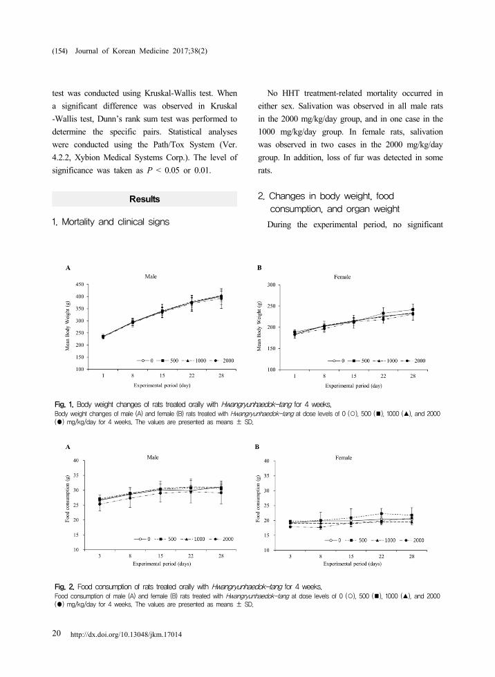
Fig. 1. Body weight changes of rats treated orally with Hwangryunhaedok-tang for 4 weeks.Body weight changes of male (A) and female (B) rats treated with Hwangryunhaedok-tang at dose levels of 0 (○), 500 (■), 1000 (▲), and 2000 (●) mg/kg/day for 4 weeks. The values are presented as means ± SD.
Fig. 2. Food consumption of rats treated orally with Hwangryunhaedok-tang for 4 weeks.Food consumption of male (A) and female (B) rats treated with Hwangryunhaedok-tang at dose levels of 0 (○), 500 (■), 1000 (▲), and 2000 (●) mg/kg/day for 4 weeks. The values are presented as means ± SD.
(154)
test was conducted using Kruskal-Wallis test. When
a significant difference was observed in Kruskal
-Wallis test, Dunn’s rank sum test was performed to
determine the specific pairs. Statistical analyses
were conducted using the Path/Tox System (Ver.
4.2.2, Xybion Medical Systems Corp.). The level of
significance was taken as P < 0.05 or 0.01.
Results
1. Mortality and clinical signs
No HHT treatment-related mortality occurred in
either sex. Salivation was observed in all male rats
in the 2000 mg/kg/day group, and in one case in the
1000 mg/kg/day group. In female rats, salivation
was observed in two cases in the 2000 mg/kg/day
group. In addition, loss of fur was detected in some
rats.
2. Changes in body weight, food consumption, and organ weight
During the experimental period, no significant

Sub-acute toxicity and effect of Hwangryunhaedok-tang on human drug-metabolizing enzymes
http://dx.doi.org/10.13048/jkm.17014 21
(155)
differences in body weight and food consumption
were observed between the vehicle control group
and HHT-treated groups for either sex (Fig. 1 and 2).
In male rats, there were no significant differences in
relative organ weights between the vehicle control
and HHT-treated groups (Table 1). In contrast, the
relative spleen weight was significantly increased in
the 1000 and 2000 mg/kg/day female groups when
compared with that in the vehicle control group
(Table 1). Additionally, the relative kidney weights
were significantly increased in the HHT-treated
female groups, and relative ovaries weights were
significantly decreased in the 1000 and 2000
mg/kg/day female groups when compared with those
in the vehicle control group (Table 1).
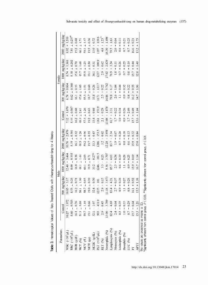
3. Urinalysis and hematology
In the 1000 mg/kg/day female group, volume of
urine was increased when compared with those in
the vehicle control group, but was within the normal
range. There were no significant changes found in
both male and female rats among any of the
HHT-treated groups for volume, SG, pH and URO
when compared with the vehicle control group
(Table 2).
In both male and female rats treated with 2000
mg/kg/day of HHT, an increase in bilirubin (+1~
+2) was detected (male, n = 5; female, n = 5) when
compared with that in the vehicle control group. and
green urine were observed (male, n = 4; female, n =
3; data not shown). In the 1000 mg/kg/day male
group, green urine was observed in one case, and
the percentage of MCHC was significantly increased
when compared with that in the vehicle control
group (Table 3). In the 2000 mg/kg/day female
group, significant decrease of RBC and increase in
the percentage of RET were detected when
compared with those in the vehicle control group
(Table 3).
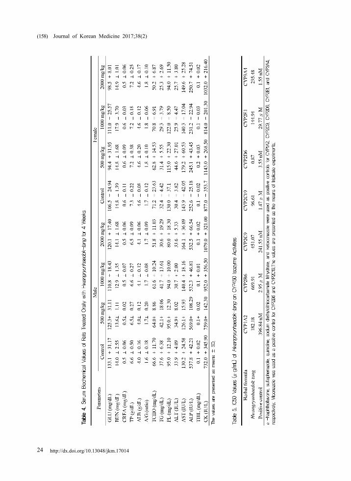
4. Serum biochemistry
No significant differences in serum biochemical
values were observed in rats treated with HHT
(Table 4).
5. Necropsy findings
Dark discoloration of the kidneys was observed at
1000 mg/kg/day (male, n = 1; female, n = 2) and
2000 mg/kg/day (male, n = 5; female, n = 5) of
HHT in both sexes. In addition, the male and
female rats treated with 2000 mg/kg/day of HHT
showed dark discoloration of the liver (male, n = 4;
female, n = 5).
6. Histopathological examination
HHT treatment did not induce histopathological
changes in the liver, kidneys, mandibular lymph
nodes, and uterus/cervix at less than 2000
mg/kg/day (data not shown).
7. Effects of HHT on the CYP450s activities
In vitro fluorescence-based CYP450 assays were
carried out to investigate the influence of HHT on
the activities of human CYP1A2, CYP2B6, CYP2C9,
CYP2C19, CYP2D6, CYP2E1, and CYP3A4. α
-Naphthoflavone, sulfaphenazole, quinidine,
diethyldithiocarbamate, and ketoconazole inhibited
the activities of CYP1A2, CYP2C9, CYP2D6,
CYP2E1, and CYP3A4 in a dose-dependent manner,
with IC50 values of 396.84 nM, 241.55 nM, 3.55
nM, 29.77 μM, and 1.55 nM, respectively (Fig. 3
and Table 5). Miconazole inhibited the activities of
CYP2B6 and CYP2C19 in a dose-dependent manner,
with IC50 values of 2.95 μM and 1.47 μM,
respectively (Fig. 3 and Table 5).
HHT potently inhibited the CYP2D6 activity,
with an IC50 value of 0.87 μg/mL, followed by the
activities of CYP2C19, CYP2E1, and CYP1A2, with
IC50 values of 96.61 μg/mL, 144.94 μg/mL, and

Journal of Korean Medicine 2017;38(2)
http://dx.doi.org/10.13048/jkm.1701422
(156)
Sub-acute toxicity and effect of Hwangryunhaedok-tang on human drug-metabolizing enzymes
http://dx.doi.org/10.13048/jkm.17014 23
(157)
Journal of Korean Medicine 2017;38(2)
http://dx.doi.org/10.13048/jkm.1701424
(158)

Fig. 3. The effects of Hwangryunhaedok-tang on the activities of CYP1A2 (A), CYP2B6 (B), CYP2C9 (C), CYP2C19 (D), CYP2D6 (E), CYP2E1 (F), and CYP3A4 (G).
Fluorescence-based enzyme assays of human microsomal CYP450 isozymes were established in vitro. α-Naphthoflavone, sulfaphenazole, quinidine, sodium diethyldithiocarbamate trihydrate, and ketoconazole were used as positive controls for CYP1A2, CYP2C9, CYP2D6, CYP2E1 and CYP3A4, respectively. Miconazole was used as a positive control for CYP2B6 and CYP2C19. The data are presented as the means ± SEM (n = 3).
Sub-acute toxicity and effect of Hwangryunhaedok-tang on human drug-metabolizing enzymes
http://dx.doi.org/10.13048/jkm.17014 25
(159)
Journal of Korean Medicine 2017;38(2)
http://dx.doi.org/10.13048/jkm.1701426
(160)
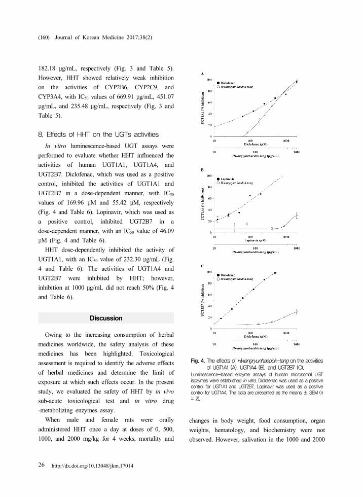
Fig. 4. The effects of Hwangryunhaedok-tang on the activities of UGT1A1 (A), UGT1A4 (B), and UGT2B7 (C).
Luminescence-based enzyme assays of human microsomal UGT isozymes were established in vitro. Diclofenac was used as a positive control for UGT1A1 and UGT2B7. Lopinavir was used as a positive control for UGT1A4. The data are presented as the means ± SEM (n = 2).
182.18 μg/mL, respectively (Fig. 3 and Table 5).
However, HHT showed relatively weak inhibition
on the activities of CYP2B6, CYP2C9, and
CYP3A4, with IC50 values of 669.91 μg/mL, 451.07
μg/mL, and 235.48 μg/mL, respectively (Fig. 3 and
Table 5).
8. Effects of HHT on the UGTs activities
In vitro luminescence-based UGT assays were
performed to evaluate whether HHT influenced the
activities of human UGT1A1, UGT1A4, and
UGT2B7. Diclofenac, which was used as a positive
control, inhibited the activities of UGT1A1 and
UGT2B7 in a dose-dependent manner, with IC50
values of 169.96 μM and 55.42 μM, respectively
(Fig. 4 and Table 6). Lopinavir, which was used as
a positive control, inhibited UGT2B7 in a
dose-dependent manner, with an IC50 value of 46.09
μM (Fig. 4 and Table 6).
HHT dose-dependently inhibited the activity of
UGT1A1, with an IC50 value of 232.30 μg/mL (Fig.
4 and Table 6). The activities of UGT1A4 and
UGT2B7 were inhibited by HHT; however,
inhibition at 1000 μg/mL did not reach 50% (Fig. 4
and Table 6).
Discussion
Owing to the increasing consumption of herbal
medicines worldwide, the safety analysis of these
medicines has been highlighted. Toxicological
assessment is required to identify the adverse effects
of herbal medicines and determine the limit of
exposure at which such effects occur. In the present
study, we evaluated the safety of HHT by in vivo
sub-acute toxicological test and in vitro drug
-metabolizing enzymes assay.
When male and female rats were orally
administered HHT once a day at doses of 0, 500,
1000, and 2000 mg/kg for 4 weeks, mortality and
changes in body weight, food consumption, organ
weights, hematology, and biochemistry were not
observed. However, salivation in the 1000 and 2000

Sub-acute toxicity and effect of Hwangryunhaedok-tang on human drug-metabolizing enzymes
http://dx.doi.org/10.13048/jkm.17014 27
Table 6. IC50 values (μg/mL) of Hwangryunhaedok-tang on UGT isozyme activities
Herbal formula UGT1A1 UGT1A4 UGT2B7
Hwangryunhaedok-tang 232.30 > 1000 > 1000
Positive control 169.96 μM 46.09 μM 55.42 μMDiclofenac was used as a positive control for UGT1A1 and UGT2B7. Lopinavir was used as a positive control for UGT1A4. The values are presented as the means of duplicate experiments.
(161)
mg/kg/day male groups and 2000 mg/kg/day female
group as well as loss of fur were observed; these
symptoms are considered natural or accidental
changes. The increase of bilirubin (+1 ~ +2) and
incidence of green urine in the 2000 mg/kg/day
male and female groups are considered changes due
to the administration of HHT. However, bilirubin
levels +1 ~ +2 are detectable in normal animals,
and there was no change in the biochemical and
histopathological examination related to the kidney
morphology or function. In addition, green urine is
reported to occur during the elimination of
administered materials such as drugs; it may also be
caused by urinary tract infection of Pseudomonas aeruginosa13,14). In the present study, green urine
was considered as a non-adverse effect because no
symptoms associated with urinary tract infection
were detected.
Significant differences were observed in the
absolute weights of the kidneys, spleen, and ovaries
of HHT-treated female group compared with those
in the vehicle control group. However, these changes
are not associated with HHT-induced abnormality
because they are within normal ranges.
In the hematological test, the percentage of RET
was significantly increased in the 2000 mg/kg/day
female group, but not in the male groups. In
addition, no change in other related hematological
levels was observed. Therefore, the increase in
percentage of RET is considered to be a non-adverse
effect.
Although black discoloration of the kidneys and
liver was considered the result of HHT treatment,
there were no associated histopathological changes.
The black discoloration of the kidney is considered
to be associated with green urine.
Histopathological examination was performed for
organs with abnormal findings in necropsy, and no
histopathological findings associated with HHT
treatment were observed.
The key elements associated with successful drug
development are efficacy and safety, and they are
dependent on drug metabolism15). Changes in the
activity or expression of drug-metabolizing enzymes
can affect the concentration of a drug in the blood
as well as its pharmacokinetic and medicinal
properties16,17). According to the report of Bush et
al., adverse effects caused by herb-drug interactions
were observed in 40% of patients receiving
conventional therapy with an herbal product18).
Therefore, it is important to study the modulation of
drug metabolizing enzymes by herbal formulas to
understand the mechanism underlying herb-drug
interactions.
Several reports have demonstrated the effects of
HHT, the medicinal herbs or components present in
HHT on the activities or levels of drug-metabolizing
enzymes. HHT has been reported to inhibit human
CYP1A2, CYP2C19, CYP2D6, and CYP3A419).
Among the medicinal herbs present in HHT,
Scutellariae Radix has been reported to inhibit
human UGT1A1 activity20). In addition, Scutellariae
Radix increase and suppresses the levels of CYP1A
and CYP2B in rats, respectively21). In our previous
study, chromatographic analysis of the five marker
components, geniposide, baicalin, coptisine, palmatine,
and berberine, in HHT was performed using an
HPLC-PDA11). Among the marker compounds,

Journal of Korean Medicine 2017;38(2)
http://dx.doi.org/10.13048/jkm.1701428
(162)
geniposide has been reported to suppress the mRNA
and protein expression of CYP2E1 in mice22), and
inhibit the human CYP1A2 activity19). Berberine
decreases the activities of CYP2C9, CYP2D6, and
CYP3A4 in humans19,23). It is reported that CYP1A2
and CYP2D6 may play a role in the metabolism of
palmatine24). However, the effects of HHT on the
activities of UGTs have not been elucidated to date.
In the present study, the effects of HHT on the
activities of human CYP450s (CYP1A2, CYP2B6,
CYP2C9, CYP2C19, CYP2D6, CYP2E1, and CYP3A4)
and UGTs (UGT1A1, UGT1A4, and UGT2B7) were
examined to assess herb-drug interactions related to
the use of HHT in clinic. HHT most potently
inhibited the activity of CYP2D6, followed by the
activities of CYP2C19, CYP2E1, and CYP1A2.
These results are consistent with the results reported
by Lee et al.19). Furthermore, HHT moderately
inhibited the CYP3A4 activity, while CYP2B6,
CYP2C9, UGT1A1, UGT1A4, and UGT2B7-mediated
metabolism was not affected. Therefore, care should
be taken when HHT is co-administered with any
inhibitor or substrate of CYP2D6. In addition, HHT
may influence the metabolic reactions mediated by
CYP2C19, CYP2E1, and CYP1A2. Attention should
be paid when high-dose HHT is simultaneously
taken with drugs that are metabolized by CYP3A4.
Ibuprofen and naproxen, which are non-steroidal
anti-inflammatory drugs (NSAIDs), are metabolized
by CYP2C925). Therefore, HHT is unlikely to cause
clinically relevant herb-drug interactions when
co-administered with ibuprofen or naproxen for
inflammatory diseases. Among the drugs used for
the treatment of hypertension, nifedipine and
simvastatin are substrates of CYP3A4, and tolbutamide
and warfarin are metabolized by CYP2C925,26). In
patients taking HHT for hypertension, the
co-administration of tolbutamide or warfarin instead
of nifedipine or simvastatin can reduce the
possibility of herb-drug interactions. Cimetidine and
omeprazole are widely used to treat gastric diseases.
Cimetidine is known to be an inhibitor of CYP1A2,
CYP2C19, and CYP3A4, and omeprazole acts as a
substrate of CYP2C19 and an inhibitor of CYP2C19
and CYP3A4. Ticlopidine, which is used to treat
cerebrovascular diseases, has been reported to inhibit
CYP1A2, CYP2C9, CYP2C19, and CYP2D625).
Therefore, to reduce any adverse effects, caution
should be exercised when HHT is co-administered
with cimetidine, omeprazole, or ticlopidine.
Conclusions
We previously reported the safety of HHT in a
13-week repeated-dose oral toxicity study in rats;
however, the side effects of HHT have not been
reported. In the present study, the NOAEL value of
HHT administration for 4 weeks is determined as
2000 mg/kg/day in both sexes. In addition, HHT
acts as an inhibitor of CYP1A2, CYP2C19,
CYP2D6, and CYP2E1. Therefore, to reduce its
adverse effects, caution should be exercised when
HHT is co-administered with substrates or other
inhibitors of CYP1A2, CYP2C19, CYP2D6 or
CYP2E1. However, HHT has a relatively low
potential to be involved in herb-drug interactions
when taken along with substrates or inhibitors of
CYP2B6, CYP2C9, CYP3A4, UGT1A1, UGT1A4,
and UGT2B7.
Acknowledgements
This research was supported by the grants
‘Construction of Scientific Evidences for Herbal
Medicine Formulas (K17251)’ from the Korea
Institute of Oriental Medicine (KIOM).
Competing interests
The authors declare that they have no competing
interests.
Sub-acute toxicity and effect of Hwangryunhaedok-tang on human drug-metabolizing enzymes
http://dx.doi.org/10.13048/jkm.17014 29
(163)
References
1. Firenzuoli F, Gori L. Herbal medicine today:
clinical and research issues. Evid Based
Complement Alternat Med. 2007;4(Suppl 1):37-40.
2. Moreira DL, Teixeira SS, Monteiro MHD,
De-Oliveira ACAX, Paumgartten FJR. Traditional
use and safety of herbal medicines. Revista
Brasileira de Farmacognosia. 2014;24(2):248-57.
3. Nebert DW, Russell DW. Clinical importance of
the cytochromes P450. Lancet. 2002;360(9340):
1155-62.
4. Wienkers LC, Heath TG. Predicting in vivo
drug interactions from in vitro drug discovery
data. Nat Rev Drug Discov. 2005;4(10):825-33.
5. Court MH, Hazarika S, Krishnaswamy S, Finel
M, Williams JA. Novel polymorphic human
UDP-glucuronosyltransferase 2A3: cloning,
functional characterization of enzyme variants,
comparative tissue expression, and gene induction.
Mol Pharmacol. 2008;74(3):744-54.
6. Mackenzie PI, Bock KW, Burchell B, Guillemette
C, Ikushiro S, Iyanagi T, et al. Nomenclature
update for the mammalian UDP glycosyltransferase
(UGT) gene superfamily. Pharmacogenet Genomics.
2005;15(10):677-85.
7. Hur J. Donguibogam. Seoul:Namsandang. 2007:
382.
8. Lu J, Wang JS, Kong LY. Anti-inflammatory
effects of Huang-Lian-Jie-Du decoction, its two
fractions and four typical compounds. J
Ethnopharmacol. 2011;134(3):911-8.
9. Ohta Y, Kongo-Nishimura M, Hayashi T,
Kishikawa T. Effect of Oren-gedoku-to (Huanglian
-Jie-Du-Tang) extract on disruption of hepatic
antioxidant defense systems in rats treated with
D-galactosamine. J Ethnopharmacol. 2004;94(2-3):
323-9.
10. Lee MY, Seo CS, Kim YB, Shin IS, Shin HK.
Non-clinical safety assessment of Hwangryunhaedok
-tang: 13-week toxicity in Crl:CD Sprague
Dawley rats. Regul Toxicol Pharmacol. 2014;
68(3):378-86.
11. Seo CS, Kim OS, Kim JH, Shin HK.
Simultaneous quantification and antiatherosclerosis
effect of the traditional Korean medicine,
Hwangryunhaedok-tang. BMC Complement Altern
Med. 2015;15:108.
12. Jin SE, Seo CS, Shin HK, Ha H. Traditional
Herbal Formulas to as Treatments for
Musculoskeletal Disorders: Their Inhibitory
Effects on the Activities of Human Microsomal
Cytochrome P450s and UDP-glucuronosyltransferases.
Pharmacogn Mag. 2016;12(48):241-52.
13. Raymond JR, Yarger WE. Abnormal urine
color: differential diagnosis. South Med J. 1988;
81(7):837-41.
14. Lee YC, Lee JN, Bae JS, Park YC. Green urine
in a patient who received a continuous infusion
of propofol. Kor J Anesthesiol. 2009;56:325-7.
15. Gonzalez Frank J, Tukey Robert H. Chapter 3,
Drug metabolism. In: Brunton LL, Lazo JS, Parker
KL. Goodman & Gilman’s the pharmacological
basis of therapeutics, 11th ed. New York:
McGraw Hill. 2006:71-91.
16. Wang BL, Li Y. Progress of pharmacokinetic
interactions and possible mechanisms of botanical
medicines-chemical drugs. Chin J Pharmacol
Toxicol. 2008;22:237-40.
17. Flockhart DA, Oesterheld JR. Cytochrome
P450-mediated drug interactions. Child Adolesc
Psychiatr Clin N Am. 2000;9(1):43-76.
18. Bush TM, Rayburn KS, Holloway SW,
Sanchez-Yamamoto DS, Allen BL, Lam T, et
al. Adverse interactions between herbal and
dietary substances and prescription medications:
a clinical survey. Altern Ther Health Med.
2007;13(2):30-5.
19. Lee SY, Jang H, Lee JY, Ma JY, Oh SJ, Kim
SK. Inhibitory effects of Hwang-Ryun-Hae-Dok
-Tang on cytochrome P450 in human liver
microsomes. Xenobiotica. 2015;45(2):131-8.
Journal of Korean Medicine 2017;38(2)
http://dx.doi.org/10.13048/jkm.1701430
(164)
20. Katoh M, Yoshioka Y, Nakagawa N, Yokoi T.
Effects of Japanese herbal medicine, Kampo,
on human UGT1A1 activity. Drug Metab
Pharmacokinet. 2009;24(3):226-34.
21. Kang JJ, Chen YC, Kuo WC, Chen T, Cheng
YW, Kuo ML, et al. Modulation of microsomal
cytochrome P450 by Scutellariae Radix and
Gentianae scabrae Radix in rat liver. Am J
Chin Med. 1996;24(1):19-29.
22. Ma T, Huang C, Zong G, Zha D, Meng X, Li
J, et al. Hepatoprotective effects of geniposide
in a rat model of nonalcoholic steatohepatitis. J
Pharm Pharmacol. 2011;63(4):587-93.
23. Guo Y, Chen Y, Tan ZR, Klaassen CD, Zhou
HH. Repeated administration of berberine inhibits
cytochromes P450 in humans. Eur J Clin
Pharmacol. 2012;68(2):213-7.
24. Vrba J, Papouskova B, Pyszkova M, Zatloukalova
M, Lemr K, Ulrichova J, et al. Metabolism of
palmatine by human hepatocytes and recombinant
cytochromes P450. J Pharm Biomed Anal.
2015;102:193-8.
25. Ogu CC, Maxa JL. Drug interactions due to
cytochrome P450. Proc (Bayl Univ Med Cent).
2000;13(4):421-3.
26. Marino MR, Vachharajani NN. Drug interactions
with irbesartan. Clin Pharmacokinet. 2001;40(8):
605-14.