STUDY ON CHARACTERISTICS AND OUTCOME OF PAEDIATRIC …
112
STUDY ON CHARACTERISTICS AND OUTCOME OF PAEDIATRIC EYE INJURIES (SCOPE) Dissertation submitted for MS (Branch III) Ophthalmology The Tamil Nadu Dr. M.G.R. Medical University Chennai April 2011
Transcript of STUDY ON CHARACTERISTICS AND OUTCOME OF PAEDIATRIC …

STUDY ON CHARACTERISTICS
AND OUTCOME OF PAEDIATRIC
EYE INJURIES (SCOPE)
Dissertation submitted for
MS (Branch III) Ophthalmology
The Tamil Nadu Dr. M.G.R. Medical University
Chennai
April 2011

CERTIFICATE
Certified that this dissertation entitled “Study on
Characteristics and Outcome of Paediatric Eye Injuries (SCOPE)”
submitted for the Master of Surgery (Branch III) Ophthalmology, is a
bonafide work done by DR.M.PURUSHOTHAMA RAJKUMAR under
our supervision and guidance in the Aravind Eye Hospital and
Postgraduate Institute of Ophthalmology, Madurai during his residency
period from May 2008 to April 2011.
DR. M. SRINIVASAN,
Director-Emeritus,
Aravind Eye Hospital,
Madurai
DR. R. D. RAVINDRAN,
Chairman,
Aravind Eye Hospital,
Madurai

ACKNOWLEDGEMENT
I acknowledge with gratitude my chief guide Dr.M.Srinivasan, Director-
Emeritus, Aravind Eye Hospital, Madurai and my mentor Dr. Jeena
Mascarenhas, Asst. Professor, Cornea Services, Aravind Eye Hospital,
Madurai for her dynamic guidance and encouragement without which this
dissertation wouldn’t have been made possible. Her valuable suggestions and
patronage has been a driving force to make this endeavor possible.
I would also like to pay my respects to our founder,
Dr. G. Venkataswamy, whose dynamism and vision brought about the origin of
this great institution.
I deem it an honour to express my heartfelt gratitude to Dr.Prajna, Chief,
Cornea services, Dr.R.D.Ravindran, Chairman, Dr.P.Namperumalsamy,
Chairman-Emeritus, Dr.G.Natchiar, Director HRD, Dr.P.Vijayalakshmi, Chief,
Paediatric Ophthalmology, Dr.Rathinam, Chief, Uvea Services, Dr.Krishnadas,
Chief, Glaucoma Services, Dr.Kim, Chief, Retina Department, Dr.Usha Kim,
Chief, Orbit and Oculoplasty, Dr. Lalitha Prajna, Chief, Department of
Microbiology and Dr.Haripriya Aravind, Chief, Cataract Services. Their
perseverance, single minded and dedicated approach to work, have been a great
source of inspiration. It was a great privilege to have been trained by them all.

I would like to thank all Cornea services sisters including Sr.Jayachandra
and all Medical Records Department (MRD) sisters including Sr.Lakshmi
Gandhi for their kind cooperation. My special thanks to the librarian Mrs.
Kumaragurupari for her help and valuable support during my study.
I would also like to thank staffs of Media Nett, for their admirable effort,
meticulous attention and patience in computer analysis.
I am grateful to my friends and colleagues for their unwavering support.
I will be failing in my duties if I do not acknowledge my sincere thanks
to the study subjects without whom this work would not have been possible.
Last but not the least, I would like to thank my parents. If not for their
love and sacrifices, I would not have been what I am this day.

CONTENTS
PART - 1
1.
2.
3.
4.
5.
6.
Introduction
Magnitude of Blindness and Eye Injuries
Magnitude of Childhood Eye Injuries
Impact of Childhood Blindness
Clinical perspective
Risk factors
Clinical difficulties in Childhood Ocular
Injuries
Classification of Ocular Injuries
Causes of Childhood Eye Injuries
Injuries Common in Our Country
Clinical Manifestations
Evaluation
Management
Prognosis in Childhood Eye Injuries
Prevention of eye injuries in children
Review of literature

1.
2.
3.
4.
5.
CONTENTS: PART – II
Aims and objectives
Materials & methods
Observation and Results
Discussion
Summary and Conclusion
Bibliography
Annexure
Proforma
Master Chart

CLASSIFICATION OF OCULAR INJURIES
MECHANICAL EYE INJURIES
CLOSED GLOBE OPEN GLOBE
Contusion Lamellar Superficial Laceration Rupture
laceration foreign body
Penetrating Intraocular
Perforating foreign body

1
INTRODUCTION
In India, ocular injuries have been identified as the major cause of
acquired monocular blindness in children.1
They account for approximately 8–
14% of total injuries in children and are the most common type requiring
hospitalization (in up to 40% cases).2 It continues to be a major public health
problem in our country and assumes special importance in children due to the
number of years of blindness and the loss of man-hours that ensues.
The weakness of the infantile cornea, the thin and elastic anterior lens
capsule, and the firm vitreous with its strong adherence to the retina bear
particular intra and post-op problems.3 Epidemiological studies are needed to
permit a more accurate planning for prevention and management measures, a
standardized international template for reporting on eye injuries might be useful
to this effect.4-5

2
MAGNITUDE OF BLINDNESS WORLDWIDE
Age-specific prevalence of blindness and number of blind people, by
age and WHO sub region, 2002 #, 6
WHO
Subregion
Prevalence Number (Millions)
<15yrs of
age 15-49 Yrs >50 yrs
<15yrs of
age 15-49 Yrs >50 yrs
Afr 0.12 0.2 9 0.39 0.67 6.23
Amr 0.05 0.15 1.4 0.12 0.56 1.74
Emr 0.08 0.18 6.3 0.08 0.26 2.14
Eur 0.05 0.14 1.1 0.07 0.58 2.09
Sear 0.08 0.18 4.9 0.49 1.76 10.31
Wpr 0.06 0.14 2.7 0.21 1.36 7.81
World 1.36 5.20 30.32
Afr, WHO African region; Amr, WHO Region of the Americas; Emr, WHO Eastern
Mediterranean Region; Sear, WHO South-East Asia Region; Wpr, WHO Western Pacific
Region
# Blindness defined as visual acuity < 3/60 - NLP in the better eye with best correction
South East Asian Region 7
Region Total no of
children
(millions)
Prevalence of
blindness in 0-15
yrs age group
Estimates of
number of blind
children
% Blind
children
worldwide
China 340 0.050 210,000 15.0
India 350 0.080 270,000 19.3
Other Asia 260 0.083 220,000 15.6
MAGNITUDE OF VISION LOSS DUE TO EYE INJURIES
Prevalence (per 100,000) of Blindness/ Low vision due to eye
trauma; Review of 10 Cross-sectional Random sample studies are
given below,

3
Country
Year Population
Examined
Blindness
Low
Vision
Monocular loss
of vision**
Due to eye injury
Congo 1982 7041 - - 216
The Gambia 1986 8174 14 - -
Mali 1985 3538 78 137 490
Morocco 1992 8878 10.5 90 392
Nepal 1980 39887 19.2 - 228
Pakistan 1990 5732 75 - 432
Saudi
Arabia 1990 4340 46.5 - 407
Togo 1984 2758 39 37 448
Tunisia 1993 8548 17 - 285
Turkey 1989 7497 - 75 315
* Blindness is defined as vision <3/60. Low vision is defined as vision <6/18 but >3/60.
** Includes monocular blindness and monocular low vision.
(Source: WHO/PBL blindness data bank)
MAGNITUDE OF CHILDHOOD EYE TRAUMA
Percentage of childhood eye trauma in studies on eye trauma in
patients of all ages (15 studies) are given below:
Authors Definition of childhood
(years) % of childhood trauma
Moukouri
Ilsar
<20
<20
32
34.6

4
Olurin
Negrel
Maltzman
Schein
Koval
Khan
Punnonen
Thordarson
Lindstsedt
Canavan
Morris
Morris
Nepal Eye Study
Fong
<20
<15
<11
<20
<17
<15
<16
<15
<20
<16
<10
<20
<15
<15
29
36
29
33.5
47
43.7
17
37
35
29.8
10
19
21.7
6
(Source: WHO/PBL blindness data bank)
IMPACT OF CHILDHOOD BLINDNESS
Studies have shown that blindness is second only to cancer in the degree of
dread with which it is regarded. 8
Definitions
WHO defines Childhood as the period of life before 15 years of age.
Visual loss is categorized according to the International Classification
of Disease (ICD) 9

5
Level of visual
impairment
Category of
vision
Visual acuity in better eye with optical
correction
Slight Normal vision 6/18 or better (LogMAR 0.4 or better)
Visual impairment (VI) Low vision Worse than 6/18 up to 6/60 (LogMAR
0.5to 1.0)
Severe visual impairment
(SVI) Low vision
Worse than 6/60 up to 3/60 (LogMAR 1.1
to 1.3)
Blind (BL) Blindness
Worse than 3/60 (worse than LogMAR 1.3)
to no light perception or visual field< 10
degrees around central fixation
Psychosocial impact
The child undergoes a complex set of feelings after the onset of
significant visual impairment: initially rejection, then bargaining, anger,
depression, and finally acceptance. The depression stage can sometimes last
months or even years.10
Visual impairment in a child affects 4 important functional aspects. 11
Orientation/mobility,Communication,Activities of daily life,Sustained near
vision tasks. In children with major visual impairments, the development of a
positive self-concept is significantly delayed. 12
Post Traumatic Stress Disorder (PTSD) is a psychological sequel that
might develop in the patient as well as in some cases in one or both of parents

6
after any psychologically distressing event outside the range of usual human
experience, including eye trauma in children. 13
Economic impact
Those blinded during childhood incur a higher economic cost to their
family members and society over their lifetime than adults blinded later in life
because of more number of man-years lost. 14-15
The economic burden of blindness in India for the year 1997 was Rs.
159 billion (US$ 4.4 billion), and the cumulative loss over lifetime of the blind
is Rs.2,787 billion (US$ 77.4 billion). Childhood blindness accounts for 28.7%
of this lifetime loss. 15

7
RISK FACTORS
Several factors place children at risk for a serious accidental eye injury 16
1. Age: Children aged between 0 and 5 years of age are probably at greater risk
for serious eye injury than older children because of their relative inexperience,
natural curiosity and immature motor skills making them more vulnerable.
2. Anatomically, children’s eyes are more forwardly displaced and exposed
because of relatively flat features.
3. For obvious reasons, the recognition of ocular injuries is often delayed in
children with the added difficulty a child faces in communicating the nature and
extent of injuries.
4. Associated presence of malnutrition, vitamin ‘A’ deficiency, delayed
milestones also contribute to the severity of injuries because of a
decompensated cornea or blepharitis.
5. The risk of a child with amblyopia sustaining blinding trauma to the normal
eye is significantly higher (3 times that of a normal adult and 16 times that of a
normal child) than for the general population.17
Definition of ocular trauma terms: The Birmingham eye trauma
terminology (BETT) 18
1. Eyewall – Sclera and cornea.
2. Closed globe – The eyewall does not have a full-thickness wound.

8
3. Open globe – The eyewall has a full-thickness wound with the choroid
and retina intact, prolapsed or damaged.
4. Rupture – Full-thickness wound caused by a blunt object by an inside-
out mechanism.
5. Laceration – Full-thickness corneal and/or scleral wound caused by a
sharp object by an outside-in mechanism.
6. Penetrating injury – Single, full-thickness wound of the eyewall usually
caused by a sharp object.
7. Perforating injury – Two full-thickness wounds (entrance and exit) of the
eyewall usually caused by a missile.
8. Intraocular foreign body – The retained foreign body causes a single
entrance wound.
9. Contusion – Closed globe injury resulting from a blunt object: injury can
occur at the site of the injury or at a distant site secondary to changes in
globe configuration or momentary intraocular pressure elevation.
10. Lamellar laceration – Closed globe injury of the eyewall or bulbar
conjunctiva usually caused by a sharp object; the wound occurs at the
impact site.
11. Superficial foreign body – Closed globe injury resulting from a
projectile; the foreign body becomes lodged into the conjunctiva and/or
eyewall but does not result in a full-thickness eyewall defect.

9
Proposed ocular injury classification schemes 19
Open globe injury
Type
A. Rupture
B. Penetrating
C. Intraocular foreign body
D. Perforating
E. Mixed
Closed globe injury
Type
A. Contusion
B. Lamellar laceration
C. Superficial foreign body
D.Mixed
Grade Visual acuity
1 >6/12
2 6/18 - 6/36
3 6/60 - 2/60
4 1/60 - PL
5 No light perception
10
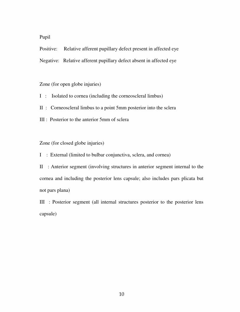
Pupil
Positive: Relative afferent pupillary defect present in affected eye
Negative: Relative afferent pupillary defect absent in affected eye
Zone (for open globe injuries)
I : Isolated to cornea (including the corneoscleral limbus)
II : Corneoscleral limbus to a point 5mm posterior into the sclera
III : Posterior to the anterior 5mm of sclera
Zone (for closed globe injuries)
I : External (limited to bulbar conjunctiva, sclera, and cornea)
II : Anterior segment (involving structures in anterior segment internal to the
cornea and including the posterior lens capsule; also includes pars plicata but
not pars plana)
III : Posterior segment (all internal structures posterior to the posterior lens
capsule)

11
CAUSES OF CHILDHOOD EYE INJURIES
Causes at various ages 16,20,21
1. Prenatal period: Contrary to the popular belief, ocular injuries can occur to a
foetus in uteri.22
Ocular injuries have been reported during amniocentesis23-28
and adnexal injuries during episiotomy, forceps-assisted delivery or with the
surgical knife during delivery with caesarean section.29-32
2. Infancy and Toddler Period: The most common ocular injury during the early
months of life is corneal scratches (abrasions) from their own fingernails,
siblings or parents.33
Another common entity we come across is injury with the
metal hook of the mother’s blouse while feeding the child resulting in injuries
and the bindi (applied on forehead by mother) getting lodged in the fornix
causing severe kerato-conjunctivitis. Alkali burns due to lime are very common
in India. Also injuries by accidental spill of raw soap nut powder, detergent
powder and liquids, hot water or soups are seen invariably resulting in
conjunctivitis and at times keratitis. Injuries associated with child abuse,
common in western literature is quite rare in our country.
3. Older Children: Young boys are more frequently injured than girls, since
they are more active in indoor and outdoor activities.34
Thorn pricks, sharp
injuries from geometry instruments, animal bites (dogs, bird beak) are common.
Firecracker injuries are also commonly seen in this age group.
Injuries Common in Our Country
Gullidanda injury 2,35
Injury from chuna packets 36
Broom stick injuries 37

12
CLINICAL MANIFESTATIONS
Mechanical - Closed Globe Injuries 39,40
A. Blunt injuries
1. Lesions of the conjunctiva
Haemorrhage, oedema, chemosis
Bruising & lacerations
2. Lesions of the Cornea
Oedematous and haemorrhagic changes
i. Epithelial opacities and erosions
ii. Blood staining of the cornea
iii. Folding of Bowman’s or Descemet’s membrane.
Pigment deposits
Corneal lacerations:
i. Partial thickness lacerations
ii. Tears in Descemet’s membrane
iii. Complete rupture of cornea
3. Lesions of the Iris & Ciliary body
Changes in pupil and accommodation
i. Traumatic miosis and accommodative spasm
ii. Traumatic iridoplegia and cycloplegia
Vascular changes
i. Reactive hyperaemia and exudation
ii. Haemorrhage
- Into the tissue of iris and ciliary body
- Traumatic hyphema

13
Lacerations of the iris and ciliary body
i. Tears at the pupillary border, sphincter tears
ii. Tears in the iris stroma
iii. Dehiscences of the pigmentary layer
iv. Iridodialysis
v. Irideremia (traumatic aniridia)
vi. Iridoschisis
vii. Traumatic cyclodialysis
viii. Retroflexion of the iris
Inflammatory and atrophic changes
i. Traumatic iridocyclitis
ii. Post traumatic atrophy of the iris and ciliary body
iii. Acute necrosis of the iris and ciliary body
iv. Pigmentary changes (traumatic heterochromia)
4. Lesions of the Lens and Zonules
Lenticular opacities
i. Vossius’s ring opacity
ii. Discrete subepithelial opacities
iii. Traumatic rosette shaped opacities
iv. Traumatic zonular cataract
v. Diffuse concussion cataract
Subluxation & dislocation of the lens
5. Lesions of the Choroid
Vascular changes
i. Choroidal hemorrhage
ii. Choroidal detachment

14
Choroidal lacerations
Traumatic choroiditis
6. Lesions of the retina
Edematous and atrophic changes
i. Concussion edema, necrosis
ii. Concussion changes at the macula
- Macular edema
- Macular cysts & holes
- Traumatic atrophy of Haab
- Acute concussion necrosis
iii. Peripheral atrophic retinal changes
Vascular changes in retina
i. Traumatic haemorrhages
ii. Embolism & thrombosis
iii. Traumatic aneurysm
Retinal tears
Traumatic retinal detachment
7. Lesions of the Optic disc
Papillitis and atrophy
Rupture and avulsion of the nerve
8. Effects on the Vitreous
Vitreous liquefaction/ opacification/ detachment/ herniation
Vitreous hemorrhages
9. Rupture of the Sclera
10. Changes in Refraction
Traumatic hypermetropia
Traumatic myopia

15
11. Changes in the Ocular tension
Traumatic instability of the tension
Traumatic glaucoma
Traumatic hypotony
B. Incised wounds
Corneal abrasions
Recurrent corneal erosions
Deep non-perforating corneal wounds
Wounds of the conjunctiva
Wounds of the sclera
C. Concussion & Contusions of the ocular adnexa
1. Contusions of the Lids
Edema & hemorrhage of the lids
2. Fractures of the orbit
3. Orbital hemorrhages
4. Orbital emphysema
5. Contusion injuries to orbital contents
Changes in the position of the eyeball
i. Luxation of the globe
ii. Traumatic enophthalmos
Concussion injuries to the orbital muscles & nerves
Traumatic aneurysm of the ophthalmic artery
Serous tendonitis
6. Injuries to the lacrimal apparatus

16
Mechanical - Open Globe Injuries 39
1. Conjunctiva - Tiny defects, localized areas of chemosis, subconjunctival
haemorrhage pigmentation, and presence of subconjunctival foreign body, any
of these can indicate a possible ocular penetration
2. Cornea - Incised wound
- Lacerations with or without loss of corneal tissue
3. Iris - Iridodialysis
- Sphincter tear
- Holes
- Foreign body hidden in iris crypts
- Prolapse through the site of perforation and
possible loss of uveal tissue.
4. Anterior chamber - Presence of small foreign body angle
5. Lens - Defect in lens capsule
- Intralenticular foreign bodies and opacities
- Localised cataract
- Subluxation
- Dislocation
6. Posterior segment - Vitreous haemorrhage
- Posterior scleral rupture
- Vitreous opacities
- Retained IOFB
- Retained tears, lacerations and detachment
- Retinal haemorrhage
- Choroidal laceration, perforation
- Expulsive choroidal haemorrhage
7. Optic nerve - Partial or complete transection by intruding object

17
Chemical Injuries 41- 49
Chemical injuries are potentially devastating ocular surface injuries that
may result in permanent visual impairment. The majority are accidental.42
Alkali burns are twice as common as acid burns since alkalis are more widely
used at home and in industry. 42
The most common involved alkalis are ammonia, sodium hydroxide and
lime. One of the most common chemical injuries is caused by chuna packets
(sodium hydroxide), which has been described in the previous section.
Ammonia being a household cleaner, is also easily accessible to children,
because it is usually stored in a cabinet beneath the sink or on a low shelf.
The commonest acids are sulphuric, acetic, chromic and hydrochloric.47
These acids can be found in various cleaning agents, rust removers, automobile
batteries and other products.
The severity of a chemical injury is related to the properties of the
chemical, the area of affected ocular surface, duration of exposure (retention of
particulate chemical on the surface of the globe) and related effects such as
thermal damage. Alkalis tend to penetrate deeper than acids, which coagulate
surface proteins, resulting in a protective barrier. Ammonia and sodium
hydroxide may produce severe damage due to rapid penetration.

18
Thermal Injuries(50-61)
Flame, flash and chemical burns account for most oculopalpebral
burns.50
Common agents are fireworks, hot liquids, head of a lighted match and
open coal fire. Fireworks remain a major source of preventable mechanical and
thermal injury in children.51
The eye is one of the most commonly involved
organs in fireworks related injuries.52,53
Fireworks are a worldwide menace, as
evidenced by reports from a wide variety of countries. 54-60
EVALUATION
Emergency room evaluation of trauma affecting lids, eye or orbit 62-65
Any life threatening injury should be treated first.
1. A quick history should be obtained : time and mode of injury; nature of
injuring object; prior treatment received; time lag between injury and treatment;
past medical and ocular history; tetanus immunization; allergies.
2. First aid should be rendered in cases of true emergency (e.g., chemical burns
of the cornea)
3. Visual acuity should be determined whenever possible. This is important for
medical and legal reasons. The method used to measure acuity must be geared
to the age and level of cooperation of the child.
4. Spectacles, if any, should be inspected (glass/ plastic)

19
5. Haemorrhages and infections of orbit, lid or conjunctiva should be noted.
Orbital rim should be palpated (discontinuity/ crepitus); facial and corneal
sensations should be checked.
6. Any real or apparent displacement of globe should be appraised: anterior,
posterior or vertical.
7. Pupillary shape, size and reactions should be recorded. Some of the steps
mentioned below might require sedation or anaesthesia
8. Partially and completely penetrating injuries of cornea and sclera should be
differentiated. Lid retractors may be used. Orbicularis muscle may be
anaesthetised if squeezing prevents atraumatic examination of the eyeball.
Uveal, vitreal, or lenticular prolapse should be noted., where possible, Siedel
test should be done in all cases of penetrating injuries.
9. Depth of all lid lacerations should be investigated, noting fat in wound.
Foreign bodies under the lid should be sought: lid should be everted & fornices
swept with cotton swab after use of topical anaesthetic.
10. Cornea should be examined for opacities, ulcers, foreign bodies, rust rings,
and abrasions, using fluorescein when required.
11. Anterior chamber should be examined for hyphema. Gonioscopy should be
done to rule out angle recession. Iridodonesis and iridodialysis should be noted.
12. Dislocation/ subluxation of lens & presence of cataract should be noted.

20
13. If traumatized globe is intact and cornea is undamaged, intraocular pressure
should be measured, at least digitally and preferably with tonometer.
14. Fundus examination using direct and indirect ophthalmoscope without
indentation: appearance of optic nerve head, macula, retinal circulation and
intraocular haemorrhage, presence of foreign bodies should be noted. Search for
retinal tears and disinsertions.
15.If diplopia is present, analysis of ocular ductions and versions; forced-
duction test under topical anaesthesia should be done.
16. X-rays should be obtained in all cases of possible retained foreign body in
globe or orbit and whenever orbital fracture is conceivable.
17. Consider value of photographing all injuries.
MANAGEMENT
Lid ecchymosis
After ruling out underlying severe globe injury, cold compresses for the initial
24-hour period, followed by warm compresses as needed.
Lid lacerations 66
Primary edge-to-edge closure in layers.
Tarsoconjunctival layer – 6-0 Vicryl
Muscle-fascial layer – 6-0 vicryl
Skin – 6-0 Silk

21
With minimal tissue loss – primary closure with lateral cantholysis
With extensive tissue loss – may require major reconstructive procedures
Canalicular lacerations – use of stents and end-to-end reapproximation of
torn canaliculi.
Traumatic ptosis 67
Surgical repair of torn Levator muscle/aponeurosis after oedema has subsided,
but before onset of fibrosis.
Blow-out fracture 68
Initial conservative management until edema subsides. Surgical repair, if
required using synthetic material such as Supramid, silicone or Teflon.
Corneal abrasions 69,70
Conservative Management: An antibiotic eye ointment with firm patch usually
suffices.
Penetrating corneal wounds
Careful corneal wound closure to achieve a water tight globe with minimal
scarring and astigmatism should be the aim.71
10-0 interrupted nylon sutures are
used. Any opaque tissue in the visual axis must be removed promptly to prevent
amblyopia in children, improve vision and prevent chronic inflammation & scar
tissue formation. Cyanoacrylate tissue adhesive can be used to close some small

22
corneal lacerations or minute leak after surgical repair.72
Severe damage to
cornea might need penetrating keratoplasty.73,74
Corneal laceration with iris incarceration
Devitalized & extremely macerated tissue and feathered or depigmented iris
should be excised. It Is safer to abscise tissue which has remained prolapsed for
more than 24 hours. After adequate iris repositioning, the corneal wound is
sutured.
Traumatic iridodialysis
A traumatic iridodialysis should be repaired when it is large enough to cause
multiplopia or glare.75,76
It is repaired by one or more mattress sutures of
double-armed 10-0 propylene sutures tied externally under a scleral flap.77
Corneal laceration with lens involvement 78
Situations in which primary lens removal is indicated:
a. A lens with disrupted capsule & flocculent cortical matter in anterior
chamber.
b. A lens & vitreous mixture.
c. A clearly cataractous lens.
Corneal laceration with vitreous involvement 79

23
The primary goal is to relieve vitreous incarceration in the wound. After closure
of wound, disrupted vitreous in the anterior chamber can be removed by
vitrectomy through limbal approach.
Corneo-scleral lacerations with or without uveal and vitreous prolapse80,81
Lacerations extending beyond the limbus and into the sclera should be explored
to determine their full extent. The wound is closed by zippering technique. It is
closed from anterior(limbal) end with interrupted sutures placed successively
proceeding posteriorly.
Hyphema 82-95
Supportive – Bed rest with patching may decrease rebleeding. 87,88
Medical – Miotics, salicylates,89-91
cycloplegics,92
antifibrinoloytics,93
fibrinolytics, estrogens and corticosteroids94
have been suggested.
Surgical
Traumatic cataract following blunt injury 96
No initial treatment is required if the capsule is intact. If capsule is ruptured,
atropine and a steroid should be used topically to control uveitis. Extra
capsular extraction is possible in most cases in older patients, but aspiration is
commonly done for patients below the age of 20 years. Implantation of
intraocular lens, as a primary or a secondary procedure, as suitable.97

24
Traumatic retinal dialysis 98,99
Surgical management can be in the form of scleral flaps, cryotherapy,
diathermy or a silicone implant, with or without encircling element. Subretinal
fluid drainage is required in most cases.
Traumatic retinal detachment 100-102
Conventional scleral buckling, with or without pars plana vitrectomy, as
suitable, has relatively good prognosis in detachments caused by ocular
contusions but poorer prognosis in those caused by penetrating injuries.
Management of chemical injuries
Emergency management
A chemical injury is the only eye injury that requires immediate treatment
without first taking a history and performing a detailed examination.
1. Copious irrigation 103-104
– crucial to minimize the duration of contact
with the chemical and to normalize the pH in the conjunctival sac as
soon as possible. Normal saline (or equivalent) 105
is used to irrigate the
eye until pH is normalized. Double eversion of lids to remove any
retained particulate.
2. Debridement – of necrotic areas of corneal epithelium should be
performed to facilitate re-epithelialisation.

25
Grading of severity
Hughes-Roper-Hall classification of chemical burns 106-108
Grade I: Clear cornea and no limbal ischemia (excellent prognosis)
Grade II: Hazy cornea but with visible iris details and less than one-third
(120 o) of limbal ischemia (good prognosis)
Grade III: Total loss of corneal epithelium, stromal haze obscuring iris
details and one-third to half (120 o – 180
o) of limbal ischemia (guarded
prognosis)
Grade IV: Opaque cornea and more than half (>180 o) of limbal ischemia
(very poor prognosis)
Other features to be noted at initial assessment are the extent of corneal
and conjunctival epithelial loss, iris changes, status of the lens and
intraocular pressure.
Medical treatment
1. Steroids – reduce inflammation and neutrophil infiltration. Should be
tailed off after 7-10 days as they impair stromal healing. 109
2. Ascorbic acid – improves wound healing by promoting collagen
synthesis.
3. Citric acid – reduces the intensity of inflammation by inhibiting
neutrophil activity.
4. Tetracyclines in the dose of 100mg OD – reduce inflammation and
ulceration.

26
Surgery 110
1. Early surgery – may be necessary to revascularise the limbus, restore the
limbal cell population and re-establish the fornices.
Advancement of Tenon capsule
Limbal stem cell transplantation 111
Amniotic membrane grafting 112
2. Late surgery -
Division of conjunctival bands and symblepharon
Conjunctival or mucous membrane grafts
Correction of eyelid deformities
Keratoplasty – should be delayed for at least 6 months
Keratoprosthesis – may be required in very severely damaged eyes

27
PROGNOSIS IN CHILDHOOD EYE INJURIES
Prognosis in eye injuries in children is debated.113
Without doubt,
anterior lacerations carry a better prognosis than posterior lacerations. But in
young children, with the risk of amblyopia, the prognosis may not be so good,
even with anterior lacerations. When anterior lacerations are combined with
cataract, the prognosis worsens.114
Several factors have been found to correlate with an unfavourable visual
outcome:115
i. Initial presenting visual acuity worse than 5/200 (1/60)
ii. Open globe injuries caused by blunt trauma
iii. Penetrating wounds involving the sclera
iv. Double penetrating (perforating) eye injuries
v. Dense vitreous haemorrhage

28
PREVENTION OF EYE INJURIES IN
CHILDREN116
Everyone in the community may play a part by showing concern for the
well-being of others. Children are often victims of trauma, not only because of
their innocence, but also because of the lack of supervision by their elders.
However, they too may play their part in making each other aware of unsafe
activities and avoid confrontational games. Adults have a larger role to play in
health promotion and accident prevention. Teachers have opportunity to
schedule health education activities in school. Parents and teachers should also
be aware of any situation, which presents opportunity for physical danger. The
use of traditional eye medicines has also resulted in varying degrees of damage
to the eyes. Not all traditional practice is harmful, but people must be made
aware of proven harmful agents.

29
REVIEW OF LITERATURE
Important studies on Epidemiology of Eye Injuries in Children
relevant to our study are summarized below in chronological order:
1. Eye injuries in children. M Niiranen and I Raivio. Br J Ophthalmol. 1981
Jun; 65(6): 436-8 117
This was a retrospective study of children's eye injuries treated during
1977 at Helsinki University Eye Hospital. There were 110 cases representing
34.5% of all eye injuries and 3% of all patients treated in 1977; 81.8% were
boys and 18.2% girls. Half of the injuries were caused by another child, one-
third were self-inflicted, and the rest were other accidents. The risk of eye injury
in girls was low and stable at all ages, but in boys the risk grew markedly at the
age of 8 years. The commonest cause of injury was a thrown missile. Other
important causes were shots, hits, and sports accidents. Two-thirds of the
injuries were concussions. The proportion of perforation was 8.9%, which is a
much lower figure than in earlier reports, suggesting that the injuries have
become milder. Some kind of complication was seen in 16% of concussions. No
secondary bleeding was found among them. Permanent impairment of vision

30
was seen in 2 cases: one had a visual acuity of 0.1 because of traumatic cataract
and the other 0.6 because of corneal scars. Although the number of perforations
was too low for statistical analysis, the final result in this group suggested that
the prognosis of perforating eye injury was still bad.
2. Penetrating ocular injuries in young patients: Initial Injuries and
Visual Results. Sternberg Paul JR et al. Retina. 4(1): 5-8, Winter/ Spring
1984 115
The records of 197 patients aged 18 years or younger who underwent
primary repair of a penetrating ocular injury at the Wilmer Ophthalmological
Institute from January 1970 through December 1981 were reviewed. The injury
was caused by sharp objects in 49% of cases, missiles in 35%, and blunt trauma
in 14%. Of 159 patients with at least 6 months follow-up, 110 (69%) achieved
final vision of 5/200 or better, and 77 patients (48%) achieved final visual
acuity of 20/50 or better. The prognosis after a penetrating injury is strongly
influenced by the nature of the injury and the extent of initial damage.
3. Epidemiological study of eye injuries in Brazilian children. C. A.
Moreira Jr, M. Debert-Ribeiro and R. Belfort Jr. Arch Ophthalmol 1988 Jun
Vol 106: 781-784 118

31
This was a year-long follow-up study of 146 eye injuries in children up
to 15 years of age. This was carried out in two emergency hospitals of a
southern Brazilian city. These injuries represented approximately 65% of the
total number of patients seeking ophthalmic care at emergency hospitals within
this city. Patients were followed up for at least four months after injury; visual
results as well as epidemiological factors were analyzed. Based on these
findings, children in the 0- to 5-year-old group were at greatest risk, regardless
of sex; among children older than 5 years, eye injuries were more frequent in
boys. Generally, the child took part in the accident as an active participant, and
adequate adult supervision decreased the number of these accidents.
4. Eye injuries in children in Israel. A nationwide collaborative study.
Rapoport I et al, Arch Ophthalmol. Vol. 108 No.3, March 1990 119
A nationwide prospective collaborative study on ocular trauma was
performed in Israel during a period of 3 years (1981 through 1983). Almost half
of the traumas (1127 [47%] of 2416 eyes) were sustained by children younger
than age 17 years, and mainly between the ages of 6 and 12 years. Most of the
injuries happened at home (38.1%) or in the street (26.8%) and during play and
sport (65.1%). The male-to-female ratio among the children was 4:1. Blunt

32
injuries accounted for 59.2%; 30.5% were perforating injuries and the rest were
chemical and radiation injuries.
5. Causes of paediatric eye injuries: A population-based study.
E. Strahlman, M. Elman, E. Daub and S. Baker. Archives of ophthalmology
Vol 108, No 4,April 1990 120
A population-based study of eye injuries requiring hospital admission for
children younger than 16 years was conducted in the state of Maryland during
the 1982 calendar year. The population-based estimate of the incidence of
ocular trauma in Maryland children was 15.2 per 100,000 per year (95%
confidence interval, 12.8 to 17.7). Male patients outnumbered female patients as
victims of eye injuries by a ratio of approximately 4:1; eye injuries in 11- to 15-
year-old children occurred at more than twice the rate than for younger children.
The most common cause of pediatric ocular trauma was accidental blows and
falls (37%). Sports and recreational activities accounted for 27% of all eye
injuries, 39% of all non-penetrating injuries, and 40% of all injuries in 11- to
15-year-old children.

33
6. Etiology of pediatric perforating eye injuries in Southern Turkey. Merih
Soylu, Nihal Demircan, Müslime Yalaz,Ismail Isigüzel. Ophthalmic
Epidemiology. Vol 5, Issue 1 March 1998,7-12 121
In this study, the records of 242 children, aged 1-14 years, admitted with
perforating ocular injury were reviewed retrospectively over a 5-year period.
There were 175 boys and 67 girls in the study group. The patients were divided
into 3 groups according to their ages. Perforating injuries occurred most
frequently in the street in all groups. The second most common place of the
injury was at home in the 1-9 year-olds and in the fields in the 10-14 year-olds.
The cause of the perforation was a metallic substance in 32.6%, wood in 15.3%,
stone in 12.0%, glass in 12.3%, pellets in 12%, and injection needles in 8.3%.
Most of the perforations occurred during unsupervised play, while all
perforations with glass occurred during traffic accidents. Surgery was
performed in 234 patients, while 8 patients in whom spontaneous closure had
already occurred during admission received only medical treatment. In 28.9%
the visual acuity was undetermined, in 25.7% the visual acuity was 0.1 or more,
in 22.7% the visual acuity was between 0.06 and light perception, and in 22.7%
there was no light perception on final evaluation.
7. Childhood eye injuries in North Jordan. Muawyah D. Al-Bdour and
Mohammed A. Azab. International Ophthalmology. Vol 22, No 5, Sep 1998 122

34
In this study, records were reviewed of 116 children who sustained
serious eye injuries that required admission to Princess Basma Teaching
Hospital between October 1995 and November 1998. The material was
analyzed retrospectively with respect to various epidemiological features.
71.5% of the injured children were male and 28.5% were female. There
was a marked preponderance of injuries in the age group 6–10 years. The
majority of injuries occurred during play and sport (74.1%). Stones and sharp
objects were the most common causes accounting for 18.1% and 17.2%
respectively. Most of the sharp objects were household instruments. Perforating
injuries were more common than non-perforating injuries. 56% of injured eyes
had a low vision with visual acuity between 6/24 and 3/60, and 13% had a
blinding outcome with visual acuity less than 3/60.
8. Eye injuries in children: The current picture. Caroline J MacEwen,
Paul S Baines, Parul Desai. Br J Ophthalmol August 1999; 83:933-936123
This was a prospective observational study of all children admitted to
hospital with ocular trauma in Scotland over a 1 year period. The commonest
mechanism of injury was blunt trauma, accounting for 65% of the total. 60% of
the patients were admitted with a hyphema. Injuries necessitating admission
occurred most frequently
at home (51%). Sporting activities were the

35
commonest cause of injury in the 5-14 age group. There were no injuries caused
by road traffic accidents or fireworks. Patients were admitted to
hospital for a
mean of 4.2 days (range 1-25 days). One (1%) child had an acuity in the
"visually impaired" range (6/18-6/60) and one (1%) was "blind" (6/60) in the
affected eye. No child was bilaterally blinded by injury and none required blind
or partial sight
registration.
9. Loss of visual acuity due to eye injuries among 6292 school
children in the Sultanate of Oman. Joan Lithander et al. Acta Ophthalmol.
Scand. 1999: 77: 697–699 124
In 1992–94 a nation-wide survey in primary schools in the Sultanate of
Oman for ocular disorders was conducted. A random selection of 6292 children
from Grades 1 and 6 from all primary schools in the country provided the
research sample. Children who failed the visual acuity screening test received a
complete ‘‘on the spot’’ eye examination by the pediatric ophthalmologist. 12
children were found to have monocular low vision (VA °0.3 to amaurosis)
caused by injury. Total prevalence for loss of vision in one eye was 0.19%, with
0.15% in 6-year-olds and 0.25% in 12-year-olds. Traumatic cataracts were
noted in 4 children, 3 of these were in need of surgery. One child had aphakia

36
after trauma surgery and needed a secondary lens implant. Altogether the
prevalence of traumatic monocular visual damage in this study was 0.19%.
10. The aetiology of perforating ocular injuries in children. C G
Thompson, N Kumar, F A Billson and F Martin. Br J Ophthalmol 2002;86:920-
922 125
All cases of perforating ocular injury presenting to a single paediatric
hospital (age less than 16 years) over a 17 year period were identified by a
medical record search A total of 72 cases were identified. The commonest
causes of perforating ocular injury were sharp tools (knives/scissors)
poked by
the child into his/her own eye (17%), or objects thrown at the child (17%).
Injuries were most likely to have occurred at home (58%). The age range for
injuries was 8 months to 14 years 8 months. Perforating ocular injury was most
frequent in the 3–6 year group (32%) followed by the 6–9
year group (25%).
Males were more frequently involved than females . There was no correlation
between the laterality of the eye, the time of day of the occurrence, or the day of
the week of the occurrence. The final acuity achieved was better or equal to
6/12 in 36% and less than 6/60 in 31%. Injuries occurred more frequently on
weekends than on weekdays. There were 6 enucleations (8%). Follow up was
for an average period of 25 months.

37
11. Mechanical eye (globe) injuries in children. Andrew Vasnaik et al.
Journal of Pediatric Ophthalmology and Strabismus. 2002; 39:5-10 126
In this study, the in-patient records of children who were admitted with
mechanical eye (globe) injuries were reviewed. Of the 68 children, the mode of
injury was child related in 12(17.7%) patients, agent related in 40(58.8%)
patients & environment related in 22(23.5%) patients. Mild injuries were seen
in 22(32.4%) patients, moderate in 31 (45.6%) and 15(22.1%) had severe
injuries. None of the patients with host-related injuries had a severe injury.
6(66.67%) patients with host-related injuries had a good visual outcome and
none had a poor outcome.
12. Severe ocular injuries in Greek children. Ephigenia K. Mela;
Constantinos D. Georgakopoulos; Athanasios Georgalis; John X. Koliopoulos;
Sotirios P. Gartaganis Ophthalmic Epidemiology, Volume 10, Issue 1
February 2003, 23 - 29 127
This was a retrospective analysis of 95 cases (103 eyes) of eye injuries in
children younger than 17 years of age admitted to the Department of
Ophthalmology, University Hospital of Patras, Greece, during a five-year
period. The data were analyzed with respect to age, sex, type, cause and mode

38
of injury, method of management, duration of hospitalization and final visual
deficit. The average age was 9.8 years and males were involved 80% of the
cases. The most common type of eye injury was mechanical closed-globe injury
(71.8%). Mechanical open-globe injuries were found in 21.3% of the eyes,
while burns comprised 6.7% of the injuries. Most injuries were agent-related,
with blows and falls being responsible most often. Multiple operations were
part of the treatment in 11.6% of the eyes; 14.5% of the eyes were blinded and
15.5% had significant final visual acuity loss.
13. Epidemiology of Childhood Ocular Trauma in a Northeastern
Colombian Region. Juan C Serrano, Patricia Chalela, Juan D Arias. Arch
Ophthalmol 2003; 121:1439-45. 128
In this study, the medical records of children 15 years and younger, who
underwent evaluation in the emergency department of a tertiary referral center
in north-eastern Colombia, during a 5-year period, were reviewed. Records of
393 children with 415 incidents of eye injury were included in the study, of
whom 22 were initially treated for bilateral ocular trauma. In this study most
patients (64.9%) were boys. The highest proportion of injuries (44.4%) occurred
at home, followed by streets and roads (28.6%). Blunt (35.1%) and sharp
(22.6%) objects represented the most frequent causes of trauma. Closed-globe

39
injuries were far more frequent than open-globe injuries for boys (82.4% vs.
17.6%) and girls (83.8% vs. 16.2%). Of those with closed-globe injuries, 253
injuries (80.0%) registered an initial visual acuity of greater than 20/60, whereas
31 open-globe injuries (52.5%) registered an initial visual acuity of less than
20/400. Most closed-globe injuries (223 [92.1%]) did not cause any final
visual
impairment in the affected eye, whereas 26 open-globe injuries (55.3%) caused
severe visual impairment or blindness.
14. Ocular injuries in children aged 0-15 years: epidemiology and
clinical aspects at the Bangui National Teaching Hospital. Yaya G et
al. J Fr Ophthal. 2005 Sep;28(7):708-12 129
A prospective study was conducted on 194 cases in the ophthalmology
department over a period of 3 years, and included children aged 0-15 years. A
total of 197 eyes were examined by the same practitioner, comprising 191
unilateral ocular injuries and three bilateral injuries. Of the children examined,
59% were males and 41% were females, with a sex ratio of 1.3. The age group
with the highest exposure (39.3%) was between 5 and 10 years. Punishments
(25.9%), accidents during games (19.3%) and fights (18.8%) were the main
sources of these ocular injuries. Consultation most often occurred long after the
incident. Only 2.0% were seen before the 6th hour and 43.7% between 48 hours
and 1 week. The clinical picture was dominated by bruises posing a therapeutic

40
problem: 25 hyphemas (12.7%), 19 conjunctival injuries (9.6%), 19 lens
dislocations with or without vitreous loss (9.6%), 18 trauma-induced cataracts
(9.4%), and 15 eye lid injuries with or without lachrymal duct ruptures (7.6%).
The most serious injuries were cornea injuries with or without hernia of the iris
(19.8%) and nine globe dislocations (4.5%)
15. Perforating ocular injuries in children: a retrospective study of 57
cases. Beby F, Kodjikian L, Roche O, Donate D, Kouassi N, Burillon C, Denis
P. J Fr Ophtalmol. 2006 Jan; 29(1): 20-3 130
The hospital records of 57 patients under 14 years of age who were
treated for open globe injuries at Edouard Herriot Hospital, Lyon, France,
between January 1999 and December 2003 were reviewed. In total they
reviewed 57 patients: 41 males and 16 females. The mean age at admission was
6.8 +/- 3.5 years. The injury involved the right eye in 27 cases and the left eye
in 30 cases. Sharp or pointed objects accounted for the majority of injuries. The
most common location for a perforating ocular injury to occur was at home.
Wounds involved the cornea in 41 cases. There was iris hernia in 21 cases,
hyphema in 15 cases, vitreous prolapse in 14 cases, lens damage in 12 cases,
and shallow anterior chamber in 11 cases. The most frequent complication was
traumatic cataract. Secondary lens removal was performed in 15 cases. Visual

41
acuity was 0.5 or better in 27 of the 57 eyes, with a mean follow-up period of
12 months.
16. Pediatric eye injury-related hospitalizations in the United States.
Brophy M, Sinclair SA, Hostetler SG, Xiang H. Pediatrics. 2006 Jun; 117(6):
e1263-71 6
This study aimed to study the demographic, medical care, and financial
characteristics associated with major categories of pediatric eye injury.
Cross-sectional data were derived from pediatric inpatient Database of the
Healthcare Cost and Utilization Project for the year 2000.
These records represented an estimated 7527 eye injury-related
hospitalizations among children aged 20 years or less in the United States
during the year 2000. Inpatient charges for the treatment of these injuries were
more than $88 million. The rate of hospitalization for pediatric eye injuries in
the United States in 2000 was 8.9 per 100,000 persons aged 20 years or less.
Young adults aged 18 to 20 years accounted for the highest percentage of
hospitalizations (23.7%). Males accounted for 69.7% of hospitalizations. A
majority of hospitalizations were for open wounds of the ocular adnexa. Motor
vehicle crash was the most common cause of injury, followed by being struck

42
by or against an object and being cut or pierced.
These findings illustrated the need for eye injury prevention efforts, like
educating parents and children about the potential for eye injuries at home and
in dangerous situations.
17. Penetrating eye injuries in South African children: aetiology and
visual outcome. Grieshaber MC, Stegmann R. Eye 2006, Jul;20(7):789-95 131
In this study,100 consecutive patients, aged 16 years and under, with
penetrating ocular injuries undergoing surgery were prospectively evaluated.
Most children (66%) were injured during play. In all, 55% of penetrating eye
injuries occurred at home, and all injuries to children under the age of 6 years
occurred there. Most injuries occurred in the absence of a caregiver (85%).
Sticks, wire, and glass caused half of all injuries (48%). The most common
mechanism of injury was impact with a sharp object (46%). Only 25% of
injured presented to the hospital within 24 h of injury; the more severe the
sustained injury and the younger the patient, the earlier was attendance at the
clinic. Most patients (71%) regained best-corrected visual acuity (Snellen
equivalent) of 20/200 or better, and 51% regained 20/40 or better. Patient age
and delay of presentation were not of prognostic value.

43
18. Severe Ocular Injury Resulting from Chuna Packets. Tushar
Agarwal, Rasik B. Vajpayee, Namrata Sharma, Radhika Tandon.
Ophthalmology 2006; 113:960–961 36
This was a retrospective study of 21 patients (25 eyes) who experienced
ocular burns as a result of bursting of chuna packets. The average age at time of
injury was found to be 8.4-5.5 years. The median visual acuity at presentation
was light perception with projection. The ocular burns were grade 4 in 23 eyes.
Eight of 25 eyes were treated medically, and the rest underwent 1 or more
surgeries in the form of symblepharon release (n=6), amniotic membrane
grafting (n =3), allograft or autograft stem cell transplantation (n =6), and large
diameter lamellar keratoplasty (n =6). At the final follow-up (mean 637-592
days), median visual acuity was 1/60.

44
AIMS AND OBJECTIVES
Following were the aims and objectives of this prospective study
conducted at Aravind Eye Hospital, Madurai between 1st June 2010 and 30
th
November 2010
1. To determine the risk factors associated with eye injuries in children.
2. To study the different agents involved.
3. To analyze the visual outcome following eye injuries in children.
4. To study the effects of prompt and delayed treatment of these cases
5. To study the effect of educational and social status of the parents on
incidence and outcome of eye injuries in children.

45
MATERIALS AND METHODS
STUDY DESIGN:
Ours was a 6 month prospective study of all children with injury
to the eye, presenting at Aravind Eye Hospital, Madurai between
1st June 2010 and 30
th November 2010.
INCLUSION CRITERIA:
1. Patients 0 – 15 yrs of age
2. Patients with definite history of trauma to the eye
3. Minimum follow up of 2 weeks
EXCLUSION CRITERIA:
1. Adult patients
2. Doubtful history of trauma
3. Unlikely to follow up (at least for one visit)
4. Earlier trauma in the same eye
STUDY PLAN:
For the purpose of this study children 15yrs of age or less were
considered as “pediatric age group”.
A detailed history focusing on circumstances of the trauma was obtained
from parents or caretakers of each child according to a standardized
form; this was followed by a detailed ophthalmic examination.

46
All patients were followed up till 30th
November 2010 or the most recent
follow up before that, follow up had to be of at least 2 weeks.
The type of injury was recorded according to the Birmingham Eye
Trauma Terminology (BETT) 18
which has been described in detail in the
section on “Classification of ocular trauma,” of this study.
Visual acuity with pinhole at the time of presentation was recorded
whenever possible with reference to patient’s age and co-operation
during the examination.
Final visual acuity was defined as the most recently recorded, best
corrected visual acuity (Snellen equivalent) of patients either discharged
from follow up, or the most recent follow up as on 30th November 2010.
Children were classified in three age groups:
Pre school age 0 – 5yrs; Primary school age 6 – 10yrs and
secondary school age, 11 – 15yrs.
Particular attention was paid to the history to determine the cause,
mechanism, locale of occurrence, presence and level of attention of an
adult at the time of trauma, and delay in seeking medical help. We
further investigated whether these factors were associated with a
favourable or unfavourable outcome.
During examination, the patients found to be requiring surgical
intervention, were immediately taken up for wound exploration or repair

47
under general anaesthesia, while medical therapy was instituted to the
rest of the patients. Patients with severe injuries, requiring close
monitoring were kept admitted.
Patients suspected to have endophthalmitis, and patients, in whom
posterior segment could not be evaluated due to hazy view, were
examined by vitreo-retina consultants and B-scan ultrasonography was
done whenever required. These cases were managed appropriately as per
the standard of case.

48
OBSERVATION AND RESULTS (Values in parenthesis are percentages)
Table 1. Age group distribution
Age Group Gender
Total Male Female
0 – 5 Yrs 10 (10.5) 9 (9.5) 19 (20.0)
6 – 10 Yrs 42 (44.2) 9 (9.5) 51 (53.7)
11 – 15 Yrs 23 (24.2) 2 (2.1) 25 (26.3)
Total 75 (78.9) 20 (21.1) 95 (100.0)
The minimum age was 3 years and the maximum was 15 years. The mean age
of injured patient was 8.5 years. 20% were in the age group 0-5 yrs, 53.7% were in
the age group 6-10 yrs and 26.3% in 11-15 yrs. 78.9% were boys and 21.1% of the
patients were girls.
Table 2. Laterality
Age Group Laterality
Total RE LE BE
0 – 5 Yrs 7 (7.4) 10 (10.5) 2 (2.1) 19 (20.0)
6 – 10 Yrs 30 (31.6) 18 (18.9) 3 (3.2) 51 (53.7)
11 – 15 Yrs 15 (15.8) 9 (9.5) 1 (1.1) 25 (26.3)
Total 52 (54.7) 37 (38.9) 6 (6.3) 95 (100.0)
Majority of the patients had unilateral injury except 6 who had bilateral injury.
Injury was slightly more common in right eye than left eye.

49
Table 3. Domicile
Domicile
Age Group
Total 0 – 5 Yrs 6 – 10 Yrs 11 – 15 Yrs
M F M F M F
Urban 1 2 4 - 3 1 11 (11.6)
Semi
Urban 2 3 17 3 9 1 35 (36.8)
Rural 7 4 21 6 11 - 49 (51.6)
Total 10 9 42 9 23 2 95 (100.0)
Half of our patients (51.6%) came from rural areas.
Table 4. Mode of Ocular Injury
Mode of
Injury
Age Group
Total 0 – 5 Yrs 6 – 10 Yrs 11 – 15 Yrs
M F M F M F
Mechanica
l 7 6 35 8 19 2 77 (81.1)
Chemical - 2 1 - - - 3 (3.1)
Thermal 3 1 6 1 4 - 15 (15.8)
Total 10 9 42 9 23 2 95 (100.0)
Majority of the injuries were mechanical(81.1%).
All 15 cases of thermal injuries were due to firecracker related injuries.

50
Table 5. Ocular Injury
Ocular Injury
Age Group
Total 0 – 5 Yrs 6 – 10 Yrs 11 – 15 Yrs
M F M F M F
Contusion 4 3 15 3 7 1 33 (34.7)
Non Penetrating
laceration 2 1 7 4 2 - 16 (16.8)
Penetrating Injuries 1 2 10 2 8 - 23 (24.2)
Intraocular Foreign
body - - 2 - - 1 3 (3.1)
Scleral perforation - - - - 1 - 1 (1.1)
Chemical burns - 2 - - - - 2 (2.1)
Rupture globe - - - - 1 - 1 (1.1)
Others 3 1 8 - 4 - 16 (16.8)
Total 10 9 42 9 23 2 95 (100.0)
The above table shows details of the number of open and closed globe injurieswith
subclassification

51
Table 6. Object of Injury
Object of Injury
Age Group
Total 0 – 5 Yrs 6 – 10 Yrs 11 – 15 Yrs
M F M F M F
Stone /Glass 1 1 5 2 3 - 12 (12.6)
Metal piece or
Fragment - - 1 - 1 - 2 (2.1)
Scissors - - - 1 1 - 2 (2.1)
Knife - - - - 1 - 1 (1.1)
Thorn 2 1 1 - 1 - 4 (4.2)
Wooden splinter - - 1 - 1 - 2 (2.1)
Firecrackers 4 1 6 1 3 - 15 (15.8)
Ball - - 4 1 - - 5 (5.3)
Chemical lime - 2 - - - - 2 (2.1)
Others 3 5 24 4 12 2 50 (52.6)
Total 10 9 42 9 23 2 95 (100.0)
Firecrackers and Stone /Glass were the most common object of injury.
List of other objects of injury
Object Frequency
Accident/Fall 8
Bangle piece/hair clip 2
Bow & arrow 4
Cow horn/tail 3
Cricket bat/hockey stick 2
Door handle 2
Gilli danda 5
Iron rod/wire 12
Pen/pencil 10
Needle 2
Total 50

52
Table 7. Associated Extraocular Injury
Type of Injury Total Extraocular
Injury Percent
Mechanical 77 15 19.5
Chemical 3 2 66.7
Thermal 15 9 60.0
Total 95 26 27.4
In total, about one-fourth of the patients had associated extraocular injury.
Among patients with thermal injuries, approximately half the patients had associated
extraocular injury.
Table 8. Use of Spectacles at the time of Injury
Wearing Frequency Percentage
Worn - -
Not Worn 95 100.0
Total 95 100.0
None of the patients were using glasses at the time of injury.

53
Table 9. Child Participation
Child
participati
on
Age Group
Total 0 – 5 Yrs 6 – 10 Yrs 11 – 15 Yrs
M F M F M F
Active 4 5 21 2 13 - 45 (47.4)
Passive 6 4 21 7 10 2 50 (52.6)
Total 10 9 42 9 23 2 95 (100.0)
This table shows the number of cases where the child actively sustained injury or got
injury as a passive or innocent bystander.
Table 10. Presence of an adult
Adult Age Group
Total 0 – 5 Yrs 6 – 10 Yrs 11 – 15 Yrs
Present, alert - 1 - 1 (1.0)
Present, not
alert 7 11 6 24 (25.3)
Absent 12 39 19 70 (73.7)
Total 19 51 25 95 (100.0)
The great majority of injuries occurred in the absence of a caregiver.
Only in 1% injuries, the child was being supervised by an adult at the time of
injury.

54
Table 11. Place of Injury
Place of
Injury
Age Group
Total 0 – 5 Yrs 6 – 10 Yrs 11 – 15 Yrs
M F M F M F
Home 8 4 24 4 14 2 56 (58.9)
School - 4 14 4 6 - 28 (29.5)
Work place - - - - 1 - 1 (1.0)
Public places 2 - 2 - 1 - 5 (5.3)
Others - 1 2 1 1 - 5 (5.3)
Total 10 9 42 9 23 2 95 (100.0)
Injuries at home were most commonly observed.
Table 12. Occupation of parent
Occupation of parent Number Percentage
Industrial Worker 4 4.2
Agricultural Worker 36 37.9
Professional 1 1.1
Business man 10 10.5
Others 44 46.3
Total 95 100.0
Most of the parents of the injured children were from agricultural background.

55
Table 13. Educational status of parent
Educational level Number Percentage
Illiterate 15 15.8
School 70 73.7
College 10 10.5
Total 95 100.0
More than half of the parents were educated up to school level.
Table 14. Educational level of parent and previous consultation
Previous
consultation
Educational level of parent
Total Illiterate School College
Ophthalmolog
ist 2 32 5 39 (65.0)
Physician 4 8 3 15 (25.0)
Quacks - 3 - 3 (5.0)
Medical Shop 2 1 - 3 (5.0)
Total 8 44 8 60 (100.0)
None of the patients whose parents were educated up to college level received
treatment from quacks or any over the counter medications.

56
Table 15. Educational level of parent and Delay in presentation
Delay Educational level of parent
Total Illiterate School College
<6 hours 3 7 3 13 (13.7)
>6 hrs but same
day 1 - - 1 (1.1)
1 Day 4 18 4 26 (27.4)
2-4 Days 4 28 2 34 (35.8)
5-7 Days 1 9 - 10 (10.5)
8 Days-1 Month 1 7 1 9 (9.5)
>1 Month 1 1 - 2 (2.1)
Total 15 70 10 95 (100.0)
30% of the patients whose parents were educated to college level were brought
to the hospital within 6 hours of injury, which was higher when compared to the other
two groups.
Table 16. Visual acuity at Presentation
Visual
Acuity
(with
pinhole)
Age
Total
0 – 5 Yrs 6 – 10 Yrs 11 – 15 Yrs
M F M F M F
Vision
>6/18 3 5 22 5 6 1 42 (44.2)
6/18 to 3/60 2 1 9 3 7 - 22 (23.2)
Vision <3/60 5 2 11 1 9 1 29 (30.5)
NOPL - 1 - - 1 - 2 (2.1)
Total 10 9 42 9 23 2 95 (100.0)

57
Table 17. Month wise distribution
MONTH Type of Injury
Total Mechanical Chemical Thermal
JUNE 10 1 - 11 (11.6)
JULY 12 1 - 13 (13.7)
AUGUST 16 - 2 18 (18.9)
SEPTEMBER 19 - - 19 (20.0)
OCTOBER 7 - 4 11 (11.6)
NOVEMBER 13 1 9 23 (24.2)
TOTAL 77 3 15 95
(100.0)
Peaks were seen in the months of September and November.
Majority of thermal injuries secondary to firecrackers were seen in November
which is the festival season.

58
Table 18. Details of treatment at first visit
a) According to age group
Age Group
Total 0 – 5 Yrs 6 – 10 Yrs 11- 15 Yrs
Medical 13 40 16 69 (72.6)
Surgical 6 11 9 26 (27.4)
Total 19 51 25 95 (100.0)
Age Group
Total 0 – 5 Yrs 6 – 10 Yrs 11- 15 Yrs
In Patient 6 16 11 33 (34.7)
Out Patient 13 35 14 62 (65.3)
Total 19 51 25 95 (100.0)
b) According to type of injury
Type of Injury
Total Mechanic
al Chemical Thermal
Medical 53 2 14 69 (72.6)
Surgical 24 1 1 26 (27.4)
Total 77 3 15 95 (100.0)
Type of Injury
Total Mechanic
al Chemical Thermal
In Patient 31 1 1 33 (34.7)
Out Patient 46 2 14 62 (65.3)
Total 77 3 15 95 (100.0)
34.7% required admission, while the remaining 65.3 % were treated on outpatient
basis.

59
Table 19. Number of follow-ups for each patient
No. of follow-
ups
Age Group
Total 0 – 5 Yrs 6 – 10 Yrs
11 – 15
Yrs
1 10 15 6 31 (32.6)
2-3 5 27 15 47 (49.5)
4-5 4 9 4 17 (17.9)
Total 19 51 25 95 (100.0)
Majority of the patients came for 2-3 follow ups.
Table 20. Final Visual Outcome of affected eye
Outcome of
affected eye
Age Group
Total 0 – 5 Yrs 6 – 10 Yrs 11 – 15 Yrs
M F M F M F
Vision >6/18 with
correction 5 8 35 8 18 2 76 (80.0)
Visual impairment
(6/18 – 3/60) - - 3 1 3 - 7 (7.4)
Blindness (<3/60) 1 - 2 - - - 3 (3.1)
Loss of eye 4 1 2 - 2 - 9 (9.5)
Total 10 9 42 9 23 2 95 (100.0)
80% patients regained a visual acuity of >6/18 with correction.

60
Table 21. Final visual acuity in relation to vision at presentation, delay in
presentation & type of injury
Final Visual
Outcome >6/18
6/18
–
3/60
<3/60
Loss
of
eye
Total p-
value
Vision at presentation
>6/18 42 - - - 42
<0.001 6/18 – 3/60 19 2 1 - 22
<3/60 15 5 2 7 29
NOPL - - - 2 2
Delay in presentation
<6 hrs 10 - 1 2 13
0.130
>6 hrs but
same day 1 - - - 1
1 day 23 - 1 2 26
2 – 4 Days 26 7 - 1 34
5 – 7 Days 8 - 1 1 10
8days – 1
month 6 - - 3 9
>1 month 2 - - - 2
Type of Injury
Mechanical 60 7 2 8 77
0.790 Chemical 3 - - - 3
Thermal 13 - 1 1 15
TOTAL 76
(80.0)
7
(7.4)
3
(3.1)
9
(9.5) 95(100.0)

61
Table 22. Final visual acuity in relation to presence of an adult and educational
level of parent
Final Visual
Outcome >6/18
6/18
–
3/60
<3/60
Loss
of
eye
Total p-
value
Presence of an adult
Absent 57 7 2 4 70
0.305 Present, not
alert 18 - 1 5 24
Present & alert 1 - - - 1
Educational level of parent
Illiterate 13 1 1 - 15
0.272 School 57 5 2 6 70
College 6 1 - 3 10
TOTAL 76
(80.0)
7
(7.4)
3
(3.1)
9
(9.5) 95(100.0)
Presence of adult (p-0.305) and education level of parent (p-0.272) did not reach
statistical significance in influencing final visual outcome.

62
Initial Visual Acuity * Final Visual Acuity Crosstabulation
Count
2 2
6 1 1 1 2 11
1 1 1 1 2 2 8
1 1 1 1 4
1 1
1 1 2
1 2 3
1 1 2
1 1 2
1 1 2
1 1 2
1 1 4 6
1 2 3
4 1 5
1 3 4
1 10 11
27 27
9 1 2 1 1 3 2 9 13 54 95
Initial Visual
Acuity
NOPL
PL
HM
FCF
0.5/60
1/60
2/60
3/60
4/60
5/60
6/60
6/36
6/24
6/18
6/12
6/9
6/6
Total
NOPL PL 1/60 6/60 6/36 6/24 6/18 6/12 6/9 6/6
Final Visual Acuity
Total

63
DISCUSSION
Our study had a total of 95 patients, out of which, 78.9% (75) were
boys and 21.1% (20) were girls. The minimum age was 3 years and the
maximum was 15 years. The mean age of injured patients was 8.5 years.
20% were in the age group 0-5 years (53% boys, 47% girls), 53.7% in 6-10
years (82% boys, 18% girls), 26.3% in 11-15 years (92%% boys, 8% girls).
Authors No. of cases Male (%) Female (%)
Our study 95 79 21.1
Niiranen & Raivio (Finland) 117
110 82 18
Rapoport et al (Israel) 119
242 72 28
Al-Bdour et al (North Jordan) 121
116 72 28
MacEwen et al (Scotland) 123
415 70 30
Thompson et al (Australia) 125
72 67 33
Vasnaik et al (Bangalore) 126
68 62 38
Mela et al (Greece) 127
103 80 20
Serrano et al (Colombia) 128
393 65 35
Yaga G et al (France) 129
194 59 41
Grieshaber et al (S Africa) 131
100 70 30
As can be seen from the above comparison, male preponderance is a
common feature of eye injuries in children. Thus, our findings are
consistent with that of all other similar studies, as is the marked increase of

64
the male-female ratio in children older than age of 5 yrs. A possible
explanation for this fact is the greater liberty and stimulus to aggressiveness
given to boys in almost all societies.
Most of the children in our study were older than four years. School-
aged children, in particular, are most often exposed to the environment and
tend to be more physically active. As well, they often take higher risks to
gain acceptance from their peers. This is reflected in the types of games
they play and in how they react to conflictive situations.
The right eye was little more commonly affected (54.7%) then the left
eye (38.9%). This was similar to the Australian study by Thompsom et al125
were right eye was involved in 54% of the cases and in the study done by
Mac Ewen et al in Scotland123
the right eye was involved in 52% of cases.
In our study there were 6 cases with bilateral injury (6.3%).
Half of our patients (51.6%) came from rural areas, but this could be
because of our geographical location. None of these studies so far on eye
injuries in children have concentrated on this aspect.
81.1% of the injuries were mechanical, 3.1% chemical and 15.8%
thermal. Associated extraocular injuries were found in overall 27.4%
injuries. 60% of thermal injuries had associated extraocular injuries.
All the 15 cases of thermal injury were due to fire crackers and the
peak was seen during the festival season in November. Most other studies
found slightly lesser incidence of burn injuries. The Israeli study done by

65
Rapoport et al119
found it to be 10% while the study done by Strahlman et al
in Maryland (Wilmer study ) 120
found it to be 9%. Only in the Brazilian
study by Moreira et al118
burns constituted 20% of the injuries.
The study at Maryland done by Strahlman et al120
(78%) and
Brazilian study by Moreira et al118
(70%) had a lesser incidence of
mechanical injuries, whereas Rapoport et al (Israel) 119
(89%) and Mac
Ewen et al (Scotland) 123
(93%) reported higher incidence of mechanical
injuries.
Pointed objects like sticks, thorns, knifes,scissors being poked into
the eye or thrown at the child accounted for majority of the mechanical
injuries. Injuries from sticks occurred frequently when children played
games like ‘gilli danda’ or while playing ‘war’ games. Thorns, stones and
pens were not found to be causative agents for injuries in children under the
age of 5 yrs. These finding were similar to the South African study by
Grieshaber et al131
and the Australian study by Thompsom et al125
. None of
the childrens in this study were wearing glasses at the time of injuries. The
child actively participated in 47.4% of injuries (45 cases), while in the
remaining 52.6% the child was a passive onlooker.
The great majority of injuries occurred in the absence of caregiver. In
73.7% of the cases, the child was alone at the time of injury, while in 25.3%
of cases, an adult was present but not alert. Only in 1% (1 cases), the child
was being supervised by an adult at the time of injury. In the Brazilian study

66
by Moreira et al 118
, 57% of injuries occurred in absence of an adult, in 34%
cases adult was present but not alert, while in only 9% cases, an adult was
supervising over the child at the time of injury. Similar observations were
reported by Grieshaber et al 131
were 85% of the injuries occurred in the
absence of a caregiver and by Moreira et al 118
were 53% of the injuries
occurred when the child was alone.
In terms of place, the home (58.9%) was the most common place for
eye injuries to occur. In the age group 0-5 years, almost 63% of the injuries
occurred at home. Among older children 64% injuries occurred at home,
around 30% at school and the rest in other places like street/public places.
These findings were similar to all studies conducted till now. Macewen et
al123
reported 51%, Thompson et al125
58% , Mela et al127
45%, and
Grieshaber et al131
reported 55% as the proportion of injuries occurring at
home.
We also made a note of the educational level, income level, and
occupation of the parent. 16% of the parents were illiterate, 74% were
educated upto school level & 11% upto college level or beyond. 38% of the
parents were agricultural workers, 11% were involved in business, and 46%
were employed in other occupation, majority of them being laborers
working on daily wage basis.
The time interval between injury and presentation to our institution is
shown in table 15. Only 13.7% patients were brought to our institution

67
within 6 hours of injury, one case came after 6 hours, but within the same
day.
Majority of the patients (35.8%) presented after 2-4 days of injury,
another 27.4% presented 1 day after injury, another 10.5% after 5-7 days,
and 9.5% after 1 week. 2 cases presented more than 1 month after injury.
There was a trend towards early attendance, within 48 hours, the more
severe the injury, & the younger the child.
30% of the patients whose parents were educated upto college level
or beyond, were brought to the hospital within 6 hours of injury, which was
only 10% in children of school-level educated & 20% in illiterate parents.
Reasons cited for delay in presentation were distance to the hospital ,
financial problem, negligence, delayed referral, no symptoms. Similar
observations were made in studies done by Serrano et all 123
& Grieshaber et
al 131
.
Observing the elapsed time between injury and medical care, it is
apparent that in the lower socio-economic strata (low education & income
levels), the period is much longer than that in higher socio-economic strata,
regardless of whether the injury was mild or severe.
Overall 60 (63%) patients had received some form of treatment prior
to presenting to our institution. 65% of these had been treated by an
ophthalmologist, 25% by a general physician, 5% consulted quacks and 5%
used over the counter drugs, without consultation. 63% of patients whose

68
parents were educated till college level or more had received prior treatment
from ophthalmologists and the remaining 37% from general practitioners.
None of the patients in this category received treatment from quacks or over
the counter drugs.
The maximum number of eye injuries occurred in the month of
November. Majority of these were firecracker injuries. This coincided with
the festival season where children had more access to fireworks. Similar
observations have been reported in other Indian studies.
At the time of presentation, 44.2% cases had vision >6/18 with
pinhole, 23.2% had 6/18 – 3/60, 30.5% had <3/60, 2 cases (2.1%) had no
perception of light. 26 patients (27.4%) required surgical intervention at
presentation while the remaining 69(72.6%) were treated medically. In the
study done by Macewen et al123
, 48% patients required surgical treatment
while the rest of the patients were managed medically. In the study by
Serrano et al128
, 23% required surgical intervention. 33 patients (34.7%)
required admission, while the remaining 65.3% were treated on outpatient
basis.
32.6% of patients came back for only 1 follow-up, 49.5% came for 2-
3 follow-ups, 17.9% for 4-5 follow-ups. For the present study, final visual
acuity was classified as vision > 6/18 with correction, “visual impairment”
(vision 6/18 – 3/60), “blindness” (vision <3/60), “loss of eye” (phthisis).

69
The final visual outcome was overall good. 80% patients regained a
visual acuity of > 6/18 with correction, 7% were rendered visually impaired,
3% were blinded, 10% of the eyes went for phthisis bulbi. The final visual
outcome in studies conducted in other countries were slightly better than
ours, pointing towards the need for better trauma management in our
country. We compared the final visual acuity with 5 variables to
determine the influence of each on the final visual outcome.
As can be expected, the relation between visual acuity at presentation
and final visual acuity was statistically significant (p<0.001). The time
elapsed between injury and consultation also was an important factor in
determining final visual outcome. Later the presentation, worse was the
prognosis ,more so in severe injuries, and injuries complicated by the use of
native medication use & sepsis. Similar findings were reported by
Grieshaber et al131
.
Other variables like type of injury (p-0.790), presence of adult (p-
0.305), education level of parent (p-0.272) did not reach statistical
significance in influencing final visual outcome.

70
SUMMARY AND CONCLUSION
The limitations in our study are,
1. The follow up is only for a brief period of time and the population size is
small. Long term follow up of a larger population of children with eye
injuries would provide information that is crucial to completely assess
the public health impact of pediatric eye trauma and its associated ocular
morbidity.
2. The Incidence of permanent visual loss cannot be predicted from the
incidence rates of eye injuries because many of the vision-threatening
complications of such injuries( eg. retinal detachment, cataract) develop
months after the initial event
3. Many of the patients in our study came from socioeconomic situations
that did not allow for regular follow-up evaluation
4. The current study included only children who presented to us with a
primary diagnosis of ocular trauma excluding those with traumatic injury
who would have sought neurosurgery or plastic surgery services.
5. This study did not investigate how the management of a condition should
be adjusted or improved based on individual etiology. This topic requires
an additional prospective study in the future.
Though there are limitations, an important point that can be concluded from
the data presented in this study is that it clearly indicates that the majority of
pediatric eye injuries are preventable.

71
Diwali crackers are more dangerous. The cornea shows the highest incidence
of involvement because it is the most exposed part of the eye ball. The
necessity of seeking professional medical help soon after the injury and the
danger of delaying treatment should be stressed. To maximize outcomes,
immediate and careful evaluation and treatment by ophthalmologists is
advocated .
The present survey confirms that elementary schoolchildren are the most
vulnerable age group who cannot be fully responsible for their actions.
Hence, parents and carers need education in preparing the home
environment to be safe for children. Adequate supervision and appropriate
ocular protection for children must be stressed especially when using sharp
tool or scissors. Safer tools such as blunt nosed scissors should be provided
and access to sharp or dangerous household utensils should be restricted.
Plants with thorns are not suitable in gardens with children. Games
involving throwing projectiles should be disallowed.
The government should take action legally to abolish the habit of bursting
crackers for Diwali. If it is not possible, children should be supervised by
responsible adults while bursting crackers.
Possible avenues of dissemination of this information would be through
schools, medical practices and baby health clinics and through media.

72
FUTURE DIRECTIONS
The findings warrant consideration of further collaborative
multicenter studies to provide a more comprehensive view of the
epidemiology of pediatric ocular trauma in our country and to create a
registry or database.
In addition, education programs can be targeted to decrease the
risk of pediatric eye injuries among various populations. For example,
children in the current study population were most likely to be injured
at home by sharp objects. Thus, it would be interesting to study
whether a public safety education program targeted toward
parents/caregivers in the home would decrease the incidence of ocular
trauma.
The results of this study can also be used to influence the safety
processes of relevant manufacturers.

BIBLIOGRAPHY
1. Thylefors B: Ocular trauma. In:Thylefors B, ed. Strategies for the
Prevention of Blindness in National Programmes – A Primary Health
Care Approach. Geneva - World Health Organization 1997; 74-8
2. Apjit Kaur and Ajai Agrawal. Paediatric ocular trauma: Current Science
July 2005; Vol 89, No.1, 43-46
3. Rohrbach JM, Szurman P, Bartz-Schmidt KU: Eye trauma in childhood
and youth. Klin Monatsbl Augenheilkd. 2004 Aug; 221(8) 636-45
4. Négrel D, Thylefors B: The global impact of eye injuries. Programme for
the Prevention of Blindness and Deafness, World Health Organization,
Geneva, Switzerland. Ophthalmic Epidemiology 1998; Vol. 5, No. 3,
143-169
5. Pediatric eye injury-related hospitalizations in the United States: Brophy
M, Sinclair SA, Hostetler SG, Xiang H. Pediatrics. 2006 Jun; 117(6):
e1263-71
6. Serge Resnikoff et al: Global data on visual impairment in the year 2002.
Bulletin of the World Health Organisation, November 2004, 82(11)
7. Preventing blindness in children:Report of a WHO/IAPB Scientific
meeting WHO/PBL/00.77. Geneva World Health Organisation (WHO)
2000

8. Lambert RM, West M. Psychology of adjustment to blindness: J Vis
Impair Blind 1981; 75:193-196
9. International Statistical Classification of Diseases and Health Problems.
10th
Revision. Geneva, World Health Organisation, 1992
10. Eichel VL: Mannerisms of the blind – A review of literature. J Vis
Impair Blind 1978; 72:125-130
11. Management of low vision in Children. WHO/PBL/93.27. Report of
WHO Consultation
12. Cook-Clampert D: The development of self-concepts in blind children. J
Vis Impair Blind 1981; 75:233-238
13. Griffith C. Steiner, Linda W. Peterson, Reno: Severe emotional response
to eye trauma in a child. Awareness and intervention. Arch Ophthamol
1992 Jun; Vol 110(6); 753
14. BR Shamanna, Lalit Dandona, Gullapalli N Rao: Economic Burden of
Blindness in India. Indian J Ophthalmol 1998;46:169-72
15. Fifty-Sixth World Health Assembly A56/26: Provisional agenda item
14.17 28 April 2003. Elimination of avoidable blindness. Report by the
Secretariat
16. Jethani J, Vijayalakshmi P: Eye safety and prevention of visual disability
in the paediatric age group. Community Eye Health Journal March 2005;
Vol 18 No. 53, s 43-46

17. Robert P Rustein: Anomalies of Binocular vision – Diagnosis and
Management. Mosby publications, 1998
18. Kuhn F, Morris R, Witherspoon CD et al: A standardized classification
of ocular trauma. Ophthalmology 1996; 103:240-243
19. Pieramici DJ, Stenberg P, Aaberg TM, Bridges WZ, Capone A, Cardillo
JA, et al: A system of classifying mechanical injuries of the eye (globe).
Am J Ophthalmol 1997;123:820-831
20. David Taylor, Creig S Hoyt: Pediatric ophthalmology and strabismus
Third Edition. Elsevier Saunders.
21. Leonard B. Nelson, Scott e. Olitsky: Harley’s Pediatric ophthalmology
Fifth Ed. Lippincott Williams & Wilkins
22. Gobert A et al: Ocular perforation in utero. Bull Soc Belge Ophthalmol
1995; 259:77
23. Hershey D: Ocular injury from amniocentesis. Ophthalmology 1993;
100:1601-2.
24. Admoni MM, BenEzra D: Ocular trauma following amniocentesis as the
cause of leukocoria. J Pediatric Ophthalmol Strabismus 1988; 25:196.
25. Naylor G, roper JP, Willshaw HE: Ophthalmic complications of
amniocentesis. Eye 1990; 4:845-9.
26. Merin S, Beyth Y: Uniocular congenital blindness as a complication of
midtrimester amniocentesis. Am J Ophthalmol 1980; 89:299-301.

27. Isenberg SJ, Heckenlively JR: Traumatized eye with retinal damage
from amniocentesis. J Pediatric Ophthalmol Strabismus 1985; 22:65-7.
28. Patel CK et al: Congenital third nerve palsy associated with midtrimester
amniocentesis. Br J Ophthalmol 1993; 77:530-3.
29. Thomson E: Discussion of birth injuries of the eye, Trans Ophthalmol
Soc UK 1926; 46:13-31
30. Dorfman MS, Benson WH: Marginal eyelid lacerations after episiotomy
Am J Ophthal 1993; 16: 778.
31. Sachs D et al: Margin eyelid lacerations at birth. Am J Ophthal 1986;
102: 539.
32. Jain IS et al. Ocular hazards during birth: J Pediatric Ophthalmol
Strabismus 1980; 17:14
33. Ackehman NW and Chidester L: "Accidental" self-injury in children.
Arch. Pediat. 1936; 53:711
34. Schlaegel TF Jr et al: Age and Sex in Eye Injuries of Children.
Psychosomatic Medicine 1953; Vol XV, No 4
35. Subhadra Jalali, David Yorston, Nikunj J. Shah, Taraprasad Das,
Ajit Babu Majji, Nazimul Hussain and Rishita Nuthet: Retinal
detachment in south India—presentation and treatment outcomes.
Graefe's Archive for Clinical and Experimental Ophthalmology Aug
2005; Vol 243, No 8: 748-753

36. Tushar Agarwal, Rasik B. Vajpayee, Namrata Sharma, Radhika Tandon:
Severe Ocular Injury Resulting from Chuna Packets. Ophthalmology
2006; 113:960–961
37. Sharma T, Agarwal P, Gopal L. et al: Penetrating ocular trauma by
Broomstick Bows and Arrows. Ophthalmologic Surg 1994; 25: 175-179
38. Srinivasan M: Ocular trauma in Tamil Nadu, an analysis of 900 cases
reported in a hospital. Community Eye Health 1997; 10:53-56,
39. Sir Stewart Duke Elder. System of ophthalmology: Vol 14. Injuries.
Part-I, Mechanical Injuries. London. Henry Kimpton. 1972
40. Canavan YM, Archer DB: Anterior segment consequences of blunt
ocular injury. Br J Ophthalmol 1982; 66:549-55
41. McCulley JP: Chemical injuries. In Smolin G and Thoft RA, editors:
The Cornea; Scientific foundations and clinical practice, ed 3, Little,
Brown and company.617-30.
42. Robert C.Arffa: Grayson’s diseases of the cornea. Fourth Ed. Mosby
Publications. 690-704
43. Jack J. Kanski: Clinical ophthalmology – a systemic approach. 5th
Ed.
2003; Butterworth- Heinemann, Elsevier Science Ltd
44. Pfister RR: Chemical injuries of the eye, Ophthalmol 1983; 90:1246-53
45. Wright P: The chemically injured eye, Trans Ophthalmol Soc UK 1982;
102:85

46. Stanley JA: Strong alkali burns of the eye. N Engl J Med 1965; 273:1265
47. Friedenwald JS et al: Acid burns of the eye, Arch Ophthalmol 1946;
35:98
48. Paterson CA et al: Intraocular pressure changes after alkali burns, Arch
Ophthalmol 1974; 91:211
49. Stein MR et al: Intraocular pressure response to experimental alkali
burns, Am J Ophthalmol 1973; 75:99
50. Thoft RA: Epitheliokeratoplasty. Am J Ophthalmol 1984; 97:1-6
51. Berger L, Kalishman S, Rivara F: Injuries from fireworks. Pediatrics
1985; 75:877-82.
52. Newell F. Firework blindness: Am J Ophthalmol 1972; 74:167-8.
53. Committee on Injury and Poison Prevention: Children and fireworks.
Pediatrics 1991; 88:652-3.
54. Dhir S, Shishko M, Krewi A, Mabruka S: Ocular fireworks injuries in
children. J Pediat Ophthalmol Strab.1991; 28:354-5.
55. Vernon S: Fireworks and the eye. J R Soc Med 1988; 81:569-71.
56. Mohan D, Marghese M: Fireworks cast a shadow on India’s festival of
lights. World Health Forum 1990; 11:323-6.
57. Wang K, Irvine A: Retinal detachment due to firecracker injury. Retina
1988; 8:3-5.
58. Hatfield E: Eye injuries due to fireworks. Sightsaving Rev 1970; 93-9.

59. Wilson R: Ocular fireworks injuries and blindness – an analysis of 154
cases and a three-state survey comparing the effectiveness of model law
regulation. Ophthalmology 1982; 89:291-7.
60. Morris R, Witherspoon C, Helms H, et al: Eye Injury Registry of
Alabama (preliminary report): demographics and prognosis of severe eye
injury. South Med J 1987; 80:810-6.
61. Ferenc Kuhn et al: Serious fireworks-related eye injuries. Ophthalmic
Epidemiology 2000; Vol. 7, No. 2, 139-48
62. Albert and Jakobeic:Prinicples and practice of ophthalmology; 2nd
edition; Vol 6
63. Shingleton BJ, Hersh PS, Kenyon KR: Eye trauma. St. Louis, Mosby
Year Book. Ed 1; 1991
64. Thomas A Deustch, Daniel B Feller: Paton and Goldberg’s
Management of ocular injuries; 2nd
edition. W.B. Saunders.
65. H. MacKenzie Freeman: Oculat trauma. Appleton-Century-Crofts. New
York.
66. Karesh JW: The evaluation and management of eyelid trauma. In:
Tasman WB, Jaeger EA,editors. Duane’s Clinical ophthalmology
Philadelphia: Lippincott-Raven Publishers, 1996; 5(75):5
67. Silkiss RZ and Baylis HI: Management of traumatic ptosis, Adv
Ophthalmic Plast Reconstr Surg 1987; 55

68. Dutton J: Management of blow-out fractures of the orbital floor. Surv
Ophthalmol 1991; 35:279
69. Wagoner MD, et al: Non infected corneal ulceration. Focal points:
Clinical modules of Ophtalmology, Vol III, no. 7, San Fransisco.
American Academy of Ophthalmology
70. Michael JG et al: Management of corneal abrasion in children: a
randomized trial. Ann Emerg Med 2002; 40:67
71. Rowsey JJ and Hays JC: Refractive reconstruction for acute eye injuries,
Ophthalmic Surg 1984; 15:569-74
72. Refojo ME, et al: Evaluation of adhesives for corneal surgery. Arch
Ophthalmol 1968; 80:645-56
73. Dana MR, et al: Outcome of penetrating keratoplasty after ocular trauma
in children. Arch Ophthalmol 1995;113:1503
74. Vajpayee RB, et al: Combined keratoplasty, cataract extraction, And
intraocular lens implantation after corneolenticular laceration in children.
Am J Ophthalmol 1994;117:507.
75. Paton D and Craig L: Management of iridodialysis, Ophthalmic surg
1973; 4(1):38-39
76. McCannel MA: A retrievable suture idea for anterior uveal problems,
Ophthalmic Surg 1976; 7(2):98-103

77. Brown SM: A technique for repair of iridodialysis in children. JAAPOS
1998; 2:380
78. Muga R, Maul E: The management of lens damage in perforating
corneal lacerations. Br J Ophthalmol 1978;62:784-87
79. Barr CC: Prognostic factors in corneo-scleral lacerations. Arch
Ophthalmol 1983; 101:919-24
80. Ryan SJ and Allen AW: Pars Plana vitrectomy in ocular trauma, Am J
Ophthalmol 1979; 88:483-91
81. Russell SR, et al: Predictors of scleral rupture and the role of vitrectomy
in severe blunt ocular trauma, Am J Ophthalmol 1988; 105:253-7
82. Crouch ER: Traumatic hyphema. J Pediatr Ophthalmol Strabismus1986;
23:95
83. Kraft SP et al: Traumatic hyphema in children. Ophthalmology 1987;
94:1232
84. Coats DK et al: Outpatient management of traumatic hyphemas in
children. Binocul Vis Strabismus Q 2000; 15:169
85. Lai JC, et al: Traumatic hyphema in children: risk factors for
complications. Arch Ophthalmol 2001;119-64
86. Agapitos PJ et al: Traumatic hyphema in children. Ophthalmology 1987;
94:1238

87. Wright KW, et al: Bed rest versus activity ad lib in the treatment of small
hyphemas, Ann Ophthalmol 1988; 20:143-45
88. Edwards WC, et al: Monocular versus bilateral patching in traumatic
hyphema, Am J Ophthalmol 1973; 76:359-62
89. Crawford JS: The effect of aspirin on rebleeding in traumatic hyphema,
Trans Am Ophthalmol Soc 1975; 73:357-62
90. Ganley JP et al: Aspirin and recurrent hyphema after blunt ocular
trauma, Am J Ophthalmol 1983; 96:797-801
91. Marcus M et al: Aspirin and secondary bleeding after traumatic
hyphema, Ann Ophthalmol 1988; 20:157-8
92. Gilbert HD and Jensen AD: Atropine in the treatment of traumatic
hyphema, Ann Ophthalmol 1973; 5:1297-1300
93. Goldberg MF: Antifibrinolytic agents in the management of traumatic
hyphema, Arch Ophthalmol 1983; 101:1029-30
94. Rynne MV and Romano PE: systemic corticosteroids in the treatment of
traumatic hyphema, J Pediatr Ophthalmol Strabismus 1980; 17:141-3
95. Read J: traumatic hyphema: surgical vs medical management, Ann
Ophthalmol 1975; 7:659-70
96. Greven CM, et al: Visual results, prognostic indicators, and posterior
segment findings following surgery for cataract/lens subluxation-
dislocation secondary to ocular contusion injuries. Retina 2002; 22:57

97. Hemo Y and Ben Ezra D: Traumatic cataracts in young children:
correction of aphakia by intraocular lens implantation, Ophthalmic
Pediatr Genet 1987; 8:203-7
98. Ross WH: Traumatic retinal dialyses, Arch Ophthalmol 1981; 99:1371-
74
99. Zion VM and Burton TC: retinal dialyses, Arch Ophthalmol 1980;
98:1971-74
100. Assaf A: Traumatic retinal detachment, J Trauma 1985; 25: 1085-89
101. Dumus JJ: Retinal detachment following contusion of the eye, Int
Opthallmol Clin 1967; 7: 19-38
102. Malbran E, et al: Traumatic retinal detachment, Mod Probl Ohthalmol
1972; 10:479-89
103. Saari KM et al: Management of chemical eye injuries with prolonged
irrigation, Acta Ophthalmologica Suppl 1984; 161:52-59
104. Brown SI et al: Treatment of the alkali burned cornea, Am J Ophthalmol
1972; 74:361
105. Herr RD et al: Clinical comparision of ocular irrigation fluids following
chemical injury, Am J Emerg med 1991; 9:228
106. Hughes WF Jr: Alkali burns of the eye. I. Review of the literature and
summary of present knowledge. Arch Ophthalmol. 1946; 35:423

107. Hughes WF Jr: Alkali burns of the eye. II. Clinical and pathological
course. Arch Ophthalmol 1946; 36:189
108. Roper-Hall MJ: Thermal and chemical burns. Trans Ophthalmol Soc UK
1965; 85:631
109. Doshnik PC et al: Effect of topical corticosteroids on ulceration in alkali-
burned corneas, Arch Ophthalmol 1978; 96:2117-20
110. Kaufman HE: Surgical approaches to corneal wound healing, Acta
Ophthalmologica Suppl 1992; 202:84-87
111. Kenyon KR and Tseng SCG: Limbal autograft transplantation for ocular
surface disorders, Ophthalmology 1989; 96:709-23
112. Tejwani S, Kolari RS, Sangwan VS, Rao GN: Role of amniotic
membrane graft for ocular chemical and thermal injuries. Cornea 2007
Jan; 26(1):21-6.
113. Sternberg P, De Juan E, Michels RG, et al: Multivariate analysis of
prognostic factors in penetrating ocular injuries. Am J Ophthalmol
1984a; 98:467-72
114. Baxter RJ et al: Visual outcome of anterior perforating eye injuries:
prognostic indicators. Eye 1994; 8: 349-52
115. Sternberg P, De Juan E, Michels RG, et al: Penetrating ocular injuries
in young patients: Initial injuries and visual results. Retina 1984; 4: 5-8

116. Sue Stevens: Eye injuries – Causes and prevention. J Comm Eye Health
1997; 10(24): 53-56
117. M Niiranen and I Raivio;l Eye injuries in children. Br J Ophthalmol.
1981 Jun; 65(6): 436-8.
118. C. A. Moreira Jr, M. Debert-Ribeiro and R. Belfort Jr: Epidemiological
study of eye injuries in Brazilian children. Arch Ophthalmol 1988; Jun
Vol 106: 781-784.
119. Rapoport I et al: Eye injuries in children in Israel. A nationwide
collaborative study. Arch Ophthalmol. March 1990; Vol. 108 No.3,
376-9
120. E. Strahlman, M. Elman, E. Daub and S. Baker: Causes of paediatric
eye injuries: A population-based study. Archives of ophthalmology
April 1990; Vol 108, No 4, 603-6
121. Merih Soylu, Nihal Demircan, Müslime Yalaz,Ismail Isigüzel: Etiology
of pediatric perforating eye injuries in Southern Turkey. Ophthalmic
Epidemiology March 1998; Vol 5, Issue 1, 7-12
122. Muawyah D. Al-Bdour and Mohammed A. Azab: Childhood eye
injuries in North Jordan. International Ophthalmology Sep 1998; Vol
22, No 5, 269-73
123. Caroline J MacEwen, Paul S Baines, Parul Desai: Eye injuries in
children: The current picture. Br J Ophthalmol Aug 1999; 83:933-936

124. Joan Lithander et al: Loss of visual acuity due to eye injuries among
6292 school children in the Sultanate of Oman. Acta Ophthalmol.
Scand. 1999; 77: 697–699
125. C G Thompson, N Kumar, F A Billson and F Martin: The aetiology of
perforating ocular injuries in children. Br J Ophthalmol 2002; 86:920-
922
126. Andrew Vasnaik et al: Mechanical eye (globe) injuries in children.
Journal of Pediatric Ophthalmology and Strabismus. 2002; 39:5-10
127. Ephigenia K. Mela; Constantinos D. Georgakopoulos; Athanasios
Georgalis; John X. Koliopoulos; Sotirios P. Gartaganis: Severe ocular
injuries in Greek children. Ophthalmic Epidemiology February 2003;
Volume 10, Issue1, 23 – 29
128. Juan C Serrano, Patricia Chalela, Juan D Arias: Epidemiology of
Childhood Ocular Trauma in a Northeastern Colombian Region. Arch
Ophthalmol 2003; 121:1439-45.
129. Yaya G et al. J Fr Ophthal: Ocular injuries in children aged 0-15 years:
epidemiology and clinical aspects at the Bangui National Teaching
Hospital. 2005 Sep; 28(7):708-12
130. Beby F, Kodjikian L, Roche O, Donate D, Kouassi N, Burillon C,
Denis P: Perforating ocular injuries in children: a retrospective study of
57 cases. J Fr Ophtalmol. 2006 Jan; 29(1): 20-3.

131. Grieshaber MC, Stegmann R: Penetrating eye injuries in South African
children: aetiology and visual outcome. Eye 2006 Jul; 20(7):789-95

PROFORMA
STUDY ON CHARACTERISTICS AND OUTCOME OF PAEDIATRIC
EYE INJURIES (SCOPE)
S.No. : MR No :
Name : ________________________ Sex : [1 – Male;2-Female]
Age :
Laterality : [1 – RE; 2-LE; 3-BE]
Domicile : [1-Urban, 2-Semi Urban; 3-Rural]
Type of Ocular Injury : [1-Contusion ; 2- Non penetrating
laceration; 3 – Penetraing Injuries;
4- Intraocular Foreign body; 5-
Scleral Perforation ; 6-Chemical
burns; 7-Rupture globe; 8- Others]
If others, describe : ________________________
Extra ocular Injury : [ 1- Yes ; 2-No]
Object of Injury : [1- Sharp; 2-Blunt; 3-Chemical and
Thermal burns; 4-Others---------------)
If the object of injury is Sharp, specify: [1-Stone Glass; 2-Metal piece of
Fragment; 3-Scissors; 4-Knife;
5-Thorn; 6-Wooden splinter; 7-Bird
peck; 8- Animal injuries; 9-
Vehicular injuries; 10-Fire crackers
explosive; 11-Ball; 12-Chemical
lime; 13-Others_______________)
Place of Injury : [1-Home; 2-School; 3-Workplace; 4-
Public Place like roads; 5-Others)
Was the child wearing spectacles: [1 – Worn; 2-Not worn]
Type of Lenses : [1- Glasses; 2- Plastic]

Child Participation : [1-Active; 2-Passive]
Presence and level of attention of an : [1-Absent; 2-Present, Not
alert, 3-Present and alert]
Occupation of Parent : [1-Industrial Worker; 2-
Agricultural Worker; 3-
Professional; 4-Business
man; 5-Others]
Educational status of parent : [1 – Illiterate; 2-School;
3-College]
Income level of parent : [1-Below 1000; 2-1000 to
3000; 3-3000 to 5000;
4- >5000]
Visual Acuity with PH : RE LE
Date of Injury : ___/___/___ Time_____
Date of reporting : ___/___/___ Time_____
History of previous treatment : [1-Yes ; 2-No]
If Yes, then : [1-Ophthalmologist;
2-Physician; 3-Quacks; 4-
Medical Shop; 5-Others]
Diagnosis : RE_________________
LE_________________
Treatment : [1 – Medical ; 2-Surgical]
: [1- In patient ; 2-Out Patient]

FOLLOW UP
SNO: MR NO: NAME:
Follow up Number :
Date of Follow-up visit : ______ /____ / _________
Visual Acuity : RE LE
Treatment : [1-Medical; 2-Surgical]
[1-In patient; 2- Out patient]
Outcome of the affected eye : [1-Vision >6/18 with correction;
2-Visual impairment (6/18-3/60);
3-Blindness (<3/60); 4- Loss of eye
(pthisis); 5- Removal of eye]

Age Distribution
Laterality

Domicile
Type of Ocular Injury

Object of Injury

Child Participation
Presence and level of attention of an adult

Place of Injury
Occupation of the Parent

Educational Status of the Parent

Relation between education level of parents and delay in presentation

Visual Acuity at Presentation
Month wise Distribution

Number of Follow-ups for each patient

Final Visual Outcome of Affected Eye

Comparison of Visual Acuity

CHEMICAL INJURY GRADE 2
CHEMICAL INJURY GRADE 4
FIRE CRACKER INJURY CAUSING THERMAL BURN

GLOBE RUPTURE
THERMAL BURN OF BOTH LIDS
TRAUMATIC CORNEAL TEAR WITH UVEAL PROLAPSE

TRAUMATIC CORNEAL TEAR WITH UVEAL PROLAPSE (1)
TRAUMATIC UVEAL PROLAPSE
OPEN GLOBE RUPTURE

POST TRAUMATIC GLOBE RUPTURE
SCLEARAL TEAR WITH TRAUMATIC CATARACT
SUTURED CORNEAL TEAR