STUDY OF CANCER GENES IN X -CHROMOSOME - JATIT expression... · study of cancer genes in x...
24
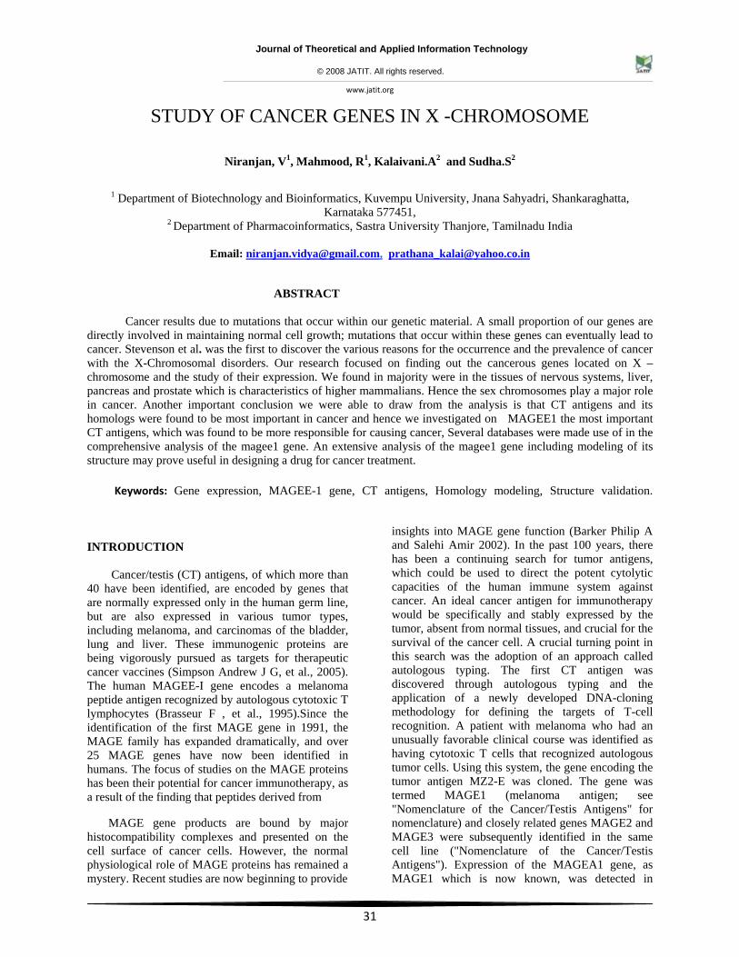
Journal of Theoretical and Applied Information Technology © 2008 JATIT. All rights reserved. www.jatit.org 31 STUDY OF CANCER GENES IN X -CHROMOSOME Niranjan, V 1 , Mahmood, R 1 , Kalaivani.A 2 and Sudha.S 2 1 Department of Biotechnology and Bioinformatics, Kuvempu University, Jnana Sahyadri, Shankaraghatta, Karnataka 577451, 2 Department of Pharmacoinformatics, Sastra University Thanjore, Tamilnadu India Email: [email protected], [email protected] ABSTRACT Cancer results due to mutations that occur within our genetic material. A small proportion of our genes are directly involved in maintaining normal cell growth; mutations that occur within these genes can eventually lead to cancer. Stevenson et al. was the first to discover the various reasons for the occurrence and the prevalence of cancer with the X-Chromosomal disorders. Our research focused on finding out the cancerous genes located on X – chromosome and the study of their expression. We found in majority were in the tissues of nervous systems, liver, pancreas and prostate which is characteristics of higher mammalians. Hence the sex chromosomes play a major role in cancer. Another important conclusion we were able to draw from the analysis is that CT antigens and its homologs were found to be most important in cancer and hence we investigated on MAGEE1 the most important CT antigens, which was found to be more responsible for causing cancer, Several databases were made use of in the comprehensive analysis of the magee1 gene. An extensive analysis of the magee1 gene including modeling of its structure may prove useful in designing a drug for cancer treatment. Keywords: Gene expression, MAGEE-1 gene, CT antigens, Homology modeling, Structure validation. INTRODUCTION Cancer/testis (CT) antigens, of which more than 40 have been identified, are encoded by genes that are normally expressed only in the human germ line, but are also expressed in various tumor types, including melanoma, and carcinomas of the bladder, lung and liver. These immunogenic proteins are being vigorously pursued as targets for therapeutic cancer vaccines (Simpson Andrew J G, et al., 2005). The human MAGEE-I gene encodes a melanoma peptide antigen recognized by autologous cytotoxic T lymphocytes (Brasseur F , et al., 1995).Since the identification of the first MAGE gene in 1991, the MAGE family has expanded dramatically, and over 25 MAGE genes have now been identified in humans. The focus of studies on the MAGE proteins has been their potential for cancer immunotherapy, as a result of the finding that peptides derived from MAGE gene products are bound by major histocompatibility complexes and presented on the cell surface of cancer cells. However, the normal physiological role of MAGE proteins has remained a mystery. Recent studies are now beginning to provide insights into MAGE gene function (Barker Philip A and Salehi Amir 2002). In the past 100 years, there has been a continuing search for tumor antigens, which could be used to direct the potent cytolytic capacities of the human immune system against cancer. An ideal cancer antigen for immunotherapy would be specifically and stably expressed by the tumor, absent from normal tissues, and crucial for the survival of the cancer cell. A crucial turning point in this search was the adoption of an approach called autologous typing. The first CT antigen was discovered through autologous typing and the application of a newly developed DNA-cloning methodology for defining the targets of T-cell recognition. A patient with melanoma who had an unusually favorable clinical course was identified as having cytotoxic T cells that recognized autologous tumor cells. Using this system, the gene encoding the tumor antigen MZ2-E was cloned. The gene was termed MAGE1 (melanoma antigen; see "Nomenclature of the Cancer/Testis Antigens" for nomenclature) and closely related genes MAGE2 and MAGE3 were subsequently identified in the same cell line ("Nomenclature of the Cancer/Testis Antigens"). Expression of the MAGEA1 gene, as MAGE1 which is now known, was detected in
Transcript of STUDY OF CANCER GENES IN X -CHROMOSOME - JATIT expression... · study of cancer genes in x...
Journal of Theoretical and Applied Information Technology
© 2008 JATIT. All rights reserved.
www.jatit.org
31
STUDY OF CANCER GENES IN X -CHROMOSOME
Niranjan, V1, Mahmood, R1, Kalaivani.A2 and Sudha.S2
1 Department of Biotechnology and Bioinformatics, Kuvempu University, Jnana Sahyadri, Shankaraghatta,
Karnataka 577451, 2 Department of Pharmacoinformatics, Sastra University Thanjore, Tamilnadu India
Email: [email protected], [email protected]
ABSTRACT Cancer results due to mutations that occur within our genetic material. A small proportion of our genes are directly involved in maintaining normal cell growth; mutations that occur within these genes can eventually lead to cancer. Stevenson et al. was the first to discover the various reasons for the occurrence and the prevalence of cancer with the X-Chromosomal disorders. Our research focused on finding out the cancerous genes located on X –chromosome and the study of their expression. We found in majority were in the tissues of nervous systems, liver, pancreas and prostate which is characteristics of higher mammalians. Hence the sex chromosomes play a major role in cancer. Another important conclusion we were able to draw from the analysis is that CT antigens and its homologs were found to be most important in cancer and hence we investigated on MAGEE1 the most important CT antigens, which was found to be more responsible for causing cancer, Several databases were made use of in the comprehensive analysis of the magee1 gene. An extensive analysis of the magee1 gene including modeling of its structure may prove useful in designing a drug for cancer treatment. Keywords: Gene expression, MAGEE-1 gene, CT antigens, Homology modeling, Structure validation. INTRODUCTION Cancer/testis (CT) antigens, of which more than 40 have been identified, are encoded by genes that are normally expressed only in the human germ line, but are also expressed in various tumor types, including melanoma, and carcinomas of the bladder, lung and liver. These immunogenic proteins are being vigorously pursued as targets for therapeutic cancer vaccines (Simpson Andrew J G, et al., 2005). The human MAGEE-I gene encodes a melanoma peptide antigen recognized by autologous cytotoxic T lymphocytes (Brasseur F , et al., 1995).Since the identification of the first MAGE gene in 1991, the MAGE family has expanded dramatically, and over 25 MAGE genes have now been identified in humans. The focus of studies on the MAGE proteins has been their potential for cancer immunotherapy, as a result of the finding that peptides derived from
MAGE gene products are bound by major histocompatibility complexes and presented on the cell surface of cancer cells. However, the normal physiological role of MAGE proteins has remained a mystery. Recent studies are now beginning to provide
insights into MAGE gene function (Barker Philip A and Salehi Amir 2002). In the past 100 years, there has been a continuing search for tumor antigens, which could be used to direct the potent cytolytic capacities of the human immune system against cancer. An ideal cancer antigen for immunotherapy would be specifically and stably expressed by the tumor, absent from normal tissues, and crucial for the survival of the cancer cell. A crucial turning point in this search was the adoption of an approach called autologous typing. The first CT antigen was discovered through autologous typing and the application of a newly developed DNA-cloning methodology for defining the targets of T-cell recognition. A patient with melanoma who had an unusually favorable clinical course was identified as having cytotoxic T cells that recognized autologous tumor cells. Using this system, the gene encoding the tumor antigen MZ2-E was cloned. The gene was termed MAGE1 (melanoma antigen; see "Nomenclature of the Cancer/Testis Antigens" for nomenclature) and closely related genes MAGE2 and MAGE3 were subsequently identified in the same cell line ("Nomenclature of the Cancer/Testis Antigens"). Expression of the MAGEA1 gene, as MAGE1 which is now known, was detected in
Journal of Theoretical and Applied Information Technology
© 2008 JATIT. All rights reserved.
www.jatit.org
32
melanomas, some breast carcinomas and other tumor types, but not in any normal tissues except testis (Mashino K, et al., (2001). Further analysis of the MAGEA family revealed 12 closely related genes clustered at Xq28. A search for the gene responsible for the sex-reversal phenotype revealed a second cluster of MAGE genes, which encoded the MAGEB genes and was located at Xp21.3. Subsequently, a third cluster, encoding the MAGEC genes, was identified at Xq26-27. The genes in all three of these families were found to exhibit expression restricted to the testis or cancers. By contrast, more distantly related clusters of genes (MAGED to MAGEL) were found to be expressed in many normal tissues.
The expression of CT-X antigens varies greatly between tumor types. According to RT-PCR analyses, bladder cancer, lung cancer, ovarian cancer, hepatocellular carcinoma and melanoma frequently express CT-X antigens. By contrast, CT-X antigen expression is rarely observed in renal cancer, colon cancer, gastric cancer, and leukemia/lymphoma cells. The CT-X mRNAs can be a dominant feature of the transcriptome in tumor cells. Of these 20 genes, six were known CT antigens: five members of the MAGEA gene family and NY-ESO-1 (Figueiredo David L A, et al., 2006).
Although CT-X antigens have taken centre stage in the development and clinical testing of experimental cancer vaccines, their biological function in both the germ line and tumors has remained poorly understood. A central question is whether their expression contributes to tumorigenesis or is a functionally irrelevant by-product of the process of cellular transformation, possibly due to global-chromatin changes. Clues have emerged, however, to indicate that expression of CT antigens such as MAGE could have a fundamental role in human.
The extensive MAGE family of CT antigens comprises more than 25 genes that are characterized by the presence of a large central region termed the MAGE homology domain (MHD). So far, the only CT-X gene product for which protein binding partners have been actively sought, through a yeast two-hybrid assay, has been MAGEA1. Using proteins expressed from a testis cDNA library, the assay identified the transcriptional regulator SKI-interacting protein (SKIP) as a binding partner for MAGEA1.Binding of MAGEA1 to SKIP depends on the extreme carboxyl terminus of the MAGE protein, a domain shared with MAGEA4, which was also shown to be able to bind to SKIP. SKIP connects DNA-binding proteins to other proteins that either activate or repress transcription, and participates in a
range of signaling pathways, including those involving vitamin D, retinoic acid, estrogens, glucocorticoids, Notch1 and transforming growth factor-ß. (Old, L. J. 2001}.
This mechanism involves key mutations in tumor-suppressor genes and oncogenes that are expressed and functional in normal cells from which the tumors arise. By contrast, CT antigen expression is aberrant in the sense that proteins normally restricted to one lineage are now expressed in another. Such aberrant expression is rare in cancer where lineage programmed fidelity is rule.
These genes remain to be identified, but important candidates are those that directly control genome demethylation. Accumulation of mutations in multiple genes is the main reason for all types of cancers which might be possible due to many factors such as environment, sudden mutations which the body can’t resist etc. After the entire survey finally we concluded that MAGEE 1 is the gene, which is linked with most of the cancers in the X-Chromosome. We carried out an extensive research using new protocols in bioinformatics to elucidate the contribution of the gene to cancer. (Scanlan, M. J et al., 2004).
There are various methods to detect the types of cancer; bioinformatics method of detection gives the faster solution than traditional, combinational biotechnology methods (www.iop.org/EJ/journal/page=featauth/-aouther=430/0031-9155)The leading approach of bioinformatics method is to identify the cancer causing genes linked in X-chromosome.
Statistical survey in India
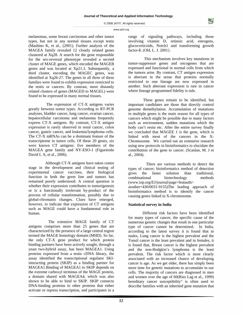
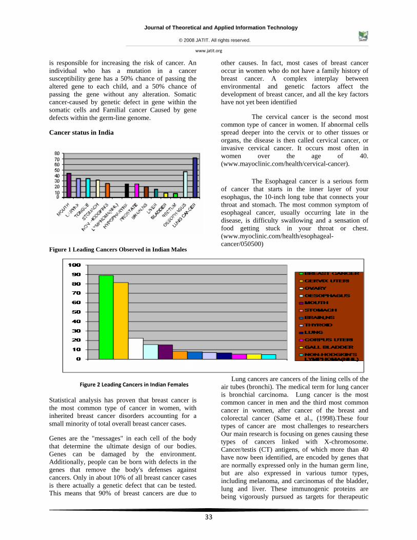
Different risk factors have been identified for many types of cancer, the specific cause of the numerous genetic changes that result in one particular type of cancer cannot be determined. In India, according to the latest survey it is found that in males, Lung cancer is the highest prevalent and the Tonsil cancer is the least prevalent and in females, it is found that, Breast cancer is the highest prevalent and the non-Hodgkin’s lymphoma is the least prevalent. The risk factor which is most clearly associated with an increased chance of developing cancer is age. As we get older, there has simply been more time for genetic mutations to accumulate in our cells. The majority of cancers are diagnosed in men and women over the age of 60(Ries Lag et al., 1999) hereditary cancer susceptibility" is often used to describe families with an inherited gene mutation that
Journal of Theoretical and Applied Information Technology
© 2008 JATIT. All rights reserved.
www.jatit.org
33
is responsible for increasing the risk of cancer. An individual who has a mutation in a cancer susceptibility gene has a 50% chance of passing the altered gene to each child, and a 50% chance of passing the gene without any alteration. Somatic cancer-caused by genetic defect in gene within the somatic cells and Familial cancer Caused by gene defects within the germ-line genome.
Cancer status in India
Figure 1 Leading Cancers Observed in Indian Males
Figure 2 Leading Cancers in Indian Females
Statistical analysis has proven that breast cancer is the most common type of cancer in women, with inherited breast cancer disorders accounting for a small minority of total overall breast cancer cases.
Genes are the "messages" in each cell of the body that determine the ultimate design of our bodies. Genes can be damaged by the environment. Additionally, people can be born with defects in the genes that remove the body's defenses against cancers. Only in about 10% of all breast cancer cases is there actually a genetic defect that can be tested. This means that 90% of breast cancers are due to
other causes. In fact, most cases of breast cancer occur in women who do not have a family history of breast cancer. A complex interplay between environmental and genetic factors affect the development of breast cancer, and all the key factors have not yet been identified
The cervical cancer is the second most common type of cancer in women. If abnormal cells spread deeper into the cervix or to other tissues or organs, the disease is then called cervical cancer, or invasive cervical cancer. It occurs most often in women over the age of 40. (www.mayoclinic.com/health/cervical-cancer).
The Esophageal cancer is a serious form of cancer that starts in the inner layer of your esophagus, the 10-inch long tube that connects your throat and stomach. The most common symptom of esophageal cancer, usually occurring late in the disease, is difficulty swallowing and a sensation of food getting stuck in your throat or chest. (www.myoclinic.com/health/esophageal-cancer/050500)
Lung cancers are cancers of the lining cells of the air tubes (bronchi). The medical term for lung cancer is bronchial carcinoma. Lung cancer is the most common cancer in men and the third most common cancer in women, after cancer of the breast and colorectal cancer (Same et al., (1998).These four types of cancer are most challenges to researchers Our main research is focusing on genes causing these types of cancers linked with X-chromosome. Cancer/testis (CT) antigens, of which more than 40 have now been identified, are encoded by genes that are normally expressed only in the human germ line, but are also expressed in various tumor types, including melanoma, and carcinomas of the bladder, lung and liver. These immunogenic proteins are being vigorously pursued as targets for therapeutic
Journal of Theoretical and Applied Information Technology
© 2008 JATIT. All rights reserved.
www.jatit.org
34
cancer vaccines. CT antigens are also being evaluated for their role in oncogenesis; recapitulation of portions of the germline gene-expression programme might contribute characteristic features to the neoplastic phenotype, including immortality, invasiveness, immune evasion, hypomethylation and metastatic capacity. MAGEE1 are members of gene families that encode CT antigens and are found in a number of melanomas and other tumors. (Simpson J.G Andrew et al., 2000).
MAGE
The melanoma antigen gene family is clustered in the chromosomal band X q28 (Rognar UC et al., 1995).The melanoma antigen gene (MAGE) family comprises 12 known genes, of which 6 are expressed in tumors. In the course of a systematic analysis of transcripts in Xq28 ,they have identified cDNAs related to different MAGE genes .Analysis of cell hybrids ,ordered YACs ,and cosmids showed that all MAGE genes are located in Xq28 and are clustered in three main intervals within 3.5Mb .A new MAGE gene with Ubiquitous expression does not code for known MAGE antigens. Identification of the MAGE1 gene product by monoclonal and polyclonal antibodies. (YT chen et al., 1996).The human MAGE1 gene encodes a melanoma peptide antigen recognized by autologous cytotoxic T lymphocytes. Genes of the MAGE family direct the expression of tumor antigens that are recognized on human melanomas by autologous cytolytic T lymphocytes .Using representational difference analysis (RDA) with a melanoma cell line showing high expression of cancer testis (CT) antigens ,(Gure et al., 2000) identified cDNA fragments corresponding to 29 genes ,including MAGEE1,which they designated CT 10.By PCR analysis of somatic cell hybrids and FISH,(Gure et al., 2000) mapped the MAGEE1 gene to Xq27. Ucas et al., (2000) mapped the gene to Xq26-27 by radiation hybrid analysis. In chromosome X the concentration of MAGE gene is higher and these genes are highly expressed in wide range of tumors. There are 19 closely related human MAGE genes have been identified and divided into four different classes based on sequence homology and X-chromosomal location.
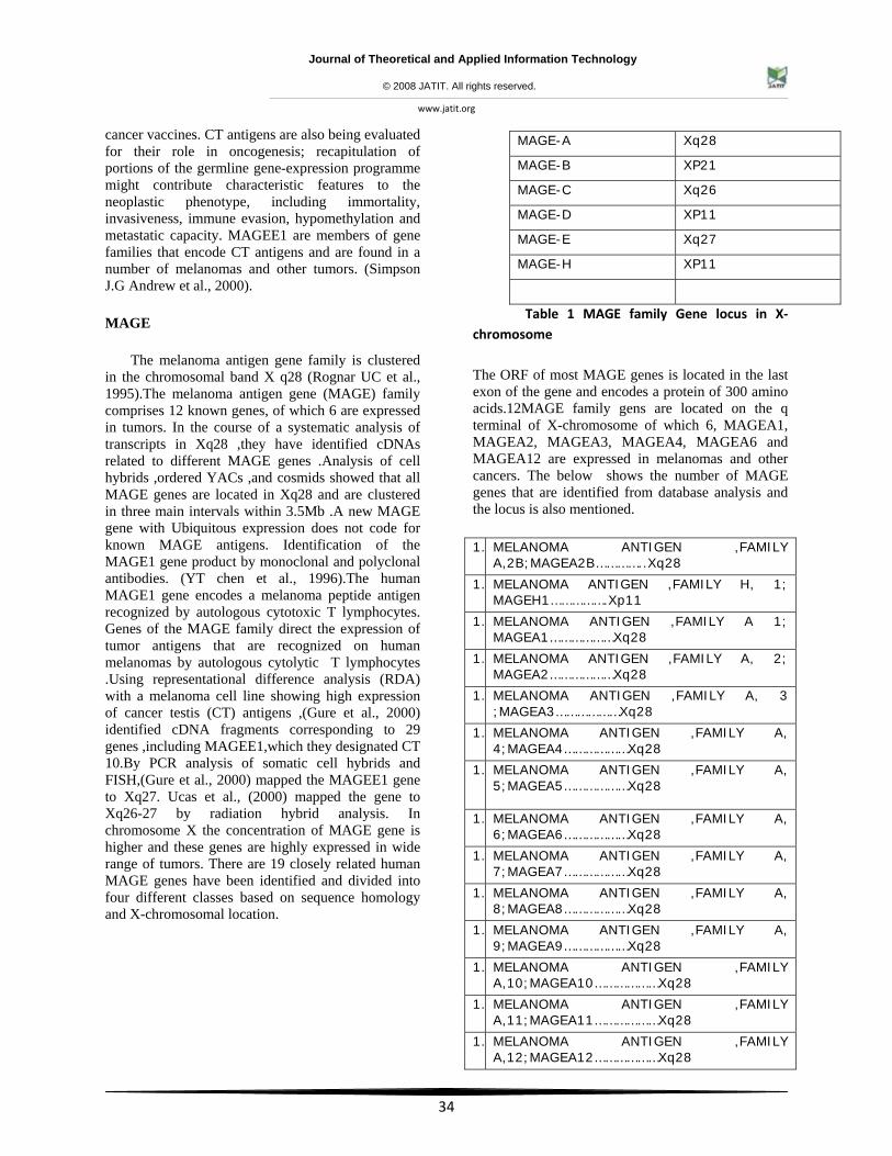
Table 1 MAGE family Gene locus in X‐chromosome
The ORF of most MAGE genes is located in the last exon of the gene and encodes a protein of 300 amino acids.12MAGE family gens are located on the q terminal of X-chromosome of which 6, MAGEA1, MAGEA2, MAGEA3, MAGEA4, MAGEA6 and MAGEA12 are expressed in melanomas and other cancers. The below shows the number of MAGE genes that are identified from database analysis and the locus is also mentioned.
1. MELANOMA ANTIGEN ,FAMILY A,2B;MAGEA2B…………..Xq28
1. MELANOMA ANTIGEN ,FAMILY H, 1; MAGEH1…………….Xp11
1. MELANOMA ANTIGEN ,FAMILY A 1; MAGEA1………………Xq28
1. MELANOMA ANTIGEN ,FAMILY A, 2; MAGEA2………………Xq28
1. MELANOMA ANTIGEN ,FAMILY A, 3 ;MAGEA3………………Xq28
1. MELANOMA ANTIGEN ,FAMILY A, 4;MAGEA4………………Xq28
1. MELANOMA ANTIGEN ,FAMILY A, 5;MAGEA5………………Xq28
1. MELANOMA ANTIGEN ,FAMILY A, 6;MAGEA6………………Xq28
1. MELANOMA ANTIGEN ,FAMILY A, 7;MAGEA7………………Xq28
1. MELANOMA ANTIGEN ,FAMILY A, 8;MAGEA8………………Xq28
1. MELANOMA ANTIGEN ,FAMILY A, 9;MAGEA9………………Xq28
1. MELANOMA ANTIGEN ,FAMILY A,10;MAGEA10………………Xq28
1. MELANOMA ANTIGEN ,FAMILY A,11;MAGEA11………………Xq28
1. MELANOMA ANTIGEN ,FAMILY A,12;MAGEA12………………Xq28
MAGE-A Xq28
MAGE-B XP21
MAGE-C Xq26
MAGE-D XP11
MAGE-E Xq27
MAGE-H XP11
Journal of Theoretical and Applied Information Technology
© 2008 JATIT. All rights reserved.
www.jatit.org
35
1. MELANOMA ANTIGEN ,FAMILY B,1;MAGEB1………………...Xp21.3
1. MELANOMA ANTIGEN ,FAMILY B,1;MAGEB1……….………..Xp21.3
1. MELANOMA ANTIGEN ,FAMILY B,2;MAGEB2………………..Xp21.3
1. MELANOMA ANTIGEN ,FAMILY B,3;MAGEB3………………..Xp21.3
1. MELANOMA ANTIGEN ,FAMILY B,4;MAGEB4………………..Xp21.3
1. MELANOMA ANTIGEN ,FAMILY B,5;MAGEB5………………..Xp22
1. MELANOMA ANTIGEN ,FAMILY B,6;MAGEB6………………..Xp22
1. MELANOMA ANTIGEN ,FAMILY C,1;MAGEC1………………..Xq26
1. MELANOMA ANTIGEN ,FAMILY C, 3;MAGEC2……………..Xq27
1. MELANOMA ANTIGEN ,FAMILY D,1;MAGED1……………….Xp11.23
1. MELANOMA ANTIGEN ,FAMILY D, 2;MAGED2……………..Xp11.2
1. MELANOMA ANTIGEN ,FAMILY E, 1; MAGEE1…………..Xq27
Table 2 MAGE gene expression in X‐chromosome
Though there are various genes responsible for different types of cancer, MAGEE1 gene is the main cancer causing gene. Our research is focused mainly on MAGEE1 gene .The techniques used to annotate this gene is by,
• Proteomics • Homology modeling • Genomics • Therapeutics
MATERIALS &METHOD
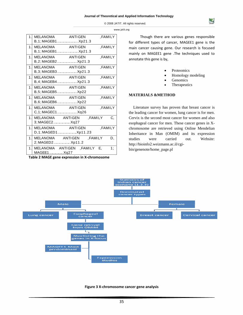
Literature survey has proven that breast cancer is the leading cancer for women, lung cancer is for men. Cervix is the second most cancer for women and also esophageal cancer for men. These cancer genes in X-chromosome are retrieved using Online Mendelian Inheritance in Man (OMIM) and its expression studies were carried out. Website: http://bioinfo2.weizmann.ac.il/cgi-bin/genenote/home_page.pl
Figure 3 X‐chromosome cancer gene analysis
Journal of Theoretical and Applied Information Technology
© 2008 JATIT. All rights reserved.
www.jatit.org
36
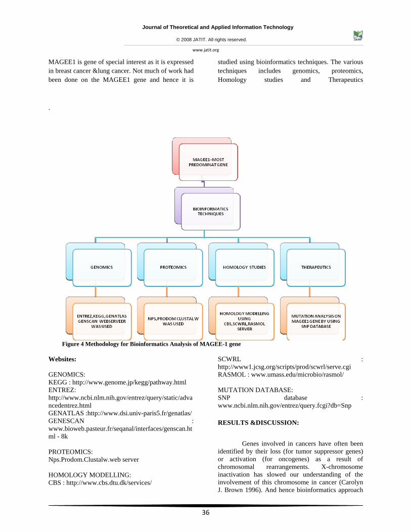
MAGEE1 is gene of special interest as it is expressed in breast cancer &lung cancer. Not much of work had been done on the MAGEE1 gene and hence it is
studied using bioinformatics techniques. The various techniques includes genomics, proteomics, Homology studies and Therapeutics
.
Figure 4 Methodology for Bioinformatics Analysis of MAGEE-1 gene
Websites: GENOMICS: KEGG : http://www.genome.jp/kegg/pathway.html ENTREZ: http://www.ncbi.nlm.nih.gov/entrez/query/static/advancedentrez.html GENATLAS :http://www.dsi.univ-paris5.fr/genatlas/ GENESCAN : www.bioweb.pasteur.fr/seqanal/interfaces/genscan.html - 8k PROTEOMICS: Nps.Prodom.Clustalw.web server HOMOLOGY MODELLING: CBS : http://www.cbs.dtu.dk/services/
SCWRL : http://www1.jcsg.org/scripts/prod/scwrl/serve.cgi RASMOL : www.umass.edu/microbio/rasmol/ MUTATION DATABASE: SNP database : www.ncbi.nlm.nih.gov/entrez/query.fcgi?db=Snp
RESULTS &DISCUSSION:
Genes involved in cancers have often been identified by their loss (for tumor suppressor genes) or activation (for oncogenes) as a result of chromosomal rearrangements. X-chromosome inactivation has slowed our understanding of the involvement of this chromosome in cancer (Carolyn J. Brown 1996). And hence bioinformatics approach
Journal of Theoretical and Applied Information Technology
© 2008 JATIT. All rights reserved.
www.jatit.org
37
attempted to crystallize the fact . The expression of the major cancer outbreaks in India were taken for study and MAGE association role in cancer were focused.
Breast Cancer
The genes located on X- chromosome which showed significant association with Breast cancer were shortlisted using OMIM.
• CTAG1B
• AR
• MAGEE1
• LDOC1
• BCAP31
• IL-13RA2
• INg2
CTAG1B Cancer testis antigen 1B is expressed in testis and ovary and in a wide variety of cancers detected in uterine myometrium .Northern blot analysis of normal human tissues revealed expression of a 0.8-kb mRNA in testis and ovary. RT-PCR analysis showed expression in a wide array of human cancers.NY-ESO-1 and CTp11 expression may correlate with stage of progression in melanoma.TRP2 is a melanoma-differentiation antigen.
Tissue Normalized Expression (%)
Cluster Clones : Tissue clones
bone: 64.54 1:53985
Placenta: 23.06 1:151135
mixed: 12.40 1:280996
Table 3 CTAG1B gene expression
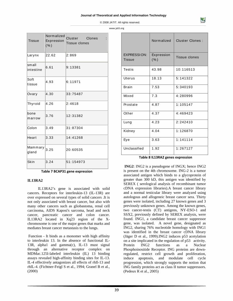
AR
This gene are expressed in various tissues such as ovary, bone marrow etc. The maximum expression of this gene was found to be in the tissues of prostate gland. Androgen receptor expression plays an important role in the proliferation of human prostate cancer and confers a better prognosis in cancer. Although androgen receptor (AR) is expressed during cancer progression but levels were reduced or absent in late stage disease.
Tissue
Normalized Cluster Clones :
Expression (%) Tissue clones
Prostate: 57.3 11:105147
bone marrow: 17.45 1:31382
ovary: 14.51 2:75487
muscle: 5.56 1:98454
Unclassified: 2.05 1:267127
Table 4 AR Cancer genes expression
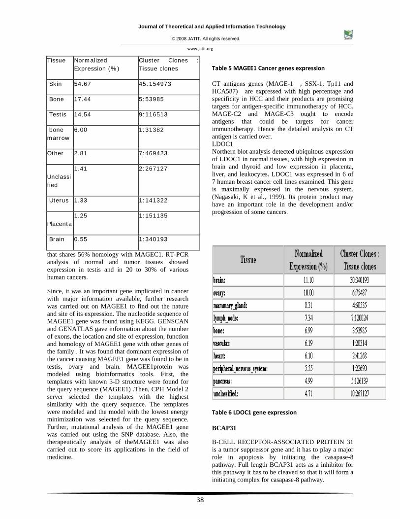
MAGEE1
This gene is expressed in the tissues of bone, testis, bone marrow, uterus, placenta, brain. The maximum expression of this gene is in the tissues of nervous system. (Gure et al., (2000)Among tumor tissues, they found that MAGEE1 was frequently expressed in seminomas, melanomas, and bladder transitional cell carcinomas. It was also expressed in a significant fraction of head and neck carcinomas, breast carcinomas, non small-cell lung carcinomas, and sarcomas. It is frequently expressed in tissues from patients with late stage diseases.
Genes of the MAGE family direct the expression of tumor antigens that are recognized on human melanomas by autologous cytolytic T lymphocytes. Using representational difference analysis (RDA) with a melanoma cell line showing high expression of cancer-testis (CT) antigens, identified cDNA fragments corresponding to 29 genes, including MAGEE1, which they designated CT10. They cloned MAGEE1 from a human placenta genomic library. MAGEE1 encodes a deduced 373-amino acid protein
Journal of Theoretical and Applied Information Technology
© 2008 JATIT. All rights reserved.
www.jatit.org
38
that shares 56% homology with MAGEC1. RT-PCR analysis of normal and tumor tissues showed expression in testis and in 20 to 30% of various human cancers.
Since, it was an important gene implicated in cancer with major information available, further research was carried out on MAGEE1 to find out the nature and site of its expression. The nucleotide sequence of MAGEE1 gene was found using KEGG. GENSCAN and GENATLAS gave information about the number of exons, the location and site of expression, function and homology of MAGEE1 gene with other genes of the family . It was found that dominant expression of the cancer causing MAGEE1 gene was found to be in testis, ovary and brain. MAGEE1protein was modeled using bioinformatics tools. First, the templates with known 3-D structure were found for the query sequence (MAGEE1) .Then, CPH Model 2 server selected the templates with the highest similarity with the query sequence. The templates were modeled and the model with the lowest energy minimization was selected for the query sequence. Further, mutational analysis of the MAGEE1 gene was carried out using the SNP database. Also, the therapeutically analysis of theMAGEE1 was also carried out to score its applications in the field of medicine.
Table 5 MAGEE1 Cancer genes expression
CT antigens genes (MAGE-1 , SSX-1, Tp11 and HCA587) are expressed with high percentage and specificity in HCC and their products are promising targets for antigen-specific immunotherapy of HCC. MAGE-C2 and MAGE-C3 ought to encode antigens that could be targets for cancer immunotherapy. Hence the detailed analysis on CT antigen is carried over. LDOC1 Northern blot analysis detected ubiquitous expression of LDOC1 in normal tissues, with high expression in brain and thyroid and low expression in placenta, liver, and leukocytes. LDOC1 was expressed in 6 of 7 human breast cancer cell lines examined. This gene is maximally expressed in the nervous system. (Nagasaki, K et al., 1999). Its protein product may have an important role in the development and/or progression of some cancers.
Table 6 LDOC1 gene expression
BCAP31
B-CELL RECEPTOR-ASSOCIATED PROTEIN 31 is a tumor suppressor gene and it has to play a major role in apoptosis by initiating the casapase-8 pathway. Full length BCAP31 acts as a inhibitor for this pathway it has to be cleaved so that it will form a initiating complex for casapase-8 pathway.
Tissue Normalized Expression (%)
Cluster Clones : Tissue clones
Skin 54.67 45:154973
Bone 17.44 5:53985
Testis 14.54 9:116513
bone marrow
6.00 1:31382
Other 2.81 7:469423
Unclassified
1.41 2:267127
Uterus 1.33 1:141322
Placenta
1.25 1:151135
Brain 0.55 1:340193
Journal of Theoretical and Applied Information Technology
© 2008 JATIT. All rights reserved.
www.jatit.org
39
Tissue Normalized Expression (%)
Cluster Clones : Tissue clones
Larynx 22.62 2:869
small intestine
6.61 9:13381
Soft tissue
4.93 6:11971
Ovary 4.30 33:75487
Thyroid 4.26 2:4618
bone marrow
3.76 12:31382
Colon 3.49 31:87304
Heart 3.33 14:41268
Mammary gland
3.25 20:60535
Skin 3.24 51:154973
Table 7 BCAP31 gene expression
IL13RA2
IL13RA2’s gene is associated with solid cancers. Receptors for interleukin-13 (IL-13R) are over expressed on several types of solid cancers .It is not only associated with breast cancer, but also with many other cancers such as gliobastoma, renal cell carcinoma, AIDS Kaposi's sarcoma, head and neck cancer, pancreatic cancer and colon cancer. IL13RA2 located in Xq23 region of the X- chromosome is one of the major genes that marks and mediates breast cancer metastasis to the lungs.
Function - It binds as a monomer with high affinity to interleukin 13. In the absence of functional IL-13R, alpha1 and gamma(c), IL-13 must signal through an alternative receptor complex on MDMac.125I-labeled interleukin (IL) 13 binding assays revealed high-affinity binding sites for IL-13. IL-4 effectively antagonizes all effects of rhIl-13 and rhIL-4. (Fichtner-Feigl S et al., 1994; Granel B et al., (2000)
EXPRESSION: Tissue
Normalized Cluster Clones :
Expression (%)
Tissue clones
Testis 43.98 10:116513
Uterus 18.13 5:141322
Brain 7.53 5:340193
Mixed 7.3 4:280996
Prostate 4.87 1:105147
Other 4.37 4:469423
Lung 4.23 2:242410
Kidney 4.04 1:126870
Eye 3.63 1:141114
Unclassified 1.92 1:267127
Table 8 IL13RA2 genes expression
ING2: ING2 is a pseudogene of INGX; hence ING2 is present on the 4th chromosome. ING-2 is a tumor associated antigen which binds to a glycoprotein of greater than 300 kD, this antigen was identified by SEREX ( serological analysis of recombinant tumor cDNA expression libraries).A breast cancer library and a normal testicular library were analyzed using autologous and allogeneic breast cancer sera. Thirty genes were isolated, including 27 known genes and 3 previously unknown genes. Among the known genes, two cancer-testis (CT) antigens, NY-ESO-1 and SSX2, previously defined by SEREX analysis, were found. ING1, a candidate breast cancer suppressor gene, was isolated. A novel gene, designated as ING2, sharing 76% nucleotide homology with ING1 was identified in the breast cancer cDNA library (Jäger D et al., 1999).ING2 induces p53 acetylation on a site implicated in the regulation of p53 activity. Protein ING2 functions as a Nuclear Phosphoinositide Receptor. ING proteins are down-regulated, restrict cell growth and proliferation, induce apoptosis, and modulate cell cycle progression, which strongly supports the notion that ING family proteins act as class II tumor suppressors. (Pedeux R et al., 2005)
Journal of Theoretical and Applied Information Technology
© 2008 JATIT. All rights reserved.
www.jatit.org
40
Cervical cancer
• HDAC28
• DKC1
• HDAC8
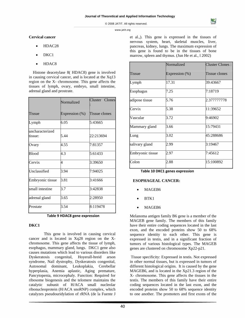
Histone deacetylase 8( HDAC8) gene is involved in causing cervical cancer, and is located at the Xq13 region on the X- chromosome. This gene affects the tissues of lymph, ovary, embryo, small intestine, adrenal gland and prostrate.
Tissue
Normalized Cluster Clones:
Expression (%) Tissue clones
Lymph 6.05 5:43665
uncharacterized tissue: 5.44 22:213694
Ovary 4.55 7:81357
Blood 4.3 5:61433
Cervix 4 3:39650
Unclassified 3.94 7:94025
Embryonic tissue 3.81 3:41666
small intestine 3.7 3:42838
adrenal gland 3.65 2:28950
Prostate 3.54 8:119478
Table 9 HDAC8 gene expression
DKC1
This gene is involved in causing cervical cancer and is located in Xq28 region on the X-chromosome. This gene affects the tissue of lymph, esophagus, mammary gland, lungs. DKC1 gene also causes mutations which lead to various disorders like Dyskeratosis congenital, Hoyerall-hreid arson syndrome, Nail dystrophy, Dyskeratosis congenital, Autosomal dominant, Leukoplakia, Cerebellar hyperplasia, Anemia aplastic, Aging premature, Pancytopenia, microcephaly. Function: Required for ribosome biogenesis and the telomere maintains the catalytic subunit of H/ACA small nucleolar ribonucleoprotein (H/ACA snoRNP) complex, which catalyzes pseudouridylation of rRNA (de la Fuente J
et al.,). This gene is expressed in the tissues of nervous system, heart, skeletal muscles, liver, pancreas, kidney, lungs. The maximum expression of this gene is found to be in the tissues of bone marrow, spleen and thymus. (Jun He et al., l 2002)
Tissue
Normalized Cluster Clones :
Expression (%) Tissue clones
Lymph 17.31 39:43667
Esophagus 7.25 7:18719
adipose tissue 5.76 2.377777778
Cervix 5.38 11:39652
Vascular 3.72 9:46902
Mammary gland 3.66 15:79431
Lung 3.02 45:288686
salivary gland 2.99 3:19467
Embryonic tissue 2.97 7:45612
Colon 2.88 15:100892
Table 10 DKC1 genes expression
ESOPHAGEAL CANCER:
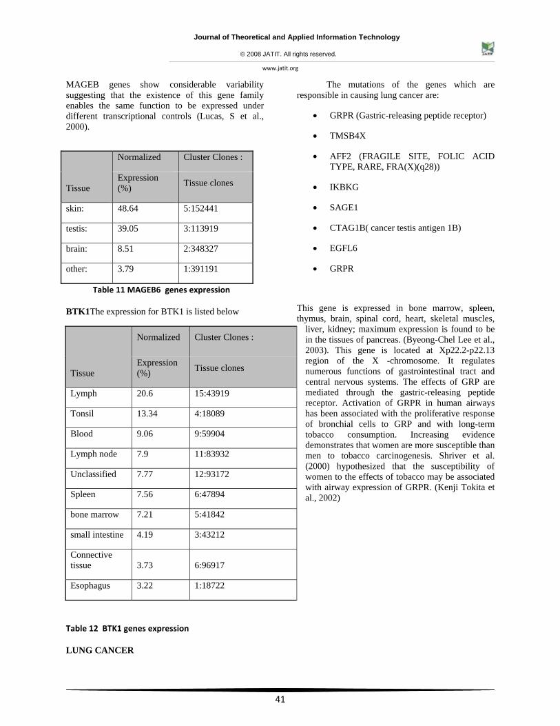
• MAGEB6
• BTK1
• MAGEB6
Melanoma antigen family B6 gene is a member of the MAGEB gene family. The members of this family have their entire coding sequences located in the last exon, and the encoded proteins show 50 to 68% sequence identity to each other. This gene is expressed in testis, and in a significant fraction of tumors of various histological types. The MAGEB genes are clustered on chromosome Xp22-p21.
Tissue specificity: Expressed in testis. Not expressed in other normal tissues, but is expressed in tumors of different histological origins. It is caused by the gene MAGEB6, and is located in the Xp21.3 region of the X- chromosome. This gene affects the tissues in the testis. The members of this family have their entire coding sequences located in the last exon, and the encoded proteins show 50 to 68% sequence identity to one another. The promoters and first exons of the
Journal of Theoretical and Applied Information Technology
© 2008 JATIT. All rights reserved.
www.jatit.org
41
MAGEB genes show considerable variability suggesting that the existence of this gene family enables the same function to be expressed under different transcriptional controls (Lucas, S et al., 2000).
Table 11 MAGEB6 genes expression
BTK1The expression for BTK1 is listed below
Table 12 BTK1 genes expression
LUNG CANCER
The mutations of the genes which are responsible in causing lung cancer are:
• GRPR (Gastric-releasing peptide receptor)
• TMSB4X
• AFF2 (FRAGILE SITE, FOLIC ACID TYPE, RARE, FRA(X)(q28))
• IKBKG
• SAGE1
• CTAG1B( cancer testis antigen 1B)
• EGFL6
• GRPR
This gene is expressed in bone marrow, spleen, thymus, brain, spinal cord, heart, skeletal muscles,
liver, kidney; maximum expression is found to be in the tissues of pancreas. (Byeong-Chel Lee et al., 2003). This gene is located at Xp22.2-p22.13 region of the X -chromosome. It regulates numerous functions of gastrointestinal tract and central nervous systems. The effects of GRP are mediated through the gastric-releasing peptide receptor. Activation of GRPR in human airways has been associated with the proliferative response of bronchial cells to GRP and with long-term tobacco consumption. Increasing evidence demonstrates that women are more susceptible than men to tobacco carcinogenesis. Shriver et al. (2000) hypothesized that the susceptibility of women to the effects of tobacco may be associated with airway expression of GRPR. (Kenji Tokita et al., 2002)
Tissue
Normalized Cluster Clones :
Expression (%) Tissue clones
skin: 48.64 5:152441
testis: 39.05 3:113919
brain: 8.51 2:348327
other: 3.79 1:391191
Tissue
Normalized Cluster Clones :
Expression (%) Tissue clones
Lymph 20.6 15:43919
Tonsil 13.34 4:18089
Blood 9.06 9:59904
Lymph node 7.9 11:83932
Unclassified 7.77 12:93172
Spleen 7.56 6:47894
bone marrow 7.21 5:41842
small intestine 4.19 3:43212
Connective tissue 3.73 6:96917
Esophagus 3.22 1:18722
Journal of Theoretical and Applied Information Technology
© 2008 JATIT. All rights reserved.
www.jatit.org
42
Tissue
Normalized Cluster Clones :
Expression (%) Tissue clones
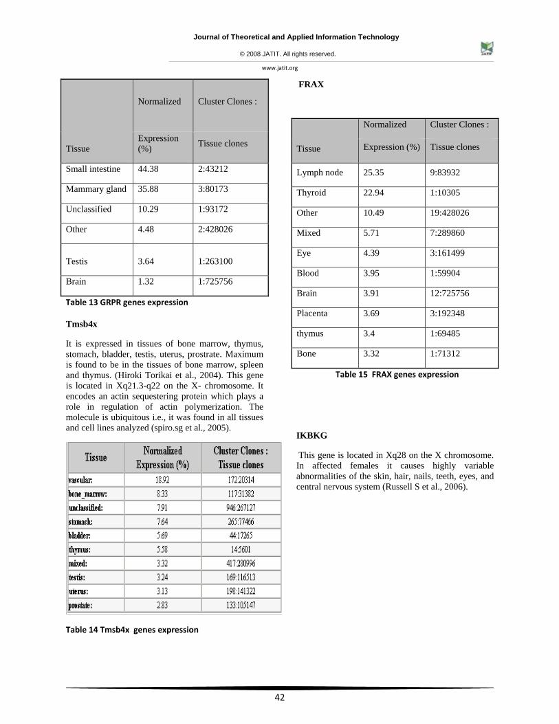
Small intestine 44.38 2:43212
Mammary gland 35.88 3:80173
Unclassified 10.29 1:93172
Other 4.48 2:428026
Testis 3.64 1:263100
Brain 1.32 1:725756
Table 13 GRPR genes expression
Tmsb4x
It is expressed in tissues of bone marrow, thymus, stomach, bladder, testis, uterus, prostrate. Maximum is found to be in the tissues of bone marrow, spleen and thymus. (Hiroki Torikai et al., 2004). This gene is located in Xq21.3-q22 on the X- chromosome. It encodes an actin sequestering protein which plays a role in regulation of actin polymerization. The molecule is ubiquitous i.e., it was found in all tissues and cell lines analyzed (spiro.sg et al., 2005).
Table 14 Tmsb4x genes expression
FRAX
Tissue
Normalized Cluster Clones :
Expression (%) Tissue clones
Lymph node 25.35 9:83932
Thyroid 22.94 1:10305
Other 10.49 19:428026
Mixed 5.71 7:289860
Eye 4.39 3:161499
Blood 3.95 1:59904
Brain 3.91 12:725756
Placenta 3.69 3:192348
thymus 3.4 1:69485
Bone 3.32 1:71312
Table 15 FRAX genes expression
IKBKG
This gene is located in Xq28 on the X chromosome. In affected females it causes highly variable abnormalities of the skin, hair, nails, teeth, eyes, and central nervous system (Russell S et al., 2006).
Journal of Theoretical and Applied Information Technology
© 2008 JATIT. All rights reserved.
www.jatit.org
43
Table 16 IKBKG genes expression
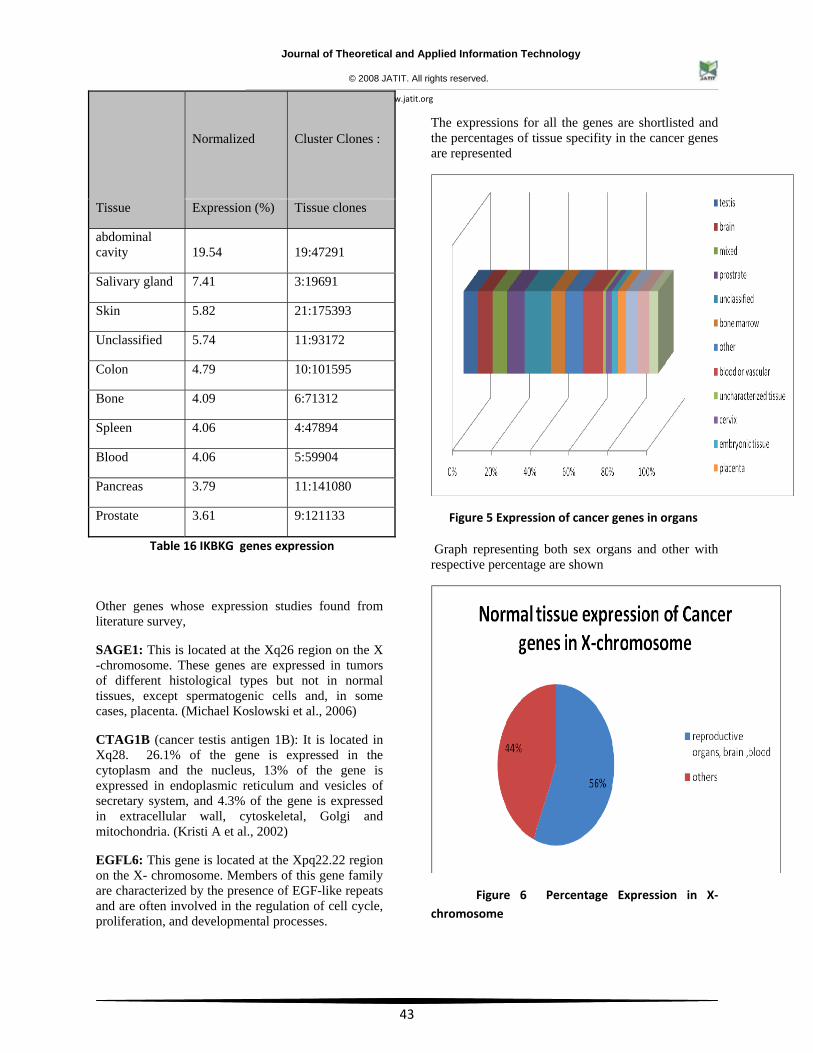
Other genes whose expression studies found from literature survey,
SAGE1: This is located at the Xq26 region on the X -chromosome. These genes are expressed in tumors of different histological types but not in normal tissues, except spermatogenic cells and, in some cases, placenta. (Michael Koslowski et al., 2006)
CTAG1B (cancer testis antigen 1B): It is located in Xq28. 26.1% of the gene is expressed in the cytoplasm and the nucleus, 13% of the gene is expressed in endoplasmic reticulum and vesicles of secretary system, and 4.3% of the gene is expressed in extracellular wall, cytoskeletal, Golgi and mitochondria. (Kristi A et al., 2002)
EGFL6: This gene is located at the Xpq22.22 region on the X- chromosome. Members of this gene family are characterized by the presence of EGF-like repeats and are often involved in the regulation of cell cycle, proliferation, and developmental processes.
The expressions for all the genes are shortlisted and the percentages of tissue specifity in the cancer genes are represented
Figure 5 Expression of cancer genes in organs
Graph representing both sex organs and other with respective percentage are shown
Figure 6 Percentage Expression in X‐ chromosome
Tissue
Normalized Cluster Clones :
Expression (%) Tissue clones
abdominal cavity 19.54 19:47291
Salivary gland 7.41 3:19691
Skin 5.82 21:175393
Unclassified 5.74 11:93172
Colon 4.79 10:101595
Bone 4.09 6:71312
Spleen 4.06 4:47894
Blood 4.06 5:59904
Pancreas 3.79 11:141080
Prostate 3.61 9:121133
Journal of Theoretical and Applied Information Technology
© 2008 JATIT. All rights reserved.
www.jatit.org
44
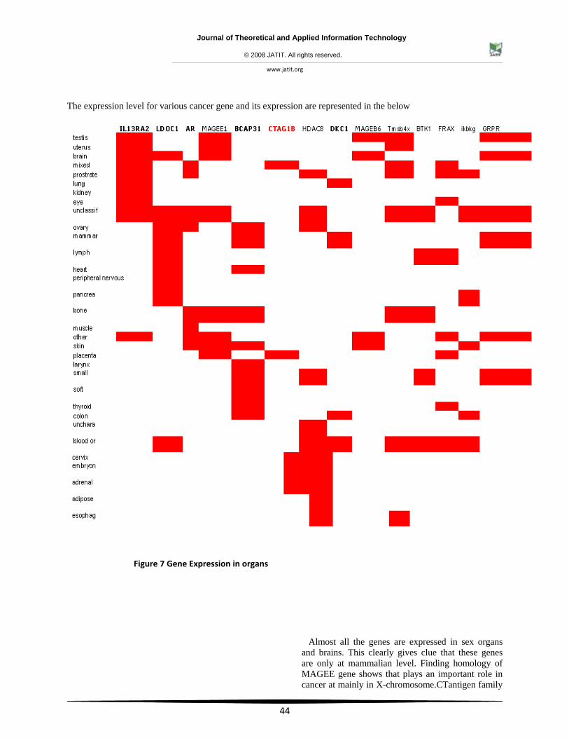
The expression level for various cancer gene and its expression are represented in the below
Figure 7 Gene Expression in organs
Almost all the genes are expressed in sex organs and brains. This clearly gives clue that these genes are only at mammalian level. Finding homology of MAGEE gene shows that plays an important role in cancer at mainly in X-chromosome.CTantigen family
Journal of Theoretical and Applied Information Technology
© 2008 JATIT. All rights reserved.
www.jatit.org
45
forms a key in X-chromosome which can act as a switch on cancer causing paths and hence detailed analysis of magee1 was shown below.
Human protein: Q6IAI7 - MAGEE1 protein
ALIASES:
CT10; HCA587; MAGEE1
CANCER-TESTIS ANTIGEN 10
MELANOMA ANTIGEN, FAMILY E, 1
MELANOMA ANTIGEN FAMILY C2
CHROMOSOMAL LOCATION:
Chromosome/Cytoband - Xq27
LOCUS LINK INFORMATION: This gene is related to members of the MAGEC gene family. It is not expressed in normal tissues, except for testis, and is expressed in tumors of various histological types. This gene and the MAGEC genes are clustered on chromosome Xq26-q27
SWISSPROT INFORMATION
SwissProt Accession No. Q9UBF1 Melanoma-associated antigen C2 (Homo sapiens); 100% similarity over 373aa
Developmental Stage:
Expressed in spermatogonia and primary spermatocytes. In later stages of maturation, expression gradually decreases and becomes undetec in mature spermatids.
Subcellular Location:
Cytoplasm and nuclear location in germ cells. Cytoplasmic location in well-differentiated hepatocellular carcinoma; nuclear location in moderately- and poorly-differentiated hepatocellular carcinoma.
Tissue Specificity:
Not expressed in normal tissues, except in germ cells in the somniferous tubules and in purkinje cells of the cerebellum. Expressed in various tumors, including melanoma, lymphoma, as well as pancreatic cancer, mammary gland cancer, non- small cell lung cancer and liver cancer. In hepatocellular carcinoma, there is an inverse correlation between tumor differentiation and protein expression, i.e. the lower the differentiation, the higher percentage of expression.
Similarity:
Contains 1 Mage Domain.
HOMOLOGY MODELING OF MAGEE1 PROTEIN USING CBS SERVER:
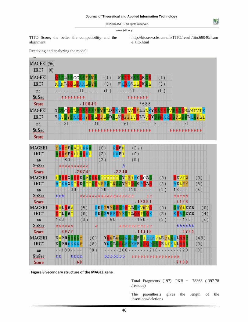
Molecular modeling of proteins is confronted with the problem of finding homologous proteins, especially when few identities remain after the process of molecular evolution. Using even the most recent methods based on sequence identity detection, structural relationships are still difficult to establish with high reliability. As protein structures are more conserved than sequences, we investigated the possibility of using protein secondary structure comparison (observed or predicted structures) to discriminate between related and unrelated proteins sequences in the range of 10%-30% sequence identity. Pair wise comparisons of secondary structures have been measured using the structural overlap (Sov) parameter. If the secondary structures likeness is >50%, most of the pairs are structurally related. Taking into account the secondary structures of proteins that have been detected by BLAST, FASTA, or SSEARCH in the noisy region (with high E: value), it is seen that distantly related protein sequences (even with <20% identity) can be still identified. This strategy can be used to identify three-dimensional templates in homology modeling by finding unexpected related proteins and to select proteins for experimental investigation in a structural genomic approach, as well as for genome annotation. Of the other modeling servers available SWISS Model is best for modeling proteins with available templates. Geno3D automatic modeling server is good as it gives a lot of other information related to the modeled protein. But in this project I have used CBS server for modeling MAGEE1 gene, as there are no readily available templates in the ExPDB.
TITO evaluates the compatibility (the pair wise alignment) between the sequence MAGEE1 and each 3D structure (PDB structure). The lower the
Ontology Annotation Evidence Source
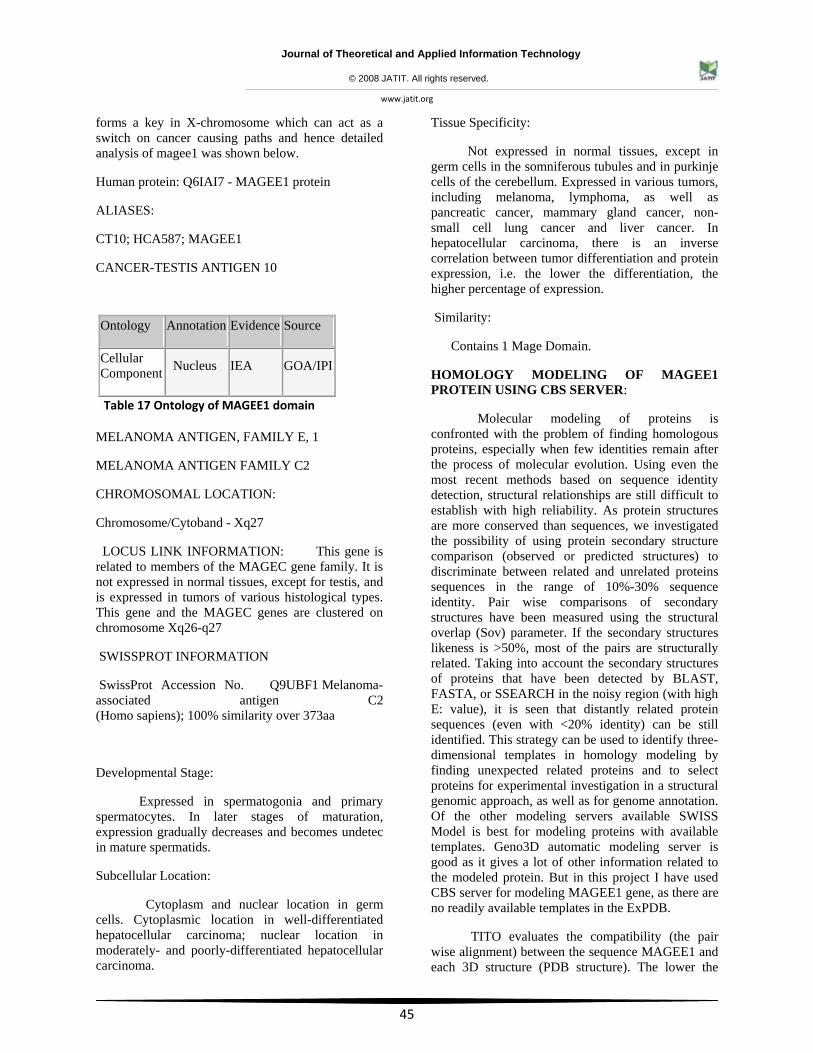
Cellular Component Nucleus IEA GOA/IPI
Table 17 Ontology of MAGEE1 domain
Journal of Theoretical and Applied Information Technology
© 2008 JATIT. All rights reserved.
www.jatit.org
46
TITO Score, the better the compatibility and the alignment.
Receiving and analyzing the model:
http://bioserv.cbs.cnrs.fr/TITO/result/tito.69040/frame_tito.html
Figure 8 Secondary structure of the MAGEE gene
Total Fragments (197): PKB = -78363 (-397.78 /residue)
The parenthesis gives the length of the insertions/deletions
Journal of Theoretical and Applied Information Technology
© 2008 JATIT. All rights reserved.
www.jatit.org
47
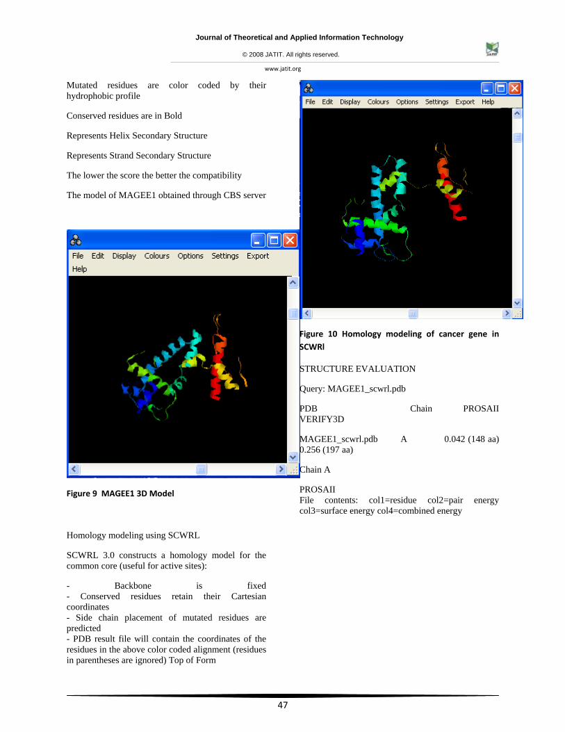
Mutated residues are color coded by their hydrophobic profile
Conserved residues are in Bold
Represents Helix Secondary Structure
Represents Strand Secondary Structure
The lower the score the better the compatibility
The model of MAGEE1 obtained through CBS server
Figure 9 MAGEE1 3D Model
Homology modeling using SCWRL
SCWRL 3.0 constructs a homology model for the common core (useful for active sites):
- Backbone is fixed - Conserved residues retain their Cartesian coordinates - Side chain placement of mutated residues are predicted - PDB result file will contain the coordinates of the residues in the above color coded alignment (residues in parentheses are ignored) Top of Form
Figure 10 Homology modeling of cancer gene in SCWRl
STRUCTURE EVALUATION
Query: MAGEE1_scwrl.pdb
PDB Chain PROSAII VERIFY3D
MAGEE1_scwrl.pdb A 0.042 (148 aa) 0.256 (197 aa)
Chain A
PROSAII File contents: col1=residue col2=pair energy col3=surface energy col4=combined energy
Journal of Theoretical and Applied Information Technology
© 2008 JATIT. All rights reserved.
www.jatit.org
48
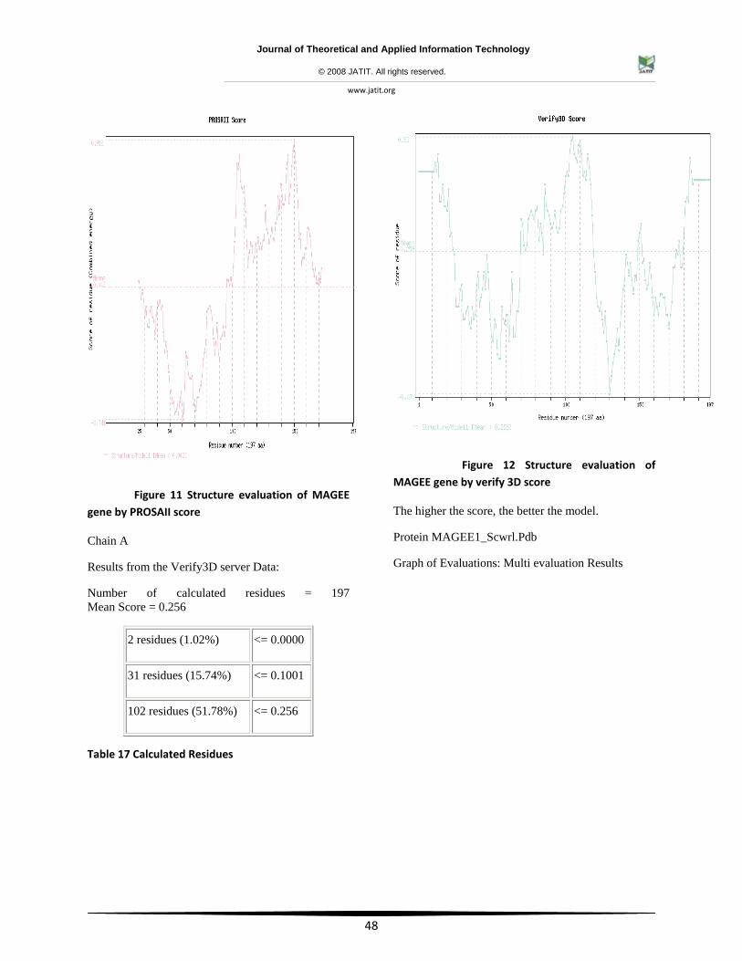
Figure 11 Structure evaluation of MAGEE gene by PROSAII score
Chain A
Results from the Verify3D server Data:
Number of calculated residues = 197 Mean Score = 0.256
Table 17 Calculated Residues
Figure 12 Structure evaluation of MAGEE gene by verify 3D score
The higher the score, the better the model.
Protein MAGEE1_Scwrl.Pdb
Graph of Evaluations: Multi evaluation Results
2 residues (1.02%) <= 0.0000
31 residues (15.74%) <= 0.1001
102 residues (51.78%) <= 0.256
Journal of Theoretical and Applied Information Technology
© 2008 JATIT. All rights reserved.
www.jatit.org
49
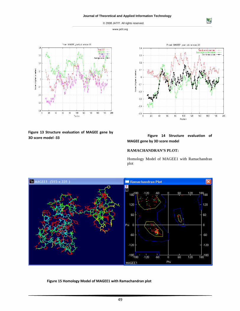
Figure 13 Structure evaluation of MAGEE gene by 3D score model ‐33
Figure 14 Structure evaluation of MAGEE gene by 3D score model
RAMACHANDRAN’S PLOT:
Homology Model of MAGEE1 with Ramachandran plot
Figure 15 Homology Model of MAGEE1 with Ramachandran plot
Journal of Theoretical and Applied Information Technology
© 2008 JATIT. All rights reserved.
www.jatit.org
50
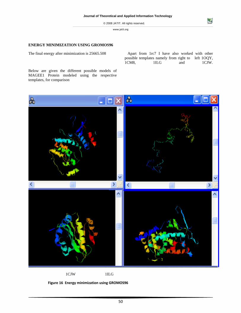
ENERGY MINIMIZATION USING GROMOS96
The final energy after minimization is 25665.508
Apart from 1rc7 I have also worked with other possible templates namely from right to left 1OQY, 1CM8, 1ILG and 1CJW.
Below are given the different possible models of MAGEE1 Protein modeled using the respective templates, for comparison
1CJW 1ILG
Figure 16 Energy minimization using GROMOS96
Journal of Theoretical and Applied Information Technology
© 2008 JATIT. All rights reserved.
www.jatit.org
51
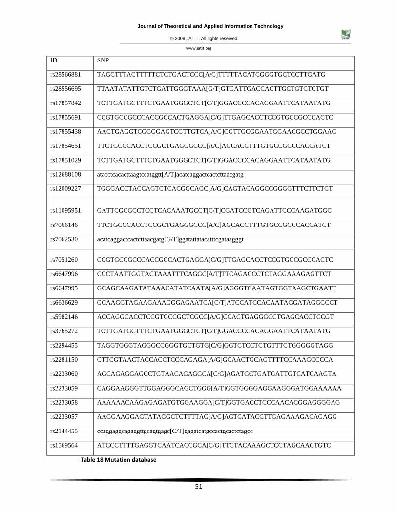
ID SNP
rs28566881 TAGCTTTACTTTTTCTCTGACTCCC[A/C]TTTTTACATCGGGTGCTCCTTGATG
rs28556695 TTAATATATTGTCTGATTGGGTAAA[G/T]GTGATTGACCACTTGCTGTCTCTGT
rs17857842 TCTTGATGCTTTCTGAATGGGCTCT[C/T]GGACCCCACAGGAATTCATAATATG
rs17855691 CCGTGCCGCCCACCGCCACTGAGGA[C/G]TTGAGCACCTCCGTGCCGCCCACTC
rs17855438 AACTGAGGTCGGGGAGTCGTTGTCA[A/G]CGTTGCGGAATGGAACGCCTGGAAC
rs17854651 TTCTGCCCACCTCCGCTGAGGGCCC[A/C]AGCACCTTTGTGCCGCCCACCATCT
rs17851029 TCTTGATGCTTTCTGAATGGGCTCT[C/T]GGACCCCACAGGAATTCATAATATG
rs12688108 atacctcacacttaagtccatggtt[A/T]acatcaggactcactcttaacgatg
rs12009227 TGGGACCTACCAGTCTCACGGCAGC[A/G]CAGTACAGGCCGGGGTTTCTTCTCT
rs11095951 GATTCGCGCCTCCTCACAAATGCCT[C/T]CGATCCGTCAGATTCCCAAGATGGC
rs7066146 TTCTGCCCACCTCCGCTGAGGGCCC[A/C]AGCACCTTTGTGCCGCCCACCATCT
rs7062530 acatcaggactcactcttaacgatg[G/T]ggatattatacatttcgataagggt
rs7051260 CCGTGCCGCCCACCGCCACTGAGGA[C/G]TTGAGCACCTCCGTGCCGCCCACTC
rs6647996 CCCTAATTGGTACTAAATTTCAGGC[A/T]TTCAGACCCTCTAGGAAAGAGTTCT
rs6647995 GCAGCAAGATATAAACATATCAATA[A/G]AGGGTCAATAGTGGTAAGCTGAATT
rs6636629 GCAAGGTAGAAGAAAGGGAGAATCA[C/T]ATCCATCCACAATAGGATAGGGCCT
rs5982146 ACCAGGCACCTCCGTGCCGCTCGCC[A/G]CCACTGAGGGCCTGAGCACCTCCGT
rs3765272 TCTTGATGCTTTCTGAATGGGCTCT[C/T]GGACCCCACAGGAATTCATAATATG
rs2294455 TAGGTGGGTAGGGCCGGGTGCTGTG[C/G]GGTCTCCTCTGTTTCTGGGGGTAGG
rs2281150 CTTCGTAACTACCACCTCCCAGAGA[A/G]GCAACTGCAGTTTTCCAAAGCCCCA
rs2233060 AGCAGAGGAGCCTGTAACAGAGGCA[C/G]AGATGCTGATGATTGTCATCAAGTA
rs2233059 CAGGAAGGGTTGGAGGGCAGCTGGG[A/T]GGTGGGGAGGAAGGGATGGAAAAAA
rs2233058 AAAAAACAAGAGAGATGTGGAAGGA[C/T]GGTGACCTCCCAACACGGAGGGGAG
rs2233057 AAGGAAGGAGTATAGGCTCTTTTAG[A/G]AGTCATACCTTGAGAAAGACAGAGG
rs2144455 ccaggaggcagaggttgcagtgagc[C/T]gagatcatgccactgcactctagcc
rs1569564 ATCCCTTTTGAGGTCAATCACCGCA[C/G]TTCTACAAAGCTCCTAGCAACTGTC
Table 18 Mutation database
Journal of Theoretical and Applied Information Technology
© 2008 JATIT. All rights reserved.
www.jatit.org
52
MAGEE1 GENE THERAPEUTICS
The melanoma antigen genes (MAGE) were found to express in a variety of tumors of different histological origin. Peptides encoded by the MAGE genes are tumor rejection antigens, which can induce specific cytotoxic T-lymphocytes (CTL) to recognize and kill tumor cells. This lead to the idea of using MAGE genes as the targets for cancer immunotherapy, and MAGE peptides are currently being investigated as immunizing agents in clinical studies. Although 23 human and 12 mouse MAGE genes have been isolated in various tumors and characterized, not much is known about their function in normal cells. In adult tissues, most MAGE genes are expressed only in the testis and expression patterns suggest that this gene family is involved in germ cell development. In contrast to the MAGE genes, more functional data have accumulated around the MAGE related gene necdin. This gene encodes a neuron-specific growth suppressor that facilitates the entry of the cell into cell cycle arrest. Necdin also interacts with p53 and works in an additive manner to inhibit cell growth. In this review we will focus on the normal functions of MAGE genes and we speculate that this gene family is involved in cell cycle regulation, especially during germ cell development.
CONCLUSION:
The study of the tissue specificity of cancer genes in human X-chromosome majorly expressed in sex organs gave a clue that the cancer has been evolved only in mammalians and hence of great importance in understanding the mechanism of cancer. The probable explanation could be the growing number of genes that escape X-chromosome inactivation and are expressed from the both otherwise inactive X-chromosome and active X-chromosome (Carolyn J. Brown 1996). The study on the homology of some of the cancer and the gene associated in X- chromosome proved that MAGE tumor-specific antigen (CT antigens) are expressed with high percentage in all cancer and their products are promising targets for antigen-specific immunotherapy. Many questions regarding CT genes remain, including the biological function of their protein products, and the elucidation of the factors that control their expression in normal tissues and cancer. Epigenetic events such as DNA methylation and histone acetylation are known to influence CT gene expression. Based on their immunogenicity and restricted expression, CT antigens are ideal for use in cancer vaccines. To date, approximately 90 CT transcripts have been identified, thus providing a substantial arsenal for active, antigen-specific immunotherapy of cancer. Many of the antigens have
been proven to be immunogenic in cancer patients. The research can continue with the crucial experiments that will test whether CT-antigen genes are tumorigenic and features generally associated with malignancy are acquired by cells that aberrantly express CT antigens. In addition, it will lead to the identification of a novel set of therapeutic targets with highly restricted expression in normal cells. These targets could be susceptible to a range of treatment modalities beyond cancer vaccines. Among the other genes Magee-1 is found be interesting due to various homolog and hence a comprehensive analysis was done to understand magee-1 and its implication in cancer. The structure was solved theoretically using homology modeling; probably MAGEE-1 can be sui drug target for cancer.
REFERENCES:
[1]. Caulfield (1999). The Commercialization of Genetic Research: Ethical, Legal, and Policy Issues. New York: Kluwer.
[2]. Stevenson BJ, Iseli C, Panji S, Zahn-Zabal M, Hide W, Old LJ, Simpson AJ, Jongeneel CV ( 2007 ). Rapid evolution of cancer/testis genes on the X chromosome. BMC Genomics. May 23;8:129.
[3]. Gure, A. O.; Stockert, E.; Arden, K. C.; Boyer, A. D.; Viars, C. S.; Scanlan, M. J.; Old, L. J.; Chen, Y.-T. CT10: (2000). A new cancer-testis (CT) antigen homologous to CT7 and the MAGE family, identified by representational-difference analysis. Int. J. Cancer 85: 726-732,PubMed ID : 10699956.
[4]. Barker Philip A & Salehi Amir (2002) . The MAGE proteins : emerging roles in cell cycle progression, apoptosis, and neurogenetic disease, J Neurosci Res, Vol:67 Issue:6 Page:705-12 Mar 15.
[5]. Mashino K , Sadanaga N, Tanaka F, Yamaguchi H, Nagashima H, Inoue H, Sugimachi K, Mori M. (2001). Expression of multiple cancer-testis antigen genes in gastrointestinal and breast carcinomas Br J Cancer, Vol:85 Issue:5 Page:713-20 Sep 01.
[6]. Figueiredo David L A , Mamede RC, Proto-Siqueira R, Neder L, Silva WA Jr, Zago MA (2006). Expression of cancer testis antigens in head and neck,squamous cell carcinomas, Head Neck, Vol:28 Issue:7 Page:614-9 Jul.
[7]. Ries Lag, et al., ( 1999). SEER Cancer Statistics Review, 1975–2001, National Cancer Institute.
Journal of Theoretical and Applied Information Technology
© 2008 JATIT. All rights reserved.
www.jatit.org
53
Bethesda, MD, 2004 (http://seer.cancer.gov/csr/1975_2001). Breast Cancer: The Complete Guide by Yashar Hirshaut, MD and Peter Pressman, MD (Bantam).
[8]. Lucas, S.et.al (2000).MAGE-B5, MAGE-B6, MAGE-C2, and MAGE-C3: four new members of the MAGE family with tumor-specific expression. Int. J. Cancer 87: 55-60, PubMed ID : 10861452
[9]. Caulfield,et.al(1999). The Commercialization of Genetic Research: Ethical, Legal, and Policy Issues. New York: Kluwer.
[10]. Fichtner-Feigl S, et.al (2005 ).Signaling through the IL-13alpha2 receptor is involved in induction of TGF-beta1 production and fibrosis. Nat Med. 2006 Jan;12(1):99-106. Epub Dec 4
[11]. Mizutani Ket.al (2005 ). WAVE3 functions as a negative regulator of LDOC1. J Biochem (Tokyo). Nov;138(5):639-46.
[12]. Murphy CE, et.al (2005). Steroid hormone receptor expression in male breast cancer. Eur J Surg Oncol. 2006 Feb;32(1):44-7. Epub Nov 2.
[13]. Jäger D,et.al (1999). Cancer-testis antigens and ING1 tumor suppressor gene product are breast cancer antigens: characterization of tissue-specific ING1 transcripts and a homologue gene. Cancer Res. Dec 15;59(24):6197-204.
[14]. Pedeux R,et.al (2005). ING2 regulates the onset of replicative senescence by induction of p300-dependent p53 acetylation. Mol Cell Biol. 2005 Aug;25(15):6639-48.
[15]. De la Fuente J,and Dokal I(2007). Dyskeratosis congenita: Advances in the understanding of the telomerase defect and the role of stem cell transplantation Pediatr Transplant. Sep;11(6):584-594.
[16]. Kenji Tokita,et.al (2002). Molecular Basis of the Selectivity of Gastrin-Releasing Peptide Receptor for Gastrin-Releasing PeptideVol. 61, Issue 6, 1435-1443, June.
[17]. Spiro SG, Silvestri GA (2005). One hundred years of lung cancer. Am J Respir Crit Care Med 172: 523–529.
[18]. Bach PB,et.al (2003). Screening for lung cancer: A review of the current literature. Chest 123: 72S–82S.
[19]. Russell S. Thomas,et.al (2006). A Comparison of Transcriptomic and Metabonomic Technologies for Identifying Biomarkers Predictive of Two-Year Rodent Cancer Bioassays ToxSci Advance Access published online on November 17.
[20]. Michael Koslowski, et.al (2006). The human X chromosome is enriched for germline genes expressed in premeiotic germ cells of both sexes. Human Molecular Genetics Advance Access originally published online on June 29.Human Molecular Genetics 2006 15(15):2392-2399; doi:10.1093/hmg/ddl163.
[21]. Kristi A. Egland, et.al (2002). Characterization of Overlapping XAGE-1 Transcripts Encoding a Cancer Testis Antigen Expressed in Lung, Breast, and Other Types of Cancers. Vol. 1, 441-450, May. Molecular Cancer Therapeutics Gene Expressed in Lung Cancer - study of EGFL6 - Brief Article.
[22]. Granel B,et.al (2006).IL13RA2 gene polymorphisms are associated with systemic sclerosisJ Rheumatol.Oct;33(10):2015-9. Epub 2006 Sep 15.
[23]. Nagasaki, K,et.al (1999).Identification of a novel gene, LDOC1, down-regulated in cancer cell lines. Cancer Lett. 140: 227-234.
[24]. E. Zoumakis,et.al (1997). Endometrial corticotropin-releasing hormone. Its potential autocrine and paracrine actions. Annals of the New York Academy of Sciences, Vol 828, Issue 1 84-94, Copyright © by New York Academy of Sciences
[25]. Dezhong Joshua Liao,et.al. Novel Perspective: Focusing on the X Chromosome in Reproductive Cancers.
[26]. Hiroki Torikai ,et.al (2004). A Novel HLA-A*3303-Restricted Minor Histocompatibility Antigen Encoded by an Unconventional Open Reading Frame of Human TMSB4Y Gene1. The Journal of Immunology, 173: 7046-7054.
[27]. Byeong-Chel Lee ,et.al (2003). P2Y-like receptor, GPR105 (P2Y14), identifies and mediates chemotaxis of bone-
marrowhematopoietic stem cells. GENES & DEVELOPMENT 17:1592-1604.
Journal of Theoretical and Applied Information Technology
© 2008 JATIT. All rights reserved.
www.jatit.org
54
[28]. Jun He et.al (2002). Targeted disruption of Dkc1, the gene mutated in X-linked dyskeratosis congenita, causes embryonic lethality in mice 31 October, Volume 21, Number 50, Pages 7740-7744.
[29]. Simpson Andrew J G ,et.al (2005). Cancer/testis antigens, gametogenesis and cancer (PMID: 16034368) Nat Rev Cancer, Vol:5 Issue:8 Page:615-25 Aug.
[30]. Brasseur F , et.al (1995). Expression of MAGE genes in primary and metastatic
cutaneous melanoma Int J Cancer, Vol:63,Issue:3, Page:375-80 Nov03.
[31]. Old, L. J. Cancer/testis (CT) antigens- a new link between gametogenesis and cancer. Cancer Immun. 1, 1 (2001).
[32]. Scanlan, M. J., Simpson, A. J. & Old, L. J. The cancer/testis genes: review, standardization, and commentary. Cancer Immun. 4, 1 (2004). A comprehensive listing and standardization of the CT-antigen families.