STUDY OF ABO HAEMOLYTIC DISEASE OF THE NEWBORN IN … · A STUDY OF ABO HAEMOLYTIC DISEASE OF THE...
36
A STUDY OF ABO HAEMOLYTIC DISEASE OF THE NEWBORN IN HOSPITAL OF UNIVERSITI SAINS MALAYSIA KELANTAN By: DR SALMAN MOHD SAHID Dissertation submitted in partial fulfillment of the requirements for the degree of Master of Medicine (Pathology). Uni ve rs iti Sains Mal ay sia Novemb er 200 I
Transcript of STUDY OF ABO HAEMOLYTIC DISEASE OF THE NEWBORN IN … · A STUDY OF ABO HAEMOLYTIC DISEASE OF THE...

A STUDY OF ABO HAEMOLYTIC DISEASE OF THE NEWBORN IN HOSPITAL OF
UNIVERSITI SAINS MALAYSIA KELANTAN
By:
DR SALMAN MOHD SAHID
Dissertation submitted in partial fulfillment of the requirements for the degree of Master of Medicine (Pathology).
Universiti Sains Malaysia
November 200 I
ACKNOWLEDGEMENTS
I am in great debt to Dr Rapiaah Mustaffa, my supervisor and lecturer of
Haematology Department HUSM for her generous help, advice and innumerable
suggestion for improvements through out this project.
Many thanks to Ass Prof Dr Normah, Dr Narazah and Dr Rosline who has
given many suggestion especially in the early part of the project. Special thank to Ass
Prof Dr Mohd Shukri Othman, Head of Obstetric and Gynaecology HUSM for
aHowing his patients to be chosen for the study.
I am also acknowledge my gratitude to doctors and nurses of the Labour
Room, HUSM for the help while collecting the specimens and laboratory
technologists for the help while processing and analysing the specimens. Not to
forget, my Community Medicine collegues who provide great help in analysing
stastitical part of the data.
:
Most of all, I am grateful for the understanding and support of my wife and
family during the hours devoted to this project.
11

TABLE OF CONTENTS
Contents Page
1. lritle ........................................................................................... 1
2. Acknowledgement •••••••••••••••••••••••••••••••••••••••••••••••••••••••••••••••••••••••• ii
3. Table of contents ••••••••••••••••••.••••••••••••.••••••••••••••••••••••••••••••••••••••••• iii
4. List of ta bles ................................................................................ v
5. List of figtJ res •••••••••••••••••••••••••••••••••••••••••••••••••••••••••••••••••••.••••••••• VII
6. List of abbreviations ••••••••••••••••••••••••••••••••••••••••••.••••••••••••••••••••••••• VUI
7. Abstract (in Malay language) •••.••.•..••..••••..••..••••...••......••••••.•..••••.•.. IX
8. A bstract (in English) •••..•••.•••••...••••......•••.••..•••.•.•.•••..••••••..•.•.•••..••• XI
9. Chapter t: Introduction
1.1 Definition ............................................................................... 2
1.2 Historical aspects ..................................................................... 4
1.3 Pathophysiology ....................................................................... 5
1.4 Incidence ............................................................................... 8
1. 5 Clinical features ...................................................................... 12
1.5.1 Spectrum oflIDN ..................................................... 12
1.5.2 Common manifestation ............................................... 14
I .6 Laboratory findings ................................................................. 15
1.6.1 Serology ................................................................ 15
1.6.2 Haematology ........................................................... 17
1.6.3 Special Tests .. , ........................................................ 19
1.7 Management .... , ............................ '" ... '" ................ , .............. 21
11]

1.8 Rationale of the study .............................................................. 23
10. Chapter 2: Research Methodology
2.1 Aim and objectives ................................................................. 27
2.2 Study design and sampling method .............................................. 28
2.3 Selection of measures .............................................................. 29
2.4 Study samples ....................................................................... 30
2.5 Sample size .......................................................................... 32
2.6 Samples collection .................................................................. 33
2.7 Sample analysis ..................................................................... 35
2.8 Selection of analysis ................................................................ 45
2.9 Data collection on clinical feature and management ........................... 46
2.10 Limitations of study ............................................................... 47
11. Chapter 3: Results
3.1 Pre-analytical data ................................................................... 49
3.2 Analysis of parameters .............................................................. 53
3.3 Analysis of clinical feature and management .................................... 62
3.4 Incidence and severity of ABO incompatibility and ABO HDN ............. 64
12. Chapter 4: Discussion .•••••••••••••••••.•••••••••••.•••••••••••••••••••••....••••.•.••• 66
13. Chapter 5: Conclusion •••••••••.••..•....•••••••••.•••••••••••••••••••••••..••.••••••..• 74
14. References •••••••••••••••••••••••. •••••••••••••••••••• ••••••••••••••••••••••••••••••••••••• 76
15. Appendix ••••••••••••••••••••••••••••••••••••••••••••••••••••••••••••••••••••••••••••••••••• 82
IV

LIST OF TABLES
Table 1.1
Table 1.3
Table l.4
Table 3.l.1
Table 3.l.2
Alloantibodies reported to cause HDN.
Properties of human IgG subclasses.
The incidence of ABO maternal - infant incompatibility among jaundice
baby.
Blood group 0 mothers.
Data of newborn babies.
Table 3.2.1a Mean and standard deviation of haemoglobin among ABO compatible and
ABO incompatible.
Table 3.2.1 b Mean and standard deviation of haemoglobin among blood group A and
B.
Table 3.2.1 cAnova test for multiple comparisons of mean of haemoglobin among
various blood groups.
Table 3.2.2 Mean and standard deviation ofreticulocytes count among ABO
compatible and ABO incompatible.
Table 3.2.3 Mean and standard deviation of serum bIiIirubin among ABO compatible
and ABO incompatible.
Table 3.2.4a Mean and standard deviation of spherocytes count among ABO compatible
and ABO incompatible.
v

Table 3.2.4b Mean and standard deviation of spherocytes count among blood group A
andB.
Table 3.2.4c Anova test for multiple comparisons of mean of spherocytes count among
various blood groups.
Table 3.2.5 Mean and standard deviation ofNRBC per 100 WC among ABO
compatible and ABO incompatible.
Table 3.2.6 Results of direct antiglobulin test among ABO compatible and ABO
incompatible.
Table 3.3.1 Number and percentage of jaundice among ABO incompatible babies.
Table 3.3.2 Data on management of the five clinically jaundice babies.
Table 3.3.3 The jaundice babies and their cord blood results.
Table I Distribution and percentage of total number of donors who donated blood
within five years.
VI

LIST OF FIGURES
Figure 1.3 Diagram showing the fetus in relation to placenta and uterine wall.
Figure 3. I .2a Bar chart showing the number of babies for each of the blood groups.
Figure 3.1.2b Bar chart showing distribution of races among ABO compatible and ABO
incompatible.
Figure 3.2.4 Peripheral blood film from one of the ABO incompatible babies in this study
showing many spherocytes.
VII

LIST OF ABBRREVIATIONS
ABOHDN
ADCC
DAT
EDTA
ELAT
ELISA
G6PD
Hb
HUSM
NADPH
NICU
NNJ
NRBC per 100 WC
SD
ABO haemolytic disease of the newborn
Antibody-dependent cell-mediated cytotoxicity
Direct antiglobulin test
Ethylenediamine tetra-acetic acid
Enzyme-linked antiglobulin test
Enzyme-linked immunosorbent assay
Glucose-6-phospatase dehyrogenase
Haemoglobin
Hospital of Universiti Sains Malaysia
Reduced nicotine adenine dinucleotide phosphate
Neonatal intensive care unit
Neonatal jaundice
Nucleated red blood cell per hundred white cells
Standard deviation
VII)
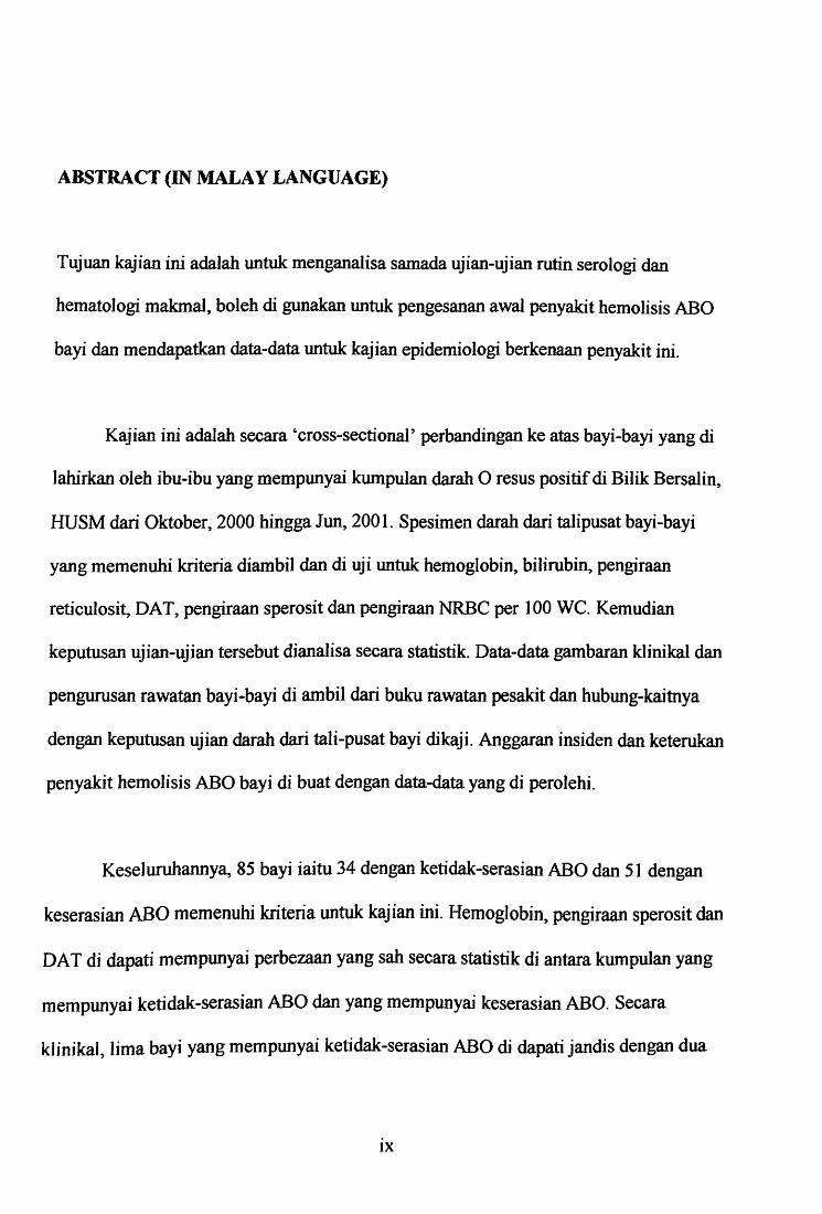
ABSTRACT (IN MALAY LANGUAGE)
Tujuan kajian ini adalah untuk menganalisa samada ujian-ujian rutin serologi dan
hemato]ogi makmal, boleh di gunakan untuk pengesanan awal penyakit hemolisis ABO
bayi dan mendapatkan data-data untuk kajian epidemiologi berkenaan penyakit ini.
Kajian ini adalah secara 'cross-sectional' perbandingan ke atas bayi-bayi yang di
lahirkan oleh ibu-ibu yang mempunyai kumpulan darah 0 resus positif di Bilik Bersalin,
HUSM dari Oktober, 2000 hingga Jun, 2001. Spesimen darah dati talipusat bayi-bayi
yang memenuhi kriteria diambil dan di uji untuk hemoglobin, bilirubin, pengiraan
reticulosit, DAT, pengiraan speros it dan pengiraan NRBC per 100 WC. Kemudian
keputusan ujian-ujian tersebut dianalisa secara statistik. Data-data gambaran klinikal dan
pengurusan rawatan bayi-bayi di ambil dari buku rawatan pesakit dan hubung-kaitnya
dengan keputusan ujian darah dari tali-pusat bayi dikaji. Anggaran insiden dan keterukan
penyakit hemolisis ABO bayi di buat dengan data-data yang di perolehi.
Keseluruhannya, 85 bayi iaitu 34 dengan ketidak-serasian ABO dan 51 dengan
keserasian ABO memenuhi kriteria untuk kajian ini. Hemoglobin, pengiraan sperosit dan
DA T di dapati mempunyai perbezaan yang sab secara statistik di antara kumpulan yang
mempunyai ketidak-serasian ABO dan yang mempunyai keserasian ABO. Secara
kJinikal, lima bayi yang mempunyai ketidak-serasian ABO di dapati jandis dengan dua
IX

daripada mereka telah menerima rawatan terapifoto dan tiada 'exchange transfusion' di
perlukan. Insiden ketidak-serasian ABO di kalangan bayi-bayi di HUSM ialah 15.6%.
Insiden penyakit hemolisis ABO bayi ialah 2.3%. Insiden bagi bayi yang mempunyai
ketidak-serasian ABO memerlukan terapifoto di sebabkan penyakit hemolisis ABO bayi
ialah 6.7% dan insiden untuk semua bayi memerlukan terapifoto di sebabkan penyakit ini
pula ialah 0.92%.
Tiada hubung-kait yang jeJas di antara gambaran klinikal dan keputusan ujian
makmal. Ujian darah menerusi talipusat bayi, tidak boleh di cadangkan sebagai cara
untuk ujian pengesanan awal untuk penyakit hemolisis ABO bayi. Ujian darah talipusat
bayi adalah tidak sensitif dan spesifik berkemungkinan kerana keadaan penyakit tersebut
yang tidak seri us di HUSM Insiden dan keterukan penyakit hemolisis ABO bayi di sini
adalah hampir menyamai peratusannya di Amerika Utara dan Eropah.
x

ABSTRACT (IN ENGLISH)
The study was to evaluate whether the routine serological and haematological laboratory
tests could be used for an early diagnosis of ABO HDN and to obtain epidermiologicaI
data.
This is a comparative cross-sectional study of newborn babies born to blood
group 0 rhesus positive mothers in labour room of HUSM from October, 2000 to June,
2001. Specimens from cord blood of suitable babies were collected and investigated for
haemoglobin, bilirubin, reticulocytes count, DAT, spherocytes count and NRBC per 100
WC. The results were analysed statistically. The clinical features and management data of
ABO incompatible babies were taken from management file and correlation made with
the cord's blood findings. The incidence and severity of ABO HDN were also estimated
from the data.
Altogether 85 babies ie 34 ABO incompatible and 51 ABO compatible. The
haemoglobin, spherocytes count and DA T showed a statistically significant difference
between ABO incompatible and ABO compatible babies. Clinically five babies of ABO
incompatible noted to be jaundice with two of them received phototherapy but exchange
transfusion was not needed in any of them. The incidence of ABO incompatible infants in
HUSM is 15.6%. The incidence of ABO HDN is 2.3%. The incidence of ABO
Xl

incompatible infants requiring phototherapy due to ABO HDN is 6.7% and the incidence
of all infants requiring phototherapy due to ABO HDN is 0.92%.
There is poor correlation between the cl inical features and laboratory findings.
The cord blood tests therefore can not be recommended as method of early diagnosis of
ABO HDN. The cord blood tests were not sensitive and specific probably due to
mildness of ABO HDN in HUSM. The incidence and severity of ABO HDN here are
almost comparable to the figures of North America and Europe.
Xll

Chapter 1
INTRODUCTION
1

1.0 INTRODUCTION
1.1 DEFINITION
Haemolytic disease of the newborn (HON) is a condition in which the life
span of the infant's red cells is shortened by the action of specific antibodies
derived from the mother by placental transfer. The disease begins in intra-uterine
life and is therefore correctly described as haemolytic disease of the fetus and
newborn but the simple term haemolytic disease of the newborn has been used for
a long time and can be taken to include haemolytic disease of the fetus (Mollison
et al.1997).
HDN is also defined as a disease characterised by the destruction of fetal /
newborn red cells resulting from the placental transfer of maternal alloantibody
(Strohm 1995).
The placental transfer of maternal alloantibody is only of IgG class.
The commonest JgG red cell antibodies in human serum are anti-A and anti-B and
relatively high concentrations are found only in group 0 subjects (Mollison et al.
1997). This is because the group 0 individuals are "naturally" pre-sensitised to A
and B antigens by exposure to ABO-I ike substances found in food or other
exogenous sources (Foerster 1992). The anti-A and anti-B occurring in group B
2

and A subjects respectively, are almost of IgM variety that cannot cross the
placental barrier.
The disease cause by these anti-A and anti-B are usually termed as ABO
HDN and for practical purposes, it is restricted to A or B individuals with mothers
of 0 (Mollison et aI.1997). However, group B infants of group A (particularly A2)
mothers occasionally are affected (Strohm 1995).
There are many other antibodies that can cause HDN. The list of antibodies
in table 1.1 below, includes most but not all of those which can occur as IgG and
known to have caused I-IDN.
Table 1.1: Alloantibodies reported to cause HDN (Adapted from Mollison et aI.
1997).
Within the Rh system Anti-D -c -C _Cw _Cx -e -E -Ew -ce -", , '" , , Ces -Rh32 -Goa _Bea -Evans -L W , , " ,
Outside the Rh system Anti-K -k, -Ku _Kpa _Kpb -Js8 -Jsb _Fya , , , , , , , -Fy3 _Jka -Jkb -M -N -S -s -u -Vw -Far , , , , "', , , _Mv -Mit, -Mta -Mur -Hi I - Hut -Ena -pp , """ pk L 0 L b L 9 D·o D·b 0 b - - U - U - U - 1 1 - Yt -Yt -, , , , , , , ,
Doa -Coa -Wr8 , ,
Antibodies to high- Anti _At8 -J ra -Lan -Ge , , , incidence antigens
Antibodies to low- Anti-Bi, -By, -Fra, -Good, -Rd, _Rea, -Zd incidence antigens
3

1.2 HISTORICAL ASPECTS
In 1609, a French midwife, Louyse Bourgeois, writing in the popular Paris
press, was the first to describe haemolytic disease of the newborn. She reported
the birth of twins: the first twin was bloated with fluid (hydropic) and died shortly
after birth; the second appeared weII but rapidly became jaundiced (icterus
gravis), lay in a position of opisthotonos and died (kernicterus). These two
conditions ie hydrops fetalis and kernicterus (yeIIow staining of the brain) were
described in detail by pathologists at the turn of the century but were not thought
to be the same entity until 1932 (Bowman 1998).
In 1932, Diamond and co-workers showed that hydrops fetalis, icterus
gravis, and kernicterus were simply different spectra of the same disease
characterised by hemolytic anaemia, extramedullary erythropoiesis,
hepatosplenomegaly and the out-pouring of immature nucleated red blood cells
(Naiman 2001). The ABO HDN was however, first described in 1944 by
Halbrecht (Brouwers et al. 1988a).
4

1.3 PATHOPHYSIOLOGY
In humans, the transfer of antibodies from mother to fetus takes place Ol'Jy
via t.1-Je placenta. The only immunoglobulin transferred is IgG, which is bound by
an Fc receptor on the plasma membrane of the placenta. The transfer ofIgG is an
active process and takes place only from mother to fetus and not in the reverse
direction.
Figure 1.3: Diagra.111 shovling the fetus in relation to placenta and uterine \vaLL
Amniotic fluid
(Adapted from Ortho Diagnostics, 1968).
5
Umbilical cord
Placenta
Fetus

The sensitisation of mother's immune system or alloimmunisation
increase \vith each incompatible pregnancy. The primary stimulus for
itl".nlunisation, in addition to exposure to ABO-like substances such as found in
food, can also be a previous blood transfusion or abortion. Formation ofRh
a.lltibodies is the most common form of alloimmunisation to give rise to clinically
import..ant disease. Hovvever during pregnancy, ABO immunisation occurs more
often t.1)an Rh immunisation (Wells and Isbister, 1997).
ABO HDN is quite different from HDN due either to anti-D or other blood
group antibodies. ft.Jlti-A and anti-B, \vhich bind complement in adults, cause
violent, life-threathening intravascular hemolysis after transfusion of ABO
incompatible blood Ho\vever fetal ABO HDN is usually much milder than D, c
and K forms ofHDN.
Several reasons can be listed for the paradoxical mildness of PJ30 HD~J.
First, there are fe\ver A and B antigenic sites on the fetal RBC membrane. ~AJso,
anti-A and anti-B (with very rare exception; Pujol et aI. 1991) do not activate
complement on the fetal RBC membrane (Wang and Desforges, 1971; Broll'Ners
et al. 1988b). Second, anti-A and anti-B are mostly IgM, ,vhich does not cross the
placenta (Ramasethu and Luban, 2001). Third, the small amounts ofIgG anti-A
and anti-B that do traverse the placenta have myriad antigenic sites other than
those on P-BCs, other tissues, and secretions to ,vruch they may bind. Only a very
sma11 proportion of the minor amount of anti-A or anti-B that crosses the placenta
6

adheres to antigen on the fetal RBC membrane (Bo\\l1l1an 1998). Fourth, the
severity may be related to IgG subclass. IgG2 constitutes a significant component
of anti-A and anti-B antibody; this subclass oflgG is transported less readily
across the placenta than are IgGl or IgG3 and is a less efficient mediator of
macrophage-induced red cell clearance (Ramasethu and Luban, 2001).
Table 1.3: Properties of human IgG subclasses (Pars!o\v 1997).
I igGl I IgG2 IgG3 IgG4 i
Abundance (% oftotai IgG) 70 I 20 6 4 I
I Half-life in serum (days) 23 I 23 7 23 I I
Placental passage -I I 1 I 1 -I I I I I I I I I
I , i
I Complement fixation + I +
I +++ -
I I I
, Binding to Fe receptors +++ + I +++ -
The relative concentrations of the four subclasses vary some,vhat among
individuals. It appears that the propensity to produce IgG antibodies of one
subclass or another is at least partly an inherited trait (parslo\v 1997). The
antibodies with the subclass with the highest titre, strongly influence the result of
the DAT, and in many cases of ABO incompatibilities these are IgG2 antibodies.
Ho\vever, it has been reported that in infants ,vith severe ABO HDN, the IgG
subclasses bound to cord red cells are IgG 1 and / or IgG3 in addition to IgG2
(Ukita eta!. 1989).
7

1.4 INCIDENCE
Incidence of ABO HDN depends on incidence of ABO incompatibility. It
should be noted however that ABO incompatibility is not synonymous with ABO
HDN. In other words, ABO HDN is the extreme end of the spectrum of ABO
incompatibility.
ABO hemolytic disease must be defined before its incidence can be
estimated. For example, taking the criterion of the development of jaundice within
24 hours of birth, the incidence was estimated by Halbrecht (1951) to be one in
180; taking the faintest trace of jaundice in the first 24 hours as the criterion the
incidence was found by Valentine (1958) to be as high as one in 70. In a study by
Meberg and Johansen (1998), the ABO incompatible babies that required
phototherapy were one in 106 or 0.94% of all term infants.
Cases ofHDN due to anti-A or anti-B which are severe enough to need
exchange transfusion are relatively rare for examples three of 8000 births (Ames
and Lloyd 1964); six of 5704 newborn infants (V oak and Bowley 1969); none
amongst 534 infants born to group 0 mothers (Meberg and Johansen 1998).
A higher incidence has been found in some other populations. The
incidence of ABO HDN was found to be substantially higher in Venezuela, in
which about 30% of ABO incompatible infants have signs of haemolytic disease
8

compared with 20% or less in European and North American populations (Cariani
et al. 1995). The disease was found to be commoner in Blacks by Kirkman
(1977). Peevy and Wiseman (1978) also support the evidence concerning racial
differences but not in the severity of ABO HDN between black and white infants.
In a survey in Nigeria, the frequency of ABO HDN is about 5% of births
(Worlledge et al. 1974).
AI-Jawad et al. (1985) in their study concluded that ABO HDN was about
as common in Arabs as in Blacks and that the disease tended to be more severe in
Arab than in Europeans; exchange transfusion for ABO HDN was carried out on
one in every 500 newborn Arab infants. Romano et al. (1994) in their study noted
about one in 300 of all newborn infants in Venezuela need exchange transfusion.
However, a study in multi-ethnic hospital in USA concluded that there
was no significant difference in prevalance of clinical disease, which requires
exchange transfusion among Asian, Black, Hispanic and Caucasian infants (Toy
et al. 1988). Their study shows that in a sample of over 10,000 infants, ethnic
differences were detectable in the laboratory but not in clinical disease.
Locally, in HUSM, a study by Siti Aesah (1995), from the period of
October, 1994 - January, 1995, found that, out of total of 58 babies with neonatal
jaundice (NNJ) who were admitted to Neonatal Intensive Care Unit (NICU), only
9

two need exchange transfusion and there were due to ABO HDN. The study also
concluded that 12.1 % of the NNJ babies were ABO HDN.
Ho (1992) has review many studies on incidence of ABO incompatibility
among jaundice baby. The review is summarised in table 1.4.
Table 1.4: The incidence of ABO maternal- infant incompatibility among
jaundice baby.
Country Year % among cases of jaundice
Australia 1983 7.1
China, Beijing 1989 20
Hong Kong 1970 15.6
Hong Kong 1986 12.5
Singapore 1988 16.6
Singapore 1991 15.9
India, New Delhi 1987 12.2
India, Madras 1987 38.2
In relation to DA T positive, the incidence in the cord blood of ABO
incompatible babies are about 25% -30% (Bowman 1989). Toy et al. (1988) have
done a prevalence study of ABO maternal-infant incompatibility in Asians,
Blacks, Hispanics and Caucasians from 10 611 consecutive infants born over a six
10

years period. They have found that the prevalence of a positive DAT among
group A infants born to 0 mothers was different among the four groups and
highest in Asians (50%), follo\ved by Hispanics (42%), Blacks (40%) and
Caucasians (31 %). The positive DAT rates were not different among the four
groups of B infants born to 0 mothers with Asians showing positivity of 39%.
The prevalence of positive DATs in all births was 6,5,5 and 4% respectively in
the four groups.
Another study in Bangkok, Thailand (Chuansumritet al. 1997) found that
the positive rate ofDAT in the ABO incompatible group were 54.5% by a
conventional spin-tube technique and 50% by a gel technique. In the ABO
compatible the positive rate of the DAT were 2.6% by the conventional and
10.5% by the gel technique.
I 1

1.5 CLINICAL FEATURES
1.5.1 SPECTRUM OF HDN
HDN irrespective of whether it cause by ABO, Rhesus or other aIloantibodies can
occur in various forms.
In its least severe form, HDN manifests itself as a mild haemolytic
anaemia. The infant's red cells, coated with maternal IgG antibodies, are removed
prematurely from the circulation, causing slight jaundice (maximum on the
second to third days of life) and mild anaemia during the second week of life.
More severely affected infants show severe hyperbilirubinaemia in the neonatal
period, a condition which was called icterus gravis neonatorum. Prompt treatment
with exchange transfusion is necessary to prevent bilirubin impregnation of the
basal ganglia and neurological damage, a condition known as kernicterus. This
condition may be fatal, or lead to serious neurological deficit, with deafness,
mental retardation, choreo-athetosis and spasticity.
In the most severely affected cases, profound anaemia develops in utero,
and intrauterine death may occur at any time from the eighteenth week of
gestation. Affected fetuses are pale and oedematous, with marked ascites. The
placenta is bulky, swollen and friable. This condition is known as hydrops fetaIis,
and had a high mortality rate until ultrasound-guided intravascular transfusions
12

and improved intensive care facilities for very premature babies were introduced.
The pathophysiology of hydrops is not fully understood, but extravascular
haemolysis with fetal anaemia seems to playa major role by stimulating
extramedullary erythropoiesis in the liver, with distortion of the hepatic
circulation, leading to portal hypertension and impaired albumin production.
Hypoalbuminaemia leads to ascites, oedema and pleural / pericardial effusions. In
addition, the severe anaemia leads to cardiac failure and tissue hypoxia, which
damages the endothelium, leading to fluid extravasation into extravascular space.
13

1.5.2 COMMON MANIFESTATION
ABO HDN is far more comman than Rh HDN but clinical presentation is usually
mild and rarely responsible for fetal deaths (Ramasethu and Luban, 2001). The
most common manifestation is jaundice which appear during first 24 hours of life,
but it is not as pronounced and rarely is sufficiently severe to cause complications
such as kernicterus. The anaemia is correspondingly mild, pallor is uncommon
and hydrops fetalis is exceedingly rare. Mild degrees of hepatosplenomegaly may
be observed (Foerster 1992).
First-born infants are affected as frequently as those born subsequently in
which they are 40% - 50% of all HDN (Ramasethu and Luban, 2001). In families
in which ABO HDN is mild, an affected infant may be followed by a clinically
unaffected infant. On the other hand, when severe disease occurs it is likely to be
followed by similarly severe disease in subsequent infants of the same blood
group (Mollison et aI. 1997).
14

1.6 LADORA TORY FINDINGS
1.6.1 SEROLOGY
ABO GROUP: Mothers of infants with ABO HDN almost invariably
belong to group O. However, in one series of 45 cases, reported by Munk
Andersen (1958), the mother was group 0 in 43 instances and subgroup
A2 in the remaining two; A2 mothers produced much stronger
'incomplete' anti-B than Al mothers. The affected infants are either
belong to group A or B.
THE DIRECT ANTIGLOBULIN TEST (DAT): The infant's red cells
often give a negative or only weakly positive reaction to the DAT (Ukita
et al. 1989). This failure of antibody-sensitised fetal cells to agglutinate
with antiglobulin sera may be a function of the smaller number of
antibody molecules sensitising these fetal cells (Ramono et al. 1973),
which in turn may, at least in part, reflect the greater distance between
sites in fetal cells as compared to the distance in adult cells (Voak and
Williams, 1971).
Merry et al. (1984) has found correlation between agglutination
strength and the number of IgG molecules bound per cell in the DAT. In
normal subjects with a negative DA T the number of IgG molecules per red
15

cell was found to be in the range 5 - 90. The findings of Jeje et al. (1984)
were almost identical to this. Some studies has found that when the spin
tube antiglobulin test is used, the minimum number of antibody molecules
which can be detected is about 100 - 150 (Romano et al. 1973; Burkart et
aI. 1974; Stratton et aI. 1983).
In a study by Chuansumrit et al. (1997) which compared DAT
using gel technique and the conventional spin-tube technique, found that
the positive rate in the ABO incompatible group was similar by both
techniques. However the scores by the gel technique were higher than
those of the conventional technique.
IgG ANTI-A AND ANTI-B OF MOTHERS: The test can be done by
treating the mother's serum with a reducing agent to inactivate IgM
antibodies and then determine the anti-A or anti-B titre by IA T using an
anti-IgG serum (American Association of Blood Banks, 1996). Using this
method a titre of 512 or more was found to be very suggestive of
haemolytic disease (Mollison et aI.1997).
16

1.6.2 HAEMATOLOGICAL
HAEMOGLOBIN CONCENTRATION: Normal cord blood value is
15.3±1.3 gldl (Diagne et aI.1995). Anaemia is taken as Hb concentration <
14.0 gldl. In moderately severe ABO HDN the Hb concentration of cord
blood may be below normal limit. ABO HDN is a shot-lived affair and it
is unusual for anaemia to be found after the first 2 weeks or so of life.
RETICULOCYTOSIS: A slight increase in reticulocytes is common
feature in HDN due to ABO incompatibility (Rosenfield 1955). In the
series of fairly severe cases collected by Crawford et al. (1953), the
reticulocytes count exceeded 15% in six of 11 cases.
ERYTROBLASTAEMIA: The same study by Crawford et al (1953) also
noted 5 of the 11 cases were having 30 or more nucleated red cells per 100
leukocytes. Latest study by Hanlon-Lundberg and Kirby (2000) found an
association between ABO incompatibility and elevation in NRBC even
with mild clinical courses.
SPHEROCYTOSIS: Spherocytes are cells which are more spheroidal (less
disc-like) than normal red cells. Their diameter is less and their thickness
greater than nonnaI. Only in extreme instances are they almost spherical in
17

shape. Spherocytes has been described as a feature of ABO HDN but not
seen in Rh HDN (Oski and Naiman, 1982).
SERUM BILIRUBIN: Fetal serum bilirubin is maintained at a low level
«2.0 mg/dl) exclusively by placental clearance. Even in situations of
markedly increased fetal bilirubin production fetal serum bilirubin
concentration rarely exceed 5 to 7 mg/dl (Whitington and Alonso, 1998).
18

1.6.3 SPECIAL TESTS
IgG SUBCLASSES OF ANTI-A AND ANTI-B: Anti-A and anti-B from
pregnant women are at least partly IgG2. Macrophages carrying the high
affinity Fcrra receptor mediate lysis of red cells sensitised with IgG2 anti
A, although not that of cells sensitised with IgG2 anti-Rh D (Kumpel et al.
1996). Presumably, therefore, IgG2 anti-A and anti-B playa part in red
cell destruction in those infants with ABO HDN whose macrophages carry
the high-affinity Fcrra receptor.
ANTIBODY-DEPENDENT CELL-MEDIA TED CYTOTOXICITY
ASSA YS (ADCC) AND ANTIGEN DENSITY OF A AND B: Brouwers
et al (1988a) in a study of ABO HDN has measured the lytic effect of IgG
anti-A or anti-B from maternal blood samples by ADCC with monocytes
as effector cells. The antibodies were considered to induce lysis when
more than 10% of the target cells were lysed. In the cord blood samples
they have also measure the relative antigen density of the A or B antigen
by ELISA technique. The relative antigen density was expressed as the
percentage of the reaction with the most strongly reacting cord-blood red
cells and was considered to be high when the antigen density was more
than 65%. They have found that the degree of lysis was strongly affected
by the number of A or B sites on the red cel1s.
19

DIRECT ENZYME-LINKED ANTIGLOBULIN TESTS (ELAT): Kiruba
et aI (1988) has studied usefulness ofELAT in detecting sensitised cells in
haemolysis of the newborn due to ABO incompatibility. The technique
which used an ELISA reader to measure absorbance of the sample was
found to be more sensitive than the DAT.
20

1.7 MANAGEMENT
1.7.1 ANTENATAL SCREENING
Many workers have tried to use immunoglobulin class and titer of
maternal ABO antibodies to predict ABO HDN. These tests are laborious
and at best demonstrate the presence of IgG maternal antibody but do not
correlate well with the degree of fetal RBC destruction (Kennedy and
Abdul Wahee<L 1999). Consequently, detection of ABO HDN is best done
after birth.
1.7.2 EXCHANGE TRANSFUSION
The key to the management of HDN is exchange transfusion, which was
introduced by Wallerstein in 1945 (Bowman, 1998). Since severe anaemia
is very uncomman, the main indication for exchange transfusion is the
threat of serious hyperbilirubinaemia, leading to kernicterus. When
exchange transfusion is judged to be necessary, group 0 blood should be
used. Provided that the donor's plasma has been screened so as to exclude
donors with potent anti-A or anti-B, the antibodies in the transfused
plasma are unlikely to exacerbate the haemolytic process.
21

1.7.3 PHOTOTHERAPY
The mode of action of phototherapy in lowering serum bilirubin
concentration is that, on exposure to light, particularly in the region of 420
- 480 run, bilirubin is converted to the non-toxic pigment, biliverdin. In
those full-term infants with ABO HDN whose serum bilirubin
concentrations threaten to rise to dangerous levels, photothempy is often
sufficient to control situation.
22

1.8 RATIONALE OF THE STUDY
Since 1944 when the ABO HDN was first described by Halbrecht, many
investigators have tried to find a simple test to predict whether the baby will be
affected.
Toyet al. (1988) considered DAT to be the best laboratory predictor of
severity of ABO HDN. Menon and Mohapatra (1987) found the quantitative
estimate of spherocytes a good predictor of ABO incompatibility, whereas the
DATwas a better predictor of severe haemolytic disease. However, Quinn et al
(1988) mentioned that, in the individual case, Coombs' positivity and / or a strong
positive elution test may be a helpful predictor of jaundice, but not of its severity.
Kiruba et al (1988) introduced ELAT as more sensitive test than DAT.
However, ELAT may be good for epidemiology study but not good enough for
routine use since it specificity still low. Later Chuansumrit et al (1997) have
compared the DAT done using the conventional spin-tube technique with a simple
and technically less demanding gel technique. They found that although the
positive rate ofDAT in the ABO incompatible group was similar, the scores by
the gel technique were higher than those of the conventional technique.
Brouwers et al (1988a) suggested that the combination of the antibody
dependent cell-mediated cytotoxicity (ADCC) assay with the density of A or B
23
antigens on cells provides a good screening test for ABO incompatibility. The
drawback of the tests was that it not widely available and expensive for routine
use. Further more in the West, some clinicians (e.g. Quinn et a11988) were
doubted whether ABO incompatibility matters. A population based study of 2463
infants with 554 ABO incompatibility by Meberg and Johansen (1998) has found
that no exchange transfusion required in any of the infants. In fact, no kernicterus
has occurred in their population since 1970.
There were however significant different of behaviour of ABO matemal
infant incompatibility among races. The study in the multiethnic hospital in USA
by Toy et al (1988) found that the prevalence of positive DAT results were
highest in Asian followed by Black, Hispanic and Caucasian. There were also
different in incidence and severity among Nigerian's Black and Arab. The Arab
for example was reported to required exchange transfusion for ABO HDN in one
out of 500 newborn infants.
There were not many studies in Malaysia in the subject of ABO lION per
se. Many studies were on neonatal hyperbiIirubinaemia or jaundice. The local
HUSM study, on neonatal indirect hyperbiIirubinaemia by Siti Aesah (1995) in
Neonatal Intensive Care Unit (NICU) from period of October, 1994 - January,
1995 has studied total of 58 babies.
24