Study Glucose Starvation in Excised Tips - Plant physiology · trapped in the center well by a...
8
Plant Physiol. (1991) 96, 619-626 0032-0889/91/96/061 9/08/$01 .00/0 Received for publication November 1, 1990 Accepted January 29, 1991 Study of Glucose Starvation in Excised Maize Root Tips Renaud Brouquisse*, Franck James, Philippe Raymond, and Alain Pradet Institut National de la Recherche Agronomique, Centre de Recherche de Bordeaux, Station de Physiologie V6g6tale, BP 81, 33883 Villenave d'Ornon Cedex, France ABSTRACT Excised maize (Zea mays) root tips were used to follow the effects of a prolonged glucose starvation. Respiration rate began to decrease immediately after excision, reaching 30 to 40% of its initial value after 20 hours, and then declined more slowly until death of the tissues, which occurred after 200 hours of starvation. During the whole process, respiration could be uncoupled by 2,4- dinitrophenol and the energy charge remained high. These results suggest that in excised maize root tips, respiration rate is essen- tially limited by the rate of biosyntheses (ATP-utilizing processes) rather than mitochondrial number. During starvation the sugar content sharply decreased for the first 20 hours and reached zero at 120 hours. Following root excision, proteins and lipids were continuously degraded and were virtually the only substrates for respiration and biosyntheses after 20 hours of starvation. Over the first 90 hours of starvation, enzymic activities related to sugar metabolic pathways and the Krebs cycle decreased to 20% or less of their initial activity. Starvation was reversible only for the first 80 to 90 hours. Between 80 and 100 hours, there was a sharp fall in intracellular osmolarity and a 25% loss in the dry weight. The irreversibility may be due, as in senescence, to a change in membrane selective permeability. Carbohydrates are the main respiratory substrates for plants, furnishing the malate and the acetyl-CoA necessary for the functioning of the Krebs cycle. Fifteen years ago it was still considered that, during the day, photosynthesis allowed starch synthesis in such amounts that the plant, particularly the root system, was never deprived of sugars. However, it is now known that carbohydrate starvation is common in most higher plants. Indeed, microbial, insect, or herbivore attacks or reduction in light intensity or temperature may cause a substantial decrease in photosynthesis, and thus lead to starvation. Since the end of the seventies, carbohydrate starvation has been studied in a number of plant species: wheat (28, 29), maize (14, 18), barley (8), pearl millet (1), pea (20, 26, 27), soybean (13, 25), sycamore (7, 10, 12, 17), etc. These studies have shown that in most cases, sugar starvation triggers the following sequence in plant cells: (a) the depletion of intra- cellular carbohydrate content and the subsequent decrease of respiration (1, 12, 18, 20, 25); (b) the breakdown of lipids and proteins (1, 7, 12, 28) and a decline in the respiratory quotient from 1 to 0.75 (18); (c) an increase in inorganic phosphate ( 12, 17), phosphorylcholine (7, 17), and free amino acids (10), and a concomitant decline in nucleotides (17, 18) and glyco- lytic enzymatic activities (12); and (d) the more or less marked disappearance of some cell ultrastructures (1, 29). The origin of the respiratory decrease during starvation was first attributed to carbohydrate depletion, by way of limitation of the substrate either for respiration or for biosynthetic processes; however, some experiments showed that root res- piration rate was not a simple function of carbohydrate supply (8). Journet and co-workers (12) reported that during starva- tion the decrease in uncoupled respiration of sycamore cells was attributable to a progressive decrease in the number of mitochondria per cell; these authors concluded that the avail- ability of respiratory substrates for the mitochondria does not determine the respiration rate of starved cells. In the present work, we investigated changes in 02 con- sumption, different organic compounds (sugars, fatty acids, proteins, adenine nucleotides), enzyme activities, and physical parameters (fresh and dry weight, osmolarity) in excised maize (Zea mays) root tips from the beginning of glucose starvation to tissue death. Our results show that during starvation the decline of the respiration rate is mainly controlled by the sugar supply, and consequently by ATP utilization, on the biosynthetic level. On the other hand, we show that sugar starvation can be reversed by sugar replenishment up to 4 d, after which starvation becomes irreversible. Such irreversibil- ity could be due to changes in the limiting membranes of the cells. MATERIALS AND METHODS Maize seeds (Zea mays L. var DEA) were germinated for 3 d in the dark at 25°C between sheets of filter paper soaked with the mineral nutrient medium (medium A) described in ref. 18. Three days after imbibition, 3 mm long primary root tips were excised and either immediately used for analysis or incubated for long-term starvation experiments. Incubation Conditions The roots were incubated at 25°C in medium A supple- mented with an antibiotic-antimycotic mixture from Sigma Chemical Co. (ref. A-7292, 10 ,L/mL) and 0.1 M Mes at pH 6.0. A gas mixture containing 50% 02 and 50% N2 was continuously bubbled through the incubation medium to maintain a partial oxygen pressure above 35 kPa, which is the critical oxygen pressure for maize roots in aqueous solu- tion (19). During long-term starvation and control (plus 0.2 M glucose) experiments, which lasted for more than 24 h and up to 200 h, the root tips were rinsed with sterile water and the incubation medium was renewed every day. Except for 619 www.plantphysiol.org on March 19, 2019 - Published by Downloaded from Copyright © 1991 American Society of Plant Biologists. All rights reserved.
Transcript of Study Glucose Starvation in Excised Tips - Plant physiology · trapped in the center well by a...
Plant Physiol. (1991) 96, 619-6260032-0889/91/96/061 9/08/$01 .00/0
Received for publication November 1, 1990Accepted January 29, 1991
Study of Glucose Starvation in Excised Maize Root Tips
Renaud Brouquisse*, Franck James, Philippe Raymond, and Alain Pradet
Institut National de la Recherche Agronomique, Centre de Recherche de Bordeaux,Station de Physiologie V6g6tale, BP 81, 33883 Villenave d'Ornon Cedex, France
ABSTRACT
Excised maize (Zea mays) root tips were used to follow theeffects of a prolonged glucose starvation. Respiration rate beganto decrease immediately after excision, reaching 30 to 40% of itsinitial value after 20 hours, and then declined more slowly untildeath of the tissues, which occurred after 200 hours of starvation.During the whole process, respiration could be uncoupled by 2,4-dinitrophenol and the energy charge remained high. These resultssuggest that in excised maize root tips, respiration rate is essen-tially limited by the rate of biosyntheses (ATP-utilizing processes)rather than mitochondrial number. During starvation the sugarcontent sharply decreased for the first 20 hours and reached zeroat 120 hours. Following root excision, proteins and lipids werecontinuously degraded and were virtually the only substrates forrespiration and biosyntheses after 20 hours of starvation. Overthe first 90 hours of starvation, enzymic activities related to sugarmetabolic pathways and the Krebs cycle decreased to 20% orless of their initial activity. Starvation was reversible only for thefirst 80 to 90 hours. Between 80 and 100 hours, there was a sharpfall in intracellular osmolarity and a 25% loss in the dry weight.The irreversibility may be due, as in senescence, to a change inmembrane selective permeability.
Carbohydrates are the main respiratory substrates forplants, furnishing the malate and the acetyl-CoA necessaryfor the functioning of the Krebs cycle. Fifteen years ago it wasstill considered that, during the day, photosynthesis allowedstarch synthesis in such amounts that the plant, particularlythe root system, was never deprived of sugars. However, it isnow known that carbohydrate starvation is common in mosthigher plants. Indeed, microbial, insect, or herbivore attacksor reduction in light intensity or temperature may cause asubstantial decrease in photosynthesis, and thus lead tostarvation.
Since the end of the seventies, carbohydrate starvation hasbeen studied in a number of plant species: wheat (28, 29),maize (14, 18), barley (8), pearl millet (1), pea (20, 26, 27),soybean (13, 25), sycamore (7, 10, 12, 17), etc. These studieshave shown that in most cases, sugar starvation triggers thefollowing sequence in plant cells: (a) the depletion of intra-cellular carbohydrate content and the subsequent decrease ofrespiration (1, 12, 18, 20, 25); (b) the breakdown of lipids andproteins (1, 7, 12, 28) and a decline in the respiratory quotientfrom 1 to 0.75 (18); (c) an increase in inorganic phosphate( 12, 17), phosphorylcholine (7, 17), and free amino acids (10),and a concomitant decline in nucleotides (17, 18) and glyco-
lytic enzymatic activities (12); and (d) the more or less markeddisappearance of some cell ultrastructures (1, 29).The origin of the respiratory decrease during starvation was
first attributed to carbohydrate depletion, by way oflimitationof the substrate either for respiration or for biosyntheticprocesses; however, some experiments showed that root res-piration rate was not a simple function ofcarbohydrate supply(8). Journet and co-workers (12) reported that during starva-tion the decrease in uncoupled respiration of sycamore cellswas attributable to a progressive decrease in the number ofmitochondria per cell; these authors concluded that the avail-ability of respiratory substrates for the mitochondria does notdetermine the respiration rate of starved cells.
In the present work, we investigated changes in 02 con-sumption, different organic compounds (sugars, fatty acids,proteins, adenine nucleotides), enzyme activities, and physicalparameters (fresh and dry weight, osmolarity) in excised maize(Zea mays) root tips from the beginning of glucose starvationto tissue death. Our results show that during starvation thedecline of the respiration rate is mainly controlled by thesugar supply, and consequently by ATP utilization, on thebiosynthetic level. On the other hand, we show that sugarstarvation can be reversed by sugar replenishment up to 4 d,after which starvation becomes irreversible. Such irreversibil-ity could be due to changes in the limiting membranes of thecells.
MATERIALS AND METHODS
Maize seeds (Zea mays L. var DEA) were germinated for3 d in the dark at 25°C between sheets of filter paper soakedwith the mineral nutrient medium (medium A) described inref. 18. Three days after imbibition, 3 mm long primary roottips were excised and either immediately used for analysis orincubated for long-term starvation experiments.
Incubation Conditions
The roots were incubated at 25°C in medium A supple-mented with an antibiotic-antimycotic mixture from SigmaChemical Co. (ref. A-7292, 10 ,L/mL) and 0.1 M Mes at pH6.0. A gas mixture containing 50% 02 and 50% N2 wascontinuously bubbled through the incubation medium tomaintain a partial oxygen pressure above 35 kPa, which isthe critical oxygen pressure for maize roots in aqueous solu-tion (19). During long-term starvation and control (plus 0.2M glucose) experiments, which lasted for more than 24 h andup to 200 h, the root tips were rinsed with sterile water andthe incubation medium was renewed every day. Except for
619
www.plantphysiol.orgon March 19, 2019 - Published by Downloaded from Copyright © 1991 American Society of Plant Biologists. All rights reserved.
Plant Physiol. Vol. 96, 1991
02 uptake and fresh weight measurements, root tip sampleswere removed at different times, washed, dried on filter paper,promptly frozen in liquid nitrogen, and stored at -80°C untilfurther analysis. After long-term incubation in the presenceof 0.2 M glucose, when further growth had occurred, 3 mmlong tips were reexcised for control analyses. Both in thepresence or absence of glucose, contamination by microor-ganisms and fungi was markedly reduced by the antibiotic-antimycotic and by daily washing.
02 Consumption
Maize root tip respiration was measured at 25°C in theincubation medium with either a Warburg respirometer or a
Clark type 02-electrode (Beckman, 802-BGA) connected toan O2 analyzer (Beckman, Field Lab). For measurements withthe Warburg respirometer, 40 to 50 root tips were transferredinto 15 mL calibrated Warburg flasks containing 1.8 mL ofincubation medium. Water rinses ofthe root tips immediatelybefore 02 uptake measurement resulted in the elimination ofany residual respiration by contaminating microorganisms.The flasks were flushed with pure 02 (30 s, 150 mL/min) andincubated with shaking. CO2 released during incubation was
trapped in the center well by a filter paper soaked with 150,ML of half-saturated KOH solution. After equilibration for 30to 40 min, readings were taken every 15 min. For 02-electrodemeasurements, 20 root tips were transferred into 3 mL ofincubation medium which had been flushed with pure O2.The root tips were enclosed in wire gauze to keep them away
from the electrode membrane and the magnetic stirrer bar.The effects of 0.2 M glucose (control and replenishment
experiments) and uncouplers (10 gM DNP' and 0.5-5 AMFCCP) were studied by adding them to the incubation me-
dium during the measurement of 02 consumption. Concen-trations ofDNP and FCCP stock solutions were, respectively,10 mm (ethyl acetate) and 2 mm (N,N-dimethylformamide).
Measurements of Enzymatic Activities
Samples of 50 maize root tips were crushed in a mortar at4°C in 0.5 mL of grinding medium containing: 50 mM Tes(pH 7.5), 1 mM cysteine, 1 mM EDTA-Na4, 1% (w/v) solublePVP (KCa 25, Serva), and 0.2% (w/v) deoxycholic acid. Thebrei was transferred to an Eppendorf tube and the mortar wasrinsed with 0.5 mL of grinding medium, which was thenpooled with the brei. The extract thus obtained was centri-fuged for 10 min at 12000g. The supernatant was removedand the pellet was resuspended with 0.5 mL of water andcentrifuged again for 10 min at 12000g. The supernatant wasremoved and pooled with the preceding one to constitute thecrude extract.
Enzymatic activities were measured in the crude extract at
'Abbreviations: DNP, 2,4-dinitrophenol; FCCP, carbonylcyanidep-trifluoromethoxy phenylhydrazone; GPC, gas phase chromatogra-phy; ICDH-NAD, isocitrate dehydrogenase NAD-dependent; SDH,succinate dehydrogenase; G6PDH, glucose-6-phosphate dehydrogen-ase; 6PGDH, gluconate-6-phosphate dehydrogenase; GAPDH-NAD,glyceraldehyde-3-phosphate dehydrogenase NAD-dependent; PK, py-
ruvate kinase; PEPC, phosphoenolpyruvate carboxylase; ADH, alco-hol dehydrogenase; AEC, adenylate energy charge.
25°C with a spectrophotometer (Kontron, Uvikon 930). Theassays were first performed on blanks to detect any unspecificdrift, endogenous substrate, or enzyme contamination. Theywere then optimized with respect to the concentration of eachcomponent and the pH of the reaction mixture. We verifiedthat under these conditions, activities were linear with respectto time for at least 2 min. Fumarase (EC 4.2.1.2) (1 1): pH7.5, 50 mM Tricine, 50 mm malate; ICDH-NAD (EC 1. 1. 1.41)(4): pH 7.6, 50 mM Tricine, 2 mM MgSO4, 0.5 mM NAD, 2mM isocitrate; SDH (EC 1.3.5.1) (22): pH 7.5, 30 mM Hepes,2 mm KCN, 20 mm succinate, 0.1 mM N-ethylmaleimide,0.033 (w/v) phenazine methosulfate, and 0.002% (w/v) 2,6-dichlorophenol indophenol; G6PDH (EC 1.1.1.49) (6): pH8.2, 50 mM Tricine, 5 mM MgC12, 5 mm G6P (here the samplewas first preincubated 2 min with 2 mM N-ethylmaleimideand the reaction was initiated by addition of 500 ztM NADP);6PGDH (1 1. 1.44) (21): pH 8.2, 50 mM Tricine, 5 mM MgC12,500 ,tM NADP, 2 mm gluconate-6-phosphate; hexokinase (EC2.7.1.1): pH 7.8, 30 mm triethanolamine, 3 mM MgSO4, 2mM ATP, 12 mm NADP, 10 mm glucose, 1 unit G6PDH;GAPDH-NAD (EC 1.2.1.12), PK (EC 2.7.1.40), and PEPC(EC 4.1.1.13) were assayed as in Journet et al. (12); ADH (EC1.1.1.2) (2).
Extraction and Determination of Soluble Sugars by GPC
Soluble sugars and polyols were extracted from maize roottips by the alcoholic extraction method in ref. 23 modified:30 root tips were incubated, 15 min at 80°C, successively with0.5 mL of 80% (v/v) ethanol, 0.5 mL of 50% ethanol, 0.5 mLof H20, and 0.5 mL of 80% ethanol. The four extracts werepooled and dried overnight in a rotary evaporator (Speed VacConcentrator, Savant). The dry residue was resuspended in 1mL of water and centrifuged for 5 min at 100OOg in a benchcentrifuge. The supernatant was kept at -20°C until GPCanalysis.
Trimethylsilyl derivatives of soluble sugars and other hy-droxylated compounds were prepared using the pyridine-silane reagent (pyridine:hexamethyldisilazane:trimethylchlo-rosilane [7.5:5:1, v/v]) according to the method of Sweeley(24). One hundred micrograms of both xylitol and trehalosewere added as internal standards to each sample before thesilylation procedure. Samples of 2 ,L were injected into a gaschromatograph (Intersmat, IGC 1-B) equipped with a 2 m x1.2 mm i.d. stainless steel column packed with 3% OV3silicone phase. Peak integration was performed with a VarianD654 data system. Glucose, fructose, and myo-inositol werequantified by comparison with xylitol (integration factors:1.1, 1.34, and 1.0, respectively) and sucrose was quantified bycomparison with trehalose (integration factor: 1.0).
Extraction and GPC Analysis of Fatty Acids
Root tip polar lipids were extracted and analyzed as de-scribed in ref. 7. Ten micrograms of heptadecanoic acid(C 17:0) and arachidonic acid (C2 1:0) were added as internalstandards to each sample before the transesterification pro-cedure. Methyl esters were extracted with hexane and ana-lyzed on an Intersmat gas chromatograph (IGC 1-B) equippedwith a 2 m x 1.2 mm i.d. stainless steel column packed with
BROUQUISSE ET AL.620
www.plantphysiol.orgon March 19, 2019 - Published by Downloaded from Copyright © 1991 American Society of Plant Biologists. All rights reserved.
GLUCOSE STARVATION IN MAIZE ROOTS
10% diethylene glycol sulfonate bound to Varaport 30 chro-mosorb. Peak integration was performed with an IntersmatIRC 1-B integrator.
Protein Analysis
Protein measurements were made according to the methoddescribed by Bradford (3) using BSA as standard.
Extraction and Estimation of Adenine Nucleotides
Frozen root tip samples were lyophilized in an FTS Systemsfreeze-dryer; the tips were maintained at -30°C and the coldtrap at -80°C. The nucleotides were extracted and assayedaccording to Saglio and Pradet (18).
Fresh and Dry Weight Determination
Three samples of 20 root tips were removed from theincubation medium, dried on filter paper, weighed on tinfoil,and stored at -80°C. Dry weight was measured after lyophi-lization for 2 d. Water content was calculated from fresh anddry weights and expressed as: water content = (fresh weight/dry weight) - 1.
Osmolarity of Root Tip Cell
Samples of frozen root tips (approximately 50 mg of freshweight or 20 tips) were transferred into 2 mL syringes, whoseextremity had been plugged with a filter paper disk; thesyringes were then frozen in liquid nitrogen. The syringeswere then allowed to thaw to 5°C and the samples were
crushed with the piston. Five to 20 ,uL of root juice extractwere taken and diluted to 50 ,uL in an Eppendorf tube forosmolarity determination. Osmolarity was measured with a
precalibrated micro-osmometer (Roebling 1 -DR). The os-
molarity values obtained for the whole root tips were extra-polated to cell osmolarity according to Xia and Saglio (30).
RESULTS
Effects of Sucrose Starvation on Respiration Rate ofMaize Root Tips
The respiration rate of maize root tips was found to declinerapidly immediately after excision to 30% of its initial valueafter 24 h glucose starvation (Fig. 1), as already reported bySaglio and Pradet ( 18). It then further decreased slowly over
200 h until it was no longer detectable. After about 100 h theroot tips became progressively brown and soft. In the presence
of 0.2 M glucose, the rate remained constant up to 200 h (Fig.1), thus indicating that the decline in O2 uptake was not dueto excision stress. At that time the root tips were 3 to 4 cmlong and looked white and turgescent.
Addition of 0.2 M glucose to the incubation medium priorto 80 to 90 h starvation resulted in a marked stimulation ofthe respiration rate, which returned gradually to its initiallevel (Figs. 1 and 2B); it may be calculated that the times for50% respiration recovery after glucose replenishment were 30min, 2 h, and 10 h after 3, 10, and 72 h starvation, respectively.Such observations have already been reported for maize roots(18) and sycamore cells (12), and may be due to the synthesis
a
1-..' 4.0E
-6 3.2
Es 2.4
z 1.6z
0
F 0.8
cc0-
C/) 0.C
a:I 0 40 80 120 160 200
TIME (hours)
Figure 1. Time course of maize root tip respiration in the absence(0) or presence (0, E) of 0.2 M glucose in the incubation medium.Respiration rates were measured with either a Warburg respirometeror an 02-electrode as described in "Materials and Methods." At thetimes designated by arrows, the root tips were transferred to incu-bation medium containing 0.2 M glucose. Each point represents themean of three (O) or six (-, 0) independent experiments.
of new mitochondrial material (12). However, beyond 80 to90 h starvation, the addition of glucose failed to restorerespiration to its original rate (Fig. 1). Although glucoseinitially caused some stimulation, respiration decreased untiltissue death. Starvation became irreversible after 80 to 90 hand led to cell death.The effects of the uncouplers DNP and FCCP were inves-
tigated using both the Clark 02-electrode and the Warburgrespirometer. We first tested the effect of FCCP, as in Journetet al. (12) and Roby et al. (17), but it was found to have no
effect on respiration, probably because this uncoupler doesnot readily penetrate the maize root cells. On the contrary,from excision to 30 h starvation (Fig. 2A) and from 30 to 200h starvation, and in controls (data not shown), maize root tiprespiration was uncoupled by the addition of 10 ,AM DNP. Itcan be calculated that during the first 30 h starvation, theratio (respiration + DNP/respiration - DNP) varied between1.17 and 1.87 and remained above 1.6 for more than 60 h(data not shown). Such an effect ofDNP on starved roots wasnot found by Saglio and Pradet (18). This discrepancy was
found to result from the use of the Warburg respirometer bythe latter. We found that the effects of 10 ,uM DNP on oxygen
uptake take place within 30 to 40 min after addition, whichis the time lag required to equilibrate the Warburg flask beforemeasurement, and then decrease. The uncoupling effect ofDNP is in agreement with the effect of FCCP reported byJournet et al. (12) in sycamore cells, although the ratio (res-piration + FCCP/respiration - FCCP) remained constant inthe latter. This shows that during starvation the respirationrate is not limited by the mitochondrial number.
Short-term experiments on glucose replenishment duringthe first 30 h starvation showed that the respiration ratefollowing the addition of glucose increased in a biphasicmanner (Fig. 2B): first rapidly, then slowly. In the first phase,
Lrs f .0*.. ..
o control (+ glucose)* starvation
.o starvation + glucose
C .
I tc
621
I k
www.plantphysiol.orgon March 19, 2019 - Published by Downloaded from Copyright © 1991 American Society of Plant Biologists. All rights reserved.
Plant Physiol. Vol. 96, 1991
5
-4_
3a
O'E1-1
cm
0.
-6El1a
w-0
c:Z 40
< 3
a-c/) 2111
00 7 14 21 28 35
TIME (hours)
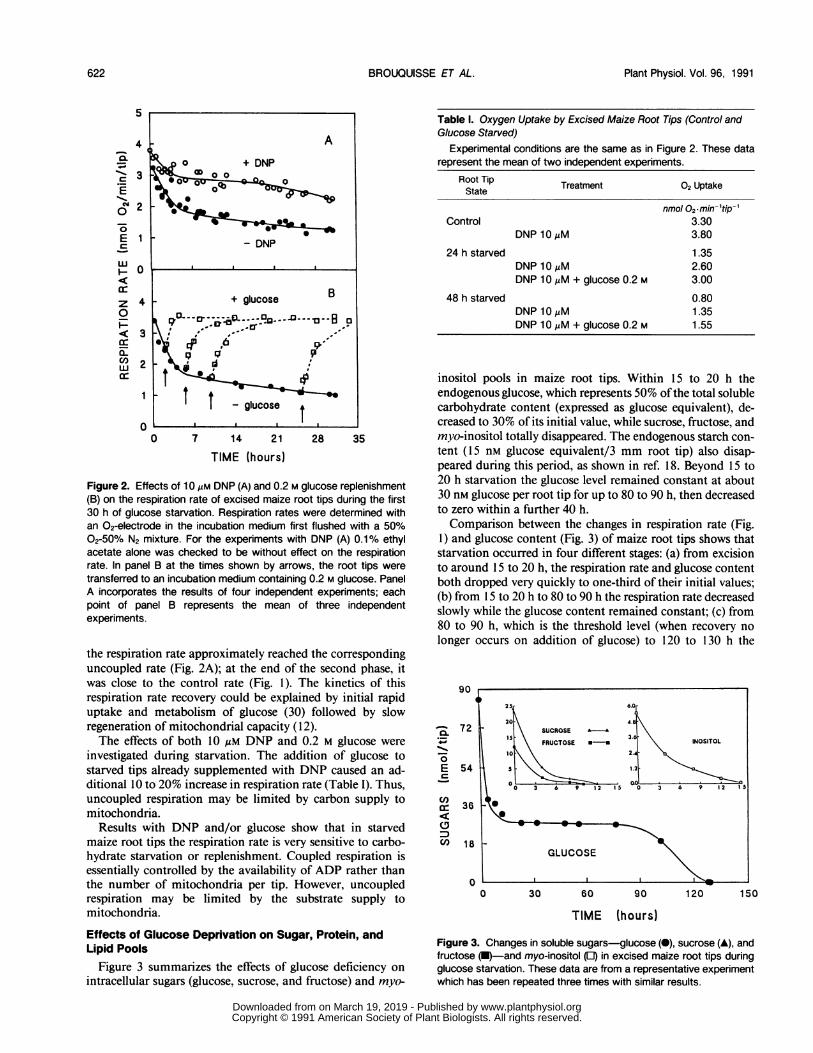
Figure 2. Effects of 10 AM DNP (A) and 0.2 M glucose replenishment(B) on the respiration rate of excised maize root tips during the first30 h of glucose starvation. Respiration rates were determined withan 02-electrode in the incubation medium first flushed with a 50%02-50% N2 mixture. For the experiments with DNP (A) 0.1% ethylacetate alone was checked to be without effect on the respirationrate. In panel B at the times shown by arrows, the root tips weretransferred to an incubation medium containing 0.2 M glucose. PanelA incorporates the results of four independent experiments; eachpoint of panel B represents the mean of three independentexperiments.
the respiration rate approximately reached the correspondinguncoupled rate (Fig. 2A); at the end of the second phase, itwas close to the control rate (Fig. 1). The kinetics of thisrespiration rate recovery could be explained by initial rapiduptake and metabolism of glucose (30) followed by slowregeneration of mitochondrial capacity (12).The effects of both 10 uM DNP and 0.2 M glucose were
investigated during starvation. The addition of glucose tostarved tips already supplemented with DNP caused an ad-ditional 10 to 20% increase in respiration rate (Table I). Thus,uncoupled respiration may be limited by carbon supply tomitochondria.
Results with DNP and/or glucose show that in starvedmaize root tips the respiration rate is very sensitive to carbo-hydrate starvation or replenishment. Coupled respiration isessentially controlled by the availability of ADP rather thanthe number of mitochondria per tip. However, uncoupledrespiration may be limited by the substrate supply tomitochondria.
Effects of Glucose Deprivation on Sugar, Protein, andLipid Pools
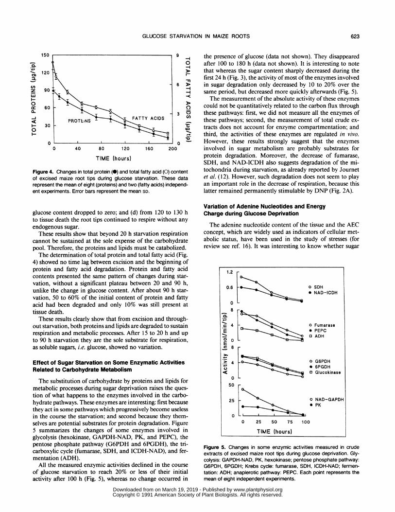
Figure 3 summarizes the effects of glucose deficiency onintracellular sugars (glucose, sucrose, and fructose) and myo-
Table I. Oxygen Uptake by Excised Maize Root Tips (Control andGlucose Starved)
Experimental conditions are the same as in Figure 2. These datarepresent the mean of two independent experiments.
Root Tip Treatment 02 UptakeState
nmol 02-min-'tip-Control 3.30
DNP 10 uM 3.80
24 h starved 1.35DNP 10,uM 2.60DNP 10 AM + glucose 0.2 M 3.00
48 h starved 0.80DNP 10 M 1.35DNP 10 ,M + glucose 0.2 M 1.55
inositol pools in maize root tips. Within 15 to 20 h theendogenous glucose, which represents 50% ofthe total solublecarbohydrate content (expressed as glucose equivalent), de-creased to 30% of its initial value, while sucrose, fructose, andmyo-inositol totally disappeared. The endogenous starch con-tent (15 nm glucose equivalent/3 mm root tip) also disap-peared during this period, as shown in ref. 18. Beyond 15 to20 h starvation the glucose level remained constant at about30 nM glucose per root tip for up to 80 to 90 h, then decreasedto zero within a further 40 h.Comparison between the changes in respiration rate (Fig.
1) and glucose content (Fig. 3) of maize root tips shows thatstarvation occurred in four different stages: (a) from excisionto around 15 to 20 h, the respiration rate and glucose contentboth dropped very quickly to one-third of their initial values;(b) from 15 to 20 h to 80 to 90 h the respiration rate decreasedslowly while the glucose content remained constant; (c) from80 to 90 h, which is the threshold level (when recovery nolonger occurs on addition of glucose) to 120 to 130 h the
90
a._
Ea
cn
a:cnc!,(0
72
54
36
18
00 30 60 so 120 150
TIME (hours)
Figure 3. Changes in soluble sugars-glucose (-), sucrose (A), andfructose (A)-and myo-inositol (E) in excised maize root tips duringglucose starvation. These data are from a representative experimentwhich has been repeated three times with similar results.
622 BROUQUISSE ET AL.
www.plantphysiol.orgon March 19, 2019 - Published by Downloaded from Copyright © 1991 American Society of Plant Biologists. All rights reserved.
GLUCOSE STARVATION IN MAIZE ROOTS
150 9
0
W ~~~~6>z 90
o60 0Fu 3C_j ~ PRTESFATTY ACIDSj- 30
0~~~~~~~~~~~~~~~~~~~*0 0
0 40 80 120 160 200
TIME (hours)
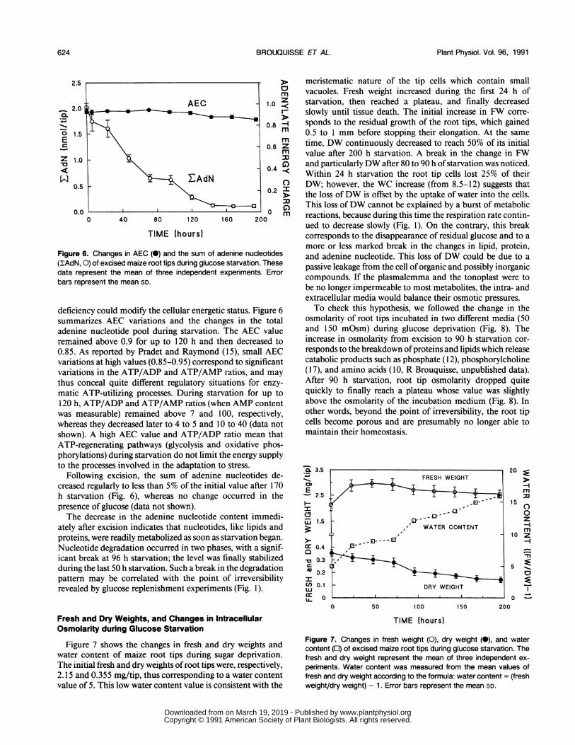
Figure 4. Changes in total protein (@)and total fatty acid (0) contentof excised maize root tips during glucose starvation. These datarepresent the mean of eight (proteins) and two (fatty acids) independ-ent experiments. Error bars represent the mean SD.
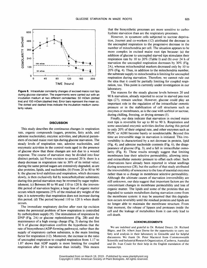
the presence of glucose (data not shown). They disappearedafter 100 to 180 h (data not shown). It is interesting to notethat whereas the sugar content sharply decreased during thefirst 24 h (Fig. 3), the activity of most ofthe enzymes involvedin sugar degradation only decreased by 10 to 20% over thesame period, but decreased more quickly afterwards (Fig. 5).The measurement of the absolute activity of these enzymes
could not be quantitatively related to the carbon flux throughthese pathways: first, we did not measure all the enzymes ofthese pathways; second, the measurement of total crude ex-tracts does not account for enzyme compartmentation; andthird, the activities of these enzymes are regulated in vivo.However, these results strongly suggest that the enzymesinvolved in sugar metabolism are probably substrates forprotein degradation. Moreover, the decrease of fumarase,SDH, and NAD-ICDH also suggests degradation of the mi-tochondria during starvation, as already reported by Journetet al. (12). However, such degradation does not seem to playan important role in the decrease of respiration, because thislatter remained permanently stimulable by DNP (Fig. 2A).
glucose content dropped to zero; and (d) from 120 to 130 hto tissue death the root tips continued to respire without anyendogenous sugar.These results show that beyond 20 h starvation respiration
cannot be sustained at the sole expense of the carbohydratepool. Therefore, the proteins and lipids must be catabolized.The determination of total protein and total fatty acid (Fig.
4) showed no time lag between excision and the beginning ofprotein and fatty acid degradation. Protein and fatty acidcontents presented the same pattern of changes during star-vation, without a significant plateau between 20 and 90 h,unlike the change in glucose content. After about 90 h star-vation, 50 to 60% of the initial content of protein and fattyacid had been degraded and only 10% was still present attissue death.
These results clearly show that from excision and through-out starvation, both proteins and lipids are degraded to sustainrespiration and metabolic processes. After 15 to 20 h and upto 90 h starvation they are the sole substrate for respiration,as soluble sugars, i.e. glucose, showed no variation.
Effect of Sugar Starvation on Some Enzymatic ActivitiesRelated to Carbohydrate Metabolism
The substitution of carbohydrate by proteins and lipids formetabolic processes during sugar deprivation raises the ques-tion of what happens to the enzymes involved in the carbo-hydrate pathways. These enzymes are interesting: first becausethey act in some pathways which progressively become uselessin the course the starvation; and second because they them-selves are potential substrates for protein degradation. Figure5 summarizes the changes of some enzymes involved inglycolysis (hexokinase, GAPDH-NAD, PK, and PEPC), thepentose phosphate pathway (G6PDH and 6PGDH), the tri-carboxylic cycle (fumarase, SDH, and ICDH-NAD), and fer-mentation (ADH).
All the measured enzymic activities declined in the courseof glucose starvation to reach 20% or less of their initialactivity after 100 h (Fig. 5), whereas no change occurred in
Variation of Adenine Nucleotides and EnergyCharge during Glucose Deprivation
The adenine nucleotide content of the tissue and the AECconcept, which are widely used as indicators of cellular met-abolic status, have been used in the study of stresses (forreview see ref. 16). It was interesting to know whether sugar
1.2
0.6 L
0
CL-.o
E0E
._-0
C
8
4
0
4
50-
25
O SDH* NAD-ICDH
0 Fumarase0 PEPCa ADH
o G6PDH* 6PGDHO Glucokinase
O NAD-GAPDH* PK
0 25 50 75 100
TIME (hours)
Figure 5. Changes in some enzymic activities measured in crudeextracts of excised maize root tips during glucose deprivation. Gly-colysis: GAPDH-NAD, PK, hexokinase; pentose phosphate pathway:G6PDH, 6PGDH; Krebs cycle: fumarase, SDH, ICDH-NAD; fermen-tation: ADH; anaplerotic pathway: PEPC. Each point represents themean of eight independent experiments.
623
c
LIIII
www.plantphysiol.orgon March 19, 2019 - Published by Downloaded from Copyright © 1991 American Society of Plant Biologists. All rights reserved.
Plant Physiol. Vol. 96, 1991
2.5
2.0 <
0 1.5Ec
Z 1.0
0.5
0.00 40 80 120 160 20C
m1.0 Z
I-
0.8 mmm
0.6 Zm
C)0.4 °
0.2 r
C)0 m
0
TIME (hours)
Figure 6. Changes in AEC (0) and the sum of adenine nucleotides(2AdN, 0) of excised maize root tips during glucose starvation. Thesedata represent the mean of three independent experiments. Errorbars represent the mean SD.
deficiency could modify the cellular energetic status. Figure 6summarizes AEC variations and the changes in the totaladenine nucleotide pool during starvation. The AEC valueremained above 0.9 for up to 120 h and then decreased to0.85. As reported by Pradet and Raymond (15), small AECvariations at high values (0.85-0.95) correspond to significantvariations in the ATP/ADP and ATP/AMP ratios, and maythus conceal quite different regulatory situations for enzy-matic ATP-utilizing processes. During starvation for up to120 h, ATP/ADP and ATP/AMP ratios (when AMP contentwas measurable) remained above 7 and 100, respectively,whereas they decreased later to 4 to 5 and 10 to 40 (data notshown). A high AEC value and ATP/ADP ratio mean thatATP-regenerating pathways (glycolysis and oxidative phos-phorylations) during starvation do not limit the energy supplyto the processes involved in the adaptation to stress.
Following excision, the sum of adenine nucleotides de-creased regularly to less than 5% of the initial value after 170h starvation (Fig. 6), whereas no change occurred in thepresence of glucose (data not shown).The decrease in the adenine nucleotide content immedi-
ately after excision indicates that nucleotides, like lipids andproteins, were readily metabolized as soon as starvation began.Nucleotide degradation occurred in two phases, with a signif-icant break at 96 h starvation; the level was finally stabilizedduring the last 50 h starvation. Such a break in the degradationpattern may be correlated with the point of irreversibilityrevealed by glucose replenishment experiments (Fig. 1).
Fresh and Dry Weights, and Changes in IntracellularOsmolarity during Glucose Starvation
Figure 7 shows the changes in fresh and dry weights andwater content of maize root tips during sugar deprivation.The initial fresh and dry weights ofroot tips were, respectively,2.15 and 0.355 mg/tip, thus corresponding to a water contentvalue of 5. This low water content value is consistent with the
meristematic nature of the tip cells which contain smallvacuoles. Fresh weight increased during the first 24 h ofstarvation, then reached a plateau, and finally decreasedslowly until tissue death. The initial increase in FW corre-sponds to the residual growth of the root tips, which gained0.5 to 1 mm before stopping their elongation. At the sametime, DW continuously decreased to reach 50% of its initialvalue after 200 h starvation. A break in the change in FWand particularly DW after 80 to 90 h ofstarvation was noticed.Within 24 h starvation the root tip cells lost 25% of theirDW; however, the WC increase (from 8.5-12) suggests thatthe loss ofDW is offset by the uptake of water into the cells.This loss ofDW cannot be explained by a burst of metabolicreactions, because during this time the respiration rate contin-ued to decrease slowly (Fig. 1). On the contrary, this breakcorresponds to the disappearance of residual glucose and to amore or less marked break in the changes in lipid, protein,and adenine nucleotide. This loss of DW could be due to apassive leakage from the cell of organic and possibly inorganiccompounds. If the plasmalemma and the tonoplast were tobe no longer impermeable to most metabolites, the intra- andextracellular media would balance their osmotic pressures.To check this hypothesis, we followed the change in the
osmolarity of root tips incubated in two different media (50and 150 mOsm) during glucose deprivation (Fig. 8). Theincrease in osmolarity from excision to 90 h starvation cor-responds to the breakdown ofproteins and lipids which releasecatabolic products such as phosphate ( 12), phosphorylcholine(17), and amino acids (10, R Brouquisse, unpublished data).After 90 h starvation, root tip osmolarity dropped quitequickly to finally reach a plateau whose value was slightlyabove the osmolarity of the incubation medium (Fig. 8). Inother words, beyond the point of irreversibility, the root tipcells become porous and are presumably no longer able tomaintain their homeostasis.
0. 3.5l-.cmE-2.5
UJI.U.i 1.5
aC 0.4
-0 0.3cU 0.2ICn) 0.1
LL
20 X
-im
15
0z-4-m
10 z-4i
5 11
0
I _.
0 50 100 150 200
TIME (hours)
Figure 7. Changes in fresh weight (0), dry weight (0), and watercontent (a) of excised maize root tips during glucose starvation. Thefresh and dry weight represent the mean of three independent ex-
periments. Water content was measured from the mean values offresh and dry weight according to the formula: water content = (freshweight/dry weight) - 1. Error bars represent the mean SD.
FRESH WEIGHT
,2 _S ~~~-43'
B I,- WATER CONTENT
DRY WEIGHTs~~~~~~~~~~~~~~~~~~~~~~~~~~~~~~~~
624 BROUQUISSE ET AL.
www.plantphysiol.orgon March 19, 2019 - Published by Downloaded from Copyright © 1991 American Society of Plant Biologists. All rights reserved.
GLUCOSE STARVATION IN MAIZE ROOTS
that the biosynthetic processes are more sensitive to carbo-hydrate starvation than are the respiratory processes.However, in sycamore cells subjected to sucrose depriva-
A IL, > F.> Ttion, Journet and co-workers (12) attributed the decrease inthe uncoupled respiration to the progressive decrease in thenumber of mitochondria per cell. The situation appears to be
.\\ more complex in excised maize root tips because: (a) theaddition of glucose to uncoupled starved tips stimulates theirrespiration rate by 10 to 20% (Table I) and (b) over 24 h of
150 mOsm starvation the uncoupled respiration decreases by 30% (Fig.2A), whereas mitochondrial markers decreased only by 10 to
50 mOsm 20% (Fig. 4). Thus, in addition to the mitochondria number,.........................................................th.sub trat.sup ly.t.mit chon ria .s.l.iti..forunco ple, , ~~~~~~~thesubstrate supply to mitochondria islimiting for uncoupled0 40 80 120 160 200 respiration during starvation. Therefore, we cannot rule out
the idea that it could be partially limiting for coupled respi-TIME (hours) ration, too. This point is currently under investigation in our
laboratory.itracellular osmolarity changes of excised maize root tips The reasons for the steady glucose levels between 20 and)se starvation. The experiments were carried out with an'nedium at two different osmolarities: 50 mOsm (dotted 90 h starvation, already reported to occur in starved pea root
tips (27), remain unclear. However, glucose could play an
and dashed lines indicate the incubation medium osmo- important role in the regulation of the intracellular osmoticpressure or in the stabilization of cell structures such asenzymes or membranes, as is the case with sorbitol or sucrose,during chilling, freezing, or drying stresses (5).
Finally, our data indicate that starvation in excised maizeDISCUSSION root tips is reversible for up to 80 to 90 h. Respiration and
some associated enzymic activities decline during this perioddy describes the continuous changes in respiration to only 20% of their original rate, and other enzymes such asiic compounds (sugars, proteins, fatty acids, and PEPC or ADH become barely or nondetectable. Beyond thisicleotides), enzymic activities, and physical param- time an irreversible stage in starvation is initiated. This irre--ised maize root tips during glucose starvation. The versibility is characterized by a net decrease in protein, lipidels of respiration rate, adenine nucleotides, and (Fig. 4), and adenine nucleotide contents (Fig. 6), the disap-activities in the control roots aged in the presence pearance of glucose (Fig. 3), and a fall in intracellular osmo-show that these changes are not due to a wound larity (Fig. 8). These results strongly suggest that limitingrhe course of starvation may be divided into four membranes lose their selective permeability, allowing intra-riods. (a) From excision to around 20 h: there is a and extracellular osmotic pressure to offset each other. Suchease in respiration rate to 30% of its initial value; observations have already been reported in wheat seedlingssame period sugars are extensively degraded, as are during senescence (28), but the author of that study attributedns, lipids, and nucleotides. (b) From 20 to 80 to 90 the irreversibility of senescence to the loss of essential enzymesose level stabilizes and respiration, which decreases rather than to a change in membrane selective permeability.hen exclusively fed by noncarbohydrate substrates; Although the ultimate causes of starvation irreversibility areperiod starvation may be reversed by sugar replen- still unknown, our data suggest that important factors are the) Between 80 to 90 and 110 to 120 h: the irrevers- concomitant changes in membrane permeability and loss ofof starvation begins; a large loss of organic matter organic matter. The lipids and some of the proteins that arech represents 25% of the residual dry weight of the degraded to sustain metabolism during starvation come fromoteworthy that respiration does not increase during the membrane system: it may be assumed that this degrada-[. (d) The period beyond I 10 to 120 h when death tion occurs reversibly until the residual proteins and lipids are
no longer able to maintain the membrane structure. Fromnediate respiratory decline after root tip excision that moment, the release of lipases and proteases inside the)erennial problem of how respiration is controlled cell and the leakage of metabolites from it can only lead to(drate supply (9). The stimulation of respiration by cell death.
DNP (Fig. 2A) or glucose replenishment (Fig. 2B) and themaintenance of a high energy charge (Fig. 5) during the first30 h of glucose deprivation confirm the hypothesis that therate of biosyntheses (ADP-forming pathways), rather than thesupply of respiratory carbon substrates, is the main limitingfactor for respiration (12). Moreover, the increase in the ratioof uncoupled respiration to coupled respiration from 1.17 to1.87 shows that ADP supply is more limiting for coupledrespiration after 20 h starvation than initially. This means
ACKNOWLEDGMENTS
We are indebted and grateful to Dr. Roland Douce, Dr. RichardBligny, and Dr. Albert Jean Dorne for the opportunity to carry outfatty acid analysis in their laboratory in Grenoble and for helpfuldiscussions. We thank Dr. Peter Macnicol from CommonwealthScientific and Industrial Research Organization, (Canberra, Australia)and Dr. Ivan Couee for their help in the English translation of themanuscript.
600
480
360
240
120
Eto0E
C,
cn0
0
Figure 8. Irduring gluccincubation n
line) and 15(The dottedlarity values
This sturate, orgaradenine nu.eters of excsteady levIenzymaticof glucoseresponse. Idistinct peisharp decriduring thealso proteiih: the gluclslowly, is t]during thisishment. (cible periodoccurs whitips; it is n(this periodoccurs.The imr
raises the rby carbohy
625
www.plantphysiol.orgon March 19, 2019 - Published by Downloaded from Copyright © 1991 American Society of Plant Biologists. All rights reserved.
Plant Physiol. Vol. 96, 1991
LITERATURE CITED
1. Baysdorfer C, Warmbrodt RD, Van Der Woude WJ (1988)Mechanisms of starvation tolerance in pearl millet. Plant Phys-iol88: 1381-1387
2. Bergmeyer HU (1974) Alcohol dehydrogenase. In HU Berg-meyer, ed, Methods of Enzymic Analysis, Ed 2 Vol 1. Aca-demic Press, New York, pp 428-429
3. Bradford MM (1976) A rapid and sensitive method for thequantification of microgram quantities of proteins utilizing theprinciple of protein-dye binding. Anal Biochem 72: 248-254
4. Cox GF, Davis DD (1967) Nicotinamide adenine dinucleotide-specific isocitrate dehydrogenase from pea mitochondria: pu-rification and properties. Biochem J 105: 729-734
5. Crowe JH, Crowe LM (1986) Stabilization of membranes inanhydrobiotic organisms. In C Leopold, ed, Membranes, Me-tabolism and Dry Organisms. Cornell University Press, Ithaca,NY, pp 188-209
6. Deutsch J (1983) Glucose-6-P dehydrogenase. In HU Bergmeyer,ed, Methods of Enzymic Analysis, Ed 3 Vol 3. Verlag Chemie,Weinheim, pp 190-197
7. Dorne AJ, Bligny R, Rebeille F, Roby C, Douce R (1987) Fattyacid disappearance and phosphorylcholine accumulation inhigher plant cells after a long period of sucrose deprivation.Plant Physiol Biochem 25: 589-595
8. Farrar JF (1981) Respiration rate of barley roots: its relation togrowth, substrate supply and the illumination of the shoot.Ann Bot 48: 53-63
9. Farrar JF (1985) The respiratory source of CO2. Plant CellEnviron 8: 427-438
10. Genix P, Bligny R, Martin JB, Douce R (1990) Transient accu-mulation of asparagine in sycamore cells after a long period ofsucrose starvation. Plant Physiol 94: 717-722
11. Hill RL, Bradshaw RA (1969) Fumarase. Methods Enzymol 13:91-99
12. Journet EP, Bligny R, Douce R (1986) Biochemical changesduring sucrose deprivation in higher plant cells. J Biol Chem261: 3193-3199
13. Kerr PS, Rufty TW Jr, Huber SC (1985) Changes in nonstruc-tural carbohydrates in different parts of soybean (Glycine max[L.] Merr.) plants during a light/dark cycle and in extendeddarkness. Plant Physiol 78: 576-581
14. Pace GGM, Volk RJ, Jackson WA (1990) Nitrate reduction inresponse to C02-limited photosynthesis. Plant Physiol 92: 286-292
15. Pradet A, Raymond P (1982) Adenylate energy charge: conceptand controversy. What's New Plant Physiol 13: 21-24
16. Raymond P, Gidrol X, Salon C, Pradet A (1987) Control involv-ing adenine and pyridine nucleotides. In PK Stumpf, EE Conn,eds, The Biochemistry of Plants, Vol 11. Academic Press, NewYork, pp 129-176
17. Roby C, Martin JB, Bligny R, Douce R (1987) Biochemicalchanges during sucrose deprivation in higher plant cells. J BiolChem 262: 5000-5007
18. Saglio PH, Pradet A (1980) Soluble sugars, respiration, andenergy charge during aging of excised maize root tips. PlantPhysiol 66: 516-519
19. Saglio PH, Rancillac M, Bruzeau F, Pradet A (1984) Criticaloxygen pressure for growth and respiration of excised andintact roots. Plant Physiol 76: 151-154
20. Sahulka J, Lisa L (1978) The influence of exogenously suppliedsucrose on glutamine synthetase and glutamate dehydrogenaselevels in excised Pisum sativum roots. Biol Plant 20: 446-452
21. Deleted in proof22. Singer TP, Oestreicher G, Hogue P, Contreiras J, Brandao I
(1973) Regulation of succinate dehydrogenase in higher plants.Plant Physiol 52: 616-621
23. Stitt M, ap Rees T (1978) Pathways of carbohydrate oxidationin leaves ofPisum sativum and Triticum aestivum. Phytochem-istry 17: 1251-1256
24. Sweeley CC (1965) Analyse des hydrates de carbone par chro-matographie en phase gazeuse. Bull Soc Chim Biol 47: 1477-1494
25. Walsh KB, Vessey JK, Layzell DB (1987) Carbohydrate supplyand N2 fixation in soybean. Plant Physiol 85: 137-144
26. Webster PL, van't Hof J (1973) Polyribosomes in proliferatingand non-proliferating root meristem cells. Am J Bot 60: 117-121
27. Webster PL, Henry M (1987) Sucrose regulation of proteinsynthesis in pea root meristem cells. Environ Exp Bot 27: 253-262
28. Wittenbach VA (1977) Induced senescence of intact wheat seed-lings and its reversibility. Plant Physiol 59: 1039-1042
29. Wittenbach VA, Lin W, Hebert RR (1982) Vacuolar localizationof proteases and degradation of chloroplasts in mesophyllprotoplasts from senescing primary wheat leaves. Plant Physiol69: 98-102
30. Xia JH, Saglio PH (1988) Characterization of the hexose trans-port system in maize root tips. Plant Physiol 88: 1015-1020
626 BROUQUISSE ET AL.
www.plantphysiol.orgon March 19, 2019 - Published by Downloaded from Copyright © 1991 American Society of Plant Biologists. All rights reserved.