Studies the Localization of the Cardiac Glycoside...
13
Studies on the Localization of the Cardiac Glycoside Receptor THOMAS W. SMrrH, HENRY WAGNER, JR., JoHN E. MARKis, and MICHAEL YOUNG From the Laboratory of Physical Biochemistry, Massachusetts General Hospital, and the Departments of Biological Chemistry, Medicine, and Pharmacology, Harvard Medical School, Boston, Massachusetts 02114 A B S TR A C T The purpose of this study was to see whether the receptor for cardiac glycosides might be localized upon or within the plasma membrane of digi- talis-sensitive cells. Ouabain and digoxin were joined covalently to several large protein molecules. These macromolecular conjugates are too large to enter intact cells; consequently, any pharmacologic or biochemical effects which they display should arise from interaction with a cell surface receptor. Conjugates were tested in several cardiac glycoside-sensitive systems: (a), con- tractility response of isolated cardiac muscle; (b), ac- tive 'Rb+ uptake by red cells; (c), enzymatic activity of isolated myocardial microsomal (Na+ + K+) -activated adenosine triphosphatase (ATPase); and (d), enzymatic activity of solubilized red cell (Na+ + K+) -activated ATPase. Results demonstrated that in all of these sys- tems, the macromolecular-glycoside conjugates were 100- to 1000-fold less active than the free glycosides. Careful chromatographic examination of the various conjugates revealed that they contained a small but persistent free cardiac glycoside contaminant. The amount of this species ranged from 0.1 to 1.0% of the total macromolecule-bound glycoside, and its pres- ence fully explains the levels of biologic activity ob- served with the conjugates. To try to minimize steric factors which could inter- fere with glycoside-receptor interaction, digoxin and ouabain were also coupled to macromolecule via long, flexible polyamide side-chains. These extended chain An account of this work was presented before the Ameri- can Society for Pharmacology and Experimental Thera- peutics, Burlington, Vt., August 1971. Dr. Smith was supported by special National Institutes of Health Postdoctoral Fellowship F03HE44673 and sub- sequently by an Established Investigatorship of the Ameri- can Heart Association. Received for publication 6 October 1971 and in revised form 28 December 1971. conjugates, in which the cardiac glycoside potentially lay some 30 A removed from the surface of the macro- molecule, also exhibited negligible digitalis-like effects when tested upon isolated cardiac muscle, red cell MRb+ uptake, and enzymatic activity of cardiac microsomal (Na' + K+) -ATPase. However, the extended chain conjugates were fully active when examined with the solubilized red cell (Nae + K+) -ATPase system. To further ensure that the chemical reactions used to couple macromolecule to glycoside did not inactivate the drug, all conjugates were subjected to extensive proteolysis with Streptomyces griseus protease. These proteolytic digests exhibited full pharmacologic ac- tivity. Digoxin was also coupled to the tripeptide ala- nylglycylglycine, and the resulting conjugate was fully active. Taken together, these results suggest that if the re- ceptor(s) for cardiac glycosides is associated with the plasma membrane, then it may lie deep within it. INTRODUCTION Digitalis glycosides exert characteristic inotropic (1, 2) and electrophysiologic (3, 4) effects upon both the intact heart and isolated cardiac tissue preparations. Although the cardiac receptor (or receptors) which mediates the pharmacologic effects of these drugs has neither been isolated nor fully identified, recent evi- dence suggests that (Na' + K+) -activated adenosine triphosphatase (ATP phosphohydrolase, E.C. 3.6.1.3) plays a role in the mechanism of action of the cardio- active steroid glycosides (5-8). Thus, ouabain and di- goxin bind to this membrane-associated enzyme, re- sulting in (a), inhibition of (Na' + K+) -activated ATPase activity; and (b), a decrease in the active transport of sodium and potassium ions across the cell membrane (9-12). It has been suggested that the The Journal of Clinical Investigation Volume 51 July 1972 1777
Transcript of Studies the Localization of the Cardiac Glycoside...

Studies on the Localization of
the Cardiac Glycoside Receptor
THOMASW. SMrrH, HENRYWAGNER,JR., JoHN E. MARKis, andMICHAELYOUNG
From the Laboratory of Physical Biochemistry, Massachusetts General Hospital,and the Departments of Biological Chemistry, Medicine, and Pharmacology,Harvard Medical School, Boston, Massachusetts 02114
A B S T R A C T The purpose of this study was to seewhether the receptor for cardiac glycosides might belocalized upon or within the plasma membrane of digi-talis-sensitive cells. Ouabain and digoxin were joinedcovalently to several large protein molecules. Thesemacromolecular conjugates are too large to enter intactcells; consequently, any pharmacologic or biochemicaleffects which they display should arise from interactionwith a cell surface receptor. Conjugates were tested inseveral cardiac glycoside-sensitive systems: (a), con-tractility response of isolated cardiac muscle; (b), ac-tive 'Rb+ uptake by red cells; (c), enzymatic activityof isolated myocardial microsomal (Na+ + K+) -activatedadenosine triphosphatase (ATPase); and (d), enzymaticactivity of solubilized red cell (Na+ + K+) -activatedATPase. Results demonstrated that in all of these sys-tems, the macromolecular-glycoside conjugates were100- to 1000-fold less active than the free glycosides.Careful chromatographic examination of the variousconjugates revealed that they contained a small butpersistent free cardiac glycoside contaminant. Theamount of this species ranged from 0.1 to 1.0% ofthe total macromolecule-bound glycoside, and its pres-ence fully explains the levels of biologic activity ob-served with the conjugates.
To try to minimize steric factors which could inter-fere with glycoside-receptor interaction, digoxin andouabain were also coupled to macromolecule via long,flexible polyamide side-chains. These extended chain
An account of this work was presented before the Ameri-can Society for Pharmacology and Experimental Thera-peutics, Burlington, Vt., August 1971.
Dr. Smith was supported by special National Institutesof Health Postdoctoral Fellowship F03HE44673 and sub-sequently by an Established Investigatorship of the Ameri-can Heart Association.
Received for publication 6 October 1971 and in revisedform 28 December 1971.
conjugates, in which the cardiac glycoside potentiallylay some 30 A removed from the surface of the macro-molecule, also exhibited negligible digitalis-like effectswhen tested upon isolated cardiac muscle, red cell MRb+uptake, and enzymatic activity of cardiac microsomal(Na' + K+) -ATPase. However, the extended chainconjugates were fully active when examined with thesolubilized red cell (Nae + K+) -ATPase system. Tofurther ensure that the chemical reactions used tocouple macromolecule to glycoside did not inactivatethe drug, all conjugates were subjected to extensiveproteolysis with Streptomyces griseus protease. Theseproteolytic digests exhibited full pharmacologic ac-tivity. Digoxin was also coupled to the tripeptide ala-nylglycylglycine, and the resulting conjugate was fullyactive.
Taken together, these results suggest that if the re-ceptor(s) for cardiac glycosides is associated with theplasma membrane, then it may lie deep within it.
INTRODUCTIONDigitalis glycosides exert characteristic inotropic (1,2) and electrophysiologic (3, 4) effects upon both theintact heart and isolated cardiac tissue preparations.Although the cardiac receptor (or receptors) whichmediates the pharmacologic effects of these drugs hasneither been isolated nor fully identified, recent evi-dence suggests that (Na' + K+) -activated adenosinetriphosphatase (ATP phosphohydrolase, E.C. 3.6.1.3)plays a role in the mechanism of action of the cardio-active steroid glycosides (5-8). Thus, ouabain and di-goxin bind to this membrane-associated enzyme, re-sulting in (a), inhibition of (Na' + K+) -activatedATPase activity; and (b), a decrease in the activetransport of sodium and potassium ions across the cellmembrane (9-12). It has been suggested that the
The Journal of Clinical Investigation Volume 51 July 1972 1777
positive inotropic effect may result from an increasein the amount of calcium ion available to the contrac-tile element at the time of excitation-contraction cou-pling and that this effect might be mediated by cardiacglycoside binding to and/or inhibition of (Nae + K+) -ATPase (13-16). Since this enzyme is believed to bea component of the sarcolemma (17), any digitalis-induced change in contractility would require an inter-action between the drug and the cell plasma membrane.Of interest in this regard are studies of the squidgiant axon (18) and the human erythrocyte ghost(19) which indicate that the cardiac glycoside recep-tor is accessible only from the external cell surface inthese two digitalis-sensitive systems. The complexstructure of the functioning myocardial cell (20) hasprecluded such direct assessment of the locus of digi-talis action.
The studies described below were undertaken to tryto localize directly the cellular receptor for digitalis,and to define further the nature of the interaction be-tween cardiac glycosides and (Na+ + K+) -ATPase. Forthis purpose, we have coupled digoxin and ouabain toseveral macromolecules. These high molecular weightconjugates would be expected to enter intact myo-cardial or red blood cells at very low rates, if at all(21, 22). Consequently, any pharmacologic activitywhich they display should arise from interaction witha cell surface receptor. The effects of these conjugateswere studied in several digitalis-sensitive systems, in-cluding contractile response of isolated cardiac muscle,active rubidiun (Rb+) uptake by erythrocytes, enzy-matic activity of isolated myocardial microsomal (Na'+ K+)-activated ATPase, and enzymatic activity ofsolubilized erythrocyte (Na' + K+) -activated ATPase.
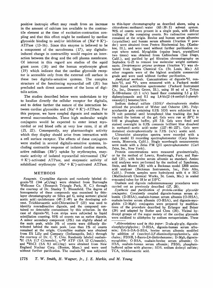
METHODSReagents. Crystalline digoxin and randomly labeled di-
goxin-3H (144 /LCi/mg) were obtained from BurroughsWellcome Co. (Research Triangle Park, N. C.) throughthe courtesy of Dr. Stanley T. Bloomfield. The degree ofhomogeneity of these compounds was examined by thin-layer chromatography on Silica gel G, using acetone: glacialacetic acid: cyclohexane (49:2:49) as the developing sol-vent. Trichloroacetic acid-Chloramine-T (23) was used toidentify nonradioactive digoxin, and the compound con-tained no detectable contaminant by this criterion. In thecase of digoxin-3H, 1-cm strips were subjected to liquidscintillation counting. 93%o of counts ran as native digoxin.A minor secondary component (3%) ran slightly ahead ofthe main peak, while 3%o of counts were diffusely dis-tributed behind the main peak. Less than 1%o of countsremained at the origin. Crystalline ouabain was obtainedfrom Eli Lilly and Company (Indianapolis, Ind.) throughthe courtesy of Dr. C. T. Chiu. Randomly labeled ouabain-'H (SA 11.7 Ci/mmole), -y-3P ATP (SA 12 Ci/mmole),and 'RbC1 (SA 9.1 mCi/mg) were obtained from NewEngland Nuclear Corp. (Boston, Mass.) and were usedwithout further purification. The ouabain-3H was subjected
to thin-layer chromatography as described above, using achloroform: methanol: water (65: 30: 5) solvent system.96%o of counts were present in a single peak, with diffusetrailing of the remaining counts. No radioactive materialremained at the origin. Bovine and human serum albumins(crystalline) and human serum albumin (fraction V pow-der) were obtained from Pentex Biochemical Inc. (Kanka-kee, Ill.), and were used without further purification ex-cept where noted. Myoglobin (equine heart, crystallizedtwo times) was obtained from Calbiochem (San Diego,Calif.), and purified by gel filtration chromatography onSephadex G-25 to remove low molecular weight contami-nants. Streptomyces griseus protease (fraction V) was ob-tained from Sigma Chemical Co. (St. Louis, Mo.) Allother reagents were of the highest available commercialgrade and were used without further purification.
Analytical methods. Concentrations of digoxin-SH, oua-bain-'H, and '3Pi were measured with a Packard model3003 liquid scintillation spectrometer (Packard InstrumentCo., Inc., Downers Grove, Ill.), using 10 ml of a TritonX-100-toluene (2:1 v/v) based fluor containing 5.5 g 2,5-diphenyloxazole and 0.1 mg 1,4-bis [2- (4-methyl-5-phenyl-oxazolyl)] benzene/litre.
Sodium dodecyl sulfate (SDS)' electrophoresis studiesutilized the procedure of Weber and Osborne (24). Poly-acrylamide gels containing 0.1% SDS were subjected to a70 volt potential until bromphenol blue tracking dye hadreached the bottom of the gel. Gels were run at 20'C in0.05 M phosphate buffer, pH 7.0. Gels were fixed andstained overnight in 0.3% comassie brilliant blue dissolvedin methanol: acetic acid: water (227: 46: 227). Gels weredestained electrophoretically in 7.5% (v/v) acetic acid.
Ultraviolet absorption spectra were recorded with aCary model 15 recording spectrophotometer (Cary Instru-ments, Monrovia, Calif.). Single wavelength measurementswere made with a Zeiss PM QII spectrophotometer (CarlZeiss, Inc., New York).
Protein concentrations were measured gravimetrically,or by the method of Lowry, Rosebrough, Farr, and Ran-dall (25), with bovine serum albumin as standard. Aminoacid analyses were performed by the method of Spackman,Stein, and Moore (26) with a Beckman model 120B aminoacid analyzer (Beckman Instruments, Inc., Palo Alto,Calif.). Protein samples were hydrolyzed with 6 N HCl(Mallinckrodt Chemical Works, St. Louis, Mo.) in sealedevacuated tubes for 18 hr at 110'C.
Ouabain and digoxin radioimmunoassay procedures werecarried out as previously described (27, 28).
Synthesis and purification of protein-cardiac glycosideconjugates. Covalently coupled digoxin-human serum al-bumin (D-HSA), ouabain-human serum albumin (O-HSA),ouabain-bovine serum albumin (O-BSA), and digoxin-myo-globin (D-Mgb) conjugates were prepared by modifica-tions of the procedure described by Erlanger and Beiser(29) and adapted by Butler and Chen (30). Vicinal hy-droxyl groups of the sugar moiety of the cardiac glycosidewere oxidized to aldehydes by sodium metaperiodate. These
1Abbreziations used in this paper: D-ala-gly-gly, digoxin-alanylglycylglycine; D-HSA, digoxin-human serum albu-min; DA-S-DA-S-BSA, bovine serum albumin modifiedby addition of (succinyl-3,3'-diaminodipropylamine)2 sidechains; FDNB, 1-fluoro-2,4-dinitrobenzene; D-Mgb, digoxin-myoglobin; O-B SA, ouabain-bovine serum albumin; 0-HSA, ouabain-human serum albumin; PBSG, phosphate-buffered saline with glucose; SDS. sodium dodecyl sulfate;TCA, trichloroacetic acid.
1778 T. W. Smith, H. Wagner, Jr., J. E. Markis, and M. Young

reactive aldehydes were then coupled to amino groups ofthe carrier protein by Schiff-base formation, followed byreduction with sodium borohydride.
The coupling step was carried out for 2 hr at pH 9.5-9.0.In some of the syntheses, a stoichiometric amount of so-dium metaperiodate was used to oxidize the sugar, and theethylene glycol step used to consume excess periodate wasomitted. In all syntheses, suitable amounts of digoxin-'Hor ouabain-3H were used to facilitate identification andquantitation of conjugated and free cardiac glycoside.
In a typical synthesis, 500 mg ouabain and 0.125 mCiouabain-8H were dissolved in 30 ml water. To this solution,0.428 g NaIO. dissolved in 20 ml water was added drop-wise. After reaction for 30 min at room temperature, theoxidized ouabain was added dropwise to 500 mg of proteindissolved in 25 ml water, maintained at pH 9.5 with 5%aqueous K2C03. The reactants were magnetically stirred atroom temperature. After 3 hr 0.30 g NaBH4, freshly dis-solved in 20 ml water, was added. 3 hr later, the pH waslowered to 6.5 with concentrated formic acid. After 1 hr,sodium hydroxide was added to raise the pH to 8.5 andthe reaction mixture dialyzed exhaustively against distilledwater and lyophilized.
Two types of conjugates were prepared in which cardiacglycoside was not attached directly to the carrier proteinbut was extended from it by a long, flexible side-arm (Fig.1). Two of these consisted of poly(D,L-alanyl) derivativesof human serum albumin and poly-L-lysine, synthesized byreaction with N-carboxy D,L-alanine anhydrides accordingto the method of Schechter, Bauminger, Sela, Nachtigal,and Feldman (31). Amino acid analysis of the poly (D,L-alanyl)-HSA product before and after deamination (32)showed that an average of 50 D,L-alanyl chains with a meanlength of 9.5 alanyl residues had been attached per albuminmolecule. The calculated molecular weight of the poly(D,L-alanyl)-HSA carrier was 98,000 and that of the poly(D,L-alanyl) -poly-L-lysine, 120,000.
The other extended chain conjugate was synthesized asfollows. Bovine serum albumin was succinylated accordingto the procedure of Habeeb, Cassidy, and Singer (33),using a 12-fold molar excess of succinic anhydride overprotein amino groups. Reaction was allowed to proceed for30 min at 0°C, pH 8.0-8.5. After succinylation, the proteinwas exhaustively dialyzed against distilled water at 4°Cand lyophilized. The succinylated albumin was then dis-solved in distilled water to a concentration of 5 mg/mland adjusted to pH 5.0 with HCl. To this solution werethen added a 50-fold molar excess (relative to protein car-boxyl groups) of 3,3'-diaminodipropylamine hydrochloride
DA-S-DA-S-Albumin0H R1H-[NCH2CH2CH2 NH CH2 CH2 CH2NNCCH2CH2rC]-Albumin
H 11 20
Poly (D, L-alanyl )-Albumin
H O 011 H 11
H2N-C-C-[N-C-C] -AlbuminI H 8.5
CH3 CH3
FIGURE 1 Structures of side-arms joined to serum albu-min to produce a flexible extended chain. Cardiac glycosideswere coupled to the terminal amino groups. (Synthesis ofthese derivatives is described in the text.)
TABLE I
Characterization of Intermediates in the Synthesis ofDA-S-DA-S-BSA
FDNBcolor yieldProduct (% of BSA color yield)
BSA 100S-BSA 17.3DA-S-BSA 117.6S-DA-S-BSA 16.0DA-S-DA-S-BSA 119.1
Products were reacted with excess FDNB, purified, andhydrolyzed as described in the text. Optical densities of thehydrolysates were measured at 360 nmand compared with theoptical density of the hydrolyzed BSA derivative.
and a 30-fold molar excess of 1-cyclohexyl-3-(2-morpho-linoethyl)-carbodiimide methyl p-toluenesulfonate, and pHreadjusted to 5.0 (34). The solution was stirred overnightat room temperature.
After dialysis against distilled water and lyophilization,the product was resuccinylated and again reacted with thediamine by the same methods. The resulting compound issubsequently referred to as DA-S-DA-S-BSA. Extent ofreaction at each stage of the synthesis was measured byreaction with fluorodinitrobenzene (35). Derivatized pro-teins were reacted with excess 1-fluoro-2,4-dinitrobenzene,hydrolyzed in 6 N HCl for 18 hr at 110'C, and reac-tive amino groups determined by absorption at 360 nm. Re-sults of these analyses, which indicated a good couplingyield at each step, are summarized in Table I. Amino acidanalysis of the final product before and after deaminationshowed that about 90 side-arms had been attached, re-sulting in a calculated molecular weight of 106,000. Couplingof ouabain and digoxin to the amino groups of the poly(D,L-alanyl) carriers and of ouabain to the DA-S-DA-S-BSA carrier was accomplished as described above.
Conjugates were purified by exhaustive dialysis, iso-electric precipitation (36), and repeated gel-sieve chroma-tography on polyacrylamide (Bio-Gel P-60) or cross-linkeddextran (Sephadex G-75 and G-25) in neutral 0.1 M NH4-HCO3and 0.01 M Na phosphate buffer systems. These col-umns were first calibrated with a mixture of proteincarrier and free glycoside, and in all cases the two specieswere cleanly resolved by at least 20 fractions. 5-ml columnfractions were collected and digoxin-3H or ouabain-8H mea-sured by liquid scintillation counting. Radioimmunoassaywas also used to verify the identity, position, and concen-tration of free cardiac glycoside peaks in some columneffluents.
To ensure that the sequence of reactions used to couplecardiac glycoside to carrier protein did not alter the bio-logical activity of the glycoside, conjugates were treatedwith S. griseus protease. This proteolytic enzyme prepara-tion cleaves proteins into free amino acids and di- and tri-peptides (37). Conjugate (10 mg/ml) was exposed to S.griseus protease (1 mg/ml) in tris-buffered normal saline(pH 7) at 37°C for 14-16 hr. To exclude the possibilitythat presence of protease and/or the oligopeptide fragmentsreleased would affect the various assays, control digests ofcarrier proteins were prepared in the same way.
Digoxin was also coupled to the tripeptide alanylglycyl-glycine (Mann Research Labs, Inc., New York) by oxida-
Site of Action of the Cardiac Glycosides 1779

tion with NaIO followed by Schiff-base formation andNaBH4 reduction as described for the macromolecular con-jugation reactions. Digoxin-alanylglycylglycine (D-ala-gly-gly) was then separated from unreacted digoxin on aDowex-1 column. Digoxin-2H tracer was used to identifythe location of free and conjugated digoxin. After elutionof all unreacted digoxin from the resin with 0.1 M am-monium acetate at pH 6.4, the tripeptide derivative waseluted with 0.1 M acetic acid. Radioactivity measurementsand amino acid analyses of this product revealed a molarratio of digoxin: alanine: glycine of 1.0: 1.2: 2.5.
Assay proceduresFour systems were used to measure the biochemical and
pharmacologic effects of the macromolecular digoxin andouabain conjugates.
Kitten papillary muscles and left atrial strips. Theability of digoxin and ouabain macromolecular conjugatesto produce positive inotropic effects in cardiac muscle wastested. Right ventricular papillary muscles and left atrialstrips from 400- to 1200-g kittens were used. Papillarymuscles of less than 1.0 mm2 cross-sectional area wereselected to ensure adequate oxygenation of central fibers(38). After the animals were killed by a sharp blow on the
skull, their hearts were rapidly removed and immersed ina physiological salt solution at room temperature for dis-section of papillary muscles or atrial preparations. Papillarymuscles were removed together with a small button ofadjacent ventricular wall and a short piece of chordatendinea. Stretching of the muscle during dissection wascarefully avoided since this impaired subsequent contractileperformance.
The mural end of papillary muscle was clamped in aplastic electrode block, and the tendinous end was tied bya cotton thread to a stainless steel wire hook extendingupward to a strain-gauge transducer (39). The long axisof the atrial strips was similarly fixed. Length-tensioncurves were determined for each papillary muscle andatrial strip, and each preparation was maintained for theremainder of the experiment at one-half of the restingtension necessary for maximal tension development. Allpreparations were allowed to equilibrate at this restingtension for at least i hr after developed tension had be-come stable.
The preparations were mounted in a 50 ml organ bathcontaining a physiologic salt solution of the following com-position in mmoles/liter: Na+, 140; K+, 5; Ca++, 4.5;Mg++, 2; C1, 98.5; SOS 2; HCO3- and H2COs, 29;HPO4= and H2PO-, 2; fumarate, 5; pyruvate, 5; L-gluta-mate, 5; glucose, 10. Demineralized water, double-distilledin glass, was used. The bath solution was continuously oxy-genated and stirred by a gas mixture of 95% 02 and 5%CO2. After equilibration with this mixture the bath pH was7.4. All experiments were carried out at 32.5°C.
A driving stimulus was applied through a pair of punc-tate platinum electrodes in contact with the nontendinousend of the papillary muscle or left atrial strip. Muscleswere driven at 2-sec intervals by square-wave pulses of5 msec duration. Stimulus intensity was just above thresholdin order to prevent release of norepinephrine from adren-ergic endings (40, 41). Isometric tension was measuredwith strain-gauge transducers (Statham G7B-1-350, Sta-tham Instruments, Inc., Oxnard, Calif.) and recorded on adirect-writing oscillograph (Sanborn 964, Sanborn Div.,Hewlett-Packard Co., Palo Alto, Calif.) equipped with350-1100 carrier preamplifiers. Base line active tension de-
velopment was between 0.4 and 1.0 g in all papillarymuscle preparations studied.
The volume of cardiac glycoside conjugate or free car-diac glycoside solution added never exceeded 1.%o of thetotal bath volume. Foaming produced by the addition ofprotein to the bath was controlled by the use of a loosesilicone-coated stainless steel wire mesh inserted in thetop of the open side of the muscle chamber. This deviceproduced no detectable change in resting or developed ten-sion over periods greater than 4 hr. All interventions werecarried out on at least four muscle preparations. Changesin developed tension were measured only when no furthereffect was observed for at least i hr after a plateau in re-sponse was reached.
Erythrocyte 8"Rb+ uptake. Measurements of 'Rb+ uptakeby washed human erythrocytes were made by a modifica-tion of the method of Lowenstein (42). Freshly drawn,heparinized human blood was centrifuged at 1000 g for 15min and the serum and buffy coat removed. The red cellswere washed three times with 0.01 M Na phosphate, 0.15 MNaC1, pH 7.4, containing 1 mg/ml glucose (PBSG).Preparations were used the same day. In a typical experi-ment, 0.6-ml packed red cells was mixed with 0.4 mlPBSG containing varying concentrations of free or con-jugated cardiac glycosides and preincubated for 2 hr at20'C. The uptake assay was started by addition of 'RbCland the cell suspension was gently shaken at 370C in awater bath for 2 hr. The cells were then chilled in an icebath after addition of 3 ml of ice-cold PBSG, and sepa-rated by centrifugation for 15 min at 2000 g followed bycareful removal of the supernatant phase. This washingprocedure was repeated three times. The uptake of 'Rb+was measured with a Nuclear-Chicago gamma spectrom-eter (Nuclear-Chicago Corp., Des Plaines, Ill.).
Canine myocardial microsomal (Na+ + K+) -activatedATPase. ATPase was prepared by the method of Akera,Larsen, and Brody (43) with the following modificationskindly suggested by Dr. Brody. Dithiothreitol (10' M)was included in all of the solutions used in the preparationof the enzyme. Minced left ventricular muscle was homoge-nized twice in a Dounce glass ball homogenizer, first witha 0.012 inch tolerance pestle, then with a 0.004 inch toler-ance pestle, before the first centrifugation. ATPase activitywas measured in duplicate by the liberation of inorganicphosphate using a modification of the method of Akeraand Brody (44). The assay medium contained 50 mmTris-HCl, 5 mmMgCl2, pH 7.5, and for assay of (Na+ + K+) -activated ATPase, 100 mmNaCl and 15 mmKCl. Free orconjugated cardiac glycosides were added in several finalconcentrations, and these solutions (containing 100 ,ug ofmicrosomal protein) were preincubated for 5 min at 37°C.The reaction was started by addition of Tris-ATP to afinal concentration of 5 mm. After 15 min, the reactionwas stopped by addition of ice-cold trichloroacetic acid(TCA) to a final concentration of 10%. The precipitatedprotein was spun down, and portions of the supernatantsolution assayed for inorganic phosphate by the method ofFiske and Subbarow (45). All activities were correctedfor spontaneous hydrolysis of ATP by subtraction of theamount of inorganic phosphate released by samples treatedexactly as in the corresponding enzyme assay, but to whichenzyme was added just before addition of TCA. Eachenzyme preparation was used for assay purposes within 48hr of its preparation, since significant loss of activity wasnoted after storage for longer periods. The specific activityof preparations used was 10±2 ,umoles Pi released/mg
1780 T. W. Smith, H. Wagner, Jr., J. E. Markis, and M. Young

TABLE I IChemical Characteristics of Cardiac Glycoside Conjugates
Moles cardiac glycosideConjugate coupled per mole
[Abbreviation] carrier Free contaminant*
Digoxin-human serum albumin (D-HSA) 4-5t 1Ouabain-human serum albumin (O-HSA) 4-51: 0.2-0.6tOuabain-bovine serum albumin (O-BSA) 4-5t 0.1-0.6tDigoxin-myoglobin (D-Mgb) 1.4 0.9Ouabain-poly (D,L-alanyl)-human serum albumin
[O-poly (D,L-alanyl)-HSA] 4-5t 1Digoxin-poly (D,L-alanyl)-poly-L-
lysine [D-poly(D,L-alanyl)poly-L-lysine] 17 1
Digoxin-poly (D,L-alanyl)-human serum albumin[D-poly (D,L-alanyl)-HSA] 4-51 1
Ouabain- (3,3'-diaminodipropylamino-succinyl) rbovine serum albumin(O-DA-S-DA-S-BSA) 4.6 0.7
Digoxin-alanylglycylglycine (D-ala-gly-gly) 0.83
* Per cent of total counts applied to the column which emerged in the salt volume after repeated gel filtration chroma-tography.t Values in two conjugate preparations studied.
protein per hr, of which 81±2% was Na' - K+ dependentand inhibitable in the presence of 10' M ouabain.
Human erythrocyte solubilized (Nat + K+)-ATPase.(Na++K+)-ATPase was prepared by a slight modificationof the method of Dunham and Hoffman (46). Fresh hepa-rinized human blood was centrifuged at 15,000 g for 5 min.The serum and buffy coat were removed, and the packedred cells added to 10 vol of cold hemolysis buffer (0.1 mmNa2EDTA, adjusted to pH 7.4 with Tris), shaken vigor-ously by hand, and allowed to stand for 5 min. They werethen centrifuged at 20,000 g for 20 min, and the ghostswashed with 0.1 mm Na2EDTA, 15 mm NaCl, adjustedto pH 7.4 with Tris and centrifuged at 20,000 g for 20min. This washing procedure was repeated four times.At this stage the ghosts, which were faint pink in color,were rapidly frozen and thawed five times and again cen-trifuged at 20,000 g for 20 min. The nearly white ghostswere then solubilized with SDS. In a typical experiment, 4ml of packed ghosts were mixed with 6 ml of 0.2% (w/v)SDS in 0.5 mmNa2 EDTA, 0.5 mmTris-HCl, pH 8.5, anddialyzed against several changes of 0.5 mmNa2EDTA, 0.5mMTris-HCl, pH 8.5 for periods of 48-72 hr. The SDS-treated ghosts were then centrifuged at 140,000 g for 1 hr.The supernatant solution from this centrifugation was usedas the "soluble" ATPase.
(Na + K+) -ATPase activity of this preparation wasassayed according to Dunham and Hoffman (46) withminor modifications. The assay solution contained 40 mmNaCl, 20 mmKCl, 1.25 mmMgC12, 0.25 mMNa2EDTA,10 mM Tris-HCl, pH 8.5. All measurements were per-formed in duplicate. The enzyme (final concentration 100pg protein/ml) was incubated with free or conjugated car-diac glycoside for 30 min at 20'C. Reaction was started byaddition of Na2ATP (containing 0.2 uCi tracer ATP--y-'P) to a final concentration of 1 mm. Enzyme sampleswere incubated at 37°C for 2 hr, at which time reactionwas stopped by addition of 3 vol of a cold 4% suspension
of Norit-A in 0.1 M HCl, 1 mmNaH2PO4, 1 mmNaP2O.After standing 5 min, samples were centrifuged at 3500 gfor 15 min and portions of the supernatant solution countedfor 'P t. Enzyme activity values were corrected for spon-taneous hydrolysis of ATP and for displacement of ATPfrom charcoal by protein or ouabain by the inclusion ofsamples in which the enzyme was added just before thereaction was stopped.
RESULTSCharacterization of cardiac glycoside conjugates.
Table II summarizes data which characterize the sev-eral conjugates which were synthesized. Synthesis ofconjugates of digoxin or ouabain coupled directly toe-amino groups of lysine routinely gave products afterpurification which contained between 4 and 5 molesof cardiac glycoside per mole of serum albumin, asmeasured by incorporation of ouabain-H or digoxin-2Hof known specific activity. Similar results were obtainedby spectral measurement in concentrated sulfuric acid(47, 36). The serum albumin-poly(D,L-alanine)-ouabainconjugate also contained 4-5 moles of cardiac glycosideper mole of carrier, while the digoxin conjugate withpoly(D,L-alanyl)-poly-L-lysine contained 17 digoxinresidues/mole. The purified O-DA-S-DA-S-BSA con-jugate contained 4.6 moles of ouabain/mole of carrier.
Despite the several purification methods used to elimi-nate free cardiac glycoside from the glycoside-proteinconjugates, all conjugates studied were invariably foundto contain a small amount of low molecular weightradioactive material which emerged in the salt volume
Site of Action of the Cardiac Glycosides 1781

2200 _
1800
1400
1000 _
600 _
200 _ A
0 40 80 120 160 200 240 280
ELUT7ON VOLUME(ml)
FIGURE 2 Separation of O-BSA from free ouabain con-taminant by chromatography on Sephadex G-75. The 2 X 50cm column was eluted with 0.01 M Na phosphate buffer,pH 8.0, at 4°C. The sample shown here had been chromato-graphed twice previously with identical results. 50-IAI por-tions of each fraction were counted ( * ). In theregion of the salt volume, 1-ml samples were counted(0 - - - 0) to show more clearly the presence of freeouabain.
on gel filtration chromatography in a manner identicalwith free, unreacted cardiac glycoside (Fig. 2). Evenafter seven cycles of gel filtration followed by concen-tration of the conjugate fraction by lyophilization, thislow molecular weight material was still present inamounts varying from 0.1 to 1.0% of the total cardiacglycoside content present in the sample (as calculatedfrom area measurements of the two tritium-containingpeaks corresponding to conjugated and free ouabainor digoxin). This material contained the expectedamounts of ouabain or digoxin by radioimmunoassay.Thin-layer chromatography revealed the presence oftwo components-one with Rf corresponding to freenative ouabain or digoxin, and another which did notmove from the origin. The latter component absorbedin the ultraviolet (X max= 265 nm), and was nin-hydrin positive. Amino acid analysis of acid hydroly-sates of the second peak revealed an amino acid com-position quite different from that of the carrier protein.
The possibility that small amounts of free cardiacglycoside contaminants repeatedly chromatographedwith protein carrier due to noncovalent binding waseliminated by observation of complete separation ofsimultaneously chromatographed protein carrier andnative, free tritiated cardiac glycoside under the condi-tions used for conjugate purifications. The low molecu-lar weight material eluted from gel filtration columns
in the salt volume, comprising both free digoxin orouabain and oligopeptides, will subsequently be referredto as free cardiac glycoside.
To evaluate the possibility that persistent presence offree cardiac glycoside was due to cleavage of unstablehemithioacetal linkages between cysteine sulfhydrylgroups and aldehyde groups of the NaIO&-oxidized gly-coside, myoglobin (which contains no cysteine or cys-tine residues) was used as carrier. This conjugatedemonstrated the same degree of slight but persistentinstability noted above. Another possible source of thelow molecular weight material was peptide bond cleav-age, which might arise either during NaBH, reductionor from a protease contaminant present in the proteincarriers. Treatment of chromatographically-purifiedcrystalline myoglobin with NaBH4 under the conditionsused in the actual coupling reaction gave a productwhich, on SDS gel electrophoresis, showed one majorband corresponding to intact myoglobin and at leastthree bands of lower molecular weight material. Un-treated myoglobin yielded a single band. SDS gelelectrophoresis of the second peak from D-HSA showedseveral low molecular weight components which prob-ably represented peptide fragments of the carrier pro-tein molecule.
In further efforts to remove this free glycoside, con-jugates were chromatographed on columns of Sepha-dex G-75 in 8 M urea. This was done with the idea thatproteolytic contaminants cochromatographing with thecarrier proteins (48) might be irreversibly denaturedby urea. The conjugate peak was dialyzed to removeurea, lyophilyzed, and rechromatographed on SephadexG-75. This procedure had no effect on the amount offree cardiac glycoside found in subsequent chromato-graphic analyses of these conjugates. In addition, aconjugate was synthesized using as a carrier proteinbovine serum albumin which had been precipitated with10% aqueous trichloroacetic acid, dissolved in 95%ethanol, and dialyzed against distilled water (49).After this procedure (also intended to inactivate pos-sible proteolytic enzyme contaminants), conjugates wereobtained which showed the same amount of free glyco-side contaminant previously noted.
In the studies to be described, conjugates were usedwhich had been rechromatographed to stable minimallevels of free cardiac glycoside contaminant. In allcases, conjugate preparations were used which exhibited0.1-1.0% free contaminant (Table II). Thus, in inter-preting results, the percentage of free cardiac glycosidecontaminant for each conjugate shown in Table II mustbe considered.
Effects of macromolecular conjugates upon isolatedkitten papillary muscles and atrial strips. As summa-rized in Table III, D-HSA, O-HSA, D-poly(D,L-ala-
1782 T. W. Smith, H. Wagner, Jr., J. E. Markis, and M. Young

TABLE I I ISummary of Isolated Cardiac Muscle Studies
Number of Mean %changemuscle in active
Molar Tissue specimens tension developedCardiac glycoside preparation* concentrationt preparations studied 5SEM
D-HSA
O-HSA
D-poly(D,L-alanyl)-HSAO-poly(D,L-alanyl)-HSAO-DA-S-DA-S-BSA
D-poly(D,L-alanyl)-poly-L-lysine
D-ala-gly-gly
Digoxinli
Ouabainl
D-HSA/protease¶
O-HSA/protease¶
O-poly(D,L-alanyl)-HSA/protease
O-DA-S-DA-S-BSA/protease
2.4 X I102.4 X 10-62.4 X 10⁻⁶
4 X 10-62 X 10-62 X 10-64 X 10-64 X 10-65 X 10-6
5 X 10-75 X 10-7
5 X 10-75 X 10-72 X 10-72 X 10-7
5 X 10⁻⁷
4 X 10-74 X 10-74 X 10-74 X 10-72 X 10-72 X 10-7
KPMKLASKPM
KPMKPMKLASKPMKLASKPM
KPMKLAS
KPMKLASKPMKLAS
KPM
KPMKLASKPMKLASKPMKLAS
444
442324
34
4242
4
223222
01:2+2 1-4-1 413
-4=15+3416-2:15
0414+4+4-51:7
+78:1:7+1204:14
+56:1:10+105:1:18
+90:1:15+138:120
+70:1-12
+2154:30+240417+160=120+20041:28+90:1 15+152-:420
* Abbreviations as in Table II.t Conjugate molarities are expressed in terms of carrier molecule concentration.§ KPM, kitten papillary muscles; KLAS, kitten left atrial strips.11 In presence of carrier, which by itself produced no change in resting or active tension.¶ Addition of equivalent amounts of protease-digested carrier produced no significant change in resting oractive tension.
nyl)-HSA, O-poly (D,L-alanyl) -HSA, D-poly(D,L,-ala-nyl)-poly-L-lysine, and O-DA-S-DA-S-BSA producedno positive inotropic responses or rhythm disturbanceswith exposure to conjugate concentrations as high as5 X 10' M for periods up to 4 hr. After .this timeperiod, the same muscle preparations were exposed tofree ouabain or digoxin. An increase in active tensionat least 50% above base line levels was observed inresponse to 5 X 10⁷ M free digoxin or an increase ofat least 90% with 2 X 10- M free ouabain in the pres-ence of carrier protein. The carrier compounds aloneexerted no significant effects on contractility in theconcentrations used in these studies. At high conjugateconcentrations, the effects of the small amounts of freecardiac glycoside contaminants were manifest, resultingin positive inotropic responses quantitatively similarto those obtained with equivalent free concentrations
of native ouabain or digoxin. The positive inotropicresponses observed in the presence of these higher con-centrations could always be quantitatively accounted forsolely on the basis of the free glycoside contaminant.
In marked contrast to the intact macromolecularforms, S. griseus protease digests of cardiac glycosideconjugates produced typical positive inotropic responsesin isolated cardiac muscle (see Table III). These re-sponses were equivalent in magnitude to those ob-tained with the same concentrations of native freedigoxin or ouabain. The tripeptide derivative of di-goxin, D-ala-gly-gly, also produced a marked increasein active tension development, somewhat greater inmagnitude than that obtained with similar concentra-tions of native digoxin. Thus, since small proteolyticfragments of the conjugates as well as purified D-ala-gly-gly produced full inotropic responses, we conclude
Site of Action of the Cardiac Glycosides 1783

that the sequence of reactions used in the glycoside-macromolecule coupling process has not altered thepotential biological activity of the drug.
Human erythrocyte "Rb+ uptake. None of the directconjugates of digoxin or ouabain to albumin (D-HSA,O-HSA, O-BSA, D-Mgb) inhibited red cell 'Rb' up-take to an extent greater than that expected from theamount of free cardiac glycoside contaminant. More-over, the conjugates to extended chain carriers (D-poly-(D,L-alanyl) -HSA, O-poly (D,L-alanyl) -HSA, O-DA-S-DA-S-BSA) showed no activity in this preparation.As shown in Fig. 3, the concentration-response curveof O-DA-S-DA-S-BSA was shifted to the right by aconcentration factor of 100. This activity level is fullyaccounted for by the 1% free contaminant present inthis preparation. The concentration-response curve forO-BSA, which contained only 0.1% free contaminant,was shifted to the right by a factor of 1000, as shown inFig. 4. Nevertheless, after extensive proteolytic degra-dation of the carrier protein with S. griseus protease,full MRb+ uptake inhibition occurred at concentrationscomparable with those of free ouabain (Fig. 4).
Control experiments demonstrated that the BSA andDA-S-DA-S-BSA carriers did not inhibit 'Rb+ uptakeexcept at very high concentrations (10 mg/ml), nordid they affect the inhibition produced by free ouabain.
Canine myocardial microsomal (Na+ + K+) -activatedATPase. The cardiac glycoside-sensitive microsomalATPase was not inhibited by O-BSA, D-poly(D,L-ala-
100
80
A 60 -
40-
20K
lo-" 10-8 1 ~ 106 0 1 -4
MOLARCONCENTRATION
FIGURE 3 Inhibition of human erythrocyte 'Rb+ uptakeby free ouabain (0) and by O-DA-S-DA-S-BSA (0.7%free contaminant (A). The concentration-response curveof the conjugate is shifted about 100-fold to the right, aswould be expected on the basis of the amount of freeouabain contaminant. Each point is the mean of duplicatedeterminations which agreed with 5% or less. RbCl con-centration was 0.08 mm. Maximal uptake in the absence ofinhibitor was 4.7 X 10' moles Rb/ml packed RBC per hr.100%o inhibition was taken as the degree of inhibition(82%o of maximal uptake) which occurred in the presenceof 10-' ouabain. Values were corrected for the 1-2% ofcounts retained after wash cycles when MRb was addedimmediately before washing.
MOLARCONCENTRATION
FIGURE 4 Inhibition of human erythrocyte 'Rb+ uptakeby free ouabain (0), O-BSA (0.1% free ouabain con-taminant) (El) and by S. griseus protease digest of 0-BSA (U). The inhibition in the presence of intact con-jugate is entirely accounted for by the amount of freecontaminant, while the inhibition by the proteolytic digestis comparable with that of similar concentrations of freeouabain. Each point is the mean of duplicate determinationswhich differed by 6% or less. RbCl concentration was 0.1mm. Calculations as in Fig. 3; maximal uptake in theabsence of inhibitor was 7.2 X 10V moles Rb/ml packedRBCper hr.
nyl)-HSA or O-DA-S-DA-S-BSA beyond the extentexpected from the amount of free cardiac glycosidepresent in each preparation. As in the erythrocyte 'Rb4uptake studies, concentration-response curves for en-zyme inhibition showed that at least 100-fold higherconcentrations of conjugate were required relative tofree glycoside to produce a given degree of enzymeinhibition. The inhibition that was observed was fullyaccounted for by the 1% free contaminant. Even lessinhibition occurred with preparations such as O-BSAwhich contained still less free cardiac glycoside. Pro-tease digests of the O-BSA conjugate were fully activein this system. The ouabain and ouabain-peptide frag-ments resulting from protease digestion produced thesame degree of enzyme inhibition observed with equiva-lent molar concentrations of native free ouabain. The50% inhibitory concentration for ouabain plus ouabainpeptides from protease-treated conjugates and for na-tive free ouabain was 5.1 X 10⁷ M. This value is inreasonable agreement with the half-inhibitory ouabainconcentration of 7.9 X 10⁻⁷ M reported by Akera et al.(43).
Solubilized erythrocyte (Nat + K+) -ATPase. Thenonextended conjugate O-BSA inhibited ATPase ac-tivity to an extent only slightly greated than that ex-pected from its 0.7% free ouabain contaminant. How-ever, O-DA-S-DA-S-BSA was fully active in inhibitingthe solubilized (Nat + K) -ATPase; its concentration-response curve was closely similar to that of free oua-bain. Results of a typical experiment are shown in Fig.
1784 T. W. Smith, H. Wagner, Jr., J. E. Markis, and M. Young

5. The extended chain carrier without ouabain wasunable to inhibit this ATPase within a comparable con-centration range, indicating that the observed inhibitionwas the result of the ouabain coupled to the carrier.
DISCUSSION
The best-defined biochemical effect of the cardiac glyco-sides is their ability to inhibit the (Na+ + K+) -activatedATPase which is involved in the active transport ofsodium and potassium across cell membranes (9, 50,51). It has been suggested that the (Nae + K+) -ATP-ase system is the pharmacologic receptor for cardiacglycosides (5), and recent studies (7, 8) have shownthat (Nat + K+) -ATPase of canine myocardial micro-somal preparations is indeed inhibited at positive ino-tropic but subtoxic doses of ouabain. Others have re-ported experiments consistent with a cardiac glycosideeffect on certain characteristics of calcium binding bythe sarcoplasmic reticulum (52, 53), although this hasnot been a consistent finding at concentrations suf-ficient to produce significant inhibition of (Na+ + K+)-ATPase (15). Both of these mechanisms might serveto increase the availability of calcium to the contractileelement at the time of excitation-contraction coupling,resulting in a positive inotropic response (6, 13, 14,54-56).
Insight into the mechanism of cardiac glycoside ef-fects on the heart might be gained if one could definethe location of the receptor which mediates these ac-tions. Radioautography has yielded conflicting resultswith regard to the ultrastructural localization of radio-actively labeled digitalis glycosides in cardiac tissue(57). Observations of inhibition of ouabain binding andinhibition of ouabain effect by extracellular potassiumion were initially interpreted as suggesting a competi-tion between potassium and ouabain for a site at theexternal surface of the cell (58, 59, 10). More recentevidence, however, indicates that potassium and ouabaindo not compete directly for the same binding site (19,11,60,61).
Two studies provide direct evidence that the cardiacglycoside-sensitive portion of the Na' - Ki transportmechanism is accessible only from the external surfaceof the cell membrane. Caldwell and Keynes showedthat ouabain inhibition of Na' flux in the squid giantaxon occurs only when the drug is present in the ex-ternal milieu. No effect was observed at a 100-foldhigher concentration placed inside the axon (18). Hoff-man, studying erythrocyte ghosts, found that cells lysedin the presence of the cardioactive aglycone strophan-thidin (and resealed with the agent on the inside)showed no inhibition of active sodium efflux, whereasthe strophanthidin-containing lysate from these cellswas able to inhibit sodium transport when added to
100
so
60
40
20
II II0 l0. 10- 10-6 10-5 10-4
MOLARCONCENTRAMTON
FIGuRE 5 Inhibition of solubilized human erythrocyte (Na'+ K+)-activated ATPase by ouabain (0), O-DA-S-DA-S-BSA (0.7% free ouabain contaminant) (A), and O-BSA(0.6% free contaminant) (i). Each point is the mean ofduplicate determinations agreeing within 8% or less. Theextended chain conjugate is fully active compared withfree ouabain while ouabain coupled directly to serum al-bumin inhibits the enzyme to a comparable extent only at80-fold higher concentrations. Total specific ATPase ac-tivity in the absence of any inhibitor was 1.05 pmoles Pireleased/stg protein per hr, of which 90% was inhibitableby 10' M ouabain. (Per cent inhibition is relative to thatproduced by 10' M ouabain taken as 100%o.)
intact cells and available at the external cell surface(19).
Because of the structural complexity of the myocar-dial cell (20), no direct demonstration of the locus ofdigitalis action has been possible. The approach em-ployed in the studies reported here is based on theidea that an agent which interacts with a receptor atthe external cell surface may still do so when covalentlycoupled to a macromolecular carrier too large to pene-trate the plasma membrane. This general approachenabled Cuatrecasas (62) to obtain evidence that theinsulin receptor is localized on the cell surface.
In the studies presented above, digoxin was co-valently linked to bovine and human serum albumin totest the hypothesis that the receptor which mediatesinotropic and/or electrophysiologic effects might belocalized on or within the cell membrane in such away to permit interaction with these macromolecularglycoside conjugates. All of these derivatives, however,were found to have digitalis-like effects only whenpresent at 100- or 1000-fold greater molar concentra-tions than those of native digoxin in each systemstudied.
Because of the possibility that, once coupled, therelatively apolar genin portion of digoxin might liespatially within a relatively hindered hydrophobic en-vironment of the albumin molecule, conjugates of themore polar compound ouabain were prepared. Yet,these conjugates also did not exert biochemical orpharmacologic effects which were quantitatively com-
Site of Action of the Cardiac Glycosides 1785

parable with those observed with free glycosides. De-tailed analysis of these conjugate preparations thenrevealed the presence of a minor component of lowmolecular weight which emerged from cross-linkeddextran and polyacrylamide gel columns in the saltvolume and contained free digoxin or ouabain bythin-layer chromatography and by radioimmunoassay.The latter method yielded free cardiac glycoside con-centrations which were in agreement with those cal-culated from the direct measurement of radioactivity.Material present in this fraction also contained aminoacids and, in all likelihood, oligopeptides of digoxin orouabain since about 50% of counts in labeled digoxin orouabain remained at the origin in thin-layer chroma-tography systems with a mobile organic phase. Theamino acid composition of this fraction differed sub-stantially from that of native serum albumin, and thisfeature excludes the possibility of an anomalously slow-running fraction of the intact conjugate during gelfiltration. Since this free glycoside fraction persisted,quantitatively unchanged, after seven serial gel filtra-tion chromatography steps, it appears that a slow cleav-age of cardiac glycoside and/or oligopeptides of cardiacglycoside from protein carrier occurs. Slight instabilityof the covalent bond between cardiac glycoside andcarrier cannot be excluded on the basis of these data.
Reduction with NaBH4 is known to cleave peptidebonds under certain conditions (63) as we found withmyoglobin, but this would not account for the persistentlow molecular weight contaminant after multiple highresolution gel filtration steps. The possibility that aproteolytic enzyme contaminant present in the carrierprotein (48) might persist through the various puri-fication steps was also considered. Presence of thesame amount of free cardiac glycoside and oligopeptidefragments after treatment of the conjugate with 8 Murea or of the carrier with 10% TCA indicates thatsuch a proteolytic contaminant would have to be un-usually resistant to these denaturants. The further pos-sibility that instability occurred due to cardiac glycosidemolecules linked to sulfhydryl groups through hemi-thioacetal linkages was also considered. Yet the digoxin-myoglobin conjugate, which contained no sulfhydrylgroups, exhibited the same instability pattern as thealbumin derivative. Thus, we are unable to explainsatisfactorily the presence of free cardiac glycoside inthese conjugate preparations after repeated gel filtra-tion chromatography.
The lack of activity attributable to intact albuminconjugates of digoxin or ouabain in each systemstudied raised the possibility that the sequence of cou-pling reactions employed might have altered the in-trinsic properties of the ouabain or digoxin molecule.This was ruled out in all cases by the observation that
extensive proteolytic digests of each conjugate wereas active as free, native cardiac glycoside on a mole formole basis in isolated cardiac muscle experiments aswell as myocardial microsomal (Na++ K+)-ATPaseand erythrocyte 'Rb+ uptake studies. In addition, di-goxin coupled to the tripeptide alanylglycylglycine pro-duced inotropic responses in isolated cardiac tissuewhich were at least equivalent to those of native di-goxin, and somewhat more rapid in onset.
Thus, conjugation of digoxin or ouabain directly toserum albumin blocks interaction between cardiac gly-coside and the molecular species to which it binds inisolated cardiac muscle preparations, intact erythro-cytes, myocardial microsomal (Na++ K+) -activatedATPase, and solubilized erythrocyte (Na++ K+) -acti-vated ATPase. The possibility of exclusion from thecellular compartment within which the receptor residesin the case of isolated cardiac muscle cannot be ex-cluded. A preliminary report (64) of a positive ino-tropic response in cultured heart cells to a digoxin-albumin conjugate may reflect an effect of unrecog-nized free cardiac glycoside contaminant, or possiblya system with different receptor topography and re-sponsiveness.
The lack of interaction between cellular receptor orin vitro ATPase preparations and the direct conju-gates of digoxin or ouabain to serum albumin has acounterpart in certain affinity chromatography systemswhere ligands coupled directly to a solid support ma-trix are unable to interact with the enzyme-bindingsite. In such systems, attachment of the ligand to thesolid support by a long, flexible chain of atoms some-times enables the ligand to interact readily with theenzyme-binding site (65, 66, 34). For this reason, theextended chain derivatives of serum albumin shown inFig. 1 were synthesized and digoxin or ouabain coupledto terminal amino groups. Yet these extended chainconjugates also exhibited no effect on isolated cardiacmuscle contractility, erythrocyte MRb+ uptake, or myo-cardial microsomal (Na+ + K) -ATPase which couldnot be fully accounted for by the magnitude of the freecardiac glycoside contaminant. This finding suggeststhat an affinity chromatography approach to the isola-tion of a sarcolemmal fraction or microsomal (Na+ +K+)-ATPase using an immobilized cardiac glycosidewould not be likely to succeed unless some more ef-fective means were found to overcome the factors whichinterfere with ligand-enzyme interaction.
To try to diminish further steric factors which mightinterfere with the interaction between cardiac glycosideand (Na+ + K+) -ATPase, solubilized red cell (Na+ +K+)-ATPase was prepared (46). As described in theresults section, when ouabain was linked directly tomacromolecule, the resulting conjugate produced little
1786 T. W. Smith, H. Wagner, Jr., J. E. Markis, and M. Young
if any inhibition of the soluble enzyme. On the otherhand, the extended chain conjugate O-DA-S-DA-S-BSA does inhibit solubilized erythrocyte (Na4 + K+) -ATPase, and the concentration-response curve is closelysimilar to that of native ouabain. Thus, the combinedeffects of at least partially freeing the (Na + K+)-ATPase enzyme complex from its membrane environ-ment and of extending the ouabain inhibitor from thecarrier molecule via a long, flexible side chain appearto allow an interaction to occur which neither conditionalone will permit. An alternative possibility is that theaddition of the extended side chains, terminating for themost part in primary amino groups, results in a poly-cation which through charge effects might facilitateinteraction between macromolecular ouabain and (Na4+ K4) -ATPase.
In light of evidence that the cardiac glycoside sensi-tive portion of the erythrocyte Na+-K4 pump isaccessible only from the outer cell surface (19), theinability of macromolecule-glycoside conjugates to in-hibit 'Rb transport supports the idea that steric fac-tors prevent interaction between conjugate and cellularreceptor. The cardiac glycoside sensitive (Na4 + K+) -ATPase of cardiac muscle, which is thought to residein sarcolemma (7, 17), may well share steric constraintssimilar to those of the red cell membrane. Hence, thefailure of digoxin and ouabain conjugates to exert evi-dent electrophysiologic or positive inotropic effects onisolated cardiac tissue cannot be considered a validargument against the hypothesis that the receptor is(Na+ + K4) -ATPase or some other sarcolemmal com-ponent. This point is underscored by the inability ofthe macromolecular cardiac glycoside conjugates studiedto inhibit isolated myocardial microsomal (Na4 + K+) -ATPase. Indeed, the striking parallelism between theability of various preparations to inhibit myocardialmicrosomal (Na+ + K+) -ATPase and to produce in-creased contractile strength in isolated cardiac tissuelends indirect support to the concept of a link between(Na+ + K+) -ATPase inhibition and positive inotropy.
Although the data summarized above do not providedirect evidence for localization of the cardiac glycosidereceptor, studies with the extended chain ouabain con-jugate O-DA-S-DA-S-BSA reveal that (a), this com-pound does inhibit solubilized red cell (Na+ + K4) -
activated ATPase; and (b), proteolytic digests of theconjugate as well as the tripeptide D-ala-gly-gly arefully active in all systems examined. Consequently, thefact that this conjugate is inactive upon cardiac musclepreparations, intact red cells, and myocardial micro-somal (Na4 + K+) -activated ATPase may be due to aninsufficiently long side chain extension from the albuminbackbone.
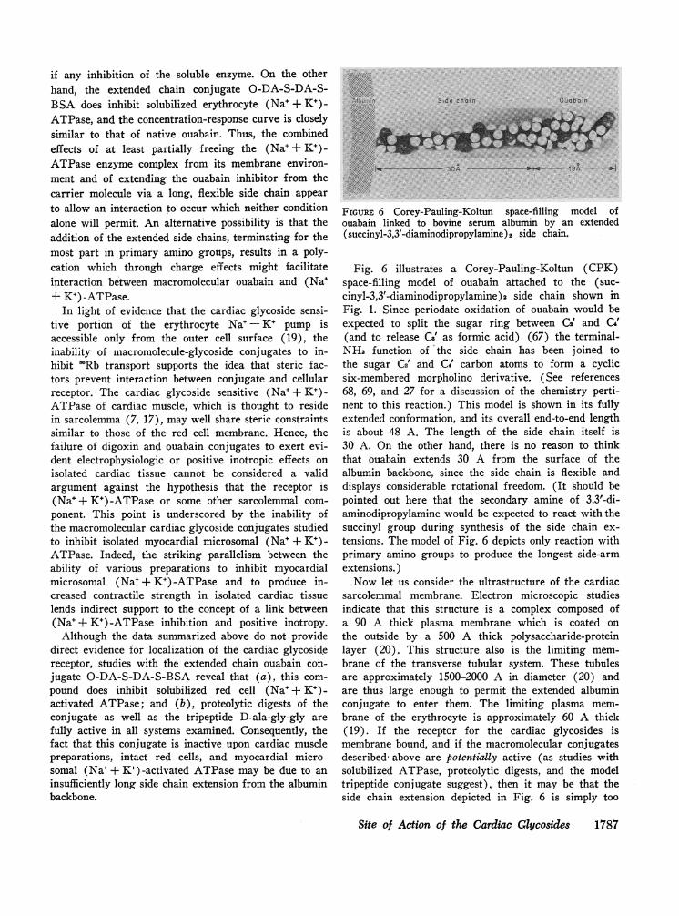
Side choin Ouaboin
3A - -- -- ISA ---_-*
FIGURE 6 Corey-Pauling-Koltun space-filling model ofouabain linked to bovine serum albumin by an extended(succinyl-3,3'-diaminodipropylamine) 2 side chain.
Fig. 6 illustrates a Corey-Pauling-Koltun (CPK)space-filling model of ouabain attached to the (suc-cinyl-3,3'-diaminodipropylamine)2 side chain shown inFig. 1. Since periodate oxidation of ouabain would beexpected to split the sugar ring between C2' and Ce'(and to release G' as formic acid) (67) the terminal-NH2 function of the side chain has been joined tothe sugar C2' and C4' carbon atoms to form a cyclicsix-membered morpholino derivative. (See references68, 69, and 27 for a discussion of the chemistry perti-nent to this reaction.) This model is shown in its fullyextended conformation, and its overall end-to-end lengthis about 48 A. The length of the side chain itself is30 A. On the other hand, there is no reason to thinkthat ouabain extends 30 A from the surface of thealbumin backbone, since the side chain is flexible anddisplays considerable rotational freedom. (It should bepointed out here that the secondary amine of 3,3'-di-aminodipropylamine would be expected to react with thesuccinyl group during synthesis of the side chain ex-tensions. The model of Fig. 6 depicts only reaction withprimary amino groups to produce the longest side-armextensions.)
Now let us consider the ultrastructure of the cardiacsarcolemmal membrane. Electron microscopic studiesindicate that this structure is a complex composed ofa 90 A thick plasma membrane which is coated onthe outside by a 500 A thick polysaccharide-proteinlayer (20). This structure also is the limiting mem-brane of the transverse tubular system. These tubulesare approximately 1500-2000 A in diameter (20) andare thus large enough to permit the extended albuminconjugate to enter them. The limiting plasma mem-brane of the erythrocyte is approximately 60 A thick(19). If the receptor for the cardiac glycosides ismembrane bound, and if the macromolecular conjugatesdescribed above are potentially active (as studies withsolubilized ATPase, proteolytic digests, and the modeltripeptide conjugate suggest), then it may be that theside chain extension depicted in Fig. 6 is simply too
Site of Action of the Cardiac Glycosides 1787
short to penetrate the membrane far enough. If so, thenthis would imply that the digitalis receptor(s) may liedeep within the plasma membrane.
ACKNOWLEDGMENTSWe are grateful to Dr. Jan Koch-Weser for the extendeduse of some of his instruments. This paper is gratefullydedicated to Mr. W. C. Ratliff.
This work was supported by NIH Research CareerAward K03-AM-18565, by NIH Research grant AM-09404,by a grant from the John A. Hartford Foundation, Inc.,and by grant 70870 from the American Heart Associationto Dr. Young; also by a grant from the MassachusettsHeart Association and by NIH Research grant lROlHE-14325 to Dr. Smith.
REFERENCES1. Mason, D. T., J. F. Spann, Jr., and R. Zelis. 1969.
New developments in the understanding of the actionsof the digitalis glycosides. Progr. Cardiovasc. Dis. 11:443.
2. Greenspan, K., and R. E. Edmands. 1969. The inotropiceffects of digitalis. In Digitalis. C. Fisch and B. Sura-wicz, editors. Grune & Stratton, Inc., New York. 65.
3. Hoffman, B. F., and D. H. Singer. 1964. Effects ofdigitalis on electrical activity of cardiac fibers. Progr.Cardiovasc. Dis. 7: 226.
4. Hoffman, B. F. 1971. Effects of digitalis on electricalactivity of cardiac membranes. In Basic and ClinicalPharmacology of Digitalis. B. Marks and A. Weissler,editors. Charles C. Thomas, Publisher, Springfield, Ill.(In press).
5. Repke, K. 1963. Metabolism of cardiac glycosides. Proc.Int. Pharmacol. Meet. 3: 47.
6. Glynn, I. M. 1969. The effects of cardiac glycosides onmetabolism and ion fluxes. In Digitalis. C. Fisch and B.Surawicz, editors. Grune & Stratton, Inc., New York.30.
7. Besch, H. R., Jr., J. C. Allen, G. Glick, and A. Schwartz.1970. Correlation between the inotropic action of ouabainand its effects on subcellular enzyme systems fromcanine myocardium. J. Pharmacol. Exp. Ther. 171: 1.
8. Akera, T., F. S. Larsen, and T. M. Brody. 1970. Cor-relation of cardiac sodium-and potassium-activatedATPase activity with ouabain-induced inotropic stimu-
lation. J. Pharmacol. Exp. Ther. 173: 145.9. Glynn, I. M. 1964. The action of cardiac glycosides
on ion movements. Pharmacol. Rev. 16: 381.10. Page, E. 1964. The actions of cardiac glycosides on
heart muscle cells. Circulation. 30: 237.11. Albers, R. W., G. J. Koval, and G. J. Siegel. 1968.
Studies on the interaction of ouabain and other cardio-active steroids with sodium-potassium-activated adeno-sine triphosphatase. Mol. Pharmacol. 4: 324.
12. Dunham, P. B., and J. F. Hoffman. 1971. Active cationtransport and ouabain binding in high potassium andlow potassium red blood cells of sheep. J. Gen. Physiol.58: 94.
13. Langer, G. A. 1968. Ion fluxes in cardiac excitationand contraction and their relation to myocardial con-tractility. Physiol. Rev. 48: 708.
14. Baker, P. F., M. P. Blaustein, A. L. Hodgkin, andR. A. Steinhardt. 1969. The influence of calcium onsodium efflux in squid axons. J. Physiol. (London). 200:431.
15. Schwartz, A., J. C. Allen, and S. Harigaya. 1969.Possible involvement of cardiac Nae, K+-adenosine tri-phosphatase in the mechanism of cardiac glycosides. J.Pharmacol. Exp. Ther. .168: 31.
16. Langer, G. A. 1970. The role of sodium ion in the regu-lation of myocardial contractility. J. Mol. Cell. Cardiol.1: 203.
17. Stam, A. C., Jr., W. B. Weglicki, Jr., D. Feldman,J. C. Shelburne, and E. H. Sonnenblick. 1970. Caninemyocardial sarcolemma-Its preparation and enzymaticactivity. J. Mol. Cell Cardiol. 1: 117.
18. Caldwell, P. C., and R. D. Keynes. 1959. The effect ofouabain on the effiux of sodium from a squid giant axon.J. Physiol. (London). 148: 8P.
19. Hoffman, J. F. 1966. The red cell membrane and thetransport of sodium and potassium. Amer. J. Med. 41:666.
20. Fawcett, D. W., and N. S. McNutt. 1969. The ultra-structure of the cat myocardium. J. Cell Biol. 42: 1.
21. Page, E. 1962. Cat heart muscle in vitro. III. Theextracellular space. J. Gen. Physiol. 46: 201.
22. Ryser, H. J.-P. 1968. Uptake of protein by mammaliancells: an underdeveloped area. Science (Washington).159: 390.
23. Waldi, D. 1959. Eine neue systematische Analyse vonAlkaloiden mit hilfe der Papierchromatographie. Arch.Pharm. Ber. Dtsch. Pharm. Ges. 292: 206.
24. Weber, K., and M. Osborne. 1969. The reliability ofmolecular weight determinations by dodecyl sulfate-polyacrylamide gel electrophoresis. J. Biol. Chem. 244:4406.
25. Lowry, 0. H., N. J. Rosebrough, A. L. Farr, and R.M. Randall. 1951. Protein measurement with the Folinphenol reagent. J. Biol. Chem. 193: 265.
26. Spackman, D. H., W. H. Stein, and S. Moore. 1958.Automatic recording apparatus for use in the chroma-tography of amino acids. Anal. Chem. 30: 1190.
27. Smith, T. W., 1972. Ouabain-specific antibodies: immu-nochemical properties and reversal of Na+, K+-activatedadenosine triphosphatase inhibition. J. Clin. Invest. 51:1583.
28. Smith, T. W., V. P. Butler, Jr., and E. Haber. 1969.Determination of therapeutic and toxic serum digoxinconcentrations by radioimmunoassay. N. Engl. J. Med.281: 1212.
29. Erlanger, B. F., and S. M. Beiser. 1964. Antibodiesspecific for ribonucleosides and ribonucleotides andtheir reaction with DNA. Proc. Nat. Acad. Sci. U. S. A.52: 68.
30. Butler, V. P., Jr., and J. P. Chen. 1967. Digoxin-spe-cific antibodies. Proc. Nat. Acad. Sci. U. S. A. 57: 71.
31. Schechter, I., S. Bauminger, M. Sela, D. Nachtigal, andM. Feldman. 1964. Immune response to polypeptidylproteins in rabbits tolerant to the protein carriers. Im-munochemistry. 1: 249.
32. Anfinsen, C. B., M. Sela, and J. P. Cooke. 1962. Thereversible reduction of disulfide bonds in poly-alanylribonuclease. J. Biol. Chem. 237: 1825.
33. Habeeb, A. F. S. A., H. G. Cassidy, and S. J. Singer.1958. Molecular structural effects produced in proteinsby reaction with succinic anhydride. Biochim. Biophys.Acta. 29: 587.
34. Berman, J. D., and M. Young. 1971. Rapid and com-plete purification of acetylcholinesterase of electric eeland erythrocyte by affinity chromatography. Proc. Nat.Acad. Sci. U. S. A. 68: 395.
1788 T. W. Smith, H. Wagner, Jr., J. E. Markis, and M. Young

35. Mills, L. 1960. Dinitro-phenyl amino acids. In Chro-matographic and Electrophoretic Techniques. I. Smith,editor. Interscience Publisher, Inc. New York. 2nd edi-tion. 143.
36. Smith, T. W., V. P. Butler, Jr., and E. Haber. 1970.Characterization of antibodies of high affinity andspecificity for the digitalis glycoside digoxin. Biochem-istrv. 9: 331.
37. Nomoto, M., Y. Narahashi, and M. Murakami. 1960. Aproteolytic enzyme of Streptomyces griseus. VI. Hy-drolysis of protein by Streptomyces griseus protease. J.Biochem (Tokyo). 48: 598.
38. Koch-Weser, J. 1963. Effect of rate changes on strengthand time course of contraction of papillary muscle.Amer. J. Physiol. 204: 451.
39. Blinks, J. R. 1965. Convenient apparatus for recordingcontractions of isolated heart muscle. J. Appl. Physiol.20: 755.
40. Koch-Weser, J. 1965. Role of norepinephrine releasein the interval-strength relationship of heart muscle.J. Pharmacol. Exp. Ther. 150: 184.
41. Blinks, J. R. 1966. Field stimulation as a means ofeffecting the graded release of autonomic transmittersin isolated heart muscle. J. Pharmacol. Exp. Ther.151: 221.
42. Lowenstein, J. M. 1965. A method for measuring plasmalevels of digitalis glycosides. Circulation. 31: 228.
43. Akera, T., F. S. Larsen, and T. M. Brody. 1969. Theeffect of ouabain on sodium- and potassium-activatedadenosine triphosphatase from the hearts of severalmammalian species. J. Pharmacol. Exp. Ther. 170: 17.
44. Akera, T., and T. M. Brody. 1968. Inhibition of brainsodium- and potassium-stimulated adenosine triphos-phatase activity by chlorpromazine free radical. Mol.Pharmacol. 4: 600.
45. Fiske, C. H., and Y. Subbarow. 1925. The colorimetricdetermination of phosphorus. J. Biol. Chein. 66: 375.
46. Dunham, P. B., and J. F. Hoffman. 1970. Partial puri-fication of the ouabain-binding component and of Na,K-ATPase from human red cell membranes. Proc. Yat.Acad. Sci. U. S. A. 66: 936.
47. Brown, B. T., and S. E. Wright. 1960. Absorptionspectra of cardiac glycosides and aglycones in sulfuricacid. J. Amer. Pharm. Ass. 49: 777.
48. Wilson, W. D., and J. F. Foster. 1971. Conformationdependent limited proteolysis of bovine plasma albuminby an enzyme present in commercial albumin prepara-tions. Biochemistry. 10: 1772.
49. Schwert, G. W. 1957. Recovery of native bovine serumalbumin after precipitation with trichloroacetic acidand solution in organic solvents. J. Amter. Chem. Soc.79: 139.
50. Skou, J. C. 1969. The role of membrane ATPase inthe active transport of ions. In The Molecular Basisof Membrane Function. D. C. Tosteson, editor. Pren-tice-Hall, Inc. Englewood Cliffs, N. J. 455.
51. Whittam, R., and K. P. Wheeler. 1970. Transportacross cell membranes. Annut. Rev. Physiol. 32: 21.
52. Klaus, W., and K. S. Lee. 1969. Influence of cardiacglycosides on calcium binding in muscle subcellularcomponents. J. Pharnmacol. Exp. Ther. 166: 68.
53. Chipperfield, D., and W. G. Nayler. 1969. The effectof ouabain on calcium in subcellular fractions of car-diac muscle. J. Pharmacol. Exp. Ther. 170: 311.
54. Koch-Weser, J. 1967. Mechanism of digitalis action onthe heart. N. Engl. J. Med. 277: 417, 469.
55. Langer, G. A., and S. D. Sarena. 1970. The effects ofstrophanthidin upon contraction and ionic exchange inrabbit ventricular myocardium: relation to control ofactive state. J. Mol. Cell Cardiol. 1: 65.
56. Besch, H. R., Jr., and A. Schwartz. 1970. On a mecha-nism of action of digitalis. J. Miol. Cell Cardiol. 1: 195.
57. Okita, G. T. 1969. Distribution, disposition and excre-tion of digitalis glycosides. In Digitalis. C. Fisch andB. Surawicz, editors. Grune & Stratton, Inc., NewYork. 13.
58. Glynn, I. M. 1957. The action of cardiac glycosides onsodium and potassium movements in human red cells.J. Physiol. (London). 136: 148.
59. Whittam, R., and M. E. Ager. 1964. Vectorial aspectsof adenosine-triphosphatase activity in erythrocytemembranes. Biochem. J. 93: 337.
60. Schwartz, A., H. Matsui, and A. H. Laughter. 1968.Tritiated digoxin binding to (Na+ +K+) -activated aden-osine triphosphatase: possible allosteric site. Scictice(Washington). 160: 323.
61. Allen, J. C., and A. Schwartz. 1970. Effects of potas-sium, temperature, and time on ouabain interactionwith the cardiac Na+, K+-ATPase: further evidencesupporting an allosteric site. J. Mol. Cell Cardiol. 1: 39.
62. Cuatrecasas, P. 1969. Interaction of insulin with thecell membrane: the primary action of insulin. Proc.Nat. Acad. Sci. U. S. A. 63: 450.
63. Crestfield, A. M., S. Moore, and W. H. Stein. 1963.The preparation and enzymatic hydrolysis of reducedand S-carboxymethylated proteins. J. Biol. Chem. 238:622.
64. Okarma, T. B.. and S. M. Kalman. 1971. The surfaceinteraction of digoxin and cultured heart cells. J. Gen.Physiol. 57: 246.
65. Cuatrecasas, P., and C. B. Anfinsen. 1971. Affinity chro-matography. Methods Enzymol. 22: 345.
65. Cuatrecasas, P. 1970. Protein purification by affinitychromatography. J. Biol. Chem. 245: 3059.
67. Bobbit, J. M. 1956. Periodate oxidation of carbohy-drates. Advan. Carbohyd. Chem. 11: 1.
68. Khym, J. X. 1963. The reaction of methylamine withperiodate-oxidized adenosine 5'-phosphate. Biochemistry.2: 344.
69. Brown, D. M., and A. P. Read. 1965. Nucleotides. Part49. The reduction of the adduct of periodate-oxidizedadenosine-5'-phosphate and methylamine. J. Chem. Soc.(London). 5072.
Site of Action of the Cardiac Glycosides 1789