Studies on Pasteurella haemolytica : comparison of ... · studies on pasteurella haemolytica:...
153
Copyright is owned by the Author of the thesis. Permission is given for a copy to be downloaded by an individual for the purpose of research and private study only. The thesis may not be reproduced elsewhere without the permission of the Author.
Transcript of Studies on Pasteurella haemolytica : comparison of ... · studies on pasteurella haemolytica:...

Copyright is owned by the Author of the thesis. Permission is given for a copy to be downloaded by an individual for the purpose of research and private study only. The thesis may not be reproduced elsewhere without the permission of the Author.

STUDIES ON PASTEURELLA HAEMOLYTICA: COMPARISON OF SEROTYPING
TECHNIQUES AND SURVEYS OF THE PREVALENCE OF SEROTYPES
IN SHEEP AND GOATS IN NEW ZEALAND
A THESIS PRESENTED IN PARTIAL FULFILMENT OF THE
REQUIREMENTS FOR THE DEGREE OF MASTER OF SCIENCE
IN MICROBIOLOGY AT MASSEY UNIVERSITY, NEW ZEALAND.
ANNE CAMILLA MIDWINTER
1987

~ssey Un~~rsity Library . Thesis Copyright Form
Title of thesis:
(a)
t . jW!I%~~ &c6p<~r= ~~<' 6'~S' oj lfJr.evcr~e-e. c;f SE'.7o'o/.;?-V" ~ ~ ~ ~...r
I give permission for my .thesis to be made availab l e to readers in the Massey University Library under c onditions determined by the Librarian.
I do not vish my thesis to be made available to readers vithout my vritten consent for months.
l agree that my thesis, or a copy, may be sent to another institution under conditions determined by the Li br arian.
I do not vish my thesis, or a copy, to be sent to another institution vithout my Yritten consent for months.
(a) I agree that my thesis may be copied for Library use .
~ I do not vish my thesis to be copied for Library months. ------
Anne Midwinter Date 2~{7!87
The copyright of this thesis belongs to the author. Readers mus t sign their name in the space below to show that they recognise this. They are asked to add their permanent address.
NAME AND ADDRESS DATE
·' "" ... I ll
" • II
·~': I I I
" •

i
ACKNOWLEDGEMENTS
I would like to thank the Department of Microbiology and Genetics For
providing the facilities and opportunity for this study.
In particular I would like to thank my supervisor Dr. J.K. Clarke, for
his enthusiastic interest and advice throughout this study.
Special thanks go to Dr. M.R. Alley for arranging the collection of CNP
lesions from sheep and nasal swabs from goats.
I would like also to thank:
George Ionas for showing me how to run SDS-PAGE gels (in lieu of 27
chocolate fish) .
My friends and colleagues in the Department (especially Chris, for his
never-failing friendship and good humour) .
And Hamish, without whose support this thesis would never have been
started, let alone finished.

ii ABSTRACT
P.haemolytica is the aetiological agent of pneumonic pasteurellosis
in sheep and goats, and, as a secondry invader it also exacerbates
lesions of chronic non-progressive pneumonia (CNP) . These diseases cause
considerable economic loss to the New Zealand farming industry.
P. haemolytica exists as 15 serotypes and immunity is serotype
specific. Vaccines against P .haemolytica are produced, but it is not
known if the serotypes contained in the vaccine are the same as those
causing disease in New Zealand as there is a lack of information on the
prevalence and distribution of the serotypes of P .haemolytica in this
country. This is largely due to the technical difficulties involved in
typing isolates because the standard method, the indirect
haemagglutination assay, ( IHA), is laborious and may give anomalous
results due to cross-reactions.
The present investigation was undertaken with two major aims: to
replace IHA with a more convenient typing system, viz. agar gel
immunodiffusion, (AGID), and to use AGID to survey the serotypes of
P.haemolytica present in CNP lesions of sheep, pneumonic pasteurellosis
of sheep, pneumonic pasteurellosis of goats and the nasal cavities of
goats.
Difficulties were encountered in the preparation of rabbit antisera
to some of the 15 prototype strains. These difficulties were overcome by
using domestic hens when necessary. Using these sera it was possible to
distinguish the 15 prototype strains by IHA, and following absorption of
sera, by AGID. The results obtained by IHA and AGID were in agreement, at
least when prototype strains were examined. It was necasary to show that
AGID is able to correctly establish the serotype of field isolates of

iii
P. haemolytica. Hence 2 5 caprine isolates of P. haemolytica from field
cases of pneumonic pasteurellosis were serotyped by both IHA and AGID. In
24 cases the results from the two tests agreed. In the remaining case IHA
indicated that the isolate was serotype A2 or All. We were able to show
that this isolate gave a line of identity with antigen prepared from the
prototype strain of All, but showed no line of identity in the AGID with
any other antigen preparation. Taking this as the critical criterion we
concluded that this isolate was serotype All, although IHA showed a 2-
fold preference for A2 over All. Since AGID was shown to be a reliable
test we used it alone for future serotyping, for two reasons: it is more
convenient, and any cross-reactions that do occur may be resolved by
looking for a line of identity between antigens of the isolate and a
prototype antigen. In the case of two serotypes involved in many cross
reactions, namely Al and A7, the capsular polysaccharide was purified by
organic solvent precipitation. This purified polysaccharide was used to
test for a line of identity with reacting isolates. This eliminates the
possibility that the line of identity seen was due to a non-serotype
specific antigen.
Four surveys (two in sheep, two in goats) of the serotypes of
P.haemolytica present in New Zealand were undertaken. The first involved
139 isolates derived from ovine lesions of CNP collected from 4 areas of
New Zealand. A total of 9 serotypes were found. Serotypes Al (31.7%), A2
(47.8%) and A7 (10%) made up 89.5% of the total.
A smaller survey of 18 isolates from pneumonic pasteurellosis of
sheep revealed 6 serotypes, including 1 isolate of TlO, a serotype and
biotype not previously found in New Zealand. Al (11.1%) and A2 (61.1%)

iv
were the predominant serotypes present and represented 72.2% of the
total.
The 25 isolates of P.haemolytica from caprine pneumonic
pasteurellosis contained only 4 serotypes. A2 represented 8 0% of the
total.
14 isolates of P.haemolytica were obtained from the nasal cavities
of 109 goats. Only 2 serotypes were isolated. 13 isolates were A2 and the
remaining isolate was All.
The implications of these results for vaccine manafucture were
discussed and it was suggested that a vaccine containing A2, Al and A7
(in order of importance) should control CNP in sheep and pneumonic
pasteurellosis in both sheep and goats.
Field isolates of P.haemolytica were compared with prototype strains
for capsule production (using Laurell Rocket test), and antibiotic
sensitivities. The total proteins of caprine and ovine strains were also
compared, using SDS-PAGE. Laurell Rocket tests showed that the prototype
strains produced more capsular polysaccharide than did any of our field
isolates. All isolates of P. haemolytica showed some resistance to
streptomycin while none were resistant to more than 4f.1g/ml
chloramphenicol or penicillin so these are the drugs of choice. No
difference was found within a serotype between the total proteins of
caprine and ovine isolates by SDS-PAGE.

v CONTENTS
Page
TITLE PAGE ....
ACKNOWLEDGEMENTS i
ABSTRACT . . . . . . . . . . . . . . . . . . . . . . . . . . . . . . . . . . . . . . . . . ii
LIST OF CONTENTS v
LIST OF FIGURES viii
LIST OF TABLES X
INTRODUCTION 1
CHAPTER 1: Historical Review
1.10 Classification of P.haemolytica . . . . • . . . • . . . . 4
1.11 Biotypes 4
1.12 Antigens of P.haemolytica .................. 6
1.13 Number of Serotypes . . . . . . . . . . . . . . . . . . . . . . 8
1.14 Untypable strains . . . . . . . . . . . . . . . . 9
1.15 Identification of Serotypes . • . . . . 9
1.16 Relationships between Serotype and Biotype ...... 10
1.20 Commensal Association of P.haemolytica with Sheep and Goat11
1.30 Diseases Associated With P.haemolytica in Sheep ...... 13
1.31 Pneumonic Pasteurellosis ................ 13
1. 32 Septicaemic Pasteurellosis . . . . . . . . . . . . . 14
1.33 Chronic Non-Progressive Pneumonia ...... 15
1.40 Diseases Associated with P.haemolytica in Goats ...... 17

vi
1.50 Importance of Pasteurellosis in Sheep and Goats ...... 13
1.60 Importance of Chronic Non-Progrssive Pneumonia . 19
1.70 Transmission of Disease
1.80 Antibiotic Treatment
1.90 Immunity to P.haemolytica .
1. 91 Vaccines . . . . . . . . .
CHAPTER 2: Comparison of AGID and IHA using Prototype Strains of
P.haemolytica and Field Isolates
19
20
24
26
2.1 Introduction . . . . . . . . . . . . . . . . . . . . . . . . 28
2. 2 Materials and Methods . . . . . . . . . . . . . . . . . . . . . . . . 2 9
2.3 Results ..
2.4 Discussion
CHAPTER 3: Survey of the Prevalence of P.haemolytica in Sheep and Goats
in New Zealand
3.1 Introduction
33
48
56
3.2 Materials and Methods ........................ 56
3. 3 Results .. 59
3. 4 Discussion . . . . . . . . . . . . . . . . . . . . . . . . . . . . . . . . 7 4

vii
CHAPTER 4: Comparison of Total Proteins, Antibiotic Sensitivities and
Capsular Antigen Production of Prototype Strains and Field
Isolates of P.haemolytica
4.1 Introduction
4.2 Materials and Methods
4.3 Results ..
4.4 Discussion
79
80
82
90
CHAPTER 5: General Discussion ............................ 93
APPENDIX . . . . . . . . . . . . . . . . . . . . . . . . . . . . . . . . . . . . . . . . . . 100
BIBLIOGRAPHY . . . . . . . . . . . . . . . . . . . . . . . . . . . . . . . . . . . . . . . 115

viii
LIST OF FIGURES
Figure Page
1 Indirect haemagglutination assay of P.haemolytica serotype A2 .... 38
2 Agar gel immunodiffusion test of P.haemolytica serotype A2 ...... 39
3 AGID of prototype Al and purified Al capsular polysaccharide . . . . 46
4 AGID of prototype A7 and purified A7 capsular polysaccharide . . . . 47
5 Comparison of total proteins of sheep and goat isolates of
P.haemolytica serotype A2 . . . . . . . . . . . ... 82
6 Comparison of total proteins of sheep and goat isolates of
P.haemolytica serotype A6 . . . . . . . . . . . . . . 83
7 Comparison of total proteins of sheep and goat isolates of
P.haemolytica serotype A7 . . . . 84

ix
8 Comparison of total proteins of sheep and goat isolates of
P.haemolytica serotype All .... 85
9 Comparison of the relative amount of capsule produced by different
strains of P. haemolytica serotype All . . . . . . . . . . . . . . . . . . . 88
10 Comparison of the relative amount of capsule produced by different
strains of P. haemolytica serotype A 7 . . . . . . . . . . . . . . . . . . . 8 9

X
LIST OF TABLES
Table Page
I Prototype strains of P.haemolytica tested by IHA .......... 34
II Prototype strains of P.haemolytica tested by AGID .......... 35
III Prototype strains of P.haemolytica tested by AGID using adsorbed
antisera .... . ............. 37
IV Serotypes of 25 caprine isolates of P.haemolytica tested by AGID 40
V Serotypes of 25 caprine isolates of P.haemolytica tested by IHA 42
VI Comparison of the results obtained by AGID and IHA .......... 44
VII Serotypes of P.haemolytica from CNP: Auckland . . . . . . . . . . . . 60
VIII Serotypes of P.haemolytica from CNP: Gisborne ............ 62
IX Serotypes of P.haemolytica from CNP: Christchurch .......... 63
X Serotypes of P.haemolytica from CNP: Invercargill .......... 65

xi
XI Recovery rate of P.haemolytica from CNP lesions .. . . 67
XII Comparison of serotypes of P.haemolytica from 4 centres . . . . 68
XIII Serotypes of P.haemolytica from caprine pneumonic pasteurellosis 69
XIV Serotypes of P.haemolytica from ovine pneumonic pasteurellosis .. 71
XV Serotypes of P.haemolytica from caprine nasal cavities . . . 72
XVI Comparison of the serotypes of P.haemolytica from acute and
chronic pneumonia and nasal cavities .... 73
XVII Antibiotic sensitivities of isolates of P.haemolytica .. . . 86
XVIII Comparison of antibiotic sensitivities of P.haemolytica . . . . 87

1
INTRODUCTION
Pasteurella haemolytica is a small Gram-negative rod found as a
commensal in the nasopharynx of sheep and goats. In both species it
causes pasteurellosis and it is also involved in chronic non-progressive
pneumonia (CNP) of sheep. Pasteurellosis occurs as two distinct disease
entities: septicaemic pasteurellosis and pneumonic pasteurellosis.
Septicaemic pasteurellosis is found in New Zealand, but is rare (Hartley
et al., 1955). The pneumonic form also occurs in New Zealand where it is
a common and widespread disease (Salisbury, 1957). It is caused by
P.haemolytica often in conjuction with parainfluenza virus type 3
(Harbourne, 1979) . It is a rapidly fatal disease of economic significance
and is found in both sheep and goats.
The aetiology of CNP is not universally agreed. However most workers
concur that M. ovipneumoniae and P. haemolytica are involved.
M.ovipneumoniae initiates lesions which are exacerbated by P.haemolytica
(Jones et al., 1978) . The disease is widespread and prevalent in New
Zealand sheep aged between 6 and 9 months. CNP was estimated to have
caused a $26 million loss in 1975 (Dysart, 1976). Treatment of
pasteurellosis or CNP is seldom a practical proposition, for economic
reasons. Furthermore acute pasteurellosis is often diagnosed only at post
mortem as it is often a rapidly fatal disease. However an effective
P.haemolytica vaccine could prevent losses due to pneumonic
pasteurellosis and CNP.
Immunity to P.haemolytica depends on a immune response to the
capsular polysaccharide (Biberstein et al., 1965b). However there are 15
serotypes of P.haemolytica based upon the capsular polysaccharide (Fraser

2
et al., 1982) . Obviously any P. haemolytica vaccine should contain the
serotype or serotypes which most frequently cause disease in New Zealand.
This requires information on the prevalence of the various serotypes in
New Zealand and their association with disease in sheep and goats. This
in turn requires a practical method by which isolates can be serotyped.
The method of serotyping P. haemolytica is, by convention, the
indirect haemagglutination assay, (IHA) (Biberstein et al., 1960) This
method is laborious and the results may be difficult to interpret due to
cross-reactions between serotypes. The agar gel immunodiffusion (AGID)
test is a more convenient method for serotyping, but it has been used
only by Murasachi et al., ( 19 65) , who found that it was less than
satisfactory due to the number of cross-reactions that occured.
This led us to undertake the following investigation.
1/ Comparison of IHA and AGID as methods of serotyping
P.haemolytica with the aim of developing AGID as a standard serotyping
method.
2 I The use of AGID to survey the prevalence of serotypes of
P.haemolytica found in:
a) The lungs of sheep with CNP
b) The lungs of sheep with pneumonic pasteurellosis
c) The lungs of goats with pneumonic pasteurellosis
d) The nasal cavities of apparently healthy goats.
3/ To examine field isolates of P.haemolytica as follows:
a) To compare the production of capsular polysaccharide
by prototype and field isolates, using the Laurell Rocket Test.
b) To compare the proteins of caprine and ovine
isolates by SDS-PAGE (only isolates from the same serotype were

3
compared) .
c) To establish the antibiotic sensitivities of
isolates of P.haemolytica from pneumonic pasteurellosis of sheep and
goats.

4
CHAPTER 1
Historical review
1.10 Classification of ~.haemol~ica
The genus Pasteurella includes 4 main species ( P. multocida,
P.haemolytica, P.pneumotropica and P.ureae) plus up to 3 more of uncertain
affiliation.
The Pasteurella are small, Gram-negative, non-motile rods. They are
aerobes or facultative anaerobes with a fermentative metabolism. They may
be differentiated from the Enterobacteriaceae as they are oxidase positive
and produce acid but no gas and no H2 S in triple-sugar-iron slants. They
may be distinguished from fe rmen ta ti ve bacteria (other than the
Enterobacteriaceae) on the basis of tests for oxidase, urease and indole
activity.
P. haemolytica can be distinguished from other Pasteurella as it
produces a zone of haemolysis on blood agar and does not produce urease.
1. 11 Biotypes
The heterogeneity P.haemolytica was first recognised by Biberstein et
al., (1958), who described two types of the organism which differed in
colonial characteristics, crystal violet uptake and serology but did not
define them further. Smith, (1959), agreed with Biberstein' s division of
P. haemolytica and designated the two forms of P. haemolytica, the «T
strains» and the «A strains». Perhaps more importantly, Smith, (1961),
found that the two strains were associated with different diseases. A

5
strains were found in the lungs of pneumonic lambs and sheep but were also
associated with septicaemia of lambs aged less than three months whereas T
strains were associated only with septicaemia of older lambs.
Smith, (1961), showed that the strains, now referred to as biotypes,
could be readily distinguished on primary isolation on the basis of colony
morphology. T biotype colonies were larger than that of A biotypes and
possessed dark centres. A biotype colonies were an even grayish colour
sometimes with a central thickening. When dispersed in water T biotypes
formed a lace-like pattern whereas A biotypes dispersed evenly. However the
biotypes are similar in the degree of haemolysis produced on blood agar
(Smith, 1961)
The biotypes can also be distinguished by fermentation reactions: the
T strains all ferment trehalose and fail to ferment arabinose within 10
days. In contrast, A strains fail to ferment trehalose but ferment
arabinose within 10 days (Smith, 1959). It was this characteristic
fermentation pattern which led to the two biotypes being called T (for
trehalose) and A (for arabinose), respectively. The biotypes may also be
differentiated on the basis of other fermentation reactions. The A biotypes
ferment starch (Smith, 1961) and xylose (Shreeve et a~., 1970) whereas the
T biotypes ferment salicin and mannose (Shreeve et al., 1970).
Growth curves of A and T strains in broth are initially similar with
both reaching similar maximum viable counts. However after this, the viable
counts of T strains decline slowly while those of A strains fall sharply.
As a consequence of this, after 24 hours incubation the viable count of T
biotypes may be 40 times that of the A biotypes (Smith, 1959)
Smith, (1959), found that the A biotype were markedly more sensitive
to penicillin than the T biotype. The A biotype is also significantly more

6
sensitive to tetracycline, ampicillin, cephalothin, chloramphenicol,
erythromycin and furadantin (Biberstein et al., 1979) A biotypes are
inhibited by much lower concentrations of basic fuchsin, methylene blue and
brilliant green than the T biotypes (Olmos et al., 1979).
The differences between the A and T biotypes extend to their capsules.
By electron-microscopy Gilmour et al., (1985), showed that while the
capsules of A biotypes are large and well defined, those of T biotypes are
thinner and less defined. Adlam et al., (1984) and (1985), chemically
analysed the capsular polysaccharides of P.haemolytica serotypes Al and T4
and found that while the capsular polysaccharide of Al was a conventional
Gram-negative polymer, that of T4 was a teichoic acid, a molecule not
usually associated with Gram-negative organisms.
The differences between the A and T biotypes of P. haemolytica are nov1
known to be more fundamental than variations in growth patterns, capsules
and antibiotic susceptibilities. Thus DNA-RNA hybridizations between the A
and T biotypes (Biberstein et al., 1968), suggest that the differences
between the two types are equivalent to those between different species.
These differences between A and T biotypes led Gilmour et al., (1985),
to suggest that the A and T biotypes should be recognised as separate
species.
1.12 Antigens of R.haemol~ica.
Biberstein et al., (1960), heated broth cultures at 56°C and used the
supernatant «soluble antigens» as the basis of a serotyping system for
P.haemolytica. They used autoclaved cells to study the somatic antigens by
agglutination tests. Six somatic groups were detected. Somatic group A (not

7
to be confused with biotype A) contains serotypes 1, 5, 6, 7, 8 and 9,
while somatic group B contains serotype 2 strains.
Immunity to P.haemolytica depends largely on the serotype (Knight et
al., 1969). Hence from a vaccine point of view, the association of serotype
(rather than somatic type) with disease is of overriding importance and has
received most attention.
Cameron, (1966), stated that the main antigen detected in the
serotyping of P.haemolytica by IHA was a polysaccharide, but that protein
antigens were also capable of producing agglutination in the IHA. Himmel et
al., (1982), extracted a highly immunogenic protein from a number of
P. haemolytica serotypes. This protein was toxic to bovine alveolar
macrophages. It was neutralized by species-specific antisera, with
homologous antisera giving better neutralization than heterologous (Shewen
etal.,1983).
Tabatabai et al., (1981), isolated from A1 a soluble, extracellular
antigen that had neuriminidase activity. This activity was inhibited by
antisera to P.haemolytica serotype A1 but not by normal serum.
Donachie et al., (1984)., made sodium salicylate extracts of
P.haemolytica serotypes A1 and A6. They determined the chemical composition
of these extracts and suggested that sodium salicylate extracted a complex
of lipid, polysaccharide and protein from the outer membrane. A number of
antigens were detected. For both serotype A1 and A6 a dominant serotype
specific antigen was detected by counter immunoelectrophoresis (CIE) .
Adlam et al., (1984) and (1985), purified the serotype-specific
capsular polysaccharide of P.haemolytica serotypes A1 and T4 and
characterized them by chemical analysis and NMR spectroscopy. The two
polymers were chemically dissimilar. The T4 capsule was composed of

8
teichoic acid. The partially purified polymers were antigenic in sheep
(although completly purified polymers were not). The purified
polysaccharide reacted with Al antisera only, in the agar gel
immunodiffusion test. Antisera raised against the partially purified
polysaccharide reacted with sodium salicylate extracts of homologous cells,
producing a line of identity with the purified polysaccharide. This led
Adlam et al., (1984), to suggest that the sodium salicylate extracts and
purified polysaccharide are similar or identical immunologically and that
this also implies that when present in the cell wall, the capsular
polysaccharide is attached to lipid components.
1.13 Number of Serotypes.
Early work on the serology of P.haemolytica established the antigenic
separation of P.haemolytica and P.multocida (Newsome et al., 1932). In
subsequent studies Tweed et al., (1930), postulated only 1 serotype whereas
Montgomerie et al., ( 1938), suggested that there were 3 serotypes of
P.haemolytica. Nearly 20 years later, Carter, (1956), reported the species
to be antigenically homogeneous.
The definitive work on the serotyping of P .haemolytica was done by
Biberstein et al., (1960). This group typed 98 isolates of P.haemolytica by
the indirect haemagglutination assay, (IHA), and found that they could be
divided into 10 serotypes (numbered 1-10) or were untypable.
Subsequently 5 additional serotypes have been identified (Biberstein
and Gills, 1962; Biberstein and Thompson, 1966; Pegram et al., 1979; Fraser
et al., 1982) It should be noted that the most recent addition to the
P.haemolytica serotypes, Tl5, is serologically related to serotype T3 and

9
it is uncertain if the two are antigenically distinct (Fraser et al.,
1982).
1.14 Untypable Strains
A number of isolates of P.haemolytica do not react in the IHA with
antisera to any of the 15 existing serotypes. Antisera raised against these
isolates do not give interpretable IHA results even when tested against the
homologous strains (Aarsleff et al., 1970; Frank, 1980). Many of these
untypable strains are also difficult to assign to a biotype as their
fermentation patterns are often anomalous (Aarsleff et al., 1970). Frank,
(1970), tested a number of untypable strains by rapid plate agglutination
and suggested that some strains may be untypable due to their lack of
serotype-specific antigen. Untypable strains are usually isolated from the
nasal cavity but are rarely, if ever, recovered from pathogenic lesions.
From a veterinary point of view they are probably unimportant.
1.15 Identification of Serotypes
The most widely used method of serotyping P. haemolytica is the
indirect haemagglutination assay (IHA), first described by Biberstein et
al., 1960). Modifications to this method include the use of glutaraldehyde
fixed sheep RBC (Sawada et al., 1982), and the use of microtitre plates
(Fraser et al., 1983).
The IHA involves the adsorption of capsular material from heat-treated
culture supernatants of the strain to be tested to glutaraldehyde-fixed
sheep RBC. These antigen-coated RBC are added to serial dilutions o::
antisera to all 15 serotypes. This test is laborious as it involves 15

10
titrations for each isolate. It may also be difficult to interpret because
cross-reactions may occur (Biberstein, 1965a) .
An agglutination test for the serotyping of P.haemolytica was
described by Frank and Wessman, (1978). It gave essentially the same
results as the IHA but more cross-reactions were found.
Muraschi et al., (1965), extracted «somatic antigens» from
P.haemolytica and used the extracts in an agar gel immunodiffusion (AGID)
test to compare serotypes. The gel precipitation lines were to a large
extent serotype specific. This is not as surprising as may be first thought
because the method of extraction used to prepare the «somatic antigens»
would also extract the serotype specific capsular polysaccharide.
It was suggested by Biberstein, ( 19 65a) that minor antigens were
responsible for cross-reactions observed in IHA. However Gonzalez-Rayos et
al., (198 6), inserted two recombinant plasmids made from the genome of
P. haemolytica serotype A1 and coding for a soluble surface antigen into
E.coli. E.coli cells carrying the plasmids were agglutinated strongly by
antiserum to P. haemolytica A1 and weakly by antisera to A2 and A 7. They
were found to have patches of extra-cellular material on their surfaces.
Southern blot hybridization using the DNA from recombinant plasmids as a
probe showed that the probe hybridized strongly to DNA from serotype A1 and
weakly to DNA from serotypes A2 and A 7. This suggests that the cross
reactions observed are due to shared determinants in the capsular material
of different serotypes rather than shared protein or other minor antigens.
1.16 Relationships Between Serotype And Biotype
Biotype and serotype are not independent. Biberstein et al., ( 19 62) ,
tested 37 strains, which represented all known serotypes of P.haemolytica

11
for fermentation reactions and antibiotic sensitivity patterns and assigned
them to either biotype A or T. Serotypes 1, 2, 5, 6, 7, 8, 9 and 11 were
all biotype A, while serotypes 3, 4 and 10 were biotype T. Serotypes 12, 13
and 14 were isolated subsequently and were found to be biotype A while the
latest serotype, 15, belongs to the T biotype, as would be expected because
it is closely related to serotype 3 (Fraser et al., 1982).
In contrast to the above, workers in Kenya (Mwangota et al., 1978)
indicated that nasal isolates, from sheep and goats, of serotypes 3, 4, 6,
10 and 12 were found to contain both A and T biotypes within a single
serotype. However this work is not convincing and has not been confirmed.
1.20 Commensal Association of ~.haemolytica with Sheep and Goats
P.haemolytica is a commensal which may be isolated from the nasal
cavity and tonsils of apparently healthy sheep and goats. The two biotypes
appear to localise in different regions. In a survey by Gilmour et al.,
(1974), P.haemolytica was isolated from 95% of the ovine tonsils examined
and from 64% of the ovine nasopharyngeal swabs. Of these isolates, 65% of
those from tonsils but only 6% of those from nasopharynxes were T biotype
while 60% of P.haemolytica isolates from the nasopharynx and 20% of those
from the tonsils were A biotype. The remaining isolates gave anomalous
fermentation patterns and thus could not be biotyped.
P. haemolytica has been isolated from the nasal cavities of normal
goats with a prevalence of from 5% (Ojo, 1970) to 64% (Ngatia et al.,
1985). The biotypes of the isolates are unknown. P.haemolytica has also
been isolated from the tonsils of goats, (Mwangota et al., 1972), although
the biotypes of these isolates are uncertain.
12
1.30 Diseases Associated with R.haemo1Y!ica in Sheep.
The most important of the P.haemolytica infections are the pneumonias:
chronic non-progressive pneumonia (CNP) and acute pneumonic pasteurellosis.
These are considered below in more detail.
P.haemolytica is also involved in a septicaemic disease of sheep
caused in young lambs by either A or T biotype and in older lambs by
biotype T.
Arthritis has been observed asted lungs and numerous blood splashes on
the heart. Lesions may be found in the alimentary tract. These lesions
appear to spread from the tons this association may be found in the field,
although P.haemolytica has not been isolated from the diseased joints.
Ovine mastitis is frequently caused by P. haemolytica. Systemic
infections following mastitis are not uncommon and sheep may die or lose a
mammary gland due to gangrene (Biberstein, 1981) .
P. haemolytica has been associated with ulcerative infections of the
oesophagus and pharynx, though it is not confirmed that the organism causes
the lesions. Also P.haemolytica has been isolated from parasitic gastro
enteritis infections, and from encephalitis and meningitis (Harbourne,
197 9) .
1.31 Pneumonic Pasteure11osis
Synonyms include enzootic pneumonia (Note: although this term is used
as a synonym for pneumonic pasteurellosis in New Zealand, elsewhere it
refers to CNP), acute necrotising pneumonia, acute exudative pneumonia and
acute pneumonia. The disease may occur in any age of sheep at all times of
the year, although outbreaks are more common in spring or early autumn
(Harbourne, 1979) .

13
Outbreaks are often sudden. The proportion of animals affected ranges
from single sporadic cases to 10% of the flock (Harbourne, 1979) .
Symptoms include respiratory distress, often accompanied by coughing,
discharges from the nose and eyes, high fever and occasionally diarrhoea.
Affected animals usually die within 24-36 hours of the onset of the disease
(Harbourne, 1979).
The pathology of pneumonic pasteurellosis is characterised by a severe
fibrinous pleurisy with pleural adhesions. The apical and cardiac lobes are
often consolidated with the remainder of the lung showing acute congestion
(Salisbury, 1957).
Histopathological examination shows a cellular exudate containing
neutrophils,macrophages and detached alveolar epithelial cells, (Alley,
1975a), with «oat cells» lying at the edge of the lesion (Harbourne, 1979).
P.haemolytica biotype A may be isolated in profusion from the lesions
and pericardial fluid of pneumonic lungs. Ocasionally a T biotype may be
isolated from a pneumonic lesion (Gilmour, 1978) .
It has been suggested that the pneumonia is associated with a viral
infection such as parainfluenza virus type 3 (PI-3) or adenovirus
(Harbourne, 1979) .
The factors governing the prevalence of pneumonic pasteurellosis are
not well known. It is not known why some flocks are affected and others not
(Harbourne, 1979).
Although spring and autumn, rather than winter show peaks in
prevalence, cold weather may be a pre-disposing factor, especially in cases
associated with a viral infection (Harbourne, 1979). As well, mustering,
over-heating and transportation, humidity and changes in diet have all been

14
implicated, as have dipping, castrating and dosing (Gilmour, 197 8) . All
these factors involve stress.
1.32 Septicaemic Pasteurellosis
Septicaemic pasteurellosis of sheep is caused by both the A and T
biotypes of P. haemolytica. In lambs under 2 months, infection with
P. haemolytica biotype A usually causes septicaemia rather than pneumonia
(Gilmour, 1978) . In older lambs and sheep, septicaemic pasteurellosis is
associated with P.haemolytica biotype T.
Septicaemic pasteurellosis occurs in all ages of sheep·throughout the
year, but appears to be more prevalent in 1-2 year old sheep during autumn,
coinciding with the movement of sheep and changes in feeding (Gilmour,
1978) . Bad weather or disease also seem to be predisposing factors (Dyson
et al., 19 81) .
Affected sheep are commonly found dead. Symptoms include dullness with
an unwillingness to move, laboured breathing, frothy discharge from the
mouth and high temperature (Gilmour, 1978).
Post mortem examination shows enlarged, haemorrhagic lymph nodes,
swollen and congested lungs and numerous blood splashes on the heart.
Lesions may found in the alimentary tract. These lesions appear to have
spread from the tonsils (Dyson et al.,1981).
Fatal septicaemic disease may also be produced in lambs by inoculation
of heat-killed bacteria (Smith, 1960) and from the pathology of the disease
it seems unlikely that it is a true septicaemia (if this is defined as
requiring multiplication of bacteria in the blood), but is a toxaemia
because the organisms multiply in the tissues. Death is due to liberated
endotoxin (Dyson et al.,1981).

15
It has been postulated that the T biotype organisms commonly found in
the tonsils, may multiply and invade surrounding tissues. From there they
spread to the liver, spleen and kidney (Dyson et al., 1981).
1.33 Chronic Non-Progressive Pneumonia
Synonyms for chronic non-progressive pneumonia or CNP include enzootic
pneumonia (Note: although this term is used as a synonym for pneumonic
pasteurellosis in New Zealand, elsewhere it refers to CNP), sub-acute
pneumonia, summer pneumonia (in Australia), acute exudative pneumonia,
hogget pneumonia, atypical pneumonia (Stamp et al., 1963), proliferative
interstitial pneumonia (Sullivan et al., 1973)
CNP is prevalent in New Zealand sheep between the ages of 3 and 10
months (hence the name hogget pneumonia) , unlike acute pneumonic
pasteurellosis which may be found in all ages of sheep.
The disease is infrequently fatal and as the name suggests, is
chronic, which distinguishs it from acute pneumonic pasteurellosis, and
non-progressive, which distinguishs it from the progressive pneumonia
produced by maedi virus and related agents.
CNP elicits few clinical signs. In experimentally induced pneumonia
(Jones et al., 1982) animals showed depression, with slight fever and
coughing. Sullivan et al., (1973), reports that an affected flock showed
poor growth and exercise intolerance.
The macroscopic appearance of the lesions shows differing degrees of
consolidation over the antereo-ventral parts of both lungs. Lesions vary in
colour between red and gray with red lines of collapse present in some
cases. In severe cases the affected lobes are noticeably firm and thicker
than normal. Occasionally fibrinous adhesions are found between the

16
affected lobes and the pleura. Abcesses may be found associated with
lesions of CNP in older lambs.
The predominant feature of CNP lesions by light microscopy is alveolar
collapse. This is associated with neutrophil infiltration and cellular
exudate composed primarily of macrophages. As well, severe hyperplasia of
both the bronchiolar and alveolar epithelium may be found (Alley, 1975a) .
Damage by CNP tends to be localised and less than that caused by
pneumonic pasteurellosis (Alley, 1975a). The aetiology of CNP is complex
and has not yet been unequivocally established, although most workers do
agree on the organisms involved.
St. George, (1972), found that the bacteria most frequently isolated
from pneumonic sheep lungs were P.haemolytica, Corynebacterium pyogenes and
Neisseria species. Mycoplasma species were also isolated, as were a low
number of viruses.
P.haemolytica can be isolated from about 60% of pneumonic lung lesions
but from only about 6% of normal lungs (Alley, 1975b) . The organism can
also be found in about 75% of nasal cavities (Alley, 1975b). P.haemolytica
produces pneumonia when inoculated intrabronchially in large (10 10
organisms) doses (Smith, 1964). However this disease (pneumonic
pasteurellosis) is not CNP and obviously the relationship of P.haemolytica
to CNP is not a straight-forward one of cause and effect.
M. ovipneumoniae was recovered from the nasal cavities of 70% of
pneumonic sheep and from 10 0% of pneumonic lungs. Titres of 10 6-10 7
organisms per gram of lung were present. M.ovipneumoniae was isolated from
only 20% of normal lungs and then with a titre of less than 10 3 (Alley et
al., 1975). M. ovipneumoniae is capable of producing macroscopic lesions
when inoculated intratracheally (Sullivan et al., 1973).

17
Alley and Clarke, (1977), found that the number of M.ovipneumoniae and
bacteria found in the lungs correlated with the histopathological lesions
such as high numbers of neutrophils and severe epithilial hyperplasia.
Jones et al., (1978), produced lung lesions identical to the field
cases by the intratracheal inoculation of pneumonic lung suspensions, and
by inoculations of a mixed culture of M. ovipneumoniae, M. arginini and
P.haemolytica. This implies that more than one organism may be necessary to
establish the disease.
1.40 Diseases Associated with ~.haemol~ica in Goats.
The majority of reports of P.haemolytica infections in goats refer to
pneumonic diseases and these appear to be the most important. However
Gourlay and Barber, (1960), report the isolation of P.haemolytiGa from
cases of septicaemia in young kids and this disease is similar to that
found in lambs.
Bhagwan and Singh, (1972), examined pneumonia in goats and concluded
that there were 6 different pathological conditions. They gave no
indication of the micro-organisms involved.
In a review on caprine pneumonia Ojo, (1977), described 2 diseases,
contagious caprine pleuropneumonia, associated with Mycoplasma species and
a pneumonic pasteurellosis infection involving either P.haemolytica or
P.multocida. Pande, {1942) and Borgman and Wilson, (1955), described
outbreaks of disease, in India and the USA respectively, but it is not
clear whether the organisms involved are P.haemolytica or P.multocida or
both and whether or not any other micro-organisms were involved. Misra et
al., (1970), reported pneumonic pasteurellosis in kids due to
P. haemolytica. This disease appears similar in sheep and goats. Symptoms

18
include nasal discharge and cough often associated with sudden death (Misra
et a1.,1970). The pathology of the disease is also similar to that of
sheep, with consolidation of the lobes of the lungs, exudate in the
thoracic cavity and inflammation of the pleural cavity (Ojo, 1976).
Predisposing factors appear to be the same; thus Ojo, (1976), reported that
the incidence of caprine pneumonia increased with increase in relative
humidity and rainfall.
It appears that the acute pneumonic pasteurellosis pneumonia in goats
and sheep are alike, in aetiology, symptoms and predisposing factors, as
well as the pathology of the lesions produced.
1.50 Importance of Pasteurellosis in Sheep and Goats
Pneumonic pasteurellosis was first described by Dungal, (1931), in
Iceland, where it caused considerable losses. The disease has subsequently
been described in the USA (Newsome et al., 1932), the UK (Montgomerie et
a1.,1938), New Zealand {Salisbury, 1957), South Africa {Cameron et al.,
1970) and Kenya (Mwangota et a1.,1978). In New Zealand the disease may
cause heavy losses in some years with up to 15% mortality (Salisbury,
1957). Between 9% {Davis, 1974) and 12% (Pyke, 1974) of all sheep deaths on
the farm have been attributed to pneumonia. In New Zealand, Davies, {1986),
estimated that pleurisy caused by pneumonic pasteurellosis alone costs
between $7 and $12 million every year in rejected carcases.
Septicaemic pasteurellosis has been reported from the UK (Stamp et
al., 1955), from New Zealand (Hartley et al., 1955) and from the USA
{Biberstein et al., 1959) and would appear to be as geographically wide
spread but not as prevalent as pneumonic pasteurellosis. In New Zealand
septicaemic pasteurellosis does not appear to be as important as overseas,
19
although up to 7% of neonatal lamb mortalities may be attributed to it
(Hartley et al., 1955).
It is difficult to assess the importance of pneumonic pasteurellosis
in goats as little work has been done in temperate countries. However if
the disease in goats follows the same pattern as that in sheep, which
appears likely, pneumonic pasteurellosis may be an important disease.
1.60 Importance of Chronic Non-Progressive Pneumonia
Chronic non-progressive pneumonia is highly prevalent in New Zealand
with 70% to 80% of some flocks being affected (Alley, 1975a). Although it
is rarely fatal it is economically important for two reasons: CNP may cause
pleural adhesions in some lambs, (Alley, 1975a) leading to downgrading or
rejection of carcasses and CNP diminishes weight gain in lambs (Jones et
al., 1982). It has been estimated that 3% of lambs killed in New Zealand
have pleural adhesions (Dysart, 1976) and this alone has been estimated to
have cost $26 million in lost revenue in the 1982/1983 season. The economic
loss due to poor weight gain is harder to assess. It was found (Jones et
al., 1982), that lambs with experimentally induced CNP needed 25% more food
and 9 additional weeks to reach the same live-weight as uninfected
controls. This has important economic consequences.
1.70 Transmission of Disease
One of the major problems in the use of sheep as experimental animals
for the investigation of immunity to P .haemolytica is the difficulty in
inducing the disease. Smith, (1964), produced an acute pneumonia in
conventionally raised sheep with large ( 6-18x10 10 ) intrabronchial
inoculations of P.haemolytica. However, inoculations of 1x10⁸ or less were

20
non-fatal. Gilmour et al., (1975), produced an experimental pneumonia,
identical to field cases of pneumonic pasteurellosis in specific pathogen
free (SPF) lambs, by exposure to 10 408 bacteria in an aerosol. However this
method was not succesful in producing pneumonia in all lambs. Sharp et al.,
(1978), consistently produced a severe pasteurellosis pneumonia (identical
to the field cases) in SPF lambs by inoculation with parainfluenza virus
type 3 followed by an aerosol of P. haemolytica serotype A1. The problem
with this method is that SPF lambs are expensive to obtain and maintain and
are only available seasonally. Gilmour et al., (1982a) produced a pneumonic
pasteurellosis, similar though not identical to the naturally occuring
disease, in conventionally raised lambs by the intravenous inoculation of
5% sterile agar, followed by an aerosol of P.haemolytica.
The above experiments refer to pneumonic pasteurellosis. CNP however
is consistently produced using an inoculum of pneumonic lung homogenate.
Jones et al., ( 19 7 8), produced «atypical pneumonia» by endobronchial
inoculations of pneumonic lung homogenate. This disease could not be
distinguished from field cases of chronic-non-progressive pneumonia. Alley
et al., (1979), transmitted CNP by aerosols of pneumonic lung homogenate.
1.80 Antibiotic Treatment
Treatment of acute pneumonic or septicaemic pasteurellosis even if it
were economic may be difficult for two reasons.
1/ The disease, especially the septicaemic form, has a rapid course.
Affected animals may die within 24-36 hours of onset of symptoms,
(Harbourne, 1979).
2/ It is difficult to assess in the time available which antibiotics
should be used.

21
Veterinary handbooks are not specific in their recomendations for
treatment of P.haemolytica infections. Whitten, (1971), suggests that
treatment with «antibiotics or sulphonamides» is useful. However no
particular antibiotic is recommended and no information on dosage is given.
The International Encyclopedia of Veterinary Medicine, (1966), advises that
early administration of streptomycin or penicillin may be beneficial.
Marsh, (1965), suggests that the regime followed in the cattle disease -
tetracycline administered intravenously, followed by penicillin and
streptomycin intramuscularly or intraperitoniall y should be used.
Salisbury, (1957), reported the successful treatment of pneumonic
pasteurellosis with penicillin, while Carter, (1967), suggested that the
sulphonamides are effective against P. haemolytica infections, at least in
cattle. Gilmour et al., (1982b), recommend the use of oxytetracycline for
the treatment of pneumonic pasteurellosis.
Reports on the minimun inhibitory concentration (MIC) of different
antibiotics for P.haemolytica vary. It should be remembered that the A and
T biotypes have different sensitivity patterns. Smith, (1959), found the A
biotype to be markedly more sensitive to penicillin than the T biotype. As
well the A biotype is significantly more sensitive to tetracycline,
ampicillin, cephalothin, chloramphenicol, erythromycin and furadantin
(Biberstein et al., 1979).

22
Unless specifically mentioned all subsequent MIC data refers to the A
biotypes.
ANTIBIOTIC MIC {~g/ml) References
Penicillin 0.008 - 12.5 Biberstein et a1.,1979, Maysson et a1.,1959.
Ampicillin 0.0125 Biberstein et a1.,1979.
Erythromycin 0.2 - 0.8 Biberstein et a1.,1979, Maysson et al.,1959.
Streptomycin 12.5 Maysson et al.,1959.
Chang et al., {1967), found that 90% of their isolates from bovine and
porcine disease were resistant to 10~g/ml streptomycin, while over 50% were
resistant to 30~g /ml tetracycline and/ or 10units/ml penicillin. However
only 1.4% were resistant to 30~g/ml chloramphenicol.
Martinet al., {1983), regarded as resistant any strain that was not killed
in growth conditions by 10 units penicillin, 5~g tetracycline or 30 ~g
chloramphenicol.
In 1980, Zimmerman et al. reported a strain of P .haemolytica, biotype T,
which had a MIC of 128~g/ml penicillin, 128~g/ml ampicillin, 32~g/ml
streptomycin and 64~g/ml tetracycline. This strain was isolated from the
lung of a feedlot calf. The tetracycline and steptomycin resistance genes
were carried on separate small, non-transmissable plasmids. In the
laboratory this resistance could be transferred by transformation to
E.coli.
Amstutz et al., {1982), found that 90% of P.haemolytica strains isolated
from nasal swabs from feedlot cattle were resistant to one or more

23
antibiotics. This included 1 strain resistant to >20 OJ.l.g /ml penicillin,
100J.l.g/ml oxytetracycline and >200J.l.g/ml streptomycin. This multiple
resistance pattern is typical of plasmid mediated resistance. These reports
are from America where the heavy use of antibiotics in feedlot cattle is
widespread.
In Britain, Wray et al., (1983), isolated a multipley resistant isolate of
P. haemolytica, serotype Al, from pneumonic lesions in a calf. Resistance
could not be transferred to E.coli, unlike the isolate made by Zimmerman et
al., (1980).
Allan et al. (1985), isolated three different serotypes of P.haemolytica
from cases of bovine pneumonic pasteurellosis, A1, (the predominant
isolate) A2 and A6. The majority of the Al strains were resistant to
lincomycin and streptomycin and some were also resistant to ampicillin,
penicillin and oxytetracycline. None were resistant to chloramphenicol. The
A2 and A6 strains were resistant to lincomycin and streptomycin but not to
any other antibiotics.
With the presence of plasmid-mediated resistance to certain antibiotics and
their widespread use in animal husbandry, the number of resistant strains
of P .haemolytica might be expected to increase. However a report from
America (Gilson et al., 1982), suggests otherwise. A comparison of
microbial susceptibility tests found that between 1974 and 1978, the
proportion of isolates of P .haemolytica (from bovine, ovine, porcine and
caprine sources) resistant to certain antibiotics actually dropped. No
reason is suggested for this.

24
1.90 Immunity to ~.haemolY!ica
Biberstein et al., (1965b), inoculated mice with serotype Al, A2, T4
or TlO, then challenged them with the homologous organism, or with strains
differing in either capsular or somatic antigens, or strains differing in
both these antigens. They found that the greatest degree of protection was
obtained when the immunizing and challenging strains were identical. A
lesser degree of protection was found when only the capsular antigens were
common, and still less when only the somatic antigens were the same.
Thus, while the capsular antigens (upon which the serological typing
is based) are the most important, they are not the only antigens involved
in protective immunity.
Cameron, (1966), showed that a phenol-water extract of polysaccharide
had immunizing properties in mice, as did a protein fraction of the cells.
Knight et al., (1969), immunized mice with serotypes A1, A2 or AS.
Their results again indicated that immunity to challenge was influenced by
both capsular antigens, and to a lesser degree, somatic antigens. However
excellent protection was afforded by vaccination with an Al strain, against
an A2 strain challenge, although the two serotypes had neither capsular nor
somatic antigens in common. This suggests that factors other than the
capsular or somatic antigens may be involved. However the results of Knight
et al., (1969) do not agree with those of other workers (Gilmour et al.,
1979; Evans et al., 1979a).
Evans et al., (1979a) inoculated mice with a trivalent vaccine which
contained capsular extracts of A1 and A6 and heat-killed whole cells of A2.
This vaccine protected against challenge with serotypes Al or A6, but not
against A2 or A9. It was concluded that immunity to P.haemolytica is type
specific, but that it is difficult to protect against A2.

25
Mice inoculated with a polyvalent vaccine including P.haemolytica
serotypes A2, T3 and T10 showed a protective response to challege with
serotype A1 or E.coli (Evans et al., 1979b), but only if the vaccine was
given 12-24 hours before challenge. This rapid response and cross
protection with E. coli suggest that this immunity was non-specific and
induced by the endotoxin rather than the capsular antigens.
Tayadon et al., (1981), prepared a number of different fractions of a
P. haemolytica strain and used these to immunize mice and hamsters. Their
results showed that capsular antigen gave the best protection. A KSCN
extract provided more protection than an NaCl extract. However the degree
of purification of these extracts did not allow unequivocal determination
of the role that the capsular antigens play in protective immunity,
especially as an ethanol-acetone purified polysaccharide produced less
immunity than the other two.
In summary it appears that while the serotype specific capsular
antigens are the most important in the production of protective immunity,
other antigens are also involved. This tends to suggest that antibody
rather than CMI confers resistance to P.haemolytica.
However as Wells et al., (1979), found lambs with passively acquired
antibody to P. haemolytica were not protected against challenge with
homologous organisms, cell-mediated immunity may be important in resistance
to disease. However, Macdonald et al., (1983), found that P.haemolytica was
sensitive to an antibody and a complement-mediated system in bovine serum.
More work is required to study the relative importance of antibody and CMI
in immunity to P.haemolytica.
26
1.91 Vaccines
Vaccines against P. haemolytica are currently available commercially,
although their efficacy has been questioned (Cameron et al., 1970; Gilmour,
1978). Gilmour et al., (1979), vaccinated SPF lambs with capsular extracts
of Al or A6 in Freund's Complete, or Freund's Incomplete Adjuvant, and
demonstrated that this gave protection against challenge with homologous
organisms. Gilmour et al., (1979), also used whole cells of serotype A2
with adjuvant as an immunizing antigen This did not protect against
challenge with the homologous serotype as well as did the other two
serotypes.
Gilmour et al., (1982c), vaccinated conventionally reared lambs and
calves with a sodium salicyate extract of Al in adjuvant and then
challenged them with an intravenous inoculation of agar plus an aerosol of
Al. This vaccine afforded protection in both species.
Wells et al., (1984), assessed the efficacy of a multivalent
commercially available vaccine produced by Hoechst. Heptavac-P contains
capsular antigens from Al, T3, T4, A6, A7, A9 and TlO, plus whole cells of
serotype A2 as well as a number of clostridial antigens. Experiments used
conventionally raised lambs and SPF lambs and compared the response of
vaccinated and unvaccinated animals to challenge by intranasal and
intrabronchial inoculations of PI-3, followed by an aerosol of
P.haemolytica. The vaccine gave a significant level of protection against
experimentally induced pneumonic pasteurellosis, although it did not
entirely prevent deaths in the vaccinated groups. When the multivalent
vaccine was compared to a monovalent A6 preparation, the multivalent
vaccine was just as effective against challenge by A6. The least protection
was given against challenge with P.haemolytica serotype A2. The difficulty

27
in producing immunity to serotype A2 was confirmed in other experiments
(Gilmour et al., 1979; Evans et al., 1979a). Unfortunately serotype A2 is
the predominant serotype involved in disease, both in Britain and New
Zealand (Rodger, 1982; Prince, 1985). Jones et al., (1986), found that the
multivalent Hoechst vaccine that provided significant protection in a PI-3
virus-P haemolytica challenge situation (Gilmour et al., 1979), did not
protect against challenge with M.ovipneumoniae-P.haemolytica. This suggests
that while vaccines may protect against acute pneumonic pasteurellosis more
work is required to produce a vaccine to protect sheep against CNP, the
lesions of which are at least partially produced by P. haemolytica. This
difficulty is complicated if serotype A2 is involved in CNP.
In summary, although the serotype-specific capsular polysaccharide is
the predominant antigen involved in immunity to P.haemolytica, other
antigens viz. endotoxin and protein do contribute. It is difficult to
produce an immunity to serotype A2, even using homologous antigens. The
importance of serotypes in immunity makes it necessary to determine the
serotypes of P. haemolytica present in New Zealand before any attempt is
made to produce a vaccine. This is the major thrust of the present work.

28
CHAPTER 2
Comparison of the Indirect Haemagglutination Assay and the Agar Gel
Immunodiffusion Test using Prototype Strains of ~ .haemolY:t:::ica and Field
Isolates
2.1 Introduction
The indirect haemaggl u tina tion assay, (I HA), as de scribed by
Biberstein et al., (1960) and later modified by Sawada et al., (1982) and
Fraser et al., (1983), is the standard technique for serotyping
P.haemolytica. Capsular material from the isolate to be typed is adsorbed
to glutaraldehyde-fixed sheep RBC. These are added to serial 2-fold
dilutions of antisera to all 15 serotypes. This test is extremely laborious
and also prone to cross-reactions (Biberstein, 1965a) .
The agar gel immunodiffusion test, (AGID) as described by Muraschi et
al., (1965) and Prince, (1985), shows a certain amount of promise as a
possible replacement for IHA. However both workers found that cross
reactions between serotypes occured. This gave rise to ambiguous results
when attempting to establish the serotypes of some isolates of
P.haemolytica by AGID.
In this chapter we compare the two serotyping methods using antigens
prepared from the prototype strains of P.haemolytica and antigens derived
from field isolates of the organism.

29
2.2 Materials and Methods
2.21 Preparation of Antigen for Immunising Rabbits
Prototype strains of serotypes A1, T4, AS, AS, A9, A14 and T15 were
inoculated into 10 ml of BHI broth and incubated at 37°C overnight. The
cultures were centrifuged at 2,000g for 10 minutes and the pellet was
resuspended in 10 ml of PBS. The suspended cells were heat-killed at S6°C
for 30 minutes and stored at 4°C.
Inoculation Schedule for Rabbits
1. 5 ml of cell suspension and 1. 5 ml of Freund's Complete Adjuvant
were mixed and emulsified using a vortex mixer to produce a water-in-oil
emulsion. 1.S ml of the emulsion was inoculated intramuscularly into each
hind leg. After 7 days a further 1 ml of cell suspension was inoculated
intramuscularly and this was repeated at 3-4 day intervals for one month.
The rabbits were bled from the ear vein 1 week after the last
inoculation and the sera were titrated using AGID. If the titre was >S the
rabbit was exsanguinated by cardiac puncture. If the titre was <S the
rabbit was given a further two inoculations and bled a week later.
Rabbits produced high titre antisera to the serotypes A1, T4, AS, AS,
A9 A14 and T1S but in our hands, did not produce adequate antisera to the
remaining S serotypes. For the latter domestic hens were used to raise
antisera.

30
Antigen Preparation for ~unising Hens
Domestic hens were used to produce antisera to P. haemolytica
serotypes A2, T3, A6, A7, TlO, All, Al2 and Al3.
The following method of antisera production is adapted from that of
Newman et al, (1982). 0.1 ml of a broth culture of P.haemolytica was placed
on a blood agar plate and streaked to give single colonies. The plates were
incubated overnight. This method gives heavier growth than the spread plate
technique. The cells were suspended in 3 ml of PBS and heat-killed at 56°C
for 30 minutes. This suspension was adjusted to a turbidity equal to
McFarland Standard 3 and stored at 4°C.
Inoculation Schedule for Hens
1 ml of suspension and 1 ml of Freund's Complete Adjuvant were mixed
to form a water-in-oil emulsion. This was inoculated intramuscularly. At
the same time 1 ml of suspension was inoculated intravenously. These
inoculations were followed by 1 ml intravenously at weekly intervals for 1
month.
Hens were bled 1 week after the last inoculation and the sera were
titrated using AGID. If the serum had a titre >8 the hen was exsanguinated
by decapitation. If the serum had a titre <8 the hen was given a further 2
inoculations and exsanguinated 1 week after the last inoculation.

31
2.22 Indirect Haemagglutination Assay (IHA)
Production of Glutaraldehyde-Fixed RBC. Sheep blood from the jugular
vein was collected in heparinised vacutainers. The RBC were washed 3 times
in PBS by centrifuging for 5 minutes at 3, 0 OOg. The final deposit was
resuspended in PBS to give a 2 0% suspension. An equal volume of 0. 2%
glutaraldehyde in PBS was added to the cell suspension and incubated at
37°C for 30 minutes. The cells were then washed 5 times in PBS and
resuspended in PBS containing 0.1% sodium azide, to give a 10% supension.
This suspension was stored at 4°C for not longer than 3 months.
Preparation of Antigen. For each P .haemolytica serotype a 10 ml
aliquot of BHI broth was inoculated and shaken at 37°C overnight. This
culture was heat-killed at 56°C for 30 minutes.
Coating of RBC with Antigen. The stored glutaraldehyde-fixed RBC were
re-washed and suspended in PBS to give a 5% suspension. An aliquot was
added to 9 times its volume of the antigen to give a final concentration of
0.5% RBC. The RBC-antigen mixture was incubated at 37°C for 30 minutes and
the RBC were then washed 3 times with PBS and suspended in PBS to give a
0.5% suspension.

32
IHA Test. 2-fold dilutions of antisera of each of the fifteen
P.haemolytica serotypes were prepared in microtitre plates, using 50 J.Ll
aliquots. 50 J.Ll of the antigen-coated RBC were added to each cavity.
Controls without antibody were included.
The titre of each antiserum was taken as the highest dilution which
showed total agglutination of the RBC.
2.23 Agar Gel Immunodiffusion Test
Agar Gel
Sodium Chloride
Noble Agar
Sodium Azide
Distilled Water to
20g
2.5g
0.25g
250 ml
This was boiled to dissolve the agar and 25 ml aliquots were dispensed
into petri plates. Wells were cut using a template.
Preparation of Antigen for AGID. Isolates were inoculated into 10 ml
aliquots of BHI broth and incubated at 37°C, with shaking overnight, after
which the cultures were heat-killed at 56°C for 30 minutes.
When an isolate did not react with antisera to any of the 15
serotypes, a concentrated antigen preparation was produced. The isolate was
streaked on blood agar and incubated overnight. Cells were removed with 3
ml of PBS and heat-killed at 56°C for 30 minutes.
Antigen from each isolate was tested against all 15 antisera.

33
Adsorption of Antisera. Antisera was adsorbed by mixing 1.0 ml of
antisera with a 0. 5 ml pellet of the appropriate heterologous cells. The
mixture was left at room temperature for 1 hour and centrifuged to remove
the cells.
2.24 Purification of Capsular Polysaccharide
See Appendix.
2.3 Results
2.31 IHA. Table I records the IHA titres of antisera to each of the 15
serotypes of P. haemolytica titrated against RBC sensitised with antigen
from each of the 15 prototype strains.

34
Titre of Antibody Raised Against the 15 ~.haemolytica Serotypes Tested by
Indirect Haemagglutination Assay Against All 15 Serotypes.
Antisera
Antigen Al A2 T3 T4 AS A6 A7 AS A9 TlO All Al2 Al3 Al4 Tl5
Al
A2
64 16 2 <2 <2
8 128 <2 2 <2
2 16
2 8
2 <2
2 <2
2
2
4
4
2
2
8 <2 <2
8 <2 <2
T3 2 8 16 2 <2 <2 4 <2 <2 2 <2 2 2 <2 16
T4 2 8 8 32 2 2 16 <2 <2 8 8 2 32 2 2
A5 <2 2 <2 <2 8 <2 <2 4 <2 <2 <2 <2 2 <2 <2
A6 4 4 2 <2 <2 32 8 <2 2 2 2 <2 8 <2 <2
A7 2 16 2 2 2 <2 64 <2 <2 8 8 4 8 <2 <2
A8 <2 2 2 2 2 32 16 32 <2 4 4 <2 4 <2 <2
A9 4 8 2 2 2 <2 4 <2 16 2 2 4 4 <2 <2
TlO <2 16 2 2 2 2 4 2 <2 64 4 8 16 <2 <2
All 2 16 <2 <2 <2 <2 8 <2 <2 2 512 <2 8 <2 <2
Al2 2 4 2 <2 <2 <2 8 <2 2 2 8 8 16 <2 <2
Al3 <2 16 2 <2 <2 <2 8 <2 <2 8 16 <2 32 <2 <2
Al4 <2 8 <2 <2 <2 <2 16 <2 <2 <2 8 <2 2 32 <2
Tl5 2 8 32 2 <2 <2 8 <2 2 2 2 2 2 <2 32
Table I
Defining a 4-fold or higher difference in titres in favour of the
homologous organism as «specific» the following antisera were specific; Al,
A2, T4, A5, A7, A8, A9, TlO, All, Al4.
Antisera to serotypes T3, A6, Al2, Al3, Tl5 showed cross-reactions with
heterologous antigens. See discussion.

35
2.32 AGID. Antisera raised against the 15 serotypes of P.haemolytica tested
by AGID with antigens prepared from all serotypes.
Reactions of Antibody to All 15 ~.haemolytica Serotypes Tested by AGID.
Antisera
Antigen Al A2 T3 T4 AS A6 A7 AS A9 TlO All Al2 Al3 Al4 TlS
Al ++a -
A2 + ++a -
T3 + +
T4 +
AS + +
A6 + ++
A7 + ++b -
A8 + +
A9 + +
TlO +
All + ++
Al2 + ++ +
Al3 + + + ++
Al4 + +
TlS + +
Table II
+ and ++ refer to the intensity of the AGID lines. ++ indicates the
more readily visible ie. stronger line.
(a) two gel precipitation lines
(b) three gel precipitation lines

36
+ and ++ refer to the intensity of the AGID lines seen.
* antiserum adsorbed with cells of a heterologous serotype as
follows:
Al was adsorbed with A7 cells.
A7 was adsorbed with A13 cells.
A12 was adsorbed with A13 cells.
Al3 was adsorbed with A12 cells.
Note: T3 and T15 were not adsorbed as they are regarded as
antigenically similar or identical. Apart from this serotype no
further cross-reactions were observed.

37
2.33 AGID Reactions of Adsorbed Antisera. Antisera were raised against all
15 P .haemolytica serotypes. Those that cross-reacted with heterologous
serotypes in the AGID test (see Table II) were adsorbed with heterologous
cells. The adsorbed antisera were retested by AGID. The results are
recorded in Table III.
Reactions of Adsorbed Antibody to all .£. haemolytica Serotypes Tested by
AGID
Adsorbed Antisera
* * * * Antigen Al A2 T3 T4 AS A6 A7 AS A9 TlO All Al2 Al3 Al4 TlS
Al ++
A2 ++
T3 + +
T4 +
AS +
A6 ++
A7 ++
AS +
A9 +
TlO +
All ++
A12 +
A13 +
A14 +
T15 + +
Table III

38
Figure 1
IHA of a field isolate of P.haemolytica, serotype A2. Rows A-H contain
dilutions of antisera to P.haemolytica serotypes Al-A8.
Note: Although the isolate is clearly serotype A2, there are cross
reactions with antisera to serotypes Al (row A), AS, (row E) and A7
(Row G) . These cross-reactions may, on occasions make IHA results
difficult to interpret.


39
Figure 2
AGID of two field isolates of P. haemolytica, serotype A2 against
antisera to A2 (central well) .
Prototype A2 (wells 2 and 5)
Field isolates (wells 3 and 4, wells 1 and 6)
Note: A line of identity can be seen between the prototype antigen and
that of the field isolate. This is the definitive test of the serotype
of an isolate.


40
2.34 Serotyping of Field Isolates. The serotypes of isolates derived from
cases of caprine pneumonic pasteurellosis were investigated by both IHA and
AGID. Table IV records the AGID results and the serotypes inferred from
them.
Investigation of the Serotypes of 25 Isolates of ~. haemolY!:ica Recovered
from Goats with Acute Pneumonia, Using Adsorbed Antisera in the AGID Test.
Antisera
Isolate A1 A2 T3 T4 AS A6 A7 A8 A9 T10 All A12 A13 A14 T15 Result
4600 + A2
3981 ++ A7
4206 ++a - A2
12445 + A2
5850 + A2
2348 ++ A2
2557 + A2
2351 ++ A2
655 + A2
631 + A2
3198 ++a - A2
113/20 - + A2
112/014- + A2
Table IV

41
Table IV continued
Isolate Al A2 T3 T4 AS A6 A7 A8 A9 TlO All Al2 Al3 Al4 TlS Result
3372 + A2
3952 + A2
2883 + A2
3068 ++ A2
3452 + A2
3056 + A2
2695 ++ A2
17585 ++a - A7
966 + A6
1847 + + A2/A6
13287 +a + A2/All
12251 + ++b - A7/All
+ and ++ refer to the AGID lines seen. ++ indicates a more readily
visible line.
(a) two gel precipitation lines.
(b) three gel precipitation lines.
42
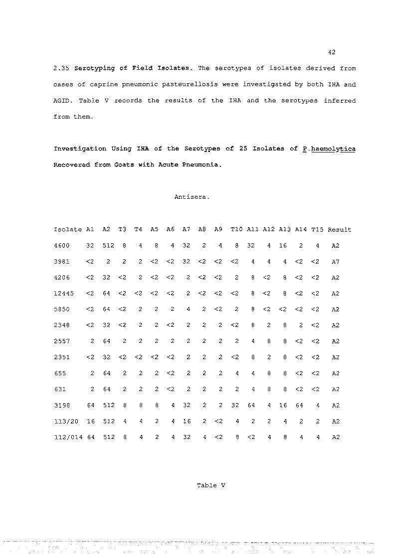
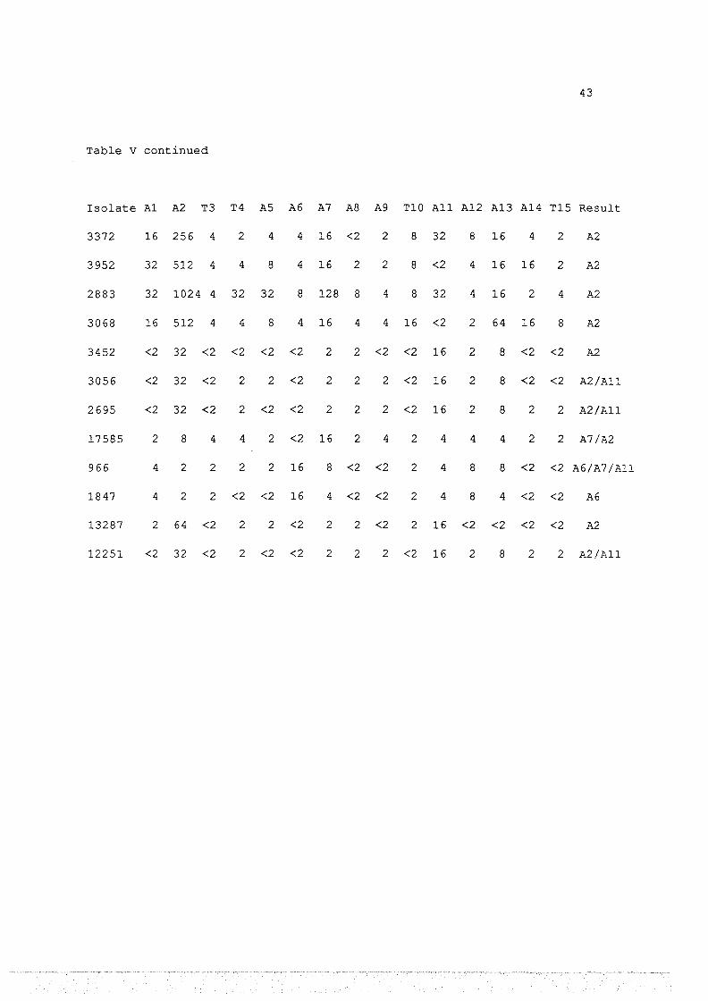
2.35 Serotyping of Field Isolates. The serotypes of isolates derived from
cases of caprine pneumonic pasteurellosis were investigated by both IHA and
AGID. Table V records the results of the IHA and the serotypes inferred
from them.
Investigation Using IHA of the Serotypes of 25 Isolates of E.haemol~ica
Recovered from Goats with Acute Pneumonia.
Antisera.
Isolate A1 A2 T3 T4 AS A6 A7 AS A9 T10 All A12 A13 A14 T15 Result
4600 32 512 8 4 8 4 32 2 4 8 32 4 16 2 4 A2
3981 <2 2 2 2 <2 <2 32 <2 <2 <2 4 4 4 <2 <2 A7
4206 <2 32 <2 2 <2 <2 2 <2 <2 2 8 <2 8 <2 <2 A2
12445 <2 64 <2 <2 <2 <2 2 <2 <2 <2 8 <2 8 <2 <2 A2
5850 <2 64 <2 2 2 2 4 2 <2 2 8 <2 <2 <2 <2 A2
2348 <2 32 <2 2 2 <2 2 2 2 <2 8 2 8 2 <2 A2
2557 2 64 2 2 2 2 2 2 2 2 4 8 8 <2 <2 A2
2351 <2 32 <2 <2 <2 <2 2 2 2 <2 8 2 8 <2 <2 A2
655 2 64 2 2 2 <2 2 2 2 4 4 8 8 <2 <2 A2
631 2 64 2 2 2 <2 2 2 2 2 4 8 8 <2 <2 A2
3198 64 512 8 8 8 4 32 2 2 32 64 4 16 64 4 A2
113/20 16 512 4 4 2 4 16 2 <2 4 2 2 4 2 2 A2
112/014 64 512 8 4 2 4 32 4 <2 8 <2 4 8 4 4 A2
Table v
43
Table v continued
Isolate A1 A2 T3 T4 AS A6 A7 A8 A9 T10 All A12 A13 A14 T15 Result
3372 16 256 4 2 4 4 16 <2 2 8 32 8 16 4 2 A2
3952 32 512 4 4 8 4 16 2 2 8 <2 4 16 16 2 A2
2883 32 1024 4 32 32 8 128 8 4 8 32 4 16 2 4 A2
3068 16 512 4 4 8 4 16 4 4 16 <2 2 64 16 8 A2
3452 <2 32 <2 <2 <2 <2 2 2 <2 <2 16 2 8 <2 <2 A2
3056 <2 32 <2 2 2 <2 2 2 2 <2 16 2 8 <2 <2 A2/A11
2695 <2 32 <2 2 <2 <2 2 2 2 <2 16 2 8 2 2 A2/All
17585 2 8 4 4 2 <2 16 2 4 2 4 4 4 2 2 A7/A2
966 4 2 2 2 2 16 8 <2 <2 2 4 8 8 <2 <2 A6/A7 /All
1847 4 2 2 <2 <2 16 4 <2 <2 2 4 8 4 <2 <2 A6
13287 2 64 <2 2 2 <2 2 2 <2 2 16 <2 <2 <2 <2 A2
12251 <2 32 <2 2 <2 <2 2 2 2 <2 16 2 8 2 2 A2/All

44
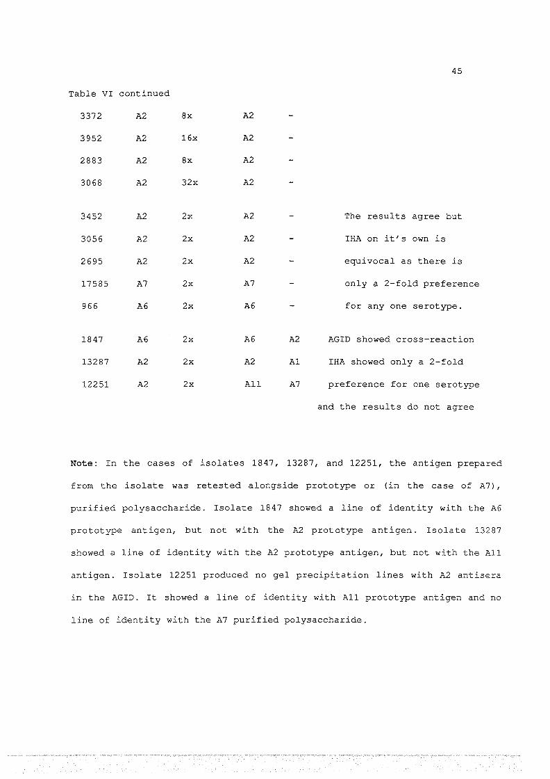
2. 35 Serotyping of Field Isolates. The serotypes of isolates derived from
cases of caprine pneumonic pasteurellosis were investigated by both IHA and
AGID. Table VI compares the results of the two tests and draws conclusions as
to the serotypes of the isolates.
Serotypes of 25 Caprine Isolates of ~.haemolY!ica: Comparison of AGID Results
with IHA Results.
Isolate IHA AGID Comments
Type Preference Type Cross-Reaction
4600 A2 16x A2 Both tests are
3981 A7 8x A7 satisfactory and the
4206 A2 4x A2 results agree.
12445 A2 8x A2
5850 A2 8x A2
2348 A2 4x A2
2557 A2 8x A2
2351 A2 4x A2
655 A2 8x A2
631 A2 8x A2
3198 A2 8x A2
113/20 A2 32x A2
112/014 A2 8x A2
Table VI
45
Table VI continued
3372 A2 8x A2
3952 A2 16x A2
2883 A2 8x A2
3068 A2 32x A2
3452 A2 2x A2 The results agree but
3056 A2 2x A2 IHA on it's own is
2695 A2 2x A2 equivocal as there is
17585 A7 2x A7 only a 2-fold preference
966 A6 2x A6 for any one serotype.
1847 A6 2x A6 A2 AGID showed cross-reaction
13287 A2 2x A2 Al IHA showed only a 2-fold
12251 A2 2x All A7 preference for one serotype
and the results do not agree
Note: In the cases of isolates 1847, 13287, and 12251, the antigen prepared
from the isolate was retested alongside prototype or (in the case of A7),
purified polysaccharide. Isolate 1847 showed a line of identity with the A6
prototype antigen, but not with the A2 prototype antigen. Isolate 13287
showed a line of identity with the A2 prototype antigen, but not with the All
antigen. Isolate 12251 produced no gel precipitation lines with A2 antisera
in the AGID. It showed a line of identity with All prototype antigen and no
line of identity with the A7 purified polysaccharide.

46
Figure 3
AGID of adsorbed antisera to serotype A1 {central well) against:
1/ Prototype A1 antigens (well 1)
2/ A1 purified capsular polysaccharide (well 2)
Note: There is a clear line of identity between the two antigens. The
precipitation line produced by the purified polysaccharide is sharper
and more distinct than that of the prototype antigen. This is probably
due to its antigenic uniformity.


47
Figure 4
AGID of unadsorbed antisera to serotype A7 (central well) against:
1/ Prototype A7 antigens (well 1)
2/ A7 purified capsular polysaccharide (well 2)
Note: There is a clear line of identity between the capsular
polysaccharide and one, but only one, of the precipitation lines
produced by the prototype A7 antigens. We conclude that the innermost
precipitation line produced by the A7 prototype antigens is due to the
capsular polysaccharide. The other two lines are probably due to
protein or endotoxin antigens.


48
2.4 Discussion
IHA is the standard method of serotyping P.haemolytica. It depends on:
1/ The ability of RBC to bind the capsular polysaccharide
strongly and the absence of binding of other cellular antigens to RBC.
2/ The availability of antisera to the capsular
polysaccharide.
Antiserum production and specificity
In our hands rabbits did not produce adequate antisera to 8 of the 15
serotypes of P.haemolytica (A2, T3, A6, A7, TlO, All, Al2 and A13). For
this reason antisera to the above types were produced in domestic hens.
It was somewhat unexpected that rabbits failed to produce reasonable
titres of antisera because this species has been used by other workers
(Sawada et al, 1982; Muraschi et al, 1965). However our experiments
initially used only one rabbit per serotype and a second rabbit was
immunised only if the first failed to respond. The literature is not clear
on how many rabbits per serotype were used by other workers, but occasional
comments on the difficulties of producing antisera (Wells, 1981; Burrells
et al, 1983) imply that we are not alone in finding that rabbits may
frequently fail to produce antibody to the capsule of P.haemolytica.
In contrast to rabbits, domestic hens never failed to produce high
titre antisera, although the avian antisera tended to cross-react with
antigens of heterologous serotypes to a greater extent than did the rabbit
antisera; eg avian antisera to serotype A13 reacted with an unidentified

49
test and the IHA (Tables I and II). Cross-reactions were resolved by
adsorbing the antisera with cells of heterologous serotypes (see Table
III). This is discussed below.
Interpretation of IHA Results
In the IHA, cross-reactions were common (Table I) and this required
the results to be interpreted on a qualitative rather than a quantitative
basis. Thus an isolate was «typed» if the titre with one of the antisera
was 4-fold greater than with any other antiserum. While this criterion has
the sanction of universal application it does not take into account the
variations in homologous titres which may occur (Table I). Thus our
antiserum to All had a titre of 512 against the All serotype and a titre of
8 against the Al2 serotype. The homologous titre of Al2 antiseum was 8 so
any Al2 isolate which happened to be identical to the prototype Al2 would
give a titre of 8 with both All and Al2 (see Table I) and thus would be of
uncertain serotype. This problem could, in theory, be avoided if antisera
were «normalized» by dilution so that they all give a titre corresponding
to that of the weakest antiserum. To our knowledge no-one has used this
approach and it would require that all isolates be typed using low titre
antibody. Futhermore the titre of an antiserum can differ when it is tested
with a prototype and an isolate. In such cases the isolate usually gave the
lower titre. As can be seen in Table V the two isolates typed as A7 gave
titres 4-fold lower than the prototype reaction with A7 antisera.
Consequently if all antisera were diluted to the titre of the lowest this
would almost certainly mean that many isolates would fail to react and
therefore could not be typed.

50
Cross-reactions of antisera with heterologous serotypes are not
unexpected since whole organisms are used as immunising antigens. These
organisms can share common endotoxins and futhermore, examination of SDS
PAGE patterns (Thompson et al., 1975) indicates that within the A and T
biotypes many proteins are common to all serotypes, at least in the sense
that they have similar banding patterns. Consequently cross-reactions may
occur in the IHA if antigens other than the capsular polysaccharide adsorb
to the RBC.
The Ability of the AGID Test to Distinguish Serotypes of Prototype Strains
of ~.haemol~ica.
Cross-reactions may occur in the AGID test because antigens other than
the capsular polysaccharide may be released from the cells and some of
these antigens will be able to diffuse into the agar. The cross-reacting
antigens can be either protein or endotoxin and this point will be
considered later. Despite the considerable theoretical potential for cross
reactions in the AGID test, in our hands 9 of the 15 serotypes produced no
cross-reactions (Table II) . These 9 antisera gave a precipitation line with
the homologous prototype antigen but failed to react with the remaining 14
antigens.
Four of the antisera to A biotypes viz. antisera to A1, A7, A12 and
A13 showed cross-reactions. However with the exception of the A1 antiserum,
the cross-reactions were limited to a reaction with only one of the
heterologous prototype strains. These cross-reactions (with the exception
of A1) were removed by adsorption of the antiserum with a deposit of cells
from the cross-reacting serotype (Table III). Antisera to serotype T3

51
cross-reacted with TlS antigen, and antisera to TlS cross-reacted with T3
antigen. Adsorption of these antisera with cells of the cross-reacting
serotype removed both the heterologous and the homologous reactions. This
is not surprising as the two serotypes are closely related if not identical
(Fraser et al.,l982).
Antiserum to Al cross-reacted with all of the A biotypes (Table II) .
However it gave two AGID lines with the homologous antigen but only one
with the heterologous A biotypes. This antiserum was adsorbed with A7 cells
which removed all cross-reactions; and also removed one of the two AGID
lines with the homologous prototype antigen (Table III) . Adsorbed antisera
were then serotype specific in the AGID test, ie. they gave an AGID line
with, and only with, the prototype serotype used as irmnunising antigen
(Table III)
These results clearly indicate that the AGID test can be used to
distinguish between all prototype strains (taking T3 and TlS as one
serotype) . However they do not show that the test will reliably identify
the serotypes of field isolates. Some such isolates may produce cross
reactions which do not occur with the prototype strains.
When cross-reactions do occur they may make it difficult to interpret
both IHA and AGID test results. However one of the advantages of the AGID
test over other serotyping methods, especially IHA, is that the
immunological relationship between two antigens can be tested by placing
them in adjacent wells. If the lines formed are contiguous, this «line of
identity» indicates that the two antigens are immunologically identical. If
the AGID lines cross this implies that the antigens are unrelated. This is
particularly useful when dealing with P.haemolytica which has many
antigens, only one of which, (the capsular antigen), is serotype specific.

52
In particular, a line of identity between a test antigen and a
purified capsular antigen allows unequivocal identification of the former
(see below) .
This provided an unequivocal test to confirm the serotype of any field
isolate which showed a reaction with more than one antiserum. However it
was still important to apply the test to establishing the serotypes of
actual field isolates and to demonstrate that it fulfilled it's function in
practice.
Purification of Capsular Polysaccharide
Serotypes A1 and A7 were among the most prevalent serotypes isolated
from disease conditions (see Chapters 3 and 4) and have also been widely
found in other studies (Fraser et al., 1982; Prince, 1985). Antisera to
both prototype strains showed cross-reactions with other serotypes (see
Table II) . This introduced a small element of uncertainty as to the
serotype of isolates because it could be argued that the single line of
identity observed with the prototype antigen did not represent a capsular
polysaccharide line but was instead due to a cross-reacting antigen such as
protein or endotoxin. This uncertainty could be resolved if purified
capsular polysaccharide was used.
Antisera to A1 was adsorbed with A7 cells. This antisera gave a
single line of identity when tested in parallel with the purified capsular
polysaccharide and a standard A1 antigen preparation
Antisera to A7 (unadsorbed) gave three gel precipitation lines when
tested with crude prototype antigen. One of these gave a single line of

53
identity when tested in parallel with the capsular polysaccharide and a
standard A7 antigen preparation.
When tested with all 15 antisera in the AGID test both purified
capsular antigens gave a single line with, and only with, the homologous
antiserum. We conclude that an isolate giving a line of identity with a
purified capsular polysaccharide provides an unequivocal determination of
the serotype of the isolate.
Attempt to Produce Mono-Specific Antisera
To circumvent the difficulties encountered when using antisera raised
against whole cells and thus to many antigens, an attempt was made to
produce mono-specific antisera. Purified capsular polysaccharide was found
to be non-antigenic (Adlam et al., 1984), so two antigen preparations were
used.
1/ An AGID line produced by the reaction between purified capsular
polysaccharide and the homologous antiserum. This line was excised and
thoroughly washed in PBS.
2/ Capsular polysaccharide adsorbed to the hens own RBC.
It was hoped that adsorption to RBC would duplicate the situation of
the capsular polysaccharide in the whole cell. The antigenic properties of
the capsular polysaccharide appear to be maximised when it is part of the
whole cell (Tadyon et al., 1981). The failure of animals to produce an
immunological reaction could alternatively be due to lack of phagocytosis
of the capsular polysaccharide so it was hoped that the combination of
purified capsular polysaccharide and antibody excised from a AGID plate
would be phagocytosed more efficiently.

54
Domestic hens failed to react to either antigen. We conclude that the
poor antigenicity of purified capsular polysaccharide was not improved by
these procedures.
Field Isolates of R.haemol~ica - Confirmation of Typing by IHA and AGID
25 isolates of P.haemolytica were obtained from goats with acute
pneumonia (Chapter 3). These isolates were typed by IHA and the AGID test.
For an isolate to be typed by IHA we initially used the standard criterion
that the antigen should show at least a 4-fold preference for one antiserum
as compared to the others. By this criterion 17 of the 25 caprine isolates
of P.haemolytica were allocated to a serotype (Table V).
However 5 isolates showed only a 2-fold preference for one antiserum.
These isolates reacted with only one antiserum in the AGID test and these
results agreed with the IHA results. If the criterion of a 4-fold
preference is relaxed ie. a 2-fold preference is accepted, then 22 of the
25 isolates were typed identically by IHA and AGID.
Two further isolates (1847 and 13287) showed a two fold preference for
the A6 and A2 antisera respectively in the IHA. These isolates also reacted
with A6 and A2 respectively in the AGID test. But in the AGID test isolate
18 47 cross-reacted with A2 antiserum and 132 87 cross-reacted with All
antiserum. However they showed lines of identity only with the A6 and A2
antigens respectively. We therefore conclude that these isolates are
serotypes A6 and A2 and within the criteria outlined above, both tests are
in agreement. This means that in the case of 24 of the 25 isolates the twc
tests agreed.
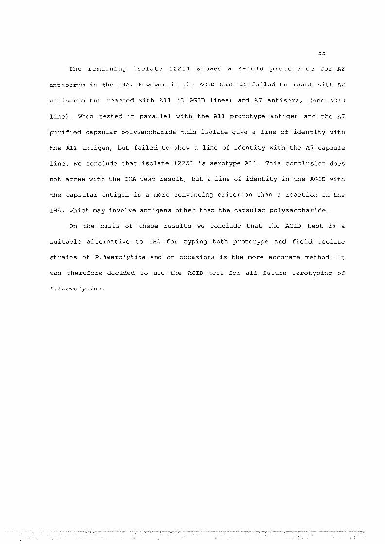
55
The remaining isolate 12251 showed a 4-fold preference for A2
antiserum in the IHA. However in the AGID test it failed to react with A2
antiserum but reacted with All (3 AGID lines) and A7 antisera, (one AGID
line) . When tested in parallel with the All prototype antigen and the A7
purified capsular polysaccharide this isolate gave a line of identity with
the All antigen, but failed to show a line of identity with the A7 capsule
line. we conclude that isolate 12251 is serotype All. This conclusion does
not agree with the IHA test result, but a line of identity in the AGID with
the capsular antigen is a more convincing criterion than a reaction in the
IHA, which may involve antigens other than the capsular polysaccharide.
On the basis of these results we conclude that the AGID test is a
suitable alternative to IHA for typing both prototype and field isolate
strains of P.haemolytica and on occasions is the more accurate method. It
was therefore decided to use the AGID test for all future serotyping of
P.haemolytica.

56
CHAPTER 3
Survey of the Prevalence of ~.haemol~ica in Sheep and Goats in New Zealand
3.1 Introduction
The specificities and relative convenience of IHA and AGID for
establishing the serotype of P. haemolytica were compared in Chapter 2,
using both prototype strains and isolates from field cases of pneumonic
pasteurellosis of goats. It was concluded that AGID was an efficient and
accurate technique for the serotyping of P.haemolytica. In this chapter we
use AGID to serotype isolates from CNP of sheep, from pneumonic
pasteurellosis of sheep and goats and from the nasal cavities of goats.
The results of this section represent a survey of the prevalence and
distribution of P.haemolytica in sheep and goats in New Zealand.
3.2 Materials and Methods
3.21 Isolation and Identification of~- haemolY!ica from Ovine Lungs
Samples of CNP lesions of ovine lungs were taken from 60 sheep in each
of four centres: Auckland, Gisborne, Christchurch and Invercargill.
A portion of lesion was smeared over half of a blood agar plate (see
Appendix) . This inoculum was then streaked over the rest of the plate and
incubated at 37°C overnight.
Colonies which had the following charateristics were provisionally
identified as P.haemolytica. 1-2 mm in diameter, smooth surface, entire

57
edge, translucent and with a zone of haemolysis directly under the colony.
Such colonies were selected and streaked on blood agar and incubated at
37°C overnight to isolate the organism in pure culture. Each such isolate
was inoculated on to MacConkey agar (using a heavy inoculum) and on to
Triple Sugar Iron Agar slants, and incubated at 37°C for 24 hours.
Isolates were also tested for Gram reaction and oxidase activity.
Isolates which
to be P.haemolytica
showed the following characteristics were considered
1/ Typical colony morphology, as above
2/ Gram negative rod
3/ Growth on MacConkey agar, from heavy inoculum
4/ Oxidase positive
5/ Produced acid throughout TSI slants but neither gas nor H2s
The production of antisera and its use in AGID and IHA were as
described in Chapter 2.
3.22 R.haemolY!ica Isolated from Pneumonic Pasteurellosis in Sheep and
Goats.
Isolates, already identified as P.haemolytica were obtained from a
number of MAF laboratories throughout New Zealand. They normally arrived by
mail on nutrient agar slopes,or in brain-heart-infusion broth (the methods
of dispatch were only partially under our control) .
A few of the broth cultures were not viable on arrival while all
cultures on slopes could be subcultured,so this was the method of choice.

58
3.23 Isolation and Identification of Nasal Isolates of R.haemolY!ica from
Goats.
Twenty goats from each of 4 flocks and 29 goats from 1 flock (a total
of 109 goats from 5 flocks), including angora and feral goats were
examined. To collect samples of nasal mucus, a sterile cotton swab was
inserted about 13cm into a nasal cavity and rotated both clockwise and
anti-clockwise. Swabs were immediately broken off into bijou bottles of
~~ies Transport Media and taken directly to the laboratory. Each swab was
smeared over half of the surface of a blood agar plate. This inoculum was
then streaked over the rest of the plate and incubated at 37°C for about 24
hours.
Colonies resembling those of P.haemolytica were streaked on blood agar
and incubated at 37°C overnight. Each isolate was inoculated on to
MacConkey agar (using a heavy inoculum) and on to Triple Sugar Iron Agar
slants and incubated for 24 hours. Identification of P.haemolytica was as
described previously.

59
3.3 Results
+ and ++ refer to the AGID lines seen. ++ indicates a strong
reaction.
Bold print + or ++ indicates the AGID lines which specify the
serotype of the isolate. Normal print + reports cross-reactions.
When an isolate reacted with antisera to one serotype only this
was deemed to denote the serotype of the isolate. When an isolate
reacted with more than one antisera it was retested alongside purified
capsular polysaccharide or prototype antigen. A line of identity was
seen with (and only with) one serotype and this was deemed to be the
serotype of the isolate.
(a) two AGID lines seen.
(*) = isolate re-tested alongside purified or prototype antigen
for a line of identity.

60
Serotyping of Isolates of R_. haemol~ica Derived from CNP Lesions, in
Auckland, Using AGID
Isolate Al A2 T3 T4 AS A6 A7 AS A9 TlO All A12 A13 Al4 Tl5 Result
Au4 + A14 Au8 ++a Al AulO + Al4 Aull + Al Au14 + Al Au16 + ++ Al3* Au20 + Al Au21 + Al Au27 + ++a A2 * Au29 + ++a A2 * Au31 + + A2 * Au32 +a A2 Au39 ++a + Al * Au41 + Al Au44 + + A2 * Au46 + + A2 * Au54 + Al Au55 + Al Au57 + Al Au59 + Al Au60 + Al
Table VII

61
+ and ++ refer to the AGID lines seen. ++ indicates a strong
reaction.
Bold print + or ++ indicates the AGID lines which specify the
serotype of the isolate. Normal print + reports cross-reactions.
When an isolate reacted with antisera to one serotype only this
was deemed to denote the serotype of the isolate. When an isolate
reacted with more than one antisera it was retested alongside purified
capsular polysaccharide or prototype antigen. A line of identity was
seen with (and only with) one serotype and this was deemed to be the
serotype of the isolate.
(a) two AGID lines seen.
(*) isolate re-tested alongside purified or prototype antigen
for a line of identity
(#) = concentrated antigen used as described in Materials and
Methods, Chapter 2.

62
Serotyping of Isolates of R.. haemoly:!;:ica Derived from CNP Lesions, in
Gisborne, Using AGID.
Isolate Al A2 T3 T4 AS A6 A7 AS A9 TlO All A12 A13 A14 Tl5 Result
Gl + ++a A2 * G3 ++ Al G5 ++ Al G7 ++a Al G9 ++ A2 Gl2 + + A2 * Gl9 + + A2 * G20 + Al G21 + Al G23 + Al G26 ++ A2 G27 + Al G28 ++a + AS # G31 + ++a A2 * G32 + + A2 * G35 + Al G41 + A2 G43 ++ A7 G44 ++ A2 G45 + Al G48 + ++a A2 * G53 + ++a + A2 * G58 + Al G59 + Al G60 + Al
Table VIII

63
Serotyping of Isolates of R_. haemolytica Derived from CNP Lesions, in
Christchurch, Using AGID
Isolate Al A2 T3 T4 AS A6 A7 A8 A9 TlO All Al2 Al3 Al4 TlS Result
Cl + A7 C2 + A2 C3 + A2 C4 + A2 cs + A7 C6 + A7 C7 + Al * C8 + + + A2 * ClO + + A2 * Cll + + A2 * C13 + A7 C15 + A2 C16 + Al * C17 + Al * C18 + A2 C20 + A2 C21 + Al C22 + A2 C23 + + Al * C25 + Al * C27 + Al C29 + A8 C31 + Al C33 + Al C34 + Al * C37 + A2 C38 + A2 C39 + A2 C40 + A7 C42 + A2 C43 + + A2 * C44 + + A2 *
Table IX

64
Table IX continued
C45 + + A2 * C46 + A7 C47 + Al C48 + Al C49 + Al cso + + Al * CSl + + Al * C52 + Al # css + A7 C56 + A7 C58 + A7 C60 + A7 C63 + A7
+ and ++ refer to the AGID lines seen. ++ indicates a strong reaction.
Bold print + or ++ indicates the AGID lines which specify the serotype
of the isolate. Normal print + reports cross-reactions.
When an isolate reacted with antisera to one serotype only this was
deemed to denote the serotype of the isolate. When an isolate reacted with
more than one antisera it was retested alongside purified capsular
polysaccharide or prototype antigen. A line of identity was seen with (and
only with) one serotype and this was deemed to be the serotype of the
isolate.
(*) = isolates re-tested alongside purified or prototype
antigens for a line of identity.
(#) concentrated antigen used as described in Materials and
Methods, Chapter 2.

65
Serotyping of Isolates of R_.haemolytica Derived from CNP Lesion, in
Invercargill, Using AGID
Isolate Al A2 T3 T4 AS A6 A7 AS A9 TlO All A12 A13 A14 TlS Result
I1 + Al I2 + A2 I3 + A2 I4 + + A2 * IS + Al I6 ++ Al I7 ++ Al I8 + Al IlO + + A2 * Ill + + A2 * I13 + + ++ A2 * I15 + ++ A2 * Il9 ++ A7 I20 + Al I21 ++ + ++a + A9 * I22 + ++a + All* I23 + ++a A7 * I24 + ++ + ++ Al3* I25 + + ++ A2 * I26 + + + ++ A2 * I27 + + + A2 * I28 + + + A2 * I29 + + + A2 * I30 + A2 I31 + + A2 * I32 + + + A2 * I33 + + + A2 * I34 + + A2 * I35 + ++ A8 * I36 + + + A9 * I37 + Al I38 + ++a A2 *
Table X
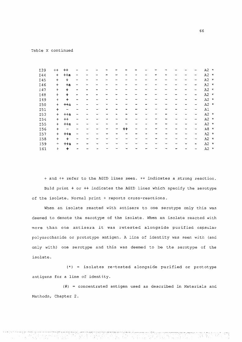
66
Table X continued
I39 ++ ++ A2 * I44 + ++a A2 * I45 + + A2 * I46 + +a A2 * I47 + + A2 * I48 + + A2 * I49 + + A2 * ISO + ++a A2 * I 51 + Al I 53 + ++a A2 * I 54 + ++ A2 * I 55 + ++a A2 * I 56 + ++ A8 * I 57 + ++a A2 * I 58 + + A2 * I 59 + ++a A2 * I61 + + A2 *
+ and ++ refer to the AGID lines seen. ++ indicates a strong reaction.
Bold print + or ++ indicates the AGID lines which specify the serotype
of the isolate. Normal print + reports cross-reactions.
When an isolate reacted with antisera to one serotype only this was
deemed to denote the serotype of the isolate. When an isolate reacted with
more than one antisera it was retested alongside purified capsular
polysaccharide or prototype antigen. A line of identity was seen with (and
only with) one serotype and this was deemed to be the serotype of the
isolate.
(*) isolates re-tested alongside purified or prototype
antigens for a line of identity.
(#) = concentrated antigen used as described in Materials and
Methods, Chapter 2.

67
Comparison of the Recovery Rate of ~.haemolY!ica from CNP Lesions Collected
from Sheep in Four Areas of New Zealand.
Centre No. Tested No. Positive Percentage Positive
AUCKLAND 60 21 35%
GISBORNE 60 25 42%
CHRISTCHURCH 60 45 75%
INVERCARGILL 60 49 82%
Table XI

68
Comparison of the Serotypes of R_ .haemolY!ica Isolated from Ovine CNP
Lesions from Four Areas of New Zealand.
Serotype AUCKLAND GIS BORNE CHRISTCHURCH INVCARGILL Totals
Al 12 (57 .1%) 11 (44%) 12 (26. 7%) 8 (16. 7%) 43 (31. 7%)
A2 6 (28. 6%) 11 ( 44%) 17 (37. 8%) 32 (66.7%) 66 (47.5%)
AS 1 (4%) 1 (0. 7%)
A7 1 (4%) 11 (24. 4%) 2 (4.2%) 14 (10%)
A8 1 (2 .2%) 2 ( 4. 2%) 3 (2.2%)
A9 2 (4.2%) 2 (1. 4%)
All 1 (4%) 2 (4.4%) 1 (2 .1%) 4 (2. 9%)
Al3 1 (4.8%) 2 ( 4. 4%) 1 (2 .1%) 4 (2. 9%)
Al4 2 (9.5%) 2 (1. 4%)
Total 21 25 45 48 139 (100%)
Table XII
Note: There are 11 A-biotypes of P.haemolytica. 9 of these 11 were isolated
from CNP lesions of sheep.

69
Serotypes of Isolates of R_.haemolytica Derived from the Lungs of Goats
which Died from Pneumonic Pasteurellosis.
Possible Serotypes Determined By:
Isolate AGID IHA Serotype
4600 A2 A2 A2
3981 A7 A7 A7
4206 A2 A2 A2
12445 A2 A2 A2
5850 A2 A2 A2
2348 A2 A2 A2
2557 A2 A2 A2
2351 A2 A2 A2
655 A2 A2 A2
631 A2 A2 A2
3198 A2 A2 A2
113/20 A2 A2 A2
112/014 A2 A2 A2
2557 A2 A2 A2
3372 A2 A2 A2
3952 A2 A2 A2
2883 A2 A2 A2
Table XIII

70
Table XIII continued
Isolate AGID IHA Serotype
3068 A2 A2 A2
3452 A2 A2/All A2 *
2695 A2 A2/All A2 *
17585 A7 A7/A2 A7 *
966 A6 A6/A7 /A13 A6 *
1847 A6/A2 A6/A12 A6 *
13287 A2/All A2 A2 *
12251 All/A7 A2/All All*
( *) = Result obtained when the isolate was re-tested alongside
purified capsular polysaccharide or prototype antigen to give a line of
identity with standard antigens as defined.

71
Serotypes of Isolates of R_.haemolytica Derived from the Lungs of Sheep
which Died from Pneumonic Pasteurellosis.
Isolate AGID Serotype
16592 A2 A2
02267 A6 A6
2451 Al Al
16792 A9/All All*
3981 Al/A2 A2 *
4411 A2 A2
13330 A2 A2
6628 A2 A2
6737 Al/A2 A2 *
1115 Al/A2 A2 *
2170 Al/A2/A6 A2 *
2217 Al/A8 A8 *
2434 Al/A2 A2 *
3354 Al/A2 A2 *
4449 Al/A2 A2 *
17514 A1 A1
11994 All All
13246 T10 T10
Table XIV
(*) = Result obtained when the isolate was re-tested alongside
purified capsular polysaccharide or prototype antigen to give a line of
identity with standard antigens as defined.

72
The Serotypes of Isolates of ~.haemolytica Obtained from the Nasal
Cavities of Goats from 5 Different Flocks.
Flock Isolate AGID Result
A Fer 1 A2
A Fer 2 A2
A Fer 7 A2
B Fer 21 A2
B Fer 25 A2
B Fer 28 A2
B Fer 33 A2
c Fer 68 A2
c Fer 70 A2
c Fer 45 A2
D Ang 16 All
D Ang 14 A2
E Ang 24 A2
E Ang 35 A2
Table XV
Note: The prefix «Fer» indicates an isolate of P. haemolytica from a feral
goat and «Ang» indicates an isolate from an Angora goat.
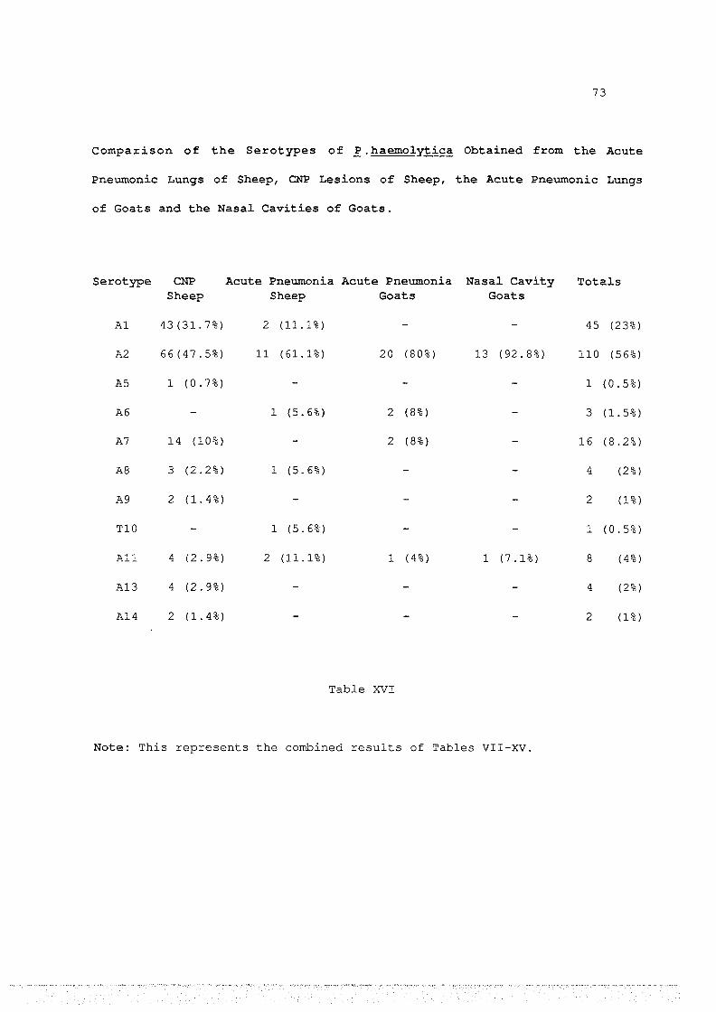
73
Comparison of the Serotypes of R. haemol~ica Obtained from the Acute
Pneumonic Lungs of Sheep, CNP Lesions of Sheep, the Acute Pneumonic Lungs
of Goats and the Nasal Cavities of Goats.
Serotype CNP Acute Pneumonia Acute Pneumonia Nasal Cavity Totals Sheep Sheep Goats Goats
Al 43 (31. 7%) 2 (11.1%) 45 (23%)
A2 66(47.5%) 11 (61.1%) 20 (80%) 13 (92.8%) 110 (56%)
AS 1 (0. 7%) 1 (0. 5%)
A6 1 (5. 6%) 2 (8%) 3 (1.5%)
A7 14 (10%) 2 (8%) 16 (8.2%)
AS 3 (2. 2%) 1 (5.6%) 4 (2%)
A9 2 (1. 4%) 2 (1%)
TlO 1 (5.6%) 1 (0.5%)
All 4 (2. 9%) 2 (11.1%) 1 (4%) 1 (7 .1%) 8 (4%)
A13 4 (2. 9%) 4 (2%)
A14 2 (1.4%) 2 (1%)
Table XVI
Note: This represents the combined results of Tables VII-XV.

74
3.4 Disscusion
The aim of the survey of P.haemolytica in CNP lesions of sheep was to
answer three questions.
1/ What percentage of CNP lesions yield P.haemolytica?
2/ Which serotypes of P. haemolytica are present, and in what
proportion?
3/ Are the serotypes found related to the geographical source of
the CNP lesion?
1/ Overall P.haemolytica was isolated from 139 (53.3%) of the 240 pneumonic
lungs. This figure is similar to, although slightly less than found in some
surveys of CNP lesions in New Zealand. Thus Pfeffer et al., (1983), recovered
P.haemolytica from 55% of CNP lesions and Prince, (1985) isolated P.haemolytica
from 67% of lesions.
Our isolation rates from the two South Island samples, Christchurch and
Invercargill, were 73.3% and 81.7% respectively, while the two North Island
samples, Auckland and Gisborne, were much lower being 35% and 41.7% respectively
(Table XI) . This difference is probably due to the seasonal nature of the
disease. Chronic non-progressive pneumonia occurs in the late summer and early
autumn months and the lesions are usually resolved by the coming of winter.
Although all our samples were taken at approximately the same time, the CNP
season begins and finishes sooner in the North than the South Island. Lesions
are healing in the north while the disease is still prevalent in the south. The
resolution of lesions is presumably accompanied by the elimination of
P.haemolytica.

7S
2 I We isolated a total of 9 serotypes from CNP lesions of sheep. All of
these were A biotypes. 43 (31.7%) were of serotype Al, 66 (47.8%) A2, 1 (0.7%)
AS, 14 (10%) A7, 3 (2.1%) A8, 2 (1.4%) A9, 4 (2.8%) All, 4 (2.8%) Al3 and 2
(1. 4%) Al4. (Table XII) .
The lack of T biotypes is not surprising as CNP is associated primarily
with the A biotypes (Smith, 1961; Gilmour, 1978).
The distribution of serotypes is similar to that found in Britain (Thompson
et al., 1977; Fraser et al., 1982), where A2 is the predominant serotype and Al,
A6, A7 and A9 are also common, making up with A2, 80% of the total. In New
Zealand, Prince, (198S), isolated P.haemolytica from lesions of CNP from sheep
in the southern part of the North Island. Only 6 serotypes were detected. Five
of these six serotypes were also detected in our survey. In addition we found
serotypes AS, All, Al3 and Al4.
The three most common isolates, Al (31.7S), A2 (47.8%) and A7 (10%) make up
89.S% of the total isolates from CNP lesions.
3/ Certain serotypes were found only in one district (AS solely in
Gisborne, Al4 only in Auckland) and the proportion of serotypes found varied
between centres. Invercargill had a higher proportion of A2 and Christchurch had
a high proportion of A7 isolated. These variations however were not dramatic and
may reflect only random sampling differences.
We conclude that P.haemolytica may be isolated from lesions of CNP with a
frequency of between 3S% and 82%, depending on the season and the severity of
the lesions. 9 of the 11 A biotypes were detected in CNP lesions, although three
(Al, A2 and A7) together accounted for 90%. These three serotypes should be
included in any vaccine for the control of chronic non-progressive pneumonia and
a vaccine containing just these three serotypes would have some relevance in 90%
76
of the affected animals. There is no major difference in the serotypes found in
various areas of the country and this is also of importance for vaccine
manufacture, as one vaccine could be used throughout the country.
Survey of R.haemol~ica Serotypes Isolated From Acute Pasteurellosis
Sheep: Six serotypes of P.haemolytica were isolated from the lungs of sheep
which died of pneumonic pasteurellosis (Table XV) . A2 was the predominant
serotype, with 11 of the 18 isolates (61.1%) being of this serotype. Also
present were Al (11.1%), A6 (5.6%), A8 (5.6%), TlO (6%) and All (11.1%). Fraser
et al, (1982) reported a large survey of serotypes of P.haemolytica isolated
from cases of ovine pasteurellosis in Britain. This survey included isolates
from septicaemic pasteurellosis, a disease not included in our surveys. A2 was
by far the most common (35.5%). No other A biotype represented more than 12.5%
of the total, hence the distribution of the serotypes in our study is similar to
that found in Britian by, Fraser et al., (1982).
Our single isolate of serotype Tl 0 is of particular interest for two
reasons. Firstly because a T biotype has not previously been isolated in New
Zealand and secondly because, although T biotypes may be isolated infrequently
from pneumonic lesions (Gilmour, 1978), they are usually associated -vlith a
septicaemia rather than pneumonic pasteurellosis. Septicaemia is not common in
New Zealand but since we now know that the T biotype is present in New Zealand
it is likely that the rarity of the disease is due to climate and management.
Goats: 25 isolates of P.haemolytica were derived from goats with pneumonic
pasteurellosis. Of these isolates 80% were serotype, A2. Also present were
serotypes A6 (8%), A7 (8%) and All (4%). Surveys of serotypes of P.haemolytica
associated with caprine pneumonia have been undertaken only in tropical

77
countries where the goats and conditions bear little similarity to New Zealand.
However Pegram, (1974), found three isolates of P .haemolytica from pneumonic
goat lungs in Somali were A2. Ojo (1977) cites a study of P.haemolytica from
goats in Nigeria (Ojo, (1975)) in which 200 isolates assigned to 6 serotypes
distributed as follows: A2 (43%), A6 (20%), A7 (20%), A8 (6.5%), TlO (3.5%) and
All (5%) . Despite the differences in conditions these are similar to our results
at least in the predominance of A2.
Survey of ~.haemolY!ica Serotypes From Caprine Nasal Cavities
The survey of P. haemolytica in the nasal cavity of goats had several
different aims.
1/ To establish the proportion of goats which carried the organism.
2/ To establish which serotypes were present.
3/ To compare the serotypes found in the nasal cavity with those causing
pneumonia.
4/ To compare the serotypes found in feral and angora goats
Of the 109 goats tested, 14 (12.8%) carried P.haemolytica in their nasal
cavity. We are aware of only two similar surveys, one in Mid-West of the USA
(Ngatia et al, 1984) and one in Nigeria (Ojo, 1976). These studies respectively
found 64% and 5% of goats examined carried P.haemolytica. Our figure of 12.8%
(ie. 14 out of 109 goats) was an intermediate one, though high enough to
indicate that P.haemolytica is not uncommon in the nasal cavity of goats.
The finding that 92.8% of nasal isolates are of the one serotype, viz. A2
clearly demonstrates that this is the most significant serotype of P.haemolytica
in nasal cavities of goats. This serotype is also the one most frequently
isolated from acute pneumonic pasteurellosis of goats.

78
There appears to be no difference in the serotypes carried in the nasal
cavities of feral and angora goats which is
run together as a single herd.
not surprising as they are often
In conclusion, P.haemolytica is found in New Zealand and is associated with
acute pneumonic pasteurellosis of sheep and goats and CNP in sheep. The
predominant serotype is A2 which accounts for nearly three-quarters of all
serotypes isolated from pneumonic pasteurellosis. Together, Al, A2 and A7 made
up 81.4% of all isolates. A2 is also the predominant serotype found in the nasal
cavities of goats, being 92.8% of the total, although this predominance of A2
may change if goats come into closer contact with sheep which carry a wider
range of P.haemolytica serotypes in their nasal cavities.
Since the serotypes of P.haemolytica causing disease in sheep and goats are
the same, any vaccine suitable for control of chronic non-progressive pneumonia
in sheep could well be suitable for use in the control of acute pneumonic
pasteurellosis in sheep and goats. It may not be economicaly viable to inoculate
sheep specificaly against acute pneumonia, but if the vaccine is also protective
against CNP it may be economically advantageous. A vaccine for goats against
acute pneumonia is more likely to be economically feasible considering the high
current value of goats.

79
CHAPTER 4
Comparison of Total Proteins, Antibiotic Sensitivities and Capsular Antigen Production Of Prototype Strains and Field Isolates of R_.haemolY!:ica
4.1 Introduction
This section examines New Zealand isolates of P. haemolytica as
follows:
1/ Comparison of total proteins.
2/ Antibiotic sensitivities
3/ Capsular antigen production
Comparison of total proteins. Pneumonic pasteurellosis is found in
both sheep and goats (Harbourne, 1979; Ojo, 1977). Immunity to
P.haemolytica is primarily dependant on the presence of antibody to the
capsular polysaccharide, but other antigens including proteins may also
play a significant role in inducing immunity (Tadayon et al., 1981). If
the proteins of isolates of P. haemolytica from sheep differed greatly
from those of isolates from goats, it may be necessary to produce
different vaccines for use in the two species. SDS-PAGE has been used to
compare P.haemolytica serotypes (Thompson et al., 1975) and we used this
method to compare the total proteins of isolates derived from sheep and
goats.
Antibiotic sensitivities. The use of antibiotics for the control of
pneumonic pasteurellosis has been advocated, but few if any
recommendations are given (Whitten, 1971) As well, antibiotic resistance
has been reported from the USA (Zimmerman et al., 1980) and the UK (Wray
et al., 1983), so it is important to establish the antibiotic
sensitivities of isolates of P.haemolytica in New Zealand.
80
Capsular antigen production. Immunity to P.haemolytica depends on an
immune response to the serotype-specific capsular antigen (Knight et al.,
1969). Thus a vaccine against P.haemolytica should contain large amounts
of the capsular polysaccharide. To determine if fresh field isolates of
P. haemolytica produced more capsular antigen than did the prototype
strains we compared them by Laurell Rocket electrophoresis.
4.2 Materials and Methods
Comparison of Isolates by SDS-PAGE Gels
The proteins of isolates from caprine and ovine sources were
compared by SDS-PAGE to see if there existed any discernable differences
in isolates from the two species.
The preparation and running of SDS-PAGE gels is described in the
Appendix.
Antibiotic Sensitivity Testing
Prototype strains and isolates from 3 4 fatal cases of pneumonic
pasteurellosis were examined. Two strains from goats and one from sheep
which had involved unsuccesful treatment with antibiotics were included.
The Minimum Inhibitory Concentration of antibiotics was determined
by the agar plate dilution method (Zimmerman et al, 1980) . Strains to be
tested were inoculated into lOmls of BHI broth and incubated at 37°C for
6 hours. 0.75ml was placed in a replicator which was used to inoculate
plates containing various concentrations of antibiotics.

81
Media for the MIC contained, per plate: 17mls of Mueller-Hinton
agar, lml of sterile defibrinated horse blood and 2mls of antibiotic
solution. Plates were used immediatly or stored at 4°C for up to a week.
Antibiotic stock solutions were made on the day of media preparation.
Control plates containing only agar and blood were inoculated before and
after inoculation of antibiotic plates to check the viability of the
inoculum.
Plates were read after 18 to 24 hours incubation at 37°C. The MIC
was taken as the lowest concentration of antibiotic without growth, a
barely visible haze, or the growth of not more than 1-2 colonies.
Comparison of Capsular Antigen Production by Different Strains of
R.haemol~ica Using the Laurell Rocket Test
Isolates of two serotypes of P.haemolytica viz. All and A7 and the
equivalent prototypes were examined by the Laurell Rocket Test to compare
the amount of capsule production.
For each serotype, four isolates from cases of CNP were selected
(one from each district), plus two isolates from acute pasteurellosis
(one from a caprine, one from an ovine source) . These were compared by
the Laurell Rocket Test using broth antigen.
The techniques used in the Laurell Rocket test are described in the
Appendix.

82
4.3 Results
Comparison of Isolates by SDS-PAGE Gels
Figure 5
Comparison of the total proteins of P.haemolytica serotype A2 (left to
right),
1/ The prototype strain (tracks 1 and 8)
2/ Three goat isolates (tracks 2-4)
3/ Three sheep isolates (tracks 5-7)
Note the overall similarity between all isolates. Some differences
occur in the middle molecular weight proteins. These differences are
not related to the source of the isolate ie. goat and sheep isolates
could not be distinguished by SDS-PAGE.

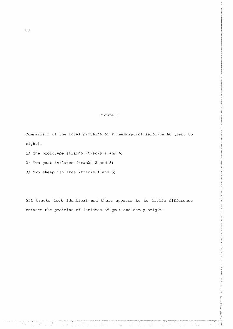
83
Figure 6
Comparison of the total proteins of P.haemolytica serotype A6 (left to
right),
1/ The prototype strains (tracks 1 and 6)
2/ Two goat isolates (tracks 2 and 3)
3/ Two sheep isolates (tracks 4 and 5}
All tracks look identical and there appears to be little difference
between the proteins of isolates of goat and sheep origin.


84
Figure 7
Comparison of the total proteins of P.haemolytica serotype A7 (left to
right),
1/ The prototype strains (tracks 1 and 4)
2/ Two goat isolates (tracks 2 and 3)
The two goat isolates are very similar to the prototype strains.
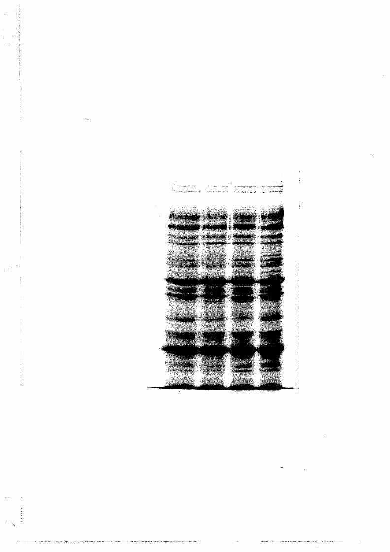

85
Figure 8
Comparison of the total proteins of P.haemolytica serotype All (left
to right),
1/ The prototype strain (tracks 1 and 4)
2/ One goat isolate (track 2)
3/ One sheep isolate (track 3)
Again there appears to be only minor differences in the proteins of
P.haemolytica from goat or sheep disease.
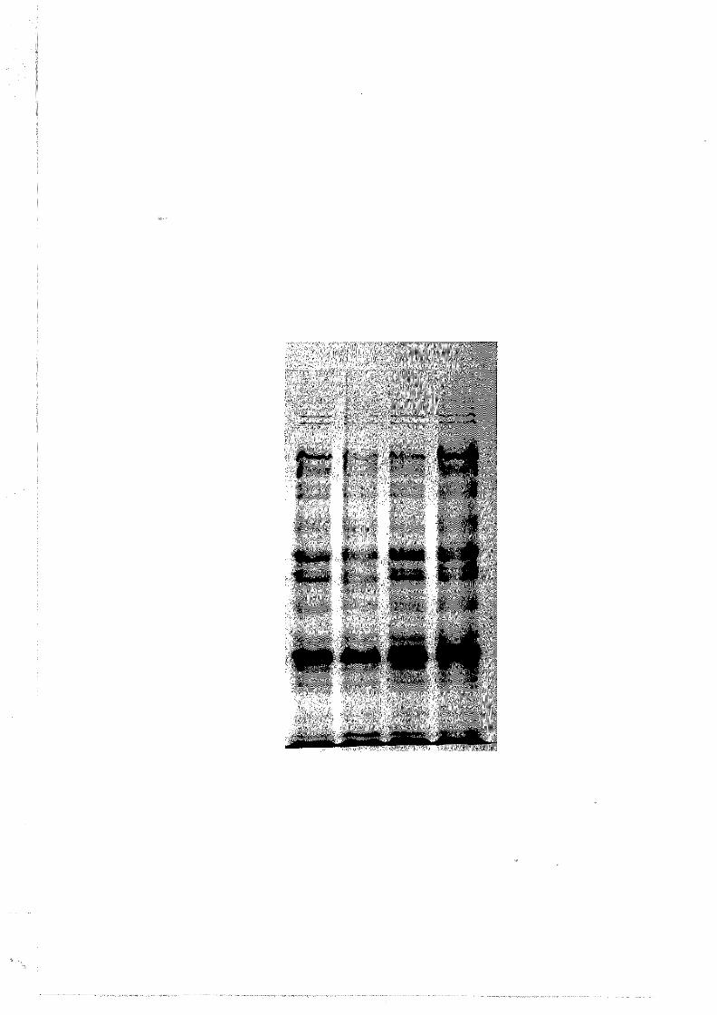

86
Antibiotic Sensitivity Testing
MIC
Isolate Penicillin Chloramphenicol Tetracycline Streptomycin
16592 <005Jlg/ml 2Jlg/ml 2Jlg/ml 16Jlg/ml 02267 <005Jlg/ml 1Jlg/ml 1Jlg/ml 8Jlg/ml
981 <005Jlg/ml <005Jlg/ml <005Jlg/ml 8Jlg/ml 4411 <005Jlg/ml 2Jlg/ml 2Jlg/ml 8Jlg/ml 13330 <0.5Jlg/ml <0.5Jlg/ml 1Jlg/ml 8Jlg/ml 6628 <005Jlg/ml 4Jlg/ml 2Jlg/ml 8Jlg/ml 6737 <005Jlg/ml 2Jlg/ml <005Jlg/ml 8Jlg/ml 1115 005Jlg/ml 4Jlg/ml 4Jlg/ml 16Jlg/ml 2170 <005Jlg/ml 2Jlg/ml 2Jlg/ml 8Jlg/ml 3354 <005Jlg/ml lJ.lg/ml lJ.lg/ml 16Jlg/ml 4449 2Jlg/ml 4Jlg/ml 4Jlg/ml 8Jlg/ml 4600 <005Jlg/ml 2Jlg/ml 2Jlg/ml 8Jlg/ml 3981 1Jlg/ml 4Jlg/ml 2Jlg/ml 8Jlg/ml 4206 2Jlg/ml 4Jlg/ml 4Jlg/ml 8Jlg/ml 12445 2Jlg/ml 4Jlg/ml 8Jlg/ml 8Jlg/ml 5850 1Jlg/ml 4Jlg/ml 4Jlg/ml 8Jlg/ml 2348 2Jlg/ml 4Jlg/ml 2Jlg/ml 8Jlg/ml 2557 2Jlg/ml 4Jlg/ml 2Jlg/ml 8Jlg/ml 2351 2Jlg/ml 4Jlg/ml 2Jlg/ml 8Jlg/ml 655 1Jlg/ml 4Jlg/ml 2Jlg/ml 8Jlg/ml 631 2Jlg/ml lJ.lg/ml 4Jlg/ml 8Jlg/ml 3198 <005Jlg/ml <005Jlg/ml 2Jlg/ml 8Jlg/ml 113/20 <005Jlg/ml 1Jlg/ml 2Jlg/ml 8Jlg/ml 112/014 lJ.lg/ml 4Jlg/ml 2Jlg/ml 8Jlg/ml 3952 2Jlg/ml 4Jlg/ml 2Jlg/ml 8Jlg/ml 2883 2Jlg/ml 2Jlg/ml 2Jlg/ml 8Jlg/ml 30 68 2Jlg/ml 4Jlg/ml 8Jlg/ml 8Jlg/ml 3452 <005Jlg/ml 4Jlg/ml 2Jlg/ml 8Jlg/ml 3056 2Jlg/ml 4Jlg/ml 2Jlg/ml 8Jlg/ml 17585 <005Jlg/ml 4Jlg/ml 2Jlg/ml 8Jlg/ml 966 <005Jlg/ml 2Jlg/ml 2Jlg/ml 8Jlg/ml 1847 <005Jlg/ml 1Jlg/ml 2Jlg/ml 8Jlg/ml 13287 <005Jlg/ml 4Jlg/ml 4Jlg/ml >1024Jlg/ml 12251 <005Jlg/ml 1Jlg/ml 4Jlg/ml 8Jlg/ml
A1 1Jlg/ml 4Jlg/ml 8Jlg/ml >1024Jlg/ml A2 <005Jlg/ml 4Jlg/ml 2Jlg/ml 8Jlg/ml A7 1Jlg/ml 4Jlg/ml 2Jlg/ml >1024Jlg/ml T10 2Jlg/ml 4Jlg/ml 8Jlg/ml 8Jlg/ml
Table XVII

87
Antibiotic Min~um Inhibitory Concentrations
Antibiotic No. Isolates
MIC Penicillin <005Jlg/ml 20
MIC Penicillin lJlg/ml 6
MIC Penicillin 2Jlg/ml 12
MIC Penicillin 4Jlg/ml
MIC Penicillin 8Jlg/ml
MIC Chloramphenicol <005Jlg/ml 3
MIC Chloramphenicol lJlg/ml 6
MIC Chloramphenicol 2Jlg/ml 7
MIC Chloramphenicol 4Jlg/ml 22
MIC Chloramphenicol 8Jlg/ml
MIC Tetracycline <005Jlg/ml 2
MIC Tetracycline lJlg/ml 3
MIC Tetracycline 2Jlg/ml 22
MIC Tetracycline 4Jlg/ml 7
MIC Tetracycline 8Jlg/ml 4
MIC Streptomycin <005Jlg/ml
MIC Streptomycin lJlg/ml
MIC Streptomycin 2Jlg/ml
MIC Streptomycin 4Jlg/ml
MIC Streptomycin 8Jlg/ml 32
MIC Streptomycin l6Jlg/ml 3
MIC Streptomycin >1024Jlg/ml 3
Table XVIII
88
Comparison of Capsule Production by Laurell Rocket Test
Figure 9
Comparison of the relative amount of capsule produced by different
strains of P.haemolytica serotype All.
Left to right,
1/ The prototype strains {wells 1 and 6)
2/ Four isolates from ovine CNP {wells 2-5)
3/ One isolate from goat pneumonic pasteurellosis (well 7)
4/ One isolate from sheep pneumonic pasteurellosis (well 8)
The prototype strain produces significantly more capsular material
then do the isolates from either CNP or pneumonic pasteurellosis.


89
Figure 10
Comparison of the relative amount of capsule produced by different
strains of P.haemolytica serotype A7.
Left to right,
1/ The prototype strains (wells 1 and 7)
2/ Four isolates from ovine CNP (wells 2-5)
3/ One isolate from goat pneumonic pasteurellosis (well 8)
The prototype strains produce significantly more capsular material
then do the isolates from either CNP or pneumonic pasteurellosis,
although isolate from CNP does come close.


90
4.4 Disscusion
Comparison of Isolates by SDS-PAGE Gels
Isolates from sheep and goats were compared by SDS-PAGE to find if the
proteins of isolates of a single serotype of P.haemolytica from different
sources (sheep and goats) differ significantly. It was found (Figures 5-8)
that the proteins of isolates from any one species show some variations.
However the variations between isolates from the two species (sheep and
goats) did not exceed the variations observed within isolates from a single
species. This suggests that transfer of P. haemolytica between sheep and
goats is not only possible but likely, especially in mixed farming
ventures. With respect to the efficacy of any vaccine the proteins may be
important. However since the proteins of isolates of P.haemolytica from
sheep and goats are similar it should not be necessary to develop two
different vaccines to protect sheep and goats.
Comparison of Capsule Production by Laurell Rocket Test
Capsular antigen is generally believed to be the major protective
antigen. It seemed reasonable to expect that fresh field isolates of
P.haemolytica may produce more capsular material than the, now laboratory
adapted, prototype strains. However our results indicate that the most
capsular material is produced by prototype strains. We therefore conclude
that for vaccine production these prototype strains should not be
substituted by any of the isolates which we tested.

91
Antibiotic Sensitivity Testing
The aims of this were two-fold.
To examine which antibiotics were most effective against
P.haemolytica in vitro and thus to recommend antibiotics for in vivo use.
To assay the level of resistance in P.haemolytica isolated from fatal
cases of pneumonic pasteurellosis.
Only 3 of the 39 strains of P. haemolytica examined showed marked
resistance to any of the antibiotics tested. This is not surprising,
because those isolates which exhibited a high level of antibiotic
resistance were isolated from cattle or pigs (Chang et al., 1976, Zimmerman
et al., 1980, Wray et al., 1983), which are more likely to have been
treated with antibiotics than the ovine and caprine sources from which the
P.haemolytica in this study were derived.
All P. haemolytica isolates showed some degree of resistance to
streptomycin, which is not a drug of choice. Three isolates showed a high
level of resistance to this drug alone. This was not correlated with a high
level of resistance to other antibiotics. Four strains of P. haemolytica
were resistant to 8~g/ml tetracycline while we found no strain resistant to
more then 4~g/ml of chloramphenicol or penicillin, so these are the drugs
of choice. However, treatment with chloramphenicol is not permitted in
animals used for human consumption, so its use is limited. In those cases,
penicillin, possibly supplemented with tetracycline should be used.
Although there does not seem to be a high incidence of antibiotic
resistance in P.haemolytica, regular assays of antibiotic resistance
patterns would be of interest.

92
CHAPTER 5
General Discussion
The present investigation was mainly concerned with serotyping
P.haemolytica and the use of this procedure to establish the prevalence
of serotypes in sheep and goats in New Zealand.
Production and Specificity of Antisera.
The first requirement for serotyping P.haemolytica is the production
of high titre antibody, specific for the capsular polysaccharide of each
of the 15 serotypes. Rabbits are the normal species used to prepare
P. haemolytica antisera and most workers seem to find them satisfactory
(Biberstein et al., 1960; Muraschi et al., 1965). However we found that
although rabbits produced antibody to the capsular polysaccharide of
several serotypes, they consistently failed to produce antisera to
others. This problem is not discussed in the literature, but has been
briefly commented on by some workers (Wells, 1981; Burrells et al.,
1983) . We conclude that the failure of rabbits to produce antibody is not
unique to this work. The reasons why Biberstein et al., (1960) were able
to produce antisera to all serotypes of P.haemolytica is not clear, but
it could depend on the breed or number of rabbits used. The fact that
other workers (Biberstein, 1960; Murasachi et al., 1965) used several
rabbits for each serotype does tend to suggest that they did not
invariably get a high response in each animal. The breed of rabbits used
is not always reported but Fraser et al., ( 19 82) and Sawada et al.,
(1982) used New Zealand White rabbits whereas with the present study a
pure strain of animal was not available.
93
For those serotypes that in our hands did not produce an adequate
immunological response in rabbits, antisera was raised in domestic hens.
In our experiments domestic hens never failed to produce high titre
antibody to P.haemolytica. It might be concluded from this that domestic
hens rather than rabbits should be routinely used for the production of
antibody to P.haemolytica. However cross-reactions with heterologous
serotypes were more commonly observed with avian antisera than with
rabbit antisera. These cross-reactions were removed by adsorbing the
antisera with cells of a heterologous serotype. However, although the
results of this procedure are satisfactory, (cross-reactions
disappeared), it is desirable to produce mono-specific antisera eg. by
the inoculation of purified serotype specific capsular antigen. This
point is returned to later.
Serotyping of ~.haemol~ica by IHA.
In the IHA, cross-reactions were numerous, so an isolate was «typed»
only if the titre with one antiserum was at least 4-fold greater than
with any other antiserum. This approach is less than satisfactory as it
does not take into account the variations in homologous tit res that
occur. Using this criterion 10 of the 15 prototype serotypes could be
correctly identified by IHA. This is insufficient to allow reliable
serotyping of isolates of P.haemolytica. The main thrust of this thesis
was to establish the AGID as a acceptable serotyping test in place of the
IHA, so the removal of IHA cross-reactions by adsorption were not
investigated.
94
Serotyping of R.haernol~ica by AGIO.
Although cross-reactions did occur between serotypes in AGIO the
majority were removed by adsorption of the antisera by homologous cells.
In the remaining case, cross-reactions between T3 and T15 were ignored
because the two serotypes are clearly related and may be treated as one
(Fraser et al., 1982). By treating T3 and T15 as one serotype, all 15
prototype serotypes could be correctly identified by AGID. One of the
advantages of AGID over IHA is that the immunological relationship can be
tested for by a «line of identity» between two antigens in adjacent
wells. This allowed unequivocal identification of P.haemolytica serotypes
by observing a line of identity between antigen from an isolate and from
the prototype serotype.
Purification of Capsular Polysaccharide.
In the AGID, serotypes, A1 and A7, showed strong cross-reactions. To
assess the validity of AGID the capsular polysaccharides of A1 and A7
were purified by organic solvent precipitation. These purified
polysaccharides gave a single line when tested by AGID using the
homologous antisera and showed a line of identity with the homologous
prototype antigen gel precipitation lines. The purified polysaccharides
gave no reaction when tested by AGID with heterologous antisera. The use
of purified polysaccharide enabled us, by looking for a line of identity
to unequivocally identify an isolate even though cross-reactions were
also observed. It also demonstrated that the AGID line obtained with the
prototype antigen was the «correct» line ie. it represented the capsular
polysaccharide and not some irrelevant antigen.
95
Attempt to Produce Mono-Specific Antisera.
To avoid the complications produced by cross-reacting antisera we
attempted to produce monospecific antisera in hens using the two antigen
preparations viz. 1/ An AGID line produced by the reaction of purified
capsular polysaccharide and the homologous antiserum 2/ Capsular
polysaccharide adsorbed to the hens own RBC.
Hens failed to react to either of these antigens. This implies that
other components of the cell are necessary to stimulate an immune
reaction to the capsular polysaccharide or that the attachment of the
capsular polysaccharide to the whole bacterial cell is necessary for
phagocytosis to occur, which in turn is a prerequisite for an immune
response. Adlam et al., (1984), suggested that the capsular
polysaccharide is attached to a lipid component of the cell wall which
could be necessary for an immune response. The failure of hens to react
to purified capsular polysaccharide meant that, even with its
shortcomings, antisera raised against whole cells must be used for
serotyping.
Comparison of IHA and AGID for the Serotyping of Field Isolates of
~.haernolY!ica.
25 isolates of P.haemolytica were serotyped by both IHA and AGID. In
17 cases the two tests agreed. The IHA showed a 4-fold or higher
preference for 1 antiserum and AGID showed a precipitation line with only
1 antiserum. A further 5 isolates showed only a 2-fold preference for 1
antiserum in the IHA test. However the AGID result agreed with the IHA
result in these cases and showed a precipitation line with one and only
one antiserum. A further 2 isolates showed cross-reactions in the AGID.
96
These were resolved by looking for a line of identity with prototype
antigens and those results agreed with the IHA results although only a 2-
fold preference was observed. In the case of the final isolate cross
reactions occured in AGID and the results of AGID and IHA did not agree.
This conflict was resolved by the unequivocal observation of a line of
identity between antigen from the isolate and one, but only one of the
prototype antigens.
We conclude that AGID is a suitable alternative to IHA for
serotyping P.haemolytica prototype strains and field isolates, and is on
occasions the more reliable test. It was therefore used for further
survey work reported here.
Survey of ~.haemolytica in Ovine CNP Lesions.
P.haemolytica was isolated from ovine CNP lesions from 4 widely
separate districts in New Zealand and serotyped by AGID. P .haemolytica
was isolated from 5303% of CNP lesions. The isolation rates were higher
in the South Island (7705%) than in the North (3804%) . All samples were
collected in March or early April. The difference may be due to the
seasonal nature of the disease and its relation to climate. CNP starts
and finishes sooner in the north than the south and hence the lesions
derived from the south probably represented earlier and as yet unresolved
lesions.
A total of nine different serotypes were isolated from CNP lesions
of sheep. All of these were A biotypes. 43 (3107%) were of serotype A1,
66 (4708%) A2, 1 (007%) AS, 14 (10%) A7, 3 (201%) A8, 2 (104%) A9, 4
(208%) All, 4 (208%) Al3 and 2 (104%) Al4. A2, Al and A7 together make up
8905%. It is interesting to note that the distribution of serotypes is

97
similar to that found in Britain (Fraser et al., 1982), where A2 is the
predominant serotype and A1, A6, A7, and A9 are also common. We found no
significant connection between the geographical source of the isolate and
the serotypes isolated. No T biotypes were found. This is not surprising
as CNP is associated primarily with A biotypes (Gilmour, 1978).
We conclude that an effective vaccine against P.haemolytica
containing only serotypes Al, A2 and A7 could contribute to the control
of ovine CNP.
Survey of R.haemol~ica from Ovine Pneumonic Pasteurellosis.
18 isolates of P.haemolytica were obtained from sheep with pneumonic
pasteurellosis. Six serotypes were found. A2 ( 61%) was the predominant
serotype. Al, A6, A8, TlO, and All were also present. The distribution of
serotypes involved in ovine pasteurellosis in Britain (Fraser et al.,
1982) is similar to that in our study, with A2 being the most prevalent.
The isolation of serotype TlO is interesing as no T biotypes have
been isolated previously in New Zealand. The T biotypes are associated
with septicaemic pasteurellosis which is not common in New Zealand
(Hartley et al., 1955). Since we now know that the T biotype is present
in New Zealand, the apparent rarity of this disease is presumably due to
climate and management, rather than absence of the causative biotype. The
presence of the T biotype in the tonsils of normal sheep and its
associations, if any, with disease in sheep deserves further
investigation.

98
Survey of R.haemol~ica from Caprine Pneumonic Pasteurellosis.
25 isolates of P.haemolytica were obtained from goats with pneumonic
pasteurellosis. Four serotypes were found. A2 (80%) was the predominant
isolate. A6, A7, and All were also present. Ojo, (1975) reported a study
of P.haemolytica from goats in Nigeria. The distribution of serotypes is
similar to those in our study with A2 again being the predominant
serotype.
Survey of R.haemol~ica from the Nasal Cavity of Goats.
14 of the 109 goats tested (1208%) carried P.haemolytica in their
nasal cavity. 11 of the isolates were serotype A2 and the remaining 1 was
serotype All. Other studies (Ojo, 1976 and Ngatia et al., 1984), found
between 5% and 64% respectively of goats carried P.haemolytica in their
nasal cavities. No information is given as to the biotypes or serotypes
of these isolates.
Relative Prevalence of ~.haemol~ica Serotypes in New Zealand.
The predominant serotype associated with pneumonic pasteurellosis in
sheep and goats is A2. It is also the most prevalent serotype found in
the nasal cavities of goats. Together Al, A2, and A7 are 8104% of the
total isolates. It follows that any vaccine suitable for the control of
P.haemolytica in chronic non-progressive pneumonia in sheep could be
suitable for use in the control of acute pneumonic pasteurellosis in both
sheep and goats.

99
Comparison of Isolates by SDS-PAGE.
The variation in total proteins seen between isolates derivied from
the two species (sheep and goats) did not exceed the variations observed
between isolates derived from a single species. Since the proteins of
P. haemolytica derived from sheep and goats are similar it is probably
unnecessary to develop different vaccines to protect sheep and goats from
P. h aemolytica infection.
Comparison of Capsule Production by Laurell Rocket Test.
Our results indicated that prototype strains o f P.haemolytica
produced more capsular antigen than did our fresh isolates from diseased
animals. While this was surprising, it suggests that prototype strains
should be used for vaccine production rather than local field isolates.
Antibiotic Sensitivity Testing.
All strains of P.haemolytica showed a degree of resistance to
steptomycin and three strains showed a high level of resistance. No
strains were resistant to more than 4Jlg/ml of chloramphenicol or
penicillin so these are the drugs of choice.
1\ifASSEY UNl'laRSITV liBRARY

100
APPENDIX
A Laurell Rocket Test.
Pre-coating of Glass Plates. Glass plates measuring Bern by 10cm were
cleaned thoroughly with detergent, rinsed in distilled water and allowed to
dry.
A 1% agarose solution in distilled water was boiled gently to dissolve
the agarose and the hot solution was spread thinly and evenly over one
complete face of the clean glass plate, using a cotton swab. The underside
of each plate was marked and the plates were dried overnight at 37°C. Once
dried the pre-coated plates could be stored, stacked between sheets of
filter paper, wrapped in aluminium foil, at 4°C.
Tris-Barbiturate Buffer
Diethylbarbituric acid
Tris-HCl buffer
Calcium lactate
Sodium azide
Distilled water
(pH 8.6)
22.4g
44.3g
0.533g
0.65g
up to 1 litre
This gives a Sx concentrated buffer. It is diluted 1:4 prior to use.
Preparation of Agarose Plates. 15ml of a 1% agarose solution in Tris
barbiturate buffer was steamed in a pressure cooker to melt the agarose.
This solution was cooled to 56°C and 0.2ml of the appropriate antisera was

101
added. A pre-coated plate was placed on a level table, coated side up and
the edges were enclosed in tape to provide a boundary for the agarose
solution. The molten agarose was quickly poured onto the plate, to cover
the entire surface. Bubbles were removed with a hot wire.
The plate was covered with a large glass petri dish and left to
solidify for 20 minutes at room temperature. The plate was either used
immediately or left overnight at 4°C in a humidified chamber.
8 wells were cut, using a template, in one end of the plate,
approximately 1.5cm from the edge. The agarose was removed from the wells
using a suction pump.
Preparation of Antigen. For each P.haemolytica serotype or isolate to
be assayed by the Laurell Rocket test, a lOml aliquot of BHI broth was
inoculated with 200J..Ll of thawed, frozen stock culture and shaken at 37°C
overnight. This culture was heat-killed at 56°C for 30 minutes
Running the Rocket Test. The buffer chambers of the electrophoresis
unit were each filled with 1 litre of Tris-barbiturate buffer. The cooling
plate was cooled to l0°C, the temperature at which the test was run. The
agarose plate was placed on the cooling plate with the wells at the cathode
end. 25J..Ll of heat-killed broth antigen was added to each well. Wicks of
double-layer Whatman No.1 filter paper measuring Bern by 13cm were dampened
in the buffer and placed on the last lcm of each end of the agarose plate
to connect the plate to the buffer.
The plate was then electrophoresed at 10 volts/em for 3 hours. Upon
completion of electrophoresis the plate was removed from the
electrophoresis chamber and dried.

Thiazine Red Stain
Thiazine Red R powder
Glacial acetic acid
Distilled water
This stain may be re-used
1.5g
5.0ml
up to 1 litre
102
Washing, Drying and Staining of Plates. The agarose plate was gently
rinsed in distilled water with all the troughs being filled. A wet piece of
Whatman No.1 fiter paper was laid over the top of the plate and both were
dried, either overnight at 37°C or for 20 minutes under a hair-dryer.
When the plate was completely dried the filter paper covering was
carefully wetted and removed. The plate was rinsed in distilled water and
any filter paper left on the plate was gently rubbed off.
The plate was completely immersed in PBS and gently agitated for 4-5
hours, with the PBS being changed at least twice during that time. The
plate was again rinsed in distilled water, for 10 minutes and immersed in
Thiazine Red R for 10 minutes.
Plates were decolourised in 1% acetic acid solution for 20 minutes,
with the 1% acetic acid being changed twice during that time. Plates were
fixed in a 1% acetic acid/1% glycerol solution for 10 minutes, removed and
allowed to dry at room temperature. Plates could then be read.

103
B Blood Agar Plates.
Blood agar plates were prepared as double layers. The base was Difco
Blood Agar Base No.2 with no blood added. The upper layer was the same, but
supplemented with horse defibrinated blood (Gibco) to 5%.
C Arnies Transport Media
Charcoal
Sodium chloride
(pH 7 .2)
10.0g
3. Og
Sodium hydrogen phosphate 1.15g
Potassium dihydrogen phosphate 0.2g
Potassium chloride 0.2g
Sodium thioglycollate 1.0g
Calcium chloride 0.1g
Magnesium chloride 0.1g
Davis agar 4.0g
Distilled water up to 1 litre
All constituents were added to the water and boiled to dissolve the
agar. 5ml aliquots were dispensed into bijou bottles with continuous
stirring to keep the charcoal evenly distributed. Bottles were sterilised
by heating at 121°C for 15 minutes. While cooling bottles were inverted to
suspend the charcoal evenly. The media were stored at 4°C.

104
D Phosphate Buffered Saline (PBS)
NaCl 8.5g
Na 2HP0 4 .12H2o 2.7g
or Na 2HP0 4 anhydrous 1.07g
NaH2Po 4 .12H2o 0.39g
Distilled water up to 1 litre
E SDS- Polyacrylamide Gel Electrophoresis
Preparation of Coomassie Brilliant Blue Protein Reagent.
Coomassie Brilliant Blue G-250 lOOmg
95% ethanol 50ml
85% (w/v) phosphoric acid lOOml
Distilled water up to 1 litre
The dye was dissolved in a mixture of the ethanol and phosphoric acid
and made up to 1 litre with distilled water. The solution was then fitered
through two layers of Whatman No.1 filter paper and stored in the dark at
room temperature.
Preparation of Standard Curve. To prepare the standard curve a range
of bovine serum albumen solutions from 0-lOO~g per O.lml were made in 0.2M
NaOH. These samples were sealed with tape and placed in a boiling water
bath for 3 minutes to simulate the treatment of the P.haemolytica proteins.
When cooled 5ml of Coomassie Brilliant Blue protein reagent was added and

105
mixed by inversion. The absorbance of the samples was read at 595nm using a
Spec 20 spectrophotometer. Each new batch of Coomassie Brilliant Blue
protein reagent required a new standard curve to be prepared.
Estimation of Protein Content of Samples. An aliquot of cell
suspension was diluted 1/3, 1/10, 1/30 and 1/100 in 0.2M NaOH. 100Jll of
each dilution was placed in a boiling water bath for 3 minutes to
solubilise the proteins. Once the samples had cooled 5ml of Coomassie
Brilliant Blue protein reagent was added and the absorbance read at 595nm.
The protein content of the diluted sample was then read off the standard
curve.
Preparation and Running of SDS-PAGE Gels
Running Gel Acrylamide
Acrylamide
Methylene bis acrylamide
Distilled water
Stacking Gel Acrylamide
Acrylamide
Methylene bis acrylamide
Distilled water
30.0g
0.5g
up to 100ml
30.0g
1. 6g
up to 100ml

10 6
Acrylamide solutions were prepared by dissolving the acrylamide in
70ml of distilled water. The methylene bis acrylamide was then dissolved
and the solution made up to lOOml.
Lower Tris Buffer
Trizma base
10% SDS in distilled water
12M HCl
Distilled water
Upper Tris Buffer
Trizma base
10% SDS in distilled water
12M HCl
Distilled water
18.17g
4.0ml
to pH 8.8
up to lOOml
6.06g
4.0ml
to pH 6. 8
up to lOOml
Trizma base was added to 70ml of distilled water and the pH was
adjusted to the appropriate value with HCl. SDS was added and the solution
was made up to lOOml with distilled water. The pH was checked and adjusted.

Ammonium Persulphate
Ammonium persulphate
Distilled water
0.1g
up to 1. Oml
A fresh solution was prepared immediatly prior to use.
Tris-Glycine Reservoir Buffer (pH 8.3)
Trizma base 6. 07g
Glycine 28.8g
SDS 2.0g
Distilled water up to 2 litres
SDS Sample Buffer
2-mercaptoethanol
SDS
Upper tris buffer
Distilled water
Bromophenol Blue Tracking Dye
Bromophenol blue
Glycerol
Distilled water
10. Oml
6.0g
25.0ml
up to 100ml
0.05g
40.0ml
up to 50ml
107

108
Isopropanol Stain
Isopropanol 250ml
Glacial acetic acid 100ml
Coomassie Brilliant Blue R-250 0.4g
Distilled water up to 1 litre
Storage. The above solutions were stored at 4°C except for the
tracking dye and the isopropanol stain which were stored at room
temperature.
Preparation of Glass Plates. The apparatus and technique used for SDS
PAGE was that of Ionas, (1983), where the vertical gel slab was polymerised
between two glass plates. One of the glass plates was a rectangle measuring
13cm by 17cm and 0.55cm thick. The second was the same size but had a notch
2cm deep and 10cm long cut 1.5cm from the corner along one of the shorter
sides. Cleanliness of the glass plates was essential. Immediately before
use the plates were scrubbed with Jif, rinsed in hot water and rubbed with
alcohol soaked tissue. A thread of petroleum jelly was piped from a syringe
along the three straight sides of the notched plate, approximatly 0. Scm
from the edge. Three 0.15cm thick perspex spacers were then pressed into
position over the threads of petroleum jelly. The thickness of the spacers
determines the thickness of the gel. Another thread of petroleum jelly was
piped along the spacers, with an extra amount placed at the junctions of
the spaces to assist liquid-tightness of the mould. The rectangular glass
plate was placed on top of the spacers and pressed firmly down to give a
good seal. To facilitate gel pouring the plates were clamped onto a

109
vertical perspex stand using bulldog clips. The notch in the glass plate
was placed uppermost, facing away from the stand.
Preparation and Pouring of Running Gel Acrylamide. 10% running gel
acrylamide was prepared by adding the following solutions in the order
given, ensuring adequate mixing after each addition.
Lower tris buffer S.Oml
Running gel acrylamide 6.7ml
Distilled water 8.3ml
Ammonium persulphate 0.3ml
N N N'N'-tetramethylethylenediamine O.Olml
The resulting solution was poured, avoiding air bubbles, between the
two glass plates to a depth of llcm. Immediately, the solution was gently
overlayed with distilled water. This provided conditions sufficiently
anaerobic to enable complete polymerisation of the acrylamide solution. The
gels were left 30-45 minutes at room temperature for polymerisation to
occur.

110
Preparation and Pouring of Stacking Gel Acrylamide. A stacking gel
solution was prepared by adding the following solutions in the order given,
ensuring adequate mixing after each addition.
Upper tris buffer 2.5ml
Stacking gel acrylamide 1.5ml
Distilled water 6.0ml
Ammonium persulphate 0.3ml
N N N'N'-tetramethylethylenediamine 0.01ml
Water and any unpolymerised acrylamide were poured off the running
gel. The exposed surface was washed twice with aliquots of freshly prepared
stacking gel acrylamide and the space above the gel was filled with the
remaining stacking gel solution. A perspex comb {with 8, 7.5mm wide teeth,
3mm apart and 17mm long) was inserted between the glass plates into the
stacking gel, leaving a gap of Bmm between the running gel and the teeth of
the comb. It is essential to avoid catching air bubbles on the teeth of the
comb as this would create air bubbles in the stacking gel. Polymerisation
of the stacking gel required 5-10 minutes at room temperature. Once
polymerisation was complete the comb was removed and the wells washed with
Tris-glycine reservoir buffer.
The gel sandwiched between the glass plates, was removed from the
vertical stand and the bottom spacer was carefully removed.
A thick thread of petroleum jelly was applied around the buffer portal
to the upper reservoir. Another thread was applied midway between the upper
and lower reservoirs. This was to provide a liquid-tight seal between the
two chambers, so the current would flow through the gel. The gel sandwich

111
was then pushed onto the electrophoresis apparatus so the notch in the
glass plate was next to the buffer portal to the upper reservoir. Bulldog
clips were applied to firmly at tach the gel onto the electrophoresis
apparatus. The reservoirs were topped up with Tris-glycine reservoir buffer
so that contact was made with the top and bottom of the gel. Bubbles were
eliminated from the lower gel surface using a syringe with a bent needle,
full of Tris-glycine reservoir buffer.
Growth of Samples for SDS-PAGE. Cultures were grown overnight in 10ml
aliquots of BHI. A Sml volume was transferred to 100ml of pre-warmed BHI in
a 500ml flask. Cells were harvested at maximum turbidity, by centrifugation
at 3, OOOg and 5°C for 30 minutes. To remove all media constituents the
cells were washed twice in PBS. The final suspension was 5% cells in PBS.
This suspension was stored at -70°C.
Sample Preparation for SDS-PAGE. To prepare the sample for SDS-PAGE
the cells were lysed and the proteins solubilised by boiling for 3 minutes,
the following:
Cell suspension
SDS sample buffer
Bromophenol blue tracking dye
lOOfll
2Sfll
12.Sfll
When cooled, samples containing 80fll of protein were applied to each
track.

112
Electrophoresis of the Protein Sample. A current of lOrnA was applied
to the gel until the tracking dye reached the stacking gel/running gel
interface. The current was then increased and maintained at lSmA until the
tracking dye was lcm from the bottom of the gel, at which point the power
was disconnected. The protein required 1. 5-2 hours to pass through the
stacking gel, and a further 4-4.5 hours to pass through the running gel.
Staining Protein Bands in Gels. On completion of the run the gel
sandwich was removed from the electrophoresis apparatus. The 2 perspex
spacers were pulled out and the glass plates levered apart. A segment of
the lower left-hand corner of the gel was removed to enable correct
orientation of the gel once stained. The gel was then carefully allowed to
fall off the glass plate into a container of isopropanol stain. This was
left rocking at least 3 hours at room temperature.
Destaining the Gel. Stain was decanted off the gel and the gel was
rinsed in 10% aqueous acetic acid. This was replaced with fresh 10% acetic
acid and left agitating. Every 2-3 hours the destaining solution was
replaced with fresh 10% acetic acid, until the background of the gel was
clear. The gel was then washed with distilled water until no acetic acid
smell remained.

113
F Purification of Capsular Polysaccharide. (Adlam et al., 1984).
(i) 2 1 of BHI broth inoculated with 0.2 ml of thawed, frozen stock
culture.
Incubated at 37°C overnight with shaking.
(ii) Culture heated-killed at 60°C for 1 hour. 4ml {=0.2% v/v) formalin
added.
(iii) Culture centrifuged 9,500g, 1 hour, 4°C to remove organisms.
Supernatant concentrated 1/50 (final volume 40 ml) by UMlO Amicon filter.
(iv) 3 volumes (120 ml) of -20°C methanol and 1% (1.2g) sodium acetate
added slowly with stirring to 4°C supernatant. Precipitate allowed to
settle under gravity at 4°C.
(v) Methanol supernatant filtered through Whatman No.1 filter and then
through Millipore 5 ~membrane.
(vi) 3 volumes (460 ml) of -20°C acetone added slowly with stirring to
methanol supernatant. Precipitate allowed to settle under gravity and
supernatant discarded. Precipitate resuspended in water and freeze-dried.

114
(vii) 1g of freeze-dried crude acetone precipate dissolved in 50 ml of 1/10
saturated aqueous sodium acetate. 25 ml of 77% (v/v) phenol added and
suspension shaken by hand for 2 minutes.
(viii) Phases split by centrifugation 5,000g, 10 minutes and the phenol
phase re-extracted with another 50 ml of 1/10 saturated sodium acetate.
(ix) Pooled aqueous phase dialysed against 0.1M CaC12 for 24 hours and then
ultra-centrifuged at 100,000g for 3 hours at 5°C.
(x) Supernatant had 3 volumes of -20°C ethanol added slowly with stirring.
The resultant precipitate was centrifuged 13,500g for 1 hour at 4°C.
(xi) Precipitate freeze-dried.
(xii) Precipitate re-dissolved in water (1mg/ml) and tested against
antisera by AGID.

115
BIBLIOGRAPHY
Aarsleff, B., Biberstein E.L., Shreeve, B.J. and Thompson, D.A. (1970). A
study of untyable strains of P.haemolytica. Journal of Comparative
Pathology 80: 493-498.
Adlam, C., Knights, J.M., Mugridge, A., Lindon, J.C., Baker, P.R., Beesley,
J.E., Spacey, B., Craig, G.R. and Nagy, L.K. (1984). Purification,
characterization and immunological properties of the serotype-specific
capsular polysaccharide of P .haemolytica (serotype A1) organisns.
Journal of General Microbiology 130: 2415-2426.
Adlam, C., Knights, J.M., Mugridge, A., Lindon, J.C. and Williams, J.M.
(1985). Purification, characterization and immunological properties of
the serotype-specific capsular polysaccharide of P. haemolytica
(serotype T4) organisns. Journal of General Microbiology 131: 387-394.
Allan, E.M., Wiseman, A., Gibbs, H.E. and Selman, I.E. (1985). Pasteurella
species isolated from the bovine respiratory tract and their
antimicrobial sensitivity patterns. Veterinary Record 117: 629-631.
Alley, M.R. (1975a). The Pathogenesis of Pneumonia in Sheep. Thesis, PhD.
Massey University.
Alley, M.R. (1975b). The bacterial flora of the respiratory tract of normal
and pneumonic sheep. New Zealand Veterinary Journal 23: 113-118.
116
Alley, M.R., Quinlan, J.R. and Clarke, J.K. (1975). The prevalence of
M.ovipneumoniae and M.arginini in the respiratory tract of sheep. New
Zealand Veterinary Journal 23: 137-141.
Alley, M.R. and Clarke, J.K. (1977). The influence of micro-organisms on
the severity of lesions in chronic ovine pneumonia. New Zealand
Veterinary Journal 25: 200-202.
Alley, M.R. and Clarke, J.K. (1979). The experimental transmission of ovine
chronic non-progressive pneumonia. New Zealand Veterinary Journal 27:
217-220.
Amstutz, H.E., Morter, R.L. and Armstrong, C.E. (1982). Antimicrobial
resistance of strains of P. haemolytica isolated from feedlot cattle.
Bovine Practitioner 17: 54-55.
Bhagwan, P.S. and Singh, N.P. (1982). Pneumonia in sheep and goats in
tarai: A pathological study. Indian Journal of Animal Science 42: 938-
942.
Biberstein, E.L.,Meyer, M.E., Kennedy, P.C. (1958). Colonial variations of
P. haemolytica isolated from sheep. Journal of Bacteriology 7 6: 445-
452.
Biberstein, E.L. and Kennedy, P.C. (1959). Septicemic pasteurellosis in
lambs. American Journal of Veterinary Research 20: 94-101.
117
Biberstein, E.L., Gills, M.J. and Knight, H. (1960). Serological types of
P.haemolytica. Cornell Veterinarian 50: 283-300.
Biberstein, E.L. and Gills, M.J. (1962). The relation of the antigenic
types to the A and T types of P.haemolytica. Journal of Comparative
Pathology 72: 316-320.
Biberstein, E.L. (1965a). Cross-reactions between types of P.haemolytica.
Cornell Veterinarian 55: 495-499.
Biberstein, E.L. and Thompson, D.A. (1965b). Type specificity of immunity
to P .haemolytica in mice. Journal of Comparative Pathology 75: 331-
337.
Biberstein, E.L. and Thompson, D.A. (1966). Epidemiological studies on
P.haemolytica in sheep. Journal of Comparative Pathology 76: 83-94.
Biberstein, E.L. and Francis, C.K. (1968) . Nucleic acid homologies between
the A and T types of P. haemolytica. Journal of Medical Microbiology l_:
105-108.
Biberstein, E.L. and Kirkham, C. (1979). Antimicrobial susceptibility
patterns of the A and T types of P.haemolytica. Research in Veterinary
Science 26: 324-328.

118
Biber stein, E. L. ( 19 81) Haemophilus-Pasteurella-Actinobacillus: Their
significance in veterinary medicine. In Haemophilus, Pasteurella and
Actinobacillus. Edited by Kilian, M., Frederiksen, W. and Biberstein,
E.L.
Borgman, R. F. and Wilson, C. E. ( 1955) . Pasteurellosis and enzootic
pneumonia in goats. Journal of the American Veterinary Medical
Association 126: 198-204.
Burrells, C., Evans, H.B. and Dawson, A. (1983). Antigenic relationships
between the serotypes of P.haemolytica demonstrable by enzyme linked
immunosorent assay (ELISA). Veterinary Microbiology 8: 187-198.
Cameron, C.M. (1966). The haemagglutination test and immunity to
P. haemolytica. Journal of the South African Veterinary Medical
Association 37: 165-170.
Cameron, C.M. and Smit, G. (1970). Immune response of rabbits, mice and
sheep to polyvalent Pasteurella vaccine. Onderstepoort Journal of
Veterinary Research. ~: 217-224.
Carter, G.R. (1956) A serological study of P.haemolytica. Canadian Journal
of Microbiology~: 483-488.
Carter, G.R. (1967). Pasteurellosis: P.multocida and P. haemolytica.
Advances in Veterinary Science 11: 321-379

119
Chang, W.H. and Carter, G.R. (1967) Multiple drug resistance in
P.multocida and P.haemolytica from cattle and swine. Journal of the
American Veterinary Medical Association 169: 710-712.
Dalling, T., Editor The International Encyclopedia of Veterinary Medicine.
England: W. Green & Son, 1966. 3168p.
Davies, H. In Viral pneumonia bad in Northland. Central Districts Farmer 3:
19.
Davis, G.B. (1974). A sheep mortality survey in Hawke's Bay. New Zealand
Veterinary Journal 22: 39-42.
Donachie, W., Gilmour, N.J., Mould, D.L. and Poxton, I.R. (1984).
Comparison of cell surface antigen extracts from two serotypes of
P.haemolytica. Journal of General Microbiology 130: 1209-1216.
Dungal, N. (1931) . Contagious pneumonia in sheep. Journal of Comparative
Pathology and Therapeutics 44: 126-143.
Dysart, T.H. (1976). Enzootic pneumonia - it's effect on the meat industry.
In Proceedings of the New Zealand Veterinary Association Sheep
Society. 6th seminar. Massey University. 94-100.
Dyson, D.A., Gilmour, N.J. and Angus, K.W. (1981). Ovine systemic
pasteurellosis caused by P.haemolytica biotype T. Journal of Medical
Microbiology 14: 89-95.

120
Evans, H.B. and Wells, P.W. (1979a). A mouse model of P.haemolytica
infection and its use in assessment of the efficacy of P.haemolytica
vaccines. Researches in Veterinary Science. 27: 213-217.
Evans, H.B. and Wells, P.W. (1979b). Non-specific resistance of mice to
P.haemolytica. Researches in Veterinary Science. 27: 297-301.
Frank, G.H. and Wessman, G.E. (1978). Rapid plate agglutination procedure
for serotyping P.haemolytica. Journal of Clinical Microbiology 7: 142-
145.
Frank, G.H. (1980}. Serological groups among untypable bovine isolates of
P.haemolytica. Journal of Clinical Microbiology 12: 579-582.
Fraser, J., Laird, S.and Gilmour, E.L. (1982a). A new serotype (biotype T)
of P.haemolytica. Research in Veterinary Science 32: 127-128.
Fraser, J., Laird, S., Donachie, W. and Gilmour, E.L. (1982b). Prevalence
of P. haemolytica serotypes isolated from ovine pasteurellosis in
Britain. Veterinary Record 110: 560-561.
Fraser, J., Donachie, W., Quirie, M. and Gilmour, E.L. (1983). Rapid
indirect haemagglutination test for serotyping P.haemolytica. Journal
of Clinical Microbiology 18: 206-207.

121
Gilmour, E.L., Fraser, J. and Thompson, D.A. (1974). The recovery of
P.haemolytica from the tonsils of adult sheep. Research in Veterinary
Science 17: 413-414.
Gilmour, N.J., Thompson, D.A., Smith, W.D. and Angus, K.W. {1975).
Experimental infection of lambs with a aerosol of P.haemolytica.
Research in Veterinary Science 18: 340-341.
Gilmour, N.J. (1978). Pastuerellosis in sheep. The Veterinary Record 102:
100.
Gilmour, N.J., Martin, W.B., Sharp, J.M. and Thompson, D.A. (1979). The
development of vaccines against pneumonic pasteurellosis. The
Veterinary Record 104: 15.
Gilmour, N.J., Angus, K.W., Donachie, W. and Fraser, J. (1982a).
Experimental pneumonic pasteurellosis in sheep and cattle. The
Veterinary Record 110: 417-418.
Gilmour, N.J., Sharp, J.M. and Gilmour, J.S. (1982b). Effect of
oxyt et racyc 1 ine therapy on experimentally induced pneumonic
pasteurellosis in lambs. The Veterinary Record. 111: 97-99.
Gilmour, N.J., Angus, K.W., Donachie, W. and Fraser, J. (1982c).
Vaccination against experimental pneumonic pasteurellosis. The
Veterinary Record 110: 450.

122
Gilmour, E.L., Menzies, E.L., Donachie, W. and Fraser J. ( 19 85)
Electronmicroscopy of the surface of P.haemolytica. Journal of Medical
Microbiology !2= 25-34.
Gilson, G.L., Steele, J.H., Whitford, H. Christensen, B.L. and Kapadia, A.
(1982). Antimicrobial resistance and susceptibility in 5 bacterial
pathogens: A comparisom of susceptibility tests in 1974 and 1978.
Journal of the American Veterinary Medical Association 180: 655-668.
Gonzalez-Rayos, C., Lo, R.Y., Shewen, P.E. and Beveridge, T.J. (1986).
Cloning of a serotype-specific antigen from P. haemolytica Al.
Infection and Immunity 53: 505-510.
Gourlay, R.N. and Barbar, L. (1960). A strain of P.haemolytica isolated
from goats in Uganda. Journal of Comparative Pathology 70: 211-216.
Harbourne, J.F. (1979). Pasteurellosis in sheep. Veterinary Annuals 19: 74-
78.
Hartley, W.J. and Boyes, B.W. (1955). The bacteriolgy of lamb mortality. In
Proceedings of the New Zealand Society of Animal Production 15: 120-
125.
Himmel, M.E., Yates, M.D., Lauerman, L.H. and Squire, P.G. (1982).
Purification and partial characterization of a macrophage cytotoxin
from P.haemolytica. American Journal of Veterinary Research 43: 764-
767.

123
Ionas, G. (1983). Studies on Mycoplasma ovipneumoniae in New Zealand sheep:
Epidemiology and comparison of isolates. Thesis, MSc. Massey
University.
Jones, G.E., Gilmour, J.S. and Rae, A. (1978). Endobronchial inoculations
of sheep with pneumonic lung-tissue suspensions and with the bacteria
and mycoplasmas isolated from them. Journal of Comparative Pathology
88: 85-96.
Jones, G.E., Field, A.C., Gilmour, J.S. Rae, A.G., Nettleton, P.F.and
McLauchlan, M. (1982). Effects of experimental chronic pneumonia on
bodyweight, feed intake and carcase composition of lambs. Veterinary
Record 110: 168-173.
Jones, G.E., Donachie, W., Gilmour, J.S. and Rae, A. (1986). Attempt to
prevent the effects of experimental chronic pneumonia in sheep by
vaccination against P.haemolytica. British Veterinary Journal 142:
189-194.
Knight, H.D., Biberstein, E.L. and Allison, M. (1969). The role of capsular
and somatic antigens in immunization of mice against P.haemolytica
infection. Cornell Veterinarian 59: 55-64.

124
MacDonald, J.T., Maheswaran, S.K., Opuda-Asibo, J., Townsend, E.L. and
Thies, E.S. (1983). suscerptibility of P.haemolytica to the
bactericidal effects of serum, nasal secretions and bronchoalveolar
washings from cattle. Veterinary Microbiology~: 585-599.
Marsh, H. Newsome's Sheep Diseases 3rd Edition. Baltimore: The Williams and
Wilkins Company, 1965. 456p.
Martin, J.W., Meek, A.H. and Curtis, R.A. (1983). Antimicrobial use in
feedlot calves: It's association with culture rates and antimicrobial
susceptibility. Canadian Journal of Comparative Medicine 47: 6-10
Maysson, J.R. and Carter, G.R. (1959). The antibiotic sensitivity in vitro
of P.haemolytica and four serological types of P.multocida. Canadian
Journal of Microbiology~: 122-124.
Misra, H.N., Pandurangarao, C.C. and Khera, S.S. (1970). An outbreak of
highly fatal pneumonia in kids, due to P.haemolytica. Indian
Veterinary Journal 47: 808-809.
Montgomerie, R.F., Bosworth, T.J. and Glover, R.E. (1938). Enzootic
pneumonia in sheep. Journal of Comparative Pathology 51: 87-107.
Muraschi, B., Lindsay, M. and Bolles, D. (1965). Identification and typing
of strains of F.meningosepticum and P.haemolytica by the gel diffusion
technic. Journal of Infectious Diseases 115: 100-104

125
Mwangota, A.W., Muhammed, E.L. and Thomson, E.L. (1978). Serological types
of P.haemolytica in Kenya. Cornell Veterinarian 68: 84-93.
Newman, P.R., Corstvet, R.E. and Panciera, R.J. (1982). Distrubution of
P.haemolytica and P.multocida in the bovine lung following vaccination
and challenge exposure as an indicator of lung resistance. American
Journal of Veterinary Resaerch. 43: 417-422.
Newsome, I.E. and Cross, F. (1932). Some bipolar organisms found in
pneumonia of sheep. Journal of American Veterinary Medical Association
80: 711-718.
Ngatia, E.L., Kimberling, E.L., Johnson, E.L., Whiteman, E.L. and
Lauermann, E.L. (1985). Nasal bacterial flora of clinically normal
goats. Journal of Comparative Pathology 95: 465-468.
Ojo, M.O. (1976) . Caprine pneumonia in Nigeria I Epidemioogy and bacterial
flora of normal and diseased respiratory tracts. Tropical Animal
Health and Production~: 85-89.
Ojo, M.O. (1977). Caprine pneumonia. The Veterinary Bulletin 47: 573-577.
Olmos, A. and Biberstein, E.L. (1979). Differentiation of P.haemolytica
biotypes A and T with growth inhibitors. Journal of Clinical
Microbiology 10: 231-234

126
Pande, P. G. (1942) . Pleuropneumonia in goats with special reference to
Pasteurella infection. Indian Journal of Veterinary Science and Animal
Husbandary 13: 44-58.
Pegram, R.G. Roeder, P.L. and Scott, J.M. (1979). Two new serotypes of
P. haemolytica from sheep in Ethiopia. Tropical Animal Health
Production 11: 29-30.
Pfeffer, A., Thurley, D.C., Boyes, B.W., Davies, D.H., Davis, G.B. and
Price, M.C. (1983) . The prevalence and microbiology of pneumonia in a
flock of lambs. New Zealand Veterinary Journal 31: 196-202.
Prince, D.V. (1985). Studies of P.haemolytica: (i) Comparison of serotyping
techniques (ii) Prevalence of serotypes in New Zealand sheep. Thesis,
M.Sc. Massey University.
Pyke, B.N. (1974). Sheep mortality in the King Country. New Zealand
Veterinary Journal 22: 196-197.
Salisbury, R.M. (1957). Enzootic pneumonia of sheep in New Zealand. New
Zealand Veterinary Journal 5: 124-127.
Sawada, T., Rimler, E.L. and Roades, E.L. (1982). Indirect
haemagglutination test that uses glutaraldehyde-fixed sheep
erythrocytes sensitized with extract antigens for detection of
Pasteurella antibody. Journal of Clinical Microbiology 15: 752-756.
127
Sharp, J.M., Gilmour, N.J., Thompson, D.A. and Rushton, B. (1978).
Experimental infection of specific-pathogen-free lambs with
parainfluenza virus type 3 and P.haemolytica. Journal of Comparative
Pathology 88: 237-243.
Shewen, P.E. and Wilkie, B.N. (1983). P.haemolytica cytotoxin: Production
by recognised seritypes and neutralization by type-specific rabbit
antisera. American Journal of Veterinary Research 44: 715-719.
Shreeve, B.J., Ivanov, I.N. and Thompson, D.A. (1970). Biochemical
reactions of different serotypes of P. haemolytica. Journal of
Comparative Pathology 80: 356-358.
Smith, G.R. (1959). Isolation of two types of P.haemolytica from sheep.
Nature 183: 1132-1133.
Smith, G.R. (1961). The characteristics of two types of P.haemolytica
associated with different pathological conditions in sheep. Journal of
Pathogenic Bacteriology 81: 431-440.
Smith, G.R. (1964) . Production of pneumonia in adult sheep with cultures of
P.haemolytica type A. Journal of Comparative Pathology 74: 241-248.
Stamp, J.T., Watt, J.A. and Thomlinson, J.R. (1955). P.haemolytica
septicaemia of lambs. Journal of Comparative Pathology~: 183-195.
Stamp, J.T. and Nisbet, D.I. (1963)
Comparative Pathology 73: 319-328.
128
Pneumonia in sheep. Journal of
St.George, T.D. (1972). Investigation of respiratory disease of sheep in
Australia. Australian Veterinary Jounal. 48: 318-322.
Sullivan, N.D., St.George, T.D. and Horsfall, N. (1973). A proliferative
interstitial pneumonia of sheep associated with Mycoplasma infection:
1. Natural history of the disease in a flock. Australian Veterinary
Journal 49: 57-62.
Tabatabai, L.B. and Frank, G.H. (1981). Neuraminidase from P.haemolytica.
Current Microbiology 2: 203-205.
Tadayon, R.A. and Lauerman, L.H. (1981). The capacity of various fractions
of P.haemolytica to stimulate protective immunity in mice and
hamsters. Veterinary Microbiology~: 85-93.
Thompson, D.A. and Mould, D.L. (1975). Protein electrophoretic paterns of
P.haemolytica. Research in Veterinary Science 18: 342-343.
Thompson, D.A., Fraser, J. and Gilmour, N.J. (1977). Serotypes of
P.haemolytica in ovine pasteurellosis. Research in Veterinary Science
22: 130-131.

129
Tweed, W. and Edington, J.W. (1930). Pneumonia of bovines due to
P.boviseptica. Journal of Comparative Pathology and Therapeutics 43:
234-252.
Wells, P.W., Evans, H.B., Burrells, C., Sharp, J.M., Gilmour, N.J.,
Thompson, D.A. and Rushton, B. (1979). Inability of passively acquired
antibody to protect lambs against experimental pasteurellosis.
Infection and Immunity 26: 25-29.
Wells, P.W. ( 1 9 81) Immunity to parainfluenza type 3 virus and
P.haemolytica in sheep. p 667-676. In The Ruminant Immune System.
Edited by Butler, J.E. Plenum Publishing Corporation.
Wells, P. W., Robinson, J. T., Gilmour, N.J., Donachie, W. and Sharp, J. T.
( 19 8 4) . Development of a combined clostridial and P .haemolytica
vaccine for sheep. Veterinary Record 114: 266-269.
Whit ten, L. K. Editor Diseases of Domestic Animals in New Zealand.
Wellington: Editorial Services Limited, 1971. 439p.
Wray, C. and Morrison, J.R. (1983). Antibiotic resistant P.haemolytica.
Veterinary Record 113: 143.
Zimmerman, M.L. and Hirsh, D.C. (1980). Demonstration of an R plasmid in a
strain of P.haemolytica isolated from feedlot cattle. American Journal
of Veterinary Research 41: 166-169.