Studies on Cryphonectria cubensis in South Africa with ...
132
Studies on Cryphonectria cubensis in South Africa with special reference to mycovirus infection by Schalk Willem van Heerden Submitted in partial fulfillment of the requirements for the degree of Philosophiae Doctor in the Faculty of Natural and Agricultural Science at the University of Pretoria, Pretoria February 2004 Supervisor: Prof. M.J. Wingfield Co-supervisors: Dr. O. Preisig Prof. B.D. Wingfield University of Pretoria etd – Van Heerden, S W (2004)
Transcript of Studies on Cryphonectria cubensis in South Africa with ...

Studies on Cryphonectria cubensis in South Africa
with special reference to mycovirus infection
by
Schalk Willem van Heerden
Submitted in partial fulfillment of the requirements for the degree of Philosophiae Doctor in
the Faculty of Natural and Agricultural Science at the University of Pretoria,
Pretoria
February 2004
Supervisor: Prof. M.J. Wingfield
Co-supervisors: Dr. O. Preisig
Prof. B.D. Wingfield
UUnniivveerrssiittyy ooff PPrreettoorriiaa eettdd –– VVaann HHeeeerrddeenn,, SS WW ((22000044))

Declaration
I, the undersigned hereby declare that no portion of the work referred to in this thesis,
submitted for the degree Philosophiae doctor to the University of Pretoria, has hitherto been
submitted for any other degree or qualification at any other university or institution of
learning.
Schalk W. van Heerden
UUnniivveerrssiittyy ooff PPrreettoorriiaa eettdd –– VVaann HHeeeerrddeenn,, SS WW ((22000044))

Table of Contents
Acknowledgements i
Preface ii
Chapter 1
Cryphonectria cubensis in South Africa, and opportunities for biological control via
hypovirulence: A review
1. Introduction: …………………………………………………………………………..……...…….……...1
2. Taxonomy and Biology of Cryphonectria cubensis ……………...……………..…..…….……....2
2.1 Geographical distribution and origin ………………………………………..……..……...3
2.2 Host range ………………………………………………………………………..…………..…4
2.3 Biology and symptoms …………………………………………………………..…...……...5
2.4 Control strategies and factors influencing their efficacy ……………………………...5
3. Hypovirulence in Fungi ………………………………………………………………………….……...7
3.1 Introduction ……………………………………………………………….................................7
3.2 Hypovirus ………………………………………………………………………………….…....8
3.2.1 Historical overview ………………………………………………………….…....8
3.2.2 Phenotypic changes associated with the presence of dsRNA ………....…8
3.2.3 Genome organisation and structure ……………………………………..….....9
3.2.4 Transmission of dsRNA ………………………………………………...……...10
3.2.5 Application of biological control ……………………………………………..11
3.2.6 Genetic engineering fungal hypoviruses ……………………………………12
3.3 Mitovirus …………………………………………………………………………................…14
4. Conclusion ………………………………………………………………………………………….…….15
Chapter 2:
Molecular characterisation of mitoviruses co-infecting South African isolates of the
Eucalyptus canker pathogen Cryphonectria cubensis. 33
UUnniivveerrssiittyy ooff PPrreettoorriiaa eettdd –– VVaann HHeeeerrddeenn,, SS WW ((22000044))

Chapter 3:
Relative pathogenicity of Cryphonectria cubensis on Eucalyptus clones differing in their
tolerance to C. cubensis. 77
Chapter 4:
Transfection studies with the Diaporthe RNA virus (DaRV) and other Cryphonectria cubensis
isolates. 91
Chapter 5:
Biological control of Cryphonectria canker of Eucalyptus using an isolate transfected with the
C. parasitica hypovirus. 107
Summary 124
UUnniivveerrssiittyy ooff PPrreettoorriiaa eettdd –– VVaann HHeeeerrddeenn,, SS WW ((22000044))

i
Acknowledgements
I wish to express my sincere thanks and appreciation to my supervisors Mike Wingfield,
Brenda Wingfield and Oliver Preisig for their valued guidance, inspiration and advice
throughout the years. I am also grateful for the help I received from various other people
throughout the duration of this study.
I thank the University of Pretoria, the National Research Foundation (NRF), members of the
Tree Protection Cooperative Programme (TPCP) and the THRIP initiative of the Department
of Trade and Industry, South Africa for financial support.
I am grateful to the Forestry and Agricultural Biotechnology Institute (FABI) and the Tree
Pathology Co-operative Programme (TPCP) and its members for providing the facilities and
material needed for this study.
I thank my family and friends for inspiration and love.
Finally, my savior and heavenly Father for none would have been possible without his mercy,
and unconditional love.
UUnniivveerrssiittyy ooff PPrreettoorriiaa eettdd –– VVaann HHeeeerrddeenn,, SS WW ((22000044))

ii
Preface
The forestry industry in South Africa is based predominantly on the planting of exotic tree
species such as Eucalyptus, Acacia and Pinus species. Planting of these trees in South
Africa has led to the introduction of numerous pests and pathogens of these species. Among
these is the notorious fungal pathogen Cryphonectria cubensis (Bruner) Hodges that causes
Cryphonectria canker on Eucalyptus. Since Cryphonectria canker causes serious losses in
Eucalyptus, it is important to have effective management strategies to reduce losses caused
by this fungus.
The first chapter of this thesis provides an overview of the literature on Cryphonectria
cubensis, emphasising the taxonomy, hosts, symptoms, biology and the control of this
fungus. Hypovirulence in fungi is also treated with the emphasis on viruses associated with
Cryphonectria.
The discovery of double stranded (ds) RNA elements in C. cubensis has raised the possibility
of using hypovirulence for biological control. In chapter two, I identified dsRNA elements in
South African C. cubensis isolates. I further determined the full nucleotide sequence for
these two mitoviruses and analysed the open reading frames for homologies to other viruses.
Furthermore, abundance of the viruses in the fungal population, quantification of the viruses
in a single C. cubensis isolate and transmission of the viruses to the conidia were considered.
The pathogenicity of mitovirus containing isolates was also investigated.
Currently, the most effective means to reduce the impact of Cryphonectria canker is by
selecting desirable planting stock with the best disease tolerance. These trees are selected for
disease tolerance by artificial inoculation with one of the most virulent C. cubensis isolates in
South Africa and monitoring disease progress. In the third chapter of this thesis, I
investigated how different South African C. cubensis isolates respond to different Eucalyptus
clones, which differ in disease susceptibility. The aim here was to determine whether disease
screening using a single C. cubensis isolate provides an effective management strategy.
A dsRNA element has been previously characterised from the fungus Diaporthe perjuncta
Niessl., which is an important pathogen of grapevine, and has been provided the name the
Diaporthe RNA virus (DaRV). DaRV was used previously in transfection studies and
UUnniivveerrssiittyy ooff PPrreettoorriiaa eettdd –– VVaann HHeeeerrddeenn,, SS WW ((22000044))
iii
successful transfection was established in D. ambigua, a related pathogen of pome and stone
fruit, as well as a Phomopsis isolate from peach. However, due to a taxonomic confusion, the
original host of DaRV (D. perjuncta) was never transfected. In chapter four I used DaRV to
transfect the original host and also attempted to establish co-infection of DaRV in a C.
parasitica hypovirus (CHV1-EP713) containing C. cubensis isolate.
In a previous study a virulent C. cubensis isolate was transfected with the C. parasitica
hypovirus (CHV1-EP713). In Chapter five of this thesis, I used this isolate as well as other
virulent C. cubensis isolates in a field trial. Here, my aim was to determine whether the
tranfected isolates show reduced virulence under field conditions. Secondly, I considered
whether already existing cankers can be reduced in sized by the treatment of these cankers
with the virus containing isolate.
All studies presented in this thesis concern the Eucalyptus fungal pathogen C. cubensis. They
were, however, conducted independently and have been written as separate publishable units,
developed over a period of four years. There is thus some repetition between parts of
chapters and they also contain a progression of knowledge accumulated over a relatively long
period of time. Nonetheless, it is my hope that they will all contribute to a better
understanding of Cryphonectria canker in South Africa, and efforts to reduce its impact.
UUnniivveerrssiittyy ooff PPrreettoorriiaa eettdd –– VVaann HHeeeerrddeenn,, SS WW ((22000044))

1
Chapter 1:
Cryphonectria cubensis in South Africa, and
opportunities for biological control via
hypovirulence: A review
1. INTRODUCTION
South African forestry depends almost exclusively on exotic species of Pinus and Eucalyptus
to produce fiber for pulp and solid timber. The planting of these trees outside their natural
habitat has made forestry in this and other countries with similar forestry programmes
especially profitable. This is largely because the trees have been separated from their natural
enemies (Bright 1998; Wingfield and Wingfield 1999).
Cryphonectria cubensis (Bruner) Hodges is a plant pathogenic ascomycetous fungus that has
a wide geographical distribution in tropical and subtropical Eucalyptus growing regions of
the world. The fungus causes a severe stem canker disease on Eucalyptus species that are
susceptible to this pathogen. This disease was first observed in South Africa in 1989
(Wingfield et al. 1989) and it has subsequently become important to implement an effective
disease management strategy to reduce its impact. There are various means to accomplish
this goal. The most effective of these has been to plant disease tolerant hybrid clones of
Eucalyptus (Alfenas et al. 1983; Wingfield 1990). Chemical control is also an option, but
due to the low economic return of individual Eucalyptus trees, it is not economical (Sharma
et al. 1985). Biological control through hypovirulence, which is linked to dsRNA
(mycovirus) infections, could be an attractive control strategy in the future (van Heerden et
al. 2001).
In this chapter I will review the literature pertaining to the taxonomy and biology of C.
cubensis. Because much of the material treated in this thesis also deals with mycoviruses and
hypovirulence, I also review that topic. In this regard emphasis is placed on the chestnut
UUnniivveerrssiittyy ooff PPrreettoorriiaa eettdd –– VVaann HHeeeerrddeenn,, SS WW ((22000044))
2
blight pathogen Cryphonectria parasitica (Murr.) Barr., a tree pathogen related to C. cubensis
and for which most knowledge pertaining to hypovirulence, is available.
2. TAXONOMY AND BIOLOGY OF CRYPHONECTRIA CUBENSIS
In 1916, Bruner described Endothia havanensis Bruner from Eucalyptus species in Cuba
(Bruner 1916). A year later he described another fungus Diaporthe cubensis Bruner also
from Eucalyptus, in the same country (Bruner 1917). Subsequent to these reports, almost 50
years passed without further mention of either of the fungi. Then in 1970, Boerboom and
Maas reported E. havanensis from Surinam on Eucalyptus saligna Sm. and Eucalyptus
grandis (Hill) Maiden (Boerboom & Maas 1970). Shortly thereafter, Hodges and Reis
(1974), observed that the same fungus identified in Surinam caused a canker disease in
Brazil. The causal agent for the diseases in Surinam and Brazil was reported as D. cubensis
(Hodges & Reis 1974). Hodges (1980) compared specimens of E. havanensis with
specimens of D. cubensis and observed significant morphological differences between the
two fungi. He also found that D. cubensis was more similar to species of Cryphonectria and,
therefore, transferred D. cubensis to Cryphonectria as C. cubensis.
Various differences exist between the South African form of C. cubensis and C. cubensis
from other parts of the world. The most prominent difference is that C. cubensis in South
Africa forms basal cankers compared to cankers which tend to be higher up in trees in other
countries (Hodges et al. 1979, Sharma et al. 1985, Wingfield et al. 1989). By comparing the
β-Tubulin and histone H3 gene sequences of C. cubensis from South America, Asia,
Australia and South Africa, it was found that the South East Asian/Australian C. cubensis
isolates formed a closely related clade. Here the South African C. cubensis isolates grouped
away from the other isolates of the fungus (Myburg et al. 2002b). However, only slight
morphological differences in the pycnidia were observed between the South African isolates
and the other isolates used. This study, has therefore, suggested that the South African C.
cubensis has a different origin than the South American, Southern Asian and Australian C.
cubensis. More recently Heath et al. (2002) confirmed these findings through the discovery
of the South African fungus on native Myrtaceae and by confirming its identity using β-
tubulin gene sequences.
UUnniivveerrssiittyy ooff PPrreettoorriiaa eettdd –– VVaann HHeeeerrddeenn,, SS WW ((22000044))

3
2.1 Geographic distribution and origin:
Subsequent to the first discovery of C. cubensis in Cuba, this fungus was recorded in
numerous other countries in tropical and sub-tropical regions of the world. These countries
include Brazil and Surinam (Boerboom & Maas 1970; Hodges 1980). Cryphonectria
cubensis has also been described from Florida, Hawaii, Puerto Rico (Hodges et al. 1979),
Kerala in India (Sharma et al. 1985) and from various African countries such as Cameroon
(Gibson 1981) and South Africa (Wingfield et al. 1989). One of the most unusual records of
C. cubensis is that from Western Australia (Davison & Coates 1991). In this situation the
fungus was isolated from roots of Eucalyptus marginata Donn: Smith (Davison & Coates
1991). The unusual aspect of this report is the fact that the environment in Western Australia
is very different to that normally associated with C. cubensis. However, using isozyme
comparison the authors of this report were able to confirm that the isolates grouped together
with C. cubensis isolates originating elsewhere in the world. This was later confirmed using
sequence data from the ITS1 and ITS2 regions and the 5.8S rRNA operon (Myburg et al.
1999) and subsequently using β-tubulin and histone H3 gene sequences (Myburg et al.
2002b).
The question of the possible origin of C. cubensis is intriguing and has been treated in various
studies. Acute die-back of clove trees Syzygium aromaticum (L.) Merr. and Perry, which is
related to Eucalyptus, in the Myrtaceae, was observed in Zanzibar in 1952, with the causal
agent Endothia eugeniae (Nutman and Roberts) Reid and Booth (Nutman & Roberts 1952).
The similarity of the pathogen to C. cubensis led to detailed comparisons, based on
morphology, cultural characteristics, pathogenicity as well as total protein and isozyme
analyses (Alfenas et al. 1984; Micales & Stipes 1984; Hodges et al. 1986; Micales et al.
1987). All of these studies confirmed that C. cubensis and E. eugeniae are similar and could
be reduced to conspecificity. In a more recent study based on sequence data from the ITS
regions and the rRNA operon Myburg et al. (1999) confirmed that the isolates from clove
formed a well-resolved clade within C. cubensis. These studies have led to the hypothesis
that C. cubensis is native to the Indonesian Molucca Islands, where it is thought to occur as a
mild pathogen on clove (Hodges et al. 1986). It has further been suggested that when
Eucalyptus was brought into Indonesia these trees became infected with C. cubensis from
native clove. However, what remains to be achieved to confirm or reject this hypothesis, is to
collect C. cubensis from clove in the Molucca islands, and to compare these collections with
those from Eucalyptus elsewhere in the world, using polymorphic molecular markers.
UUnniivveerrssiittyy ooff PPrreettoorriiaa eettdd –– VVaann HHeeeerrddeenn,, SS WW ((22000044))

4
Another possible origin for C. cubensis might be from the Melastomatalean hosts which
include Tibouchina spp. In 1999, C. cubensis was discovered in Colombia causing severe
cankers on Tibouchina urveleana (DC.) Logn., a native species in Colombia as well as on
Tibouchina lepidota Baill, which is native to Brazil (Wingfield et al. 2001). This was
followed in 1999 by the discovery of C. cubensis on an ornamental tree, Tibouchina
granulosa Cogn. in South Africa (Myburg et al. 2002a). These fungi resemble C. cubensis
based on morphology. Comparison of DNA sequence data has also revealed that the fungus
is C. cubensis, and that the C. cubensis found on Tibouchina in South Africa is closely related
to the fungus found on Eucalyptus in the same country (Myburg et al. 2002a). This study
also showed that the South American Tibouchina isolates are more closely related to those
from Eucalyptus on that continent. Furthermore, C. cubensis found in South Africa is
different to that from other parts of the world, and might have a different origin (Myburg et
al. 2002a). These studies have thus provided further evidences that C. cubensis in South
Africa has an origin different to that found elsewhere in the world.
2.2 Host range:
Cryphonectria cubensis has mainly been reported on Eucalyptus, with a wide range of
Eucalyptus species being infected (reviewed by Conradie et al. 1990). In South Africa, the
most susceptible of these is E. grandis with hybrids of E. grandis x E. urophylla S.T. Blake
and E. grandis x E. camaldulensis Dehnh., being more resistant to the fungus (Wingfield,
personal communication). Cryphonectria cubensis, has however, also been found on various
species of Syzygium including clove (S. aramaticum) (Hodges et al. 1986), and a recent
discovery in South Africa on the native S. cordatum Hachst. and S. guineense (CD.) Willd
(Heath et al. 2002). Other hosts in the Myrtaceae include Psidium guajava L. which was
shown to be susceptible using pathogenicity tests but not from natural infections (Swart et al.
1991). Another intriguing recent discovery has been the finding of C. cubensis causing a
serious stem canker disease on T. urveleana and T. lepidota in Colombia (Wingfield et al.
2001) as well as on T. granulosa in South Africa (Myburg et al. 2002a). These Tibouchina
spp. belong to the Melastomataceae which like Eucalyptus reside in the order Myrtales.
There is also substantial evidence to show that the two families are closely related (Conti et
al. 1997) and that the occurrence of the fungus on trees in the two families is perhaps not
surprising.
UUnniivveerrssiittyy ooff PPrreettoorriiaa eettdd –– VVaann HHeeeerrddeenn,, SS WW ((22000044))

5
2.3 Biology & Symptoms:
Cryphonectria canker is characterised by sunken elongated areas at the bases or higher up on
infected trees (Fig. 1A). The tissue below the bark is typically brown and dead, with kino
exudation usually observed on older cankers (Fig. 1B) (Boerboom & Maas 1970; Sharma et
al. 1985). In South Africa, only basal cankers have been observed, which is different to the
situation in other parts of the world where cankers are commonly found higher up in trees
(Hodges et al. 1979; Sharma et al. 1985; Wingfield et al. 1989). Trees react to C. cubensis
infection by producing callus around the site of invasion (Hodges et al. 1979).
Cryphonectria canker tends to be more severe on actively growing trees. Thus, the
development of cankers is limited by stress factors such as drought, which results in smaller
cambial lesions (Swart et al. 1992). This is consistent with the epidemiology of the disease
that is known to occur predominantly in higher rainfall areas in South Africa and elsewhere
in the world (Hodges et al. 1979; Sharma et al. 1985; Florence et al. 1986; Wingfield et al.
1989). Rainfall (2000-2400 mm/ annum) and temperatures above 23ºC are known to favour
Cryphonectria canker (Sharma et al. 1985; Florence et al. 1986).
Both anamorph and teleomorph states of C. cubensis are known. Pycnidia of what has been
recently referred to as the Endothiella Sacc. anamorph are cylindrical to broadly pyriform,
occurring singly or in groups (Fig. 1C) (Hodges 1980; Conradie et al. 1990). The conidia are
hyaline and one-celled, ranging from 2.5-4.0 x 1.8-2.2 μm in size (Hodges et al. 1979),
perithecia develop during drier periods and their rounded bases are embedded in the bark and
a long neck emerges in groups from the bark surface and varies in length depending on the
humidity (Fig. 1D) (Hodges 1980). The asci are 25.0-33.0 x 5.0-6.5 μm in size and contain
eight hyaline, two celled ascospores which are 5.8-8.2 x 2.2-3.0 μm in size (Hodges 1980).
2.4. Control strategies and factors influencing their efficacy:
Various strategies have been used to reduce the impact of Cryphonectria canker on
Eucalyptus. Chemical control has been tested as an immediate control measure, but due to
the low economic return of individual Eucalyptus trees, this is not a viable option (Sharma et
al. 1985). The most effective means to reduce losses due to C. cubensis is to plant resistant
or less susceptible species or clones of Eucalyptus (Alfenas et al. 1983). This approach has
UUnniivveerrssiittyy ooff PPrreettoorriiaa eettdd –– VVaann HHeeeerrddeenn,, SS WW ((22000044))

6
been shown to be effective in various parts of the world (Campinhos & Ikemori 1983;
Wingfield 1990).
Various methods exist to screen desirable Eucalyptus planting stock for tolerance to infection
by C. cubensis. Van Zyl and Wingfield (1999) used the capacity of Eucalyptus clones to
close wounds through callus production, to assess relative susceptibility to C. cubensis. In
their study, they found that tolerant clones, close wounds significantly faster than susceptible
trees. Another method that is currently used to hasten selection of disease tolerant planting
stock, is to screen trees, using artificial inoculation (Ferreira et al. 1977; Alfenas et al. 1983;
van der Westhuizen 1992). However, due to the genotype x environmental (GxE) interaction
observed for disease susceptibility, it is also important to undertake disease screening in the
areas where the clones will be commercially propagated (van Heerden & Wingfield 2002).
Another exciting, if longer term, prospect to reduce the impact of Cryphonectria canker is
potentially via biological control through hypovirulence. Hypovirulence is a pathogen
phenotype of reduced virulence, and is associated by the presence of double stranded (ds)
RNA in C. parasitica (Day et al. 1977; Nuss 1992). This topic is discussed extensively later
in this review.
Although C. cubensis was first observed in South Africa in 1989, the teleomorph is extremely
rare (Wingfield, personal communication). Studies conducted by van Heerden & Wingfield
(2001) confirmed these observations by inoculating branch sections with a diverse set of
South African C. cubensis isolates. Results indicated that only the anamorph (pycnidia) is
produced. This explains the fact that the South African C. cubensis population on Eucalyptus
has a narrow genetic diversity (van Heerden & Wingfield 2001). This is in contrast to the
situation in other parts of the world such as Brazil, Venezuela and Indonesia where the
genetic diversity is much greater (van Zyl et al. 1998; van Heerden et al. 1997).
In a study conducted by van Zyl et al. (1998), it was shown that from a relatively large
collection of Brazilian C. cubensis isolates, a high population diversity was displayed.
Similar results were also obtained from population studies done with Indonesian and
Venezuelan isolates of C. cubensis (van Heerden et al. 1997). In these countries, sexual
reproduction occurs frequently, which explains the fact that the genes can continuously
UUnniivveerrssiittyy ooff PPrreettoorriiaa eettdd –– VVaann HHeeeerrddeenn,, SS WW ((22000044))
7
recombine, giving rise to new genetic combinations and high population diversity (van
Heerden et al. 1997; van Zyl et al. 1998).
Double stranded RNA viruses are known to be able to spread through a fungal population via
hyphal anastomosis (Nuss 1996). This movement is favoured when isolates belong to the
same vegetative compatibility group (Anagnostakis 1977; Anagnostakis & Day 1979). Thus,
a biological control strategy involving hypovirulence, could be much more efficient in a
country such as South Africa, where C. cubensis on Eucalyptus has a narrow genetic
diversity.
3. HYPOVIRULENCE IN FUNGI
3.1 Introduction
Hypovirulence in fungi can result from many causes. These include mitochondrial DNA
mutations (Mahanti et al. 1993; Monteirro-Vitorello et al. 1995), nuclear genome mutations
or the presence of mycoviruses such as double stranded (ds) RNA viruses (Smart & Fulbright
1996). Double stranded RNA viruses are known to occur in various plant pathogenic fungi.
Some examples include those in Sclerotinia homoeocarpa F. T. Bennett (Zhou & Boland
1997), Sphaeropsis sapinea (Fr.:Fr.) Dyko & Sutton (Preisig et al. 1998; Steenkamp et al.
1998), Fusarium graminearum Schwabe (Chu et al. 2002), Rhizoctonia solani Kühn
(Castanho et al. 1978), Diaporthe perjuncta (Smit et al. 1996; Moleleki et al. 2002),
Leucostoma persoonii (Nits.) Hoehn (Hammar et al. 1989) and Cryphonectria parasitica
(Murr.) Barr (Day et al. 1977).
Most mycoviruses reside in the families Totiviridae, Partitiviridae, Narnaviridae and
Hypoviridae. The viruses in the families Totiviridae and Partitiviridae form isomeric
particles (20-25nm in diameter), compared to the unencapsidated viruses in the families
Hypoviridae and Narnaviridae (Ghabrial 1994; Ghabrial 1998; Hillman et al. 2000b;
Wickner et al. 2000). The Totiviridae include the genus Totivirus, that infects fungi and the
Leismaniavirus and Giardiavirus, which infect protozoan hosts (Ghabrial 1994). The
Partitiviridae include four genera. Of these Partitivirus and Chrysovirus infect fungi and
Alphacryptovirus and Betacryptovitus infects plants (Ghabrial 1998; Ghabrial et al. 2000).
The family Hypoviridae includes the single genus Hypovirus and the family Narnaviridae
includes the genera Mitovirus and Narnavirus (Hillman et al. 2000b; Wickner et al. 2000).
UUnniivveerrssiittyy ooff PPrreettoorriiaa eettdd –– VVaann HHeeeerrddeenn,, SS WW ((22000044))

8
The latter two families will be discussed extensively in this review, since they pertain to the
latter chapters of this thesis.
3.2 HYPOVIRUS
3.2.1 Historical overview:
Cryphonectria hypovirus 1 and the Cryphonectria hypovirus 2 are the only species in the
genus Hypovirus, with two other tentative species, Cryphonectria hypovirus 3/GH2 and the
Cryphonectria hypovirus 4/SR2 (Hillman et al. 2000a). These Cryphonectria hypoviruses all
infect C. parasitica, the causal agent of chestnut blight. Cryphonectria parasitica was first
reported in 1904 in North America where it has been responsible for the devastation of the
American chestnut (Castanea dentata Borkh.) (Merkel 1906). In 1938, this disease appeared
in Italy in the province of Genoa on the European chestnut Castanea sativa Mill. (Pavari
1949). However, Biraghi (1950) observed the spontaneous healing of cankers on sprouts
growing from the stem of a chestnut tree. Studying this phenomenon led to the isolation of
hypovirulent strains of C. parasitica (Grente 1965). Subsequently, the factor causing
hypovirulence was shown to be transmissible via hyphal anastomosis. Fungal isolates
harbouring this factor had the ability to heal actively growing cankers on trees after
inoculation (Grente & Sauret 1969; Grente & Berthelay-Sauret 1978). Healing blighted trees
were also observed in North America in 1976, which led to the isolation of a hypovirulent
isolate in that country (Anagnostakis 1982a).
A detailed study of C. parasitica strains from both North America and Europe has shown that
hypovirulence is consistently associated with dsRNA infections (Day et al. 1977). The
molecular weights and the concentration of the dsRNA in isolates from North America and
Europe differed, distinctly. The molecular weights were between 4.0 and 7.0 x 10⁶ and the
concentrations of dsRNA were lower in the American than the European isolates (Dodds
1980). Dot blot hybridisation has confirmed the lack of sequence homology between the ds
RNA elements of the European and the American strains of C. parasitica (L’Hostis et al.
1985). Curing these hypovirulent isolates using cycloheximide, resulted in an increase in the
virulence converting the effect of the dsRNA on virulence (Fulbright 1984).
3.2.2 Phenotypic changes associated with the presence of dsRNA:
Various phenotypic changes other than hypovirulence can be associated with the presence of
dsRNA. These include altered colony morphology (Anagnostakis 1982a; Elliston 1985a;
UUnniivveerrssiittyy ooff PPrreettoorriiaa eettdd –– VVaann HHeeeerrddeenn,, SS WW ((22000044))

9
Elliston 1985b), reduced or abolished sporulation, especially the formation of perithecia
(Anagnostakis 1982a; Elliston 1985a), reduced pigmentation (Anagnostakis 1982a), reduced
oxalate accumulation (Havir & Anagnostakis 1983) and reduced laccase production (Rigling
et al. 1989). Puhalla and Anagnostakis (1971) showed that, when C. parasitica is grown in
the dark, little or no pigment is produced. Moreover, studies by Hillman et al. (1990)
demonstrated that high light intensity can relieve most of these hypovirulence-associated
symptoms. Thus, they suggested that light intensity and hypovirulence-associated dsRNA,
might influence gene expression by the same pathways. However, not all these phenotypic
changes are consistently associated with all hypovirulent strains (Elliston 1985a). It has thus
been proposed that the symptoms are not a direct result of the response of the host due to the
presence of dsRNA, but rather to the gene products encoded by the dsRNA (Nuss & Koltin
1990).
Choi and Nuss (1992a), transformed a virus free isolate with a cDNA copy of ORF A
generated from the dsRNA genome of the virus (CHV1-EP713), under the transcriptional
control of the C. parasitica gdp1 promotor. This resulted in traits associated with the dsRNA
containing hypovirulent isolates, such as reduced pigmentation, reduced laccase
accumulation and suppressed conidiation (Choi & Nuss 1992a). These studies thus,
confirmed that the traits are caused by the viral coding domain and not the host response to
virus infection.
Other hypovirulence-associated traits in C. parasitica include: reduced levels of cutinase
(Varley et al. 1992). Some gene products can also be reduced by the presence of a hypovirus
either at mRNA level or protein level. These include: Cryparin (Carpenter et al. 1992), Vir 1
and Vir 2 (fungal sex pheromone) (Powell & van Alfen 1987; Zhang et al. 1993;
Kazmierczak et al. 1996), LAC 1 (extracellular laccase) (Larson et al. 1992), CBH 1
(Cellobiohydrolase) (Cell wall degrading enzyme) (Wang & Nuss 1995) and the CPG-1
(GTP Binding protein α subunit) (Choi et al. 1995).
3.2.3 Genome organisation and structure:
The nucleotide sequence of four viruses in the genus Hypovirus, has been determined. These
include the viruses CHV1-EP713, CHV1-Euro7, CHV2-NB58 and CHV3-GH2 (Dawe &
Nuss 2001). Subsequent to the discovery of dsRNA in C. parasitica, various studies have
been undertaken to determine the genomic structure of the viruses within the genus
UUnniivveerrssiittyy ooff PPrreettoorriiaa eettdd –– VVaann HHeeeerrddeenn,, SS WW ((22000044))

10
Hypovirus. For example, Hansen et al. (1985) have shown that dsRNA containing particles
observed in hypovirulent isolates, lack a protective protein capsid, but are associated with
membrane vesicles.
The dsRNA of CHV1-EP713 can be grouped into three size classes. The (L) large is a single
band of ~12.7 kb in size, the (M) medium ranges from 8-10 kb and the (S) small dsRNA
ranges from 0.6-1.7 kb in size (Shapira et al. 1991). Further, full sequence analysis of the L-
dsRNA of strain CHV1-EP713, revealed the existence of two open reading frames (ORF),
ORF A and ORF B (Shapira et al. 1991). Choi et al. (1991a) further showed that ORF A
encodes two polypeptides, P29 and P40, which are generated by an autoproteolytic process
governed by P29 (Fig. 2). P29 has resemblance to the potyvirus encoded protease HC-Pro
(Choi et al. 1991b) and is known to be a determinant in viral symptoms (Craven et al. 1993).
ORFB encodes a 48 kb polypeptide (protease) (P48) (Shapira et al. 1991) (Fig. 2). Other
domains have also been identified which encode a RNA-dependant RNA polymerase (RdRp)
and RNA helicase (Koonin et al. 1991) (Fig. 2).
Sequence analysis of the hypovirus species CHV2-NB58 showed a 60% nucleotide sequence
identity to CHV1-EP713. The ORFA gene product of CHV2-NB58 differs from that of
CHV1-EP713, in that it encodes a 50-kDa product and does not undergo autoproteolysis
(Hillman et al. 1994). Cryphonectria hypovirus 3 (CHV3-GH2) differs from the other two
species in this genus in that it contains only a single open reading frame, which encodes a
putitative proteinase, RNA-dependant RNA polymerase and a helicase (Smart et al. 1999).
The dsRNA of GH2 is also considerably smaller than that of CHV1-EP713, 9.8kb compared
to 12.7kb (Smart et al. 1999). Cryphonectria hypovirus 3 further contains three other smaller
dsRNAs, that represent satellite and defective RNAs, the importance of which is unknown
(Hillman et al. 2000a).
3.2.4 Transmission of dsRNA:
For the effective application of mycoviruses in any biological control system, it is important
to have a clear understanding regarding the transmission of dsRNA, through the fungal
population. DsRNAs are known to be transmitted by two means in fungal populations. This
is either vertically to a different level through spores or horizontally via hyphal anastomosis
(Nuss 1996). The movement of dsRNA via hyphal anastomosis is favoured when isolates
belong to the same vegetative compatibility group (Anagnostakis 1977).
UUnniivveerrssiittyy ooff PPrreettoorriiaa eettdd –– VVaann HHeeeerrddeenn,, SS WW ((22000044))

11
Garbelotto et al. (1992) showed that the Italian C. parasitica population consists of a few
vegetative compatibility groups (VCGs). Thus, by using five hypovirulent strains, they were
able to convert 77% of the isolates to the hypovirulent phenotype. In North America the C.
parasitica population has a much higher level of genetic diversity than it has in Europe,
which might explain the unsuccessful dissemination of hypovirulence in North America
(Anagnostakis 1982a; Anagnostakis et al. 1986; Anagnostakis 1987; Heiniger & Rigling
1994).
In C. parasitica, vegetative incompatibility is controlled by six vegetative incompatibility
(vic) loci, each with two alleles (Anagnostakis 1982b; Cortesi & Milgroom 1998). Liu &
Milgroom (1996) have further shown that a negative correlation exists between hypovirus
transmission and the number of vic genes that differ between isolates of C. parasitica. Thus,
the level of hypovirus transmission will decrease as the number of vic genes, differing
between the donor and recipient isolate, increases (Liu & Milgroom 1996). Hypovirus
transmission can still occur between fungal strains that are unable to form heterokaryons, due
to different alleles at a single vic locus (Huber & Fulbright 1994; 1995). It was later
concluded that the transmission of viruses in C. parasitica is primarily controlled by the vic
genes (Cortesi et al. 2001)
The inability of dsRNA to pass through to the conidia of C. parasitica, has been observed and
might be a reason for the ineffective spread of hypovirulence through the American chestnut
plantations (Shain & Miller 1991). In contrast, Peever et al. (2000) have shown that the
hypoviruses were transmitted to >95% of the conidia of all the European isolates tested. It is
not clear whether this variation in vertical transmission is caused by the viruses or the fungal
genotypes (Peever et al. 2000). Biological control strategies should, therefore, be focused on
using hypovirus/ fungal combinations that have the least effect on sporulation of the fungal
pathogen (Peever et al. 2000).
3.2.5 Application of biological control:
Although dsRNA can spread naturally through fungal populations, various field applications
with the Cryphonectria hypovirus have been attempted, to reduce the impact of chestnut
blight. The first of these was in France between 1966 and 1974 when Grente & Berthelay-
Sauret (1978) developed a method to treat blighted trees. After identifying the predominant
UUnniivveerrssiittyy ooff PPrreettoorriiaa eettdd –– VVaann HHeeeerrddeenn,, SS WW ((22000044))
12
VC group present in the population, they produced and distributed the appropriate mixture of
hypovirulent strains to the chestnut growers. These growers in turn removed small pieces of
bark around existing cankers, placing the inoculum in the wound and sealed these with
masking tape, to reduce desiccation (Grente & Berthelay-Sauret 1978; Heiniger & Rigling
1994). These cankers started to heal and mortality decreased. Biological control has also
been successfully applied in Italy where similar application techniques were used to those in
France (Heiniger & Rigling 1994). In North America, cankers on trees have been treated
successfully but no natural spread of the hypoviruses has been reported (Anagnostakis
1982a). However, the use of transgenic hypovirulent strains has shown effective
transmission of the virus in field trials in North America (Nuss 2000)
In a recent study Robin et al. (2000), attempted to determine whether the release of the
hypoviruses for biological control of C. parasitica in 1974 in France has led to the reduction
of blight severity. They also investigated the effect on the population structure. Results
showed that there was a low severity of chestnut blight in the areas under investigation. In
addition, the VC group diversity was lower in the C. parasitica populations than in 1981
(Robin et al. 2000). The results of these studies reflect the successful establishment of the
biological control agent in the C. parasitica population.
3.2.6 Genetic engineering of fungal hypoviruses
The completion of the full genome sequence of the C. parasitica hypovirus CHV1-EP713
(Shapira et al. 1991), allowed for the construction of a full-length cDNA clone of this virus
(Choi & Nuss 1992b). Choi and Nuss (1992b) used this full-length cDNA for transformation
into a virus-free C. parasitica strain. Transformants had the hypovirulence phenotype and
they also contained a chromosomally integrated copy of the virus as well as a cytoplasmically
replicating form (Choi & Nuss 1992b). As mentioned earlier in this review, hypoviruses are
not transmitted to the ascospore progeny and the transmission into conidia does not occur
consistently (Nuss 1996). The construction of an infectious cDNA copy of CHV1-713 has
overcome these problems. Chen et al. (1993) have used repeated rounds of conidiation to
show that the chromosomally integrated viral cDNA copy is stable and that the virus can be
transmitted to ascospore progenies. This is a novel form of transmission, since the progenies
contain a range of different VC groups due to allelic rearrangement at the vic loci (Nuss et al.
2002). The transgenenic strains, therefore, have enhanced dissemination properties and thus
enhanced biological control properties. A reporter gene was also incorporated into
UUnniivveerrssiittyy ooff PPrreettoorriiaa eettdd –– VVaann HHeeeerrddeenn,, SS WW ((22000044))

13
Cryphonectria transgenic strains. For this purpose the green fluorescent protein (GFP) gene
from Aequorea victoria was used to track the movement of hypoviruses through hyphal
anastomosis from strain to strain (Suzuki et al. 2000).
The transfection of spheroplasts produced from virus free C. parasitica isolates with the full
length in vitro produced CHV1-EP713 transcripts, using electroporation has been successful
(Chen et al. 1994). In this case, the success of the transfection protocol relies on hyphal
anastomosis. After electroporation, the spheroplasts are plated onto a regeneration medium
and the RNA present in a small number of successfully transfected speroplasts, will spread
through the colony (Nuss et al. 2002). This transfection strategy has made it possible to
expand the range of fungi that can be infected by CHV1-EP713. Three species in the genus
Cryphonectria namely C. cubensis, C. havanenesis (Bruner) Barr and C. radicalis
(Schw.:Fries) Barr, and one species in the genus Endothia namely E. gyrosa (Schw.: Fries)
Fries have been successfully transfected with the C. parasitca hypovirus RNA (Chen et al.
1994). These transfections resulted in phenotypic changes in the recipient fungi. The
phenotypic changes observed for C. radicalis were similar to those of hypovirulent C.
parasitica isolates, including reduced growth rate, reduced sporulation and a suppression of
the orange pigmentation (Chen et al. 1994). Further, Chen et al. (1994) showed that
transfected E. gyrosa and C. cubensis isolates had increased bright orange pigment, whereas
transfected C. havanensis had only slight morphological change. In addition, a study by
Chen et al. (1996) showed that virus transmission to the asexual spores ranges from 0% for
C. cubensis to 50-100% for C. parasitica.
Van Heerden et al. (2001) have recently been able to transfect a virulent South African C.
cubensis isolate with CHV1-EP713. In this study, it was shown that a blockage of the
transmission of the virus to the asexual spores occurred, and that the transfection also resulted
in the production of a bright yellow-orange pigment similar to that observed by Chen et al.
(1994). Additionally, it was shown that the virus in the transfected isolate can spread via
hyphal anastomosis into nearly half of the VC groups of C. cubensis identified from
Eucalyptus in South Africa (van Heerden et al. 2001). This study has also shown the
possibility of implementing a biological control strategy for one pathogen, using the virus
from a different pathogen (van Heerden et al. 2001).
UUnniivveerrssiittyy ooff PPrreettoorriiaa eettdd –– VVaann HHeeeerrddeenn,, SS WW ((22000044))

14
3.3 MITOVIRUS
Cryphonectria parasitca has been shown to harbour a second type of virus other than the
Hypovirus. This is a Mitovirus. This ssRNA virus has been isolated from mitochondria of C.
parasitica and belongs to the genus Mitovirus in the family Narnaviridae (Wickner et al.
2000). The type species of the genus is Cryphonectria parasitica mitovirus 1-NB631
(CpMV1-NB631) (Wickner et al. 2000). Mitoviruses are naked viruses that lack a capsid and
the genome contains a single open reading frame (Wickner et al. 2000)
The C. parasitica mitovirus is considerably smaller (2728 bp) than the C. parasitica
hypovirus and encodes for a RdRp (Polashock & Hillman, 1994; Wickner et al. 2000). The
genome is very A-U rich, with an A-U content of 63.4%. Polashock & Hillman (1994) have
shown that this mitovirus reduces virulence of C. parasitca only slightly and that it is closely
related to yeast cytoplasmic T and W dsRNAs. The C. parasitica mitovirus can be
transmitted via hyphal anastomosis, asexual (conidia) or sexual spores (ascospores)
(Polashock et al. 1997). However, ascospore transmission of the dsRNA only occurred when
the donor strain was the female in the cross (Polashock et al. 1997). These findings
confirmed those of Milgroom and Lipari (1993) which had shown that mitochondria are
maternally inherited in C. parasitica. This mode of transmission to the ascospores does not
occur for the members of the family Hypoviridae (Dawe & Nuss, 2001). This gives the
mitoviruses a better chance to spread through the fungal population than the hypoviruses.
Mitochondrial dsRNAs similar to the C. parasitica mitovirus have been found in isolates of
the Dutch elm disease fungus, Ophiostoma novo ulmi (Rodgers et al. 1987). The O. novo
ulmi isolate in that study contained twelve dsRNA segments ranging from 0.33 kb to 3.5 kb
in size (Cole et al. 1998; Rodgers et al. 1986, 1987). It was shown that the dsRNA
predominantly occurs in the positive single stranded RNA form and encodes for a RNA-
dependant RNA polymerase (RdRp) with an A-U content of 61.9% (Hong et al. 1998a; Cole
et al. 2000).
Rodgers et al. (1986) indicated that the transmission of dsRNA via hyphal anastomosis in O.
novo-ulmi resulted in the transmission of all the dsRNA segments. However, some of the
conidial isolates reverted back to the healthy phenotype and thus lost some of the dsRNA
segments (Rodgers et al. 1986). The sexual cross between a dsRNA-containing isolate,
which acted as the female parent, and a dsRNA free isolate which acted as the male parent
UUnniivveerrssiittyy ooff PPrreettoorriiaa eettdd –– VVaann HHeeeerrddeenn,, SS WW ((22000044))

15
showed that of the 20 selected ascospore progeny, 19 were dsRNA-free (Rodgers et al.
1986). This is contrary to a finding by Polashock et al. (1997) for C. parasitica where 46%
of the ascospore progeny contained the dsRNA. A possible reason for the lack of
transmission of dsRNA into ascospores of O. novo-ulmi might be that some of the dsRNA-
containing mitochondria are respiratory-deficient due to a reduction in cytochrome oxidase
levels, which may lead to the selection against these mitochondria during ascospore
formation (Buck & Brasier 2002).
The genetic structure of the O. novo-ulmi mitovirus differs from that of other dsRNA and
ssRNA viruses. The 5’and 3’ terminal sequences of O. novo-ulmi mitovirus RNA-7 are
inverted complementary repeats of each other, and could cause the ssRNA to form a
panhandle (Hong et al. 1998b). In addition, it is possible that stem-loop structures and
hairpin structures are formed (Hong et al. 1998b). Sequence comparisons of RNAs 3a, 4, 5
and 6 of O. novo-ulmi suggests that these RNA can form panhandle as well as stem loop
structures, it was also shown that these RNAs are the genomes of four different viruses,
which replicate separately in the cell (Hong et al. 1999). These structures may act as
recognition sites for the RdRp to initiate RNA replication starts (Buck & Brasier 2002).
The C. parasitica mitovirus does not have a significant effect on phenotypic or confer
hypovirulent characteristics. The virus-infected isolate appear similar in culture to the wild-
type virus-free isolates (Polashock & Hillman 1994). Virulence was shown to be slightly
reduced, but not to the levels associated with members of the Hypovirus genus (Polashock &
Hillman 1994; Polashock et al. 1997). Ophiostoma mitovirus containing isolates are
characterised by slow growth, abnormal “ameboid” colonies, a reduction in numbers of
viable asexual spores and reduced levels of mitochondrial cytochrome oxidase (Brasier 1983;
Rodgers et al. 1987). These characteristics should be taken into careful consideration when
the viruses are destined as a possible biological control agent.
4. CONCLUSION
Cryphonectria cubensis is known to cause a serious stem canker disease on Eucalyptus in the
tropics and sub-tropics. Currently, the most effective means to reduce the impact of this
disease in South Africa is to plant Eucalyptus hybrid clones, which have been selected for
disease tolerance. It is, therefore, necessary to understand the response of various Eucalyptus
UUnniivveerrssiittyy ooff PPrreettoorriiaa eettdd –– VVaann HHeeeerrddeenn,, SS WW ((22000044))

16
clones to a range of C. cubensis isolates, representing the larger part of the fungal population.
This is the most effective means to ensure that the best Eucalyptus planting stock, resistant to
diseases, is deployed.
Biological control of Cryphonectria canker involving dsRNA mediated hypovirulence
presents an exciting, if somewhat longer term opportunity to deal with this disease. In C.
parasitica a range of viruses have been identified. Among these, the hypoviruses have shown
characteristics that would make them effective bio-control agents. Mitochondrial viruses
have also been identified in this fungus, but these have not displayed any significant effect on
the host. The fact that C. parasitica and C. cubensis are closely related and since no virus has
been characterised in C. cubensis the studies presented in this thesis were undertaken to find
dsRNA genetic elements in the South African C. cubensis population. Studies also include
those concerning the biology of C. cubensis and that might, in the future be useful in
attempting biological control of the pathogen.
UUnniivveerrssiittyy ooff PPrreettoorriiaa eettdd –– VVaann HHeeeerrddeenn,, SS WW ((22000044))

17
REFERENCES
Alfenas, A. C., Hodges, C. S. & Jeng, R. (1984) Similarities in physiological characters
between Endothia eugeniae and Cryphonectria cubensis, causal agents of cankers in clove
and Eucalyptus, respectively. Phytopathology 74: 841.
Alfenas, A. C., Jeng, R. & Hubbes, M. (1983) Virulence of Cryphonectria cubensis on
Eucalyptus species differing in resistance. European Journal of Forest Pathology 13:
179-205.
Anagnostakis, S. L. (1977) Vegetative incompatibility in Endothia parasitica. Experimental
Mycology 71: 213-215.
Anagnostakis, S. L. (1982a) Biological control of chestnut blight. Science 215: 466-471.
Anagnostakis, S. L. (1982b) Genetic analysis of Endothia parasitica: Linkage data for four
single genes and three vegetative compatibility types. Genetics 102: 25-28.
Anagnostakis, S. L. (1987) Chestnut blight: the classical problem of an introduced pathogen.
Mycologia 79: 23-37.
Anagnostakis, S. L. & Day, P. R. (1979) Hypovirulence conversion in Endothia parasitica.
Phytopathology 69: 1226-1229.
Anagnostakis, S.L., Hau, B. & Kranz, J. (1986) Diversity of vegetative compatiblility groups
of Cryphonectria parasitica in Connecticut and Europe. Plant Dissease 70: 536-538.
Biraghi, A. (1950) Caratteri di resistenza in Castanea sativa nei confronti di Endothia
parasitica. Bollettino Stazione di Patologia Vegetale di Roma 7: 161-171.
Boerboom, J. H. A. & Maas, P. W. T. (1970) Canker of Eucalyptus grandis and E. saligna in
Surinam caused by Endothia havanensis. Turialba 20: 94-99.
UUnniivveerrssiittyy ooff PPrreettoorriiaa eettdd –– VVaann HHeeeerrddeenn,, SS WW ((22000044))
18
Brasier, C. M. (1983) A cytoplasmically transmitted disease of Ceratocystis ulmi. Nature
305: 220-223.
Bright, C. (1998) Life out of bounds. Bioinvasion in a borderless world. W. W. Norton &
Company, Inc., New York, USA.
Bruner, S. E. (1916) A new species of Endothia. Mycologia 8: 239-242.
Bruner, S. E. (1917) Una enfermedad gangrenosa de los eucaliptos. Estacion Experimental
Agronomica Bulletin 37: 1-33.
Buck, K. W. & Brasier, C. M. (2002) Viruses of the dutch elm disease fungus. In: dsRNA
genetic elements: Concepts and applications in agriculture, forestry and medicine.
Tavantzis, S. M. (Ed). CRC Press LCC, Florida, USA. pp. 165-190.
Campinhos, E. & Ikemori, Y.K., 1983. Mass production of Eucalyptus spp. by rooting
cuttings. Silvicultura 8: 770-775.
Carpenter, C. E., Mueller, R. J. Kazmierczak, P., Zhang, L., Villalon, D. K. & van Alfen, N.
K. (1992) Effect of a virus on accumulation of a tissue-specific cell-surface protein of the
fungus Cryphonectria (Endothia) parasitica. Molecular Plant-Microbe Interactions 6:
55-61.
Castanho, B., Butler, E. E. & Shepherd, R. J. (1978) The association of double-stranded RNA
with Rhizoctonia decline. Phytopathology 68: 1515-1519.
Chen, B., Chen, C.-H., Bowman, B.H., & Nuss, D.L. (1996) Phenotypic changes associated
with wild-type and mutant hypovirus RNA transfection of plant pathogenic fungi
phylogenetically related to Cryphonectria parasitica. Phytopathology 86:301-310.
Chen, B., Choi, G.H. & Nuss, D.L. (1993) Mitotic stability and nuclear inheritance of
integrated viral cDNA in engineered hypovirulent strains of the chestnut blight fungus.
EMBO Journal 12: 2991-2998.
UUnniivveerrssiittyy ooff PPrreettoorriiaa eettdd –– VVaann HHeeeerrddeenn,, SS WW ((22000044))

19
Chen, B., Choi, G. H. & Nuss, D. L. (1994) Attenuation of fungal virulence by synthetic
infectious hypovirus transcripts. Science 264: 1762-1764.
Choi, G. H., Chen, B. & Nuss, D. L. (1995) Virus-mediated or transgenic suppression of a G-
protein α subunit and attenuation of fungal virulence. Proceedings of the National
Academy of Sciences USA 92: 305-309.
Choi, G. H. & Nuss, D. L. (1992a) A viral gene confers hypovirulence-associated traits to the
chestnut blight fungus. The EMBO Journal 11: 473-477.
Choi, G. H. & Nuss, D. L. (1992b) Hypovirulence of the chestnut blight pathogen conferred
by an infectious viral cDNA. Science 257: 800-803.
Choi, G. H., Palwyk, D. M. & Nuss, D. L. (1991b) The autocatalytic protease p29 encoded by
a hypovirulence-associated virus of the chestnut blight fungus resembles the potyvirus-
encoded protease HC-Pro. Virology 183: 747-752.
Choi, G. H., Shapira, R. & Nuss, D. L. (1991a) Co-translational autoproteolysis involved in
gene expression from a double-stranded RNA genetic element associated with
hypovirulence of the chestnut blight fungus. Proceedings of the National Academy of
Sciences USA 88: 1167-1171.
Chu, Y., Jeon, J., Yea, S., Kim, Y., Yun, S., Lee, Y. & Kim, K. (2002) Double-stranded RNA
mycovirus from Fusarium graminearum. Applied and Environmental Microbiology 68:
2529-2534.
Cole, T. E., Hong, Y., Brasier, C. M. & Buck, K. W. (2000) Detection of an RNA-dependant
RNA polymerase in mitochondria from a mitovirus-infected isolate of the dutch elm
disease fungus, Ophiostoma novo-ulmi. Virology 268: 239-243.
Cole, T. E., Müller, B., Hong, Y., Brasier, C. M. & Buck, K. W. (1998) Complexity of virus-
like double-stranded RNA elements in a diseased isolate of the dutch elm disease fungus
fungus, Ophiostoma novo-ulmi. Journal of Phytopathology 146: 593-598.
UUnniivveerrssiittyy ooff PPrreettoorriiaa eettdd –– VVaann HHeeeerrddeenn,, SS WW ((22000044))
20
Conradie, E., Swart, W. J. & Wingfield, M. J. (1990) Cryphonectria canker of Eucalyptus, an
important disease in plantation forestry in South Africa. South African Forestry Journal
152: 43-49.
Conti, E., Litt, A., Wilson, P. G., Graham, S. A., Briggs, B. G., Johnson, L. A. S. & Systma,
K. J. (1997) Interfamilial relationships in Myrtales: Molecular phylogeny and patterns of
morphological evolution. Systematic Botany 22: 629-647.
Cortesi, P., McCulloch, C. E., Song, H., Lin, H. & Milgroom, M. G. (2001) Genetic control
of horizontal virus transmission in the chestnut blight fungus, Cryphonectria parasitica.
Genetics 159: 107-118.
Cortesi, P. & Milgroom, M. G. (1998) Genetics of vegetative incompatibility in
Cryphonectria parasitica. Applied and Environmental Microbiology 64: 2988-2994.
Craven, M. G., Palwyk, D. M., Choi, G. H. & Nuss, D. L. (1993) Papain like protease p29 as
a symptom determinant encoded by a hypovirulence-associated virus of the chestnut blight
fungus. Journal of Virology 67: 6513-6521.
Davison, E. M. & Coates, D. J. (1991) Identification of Cryphonectria cubensis and Endothia
gyrosa from eucalypts in Western Australia using isozyme analysis. Australian Plant
Pathology 20: 157-160.
Dawe, A. L. & Nuss, D. L. (2001) Hypoviruses and chestnut blight: Exploiting viruses to
understand and modulate fungal pathogenesis. Annual Review of Genetics 35: 1-29.
Day, P. R., Dodds, J. A., Elliston, J. E., Jaynes, R. A. & Anagnostakis, S. L. (1977) Double-
stranded RNA in Endothia parasitica. Phytopathology 67: 1393-1396.
Dodds, J. A. (1980) Revised estimates of the molecular weights of dsRNA segments in
hypovirulent strains of Endothia parasitica. Phytopathology 70: 1217-1220.
Elliston, J. E. (1985a) Characteristics of dsRNA-free and dsRNA-containing strains of
Endothia parasitica in relation to hypovirulence. Phytopathology 75: 151-158.
UUnniivveerrssiittyy ooff PPrreettoorriiaa eettdd –– VVaann HHeeeerrddeenn,, SS WW ((22000044))

21
Elliston, J. E. (1985b) Preliminary evidence for two debilitating cytoplasmic agents in a strain
of Endothia parasitica from Western Michigan. Phytopathology 75: 170-173.
Ferreira, F. A., Reis, M. S., Alfenas, A. C. & Hodges, C. S. (1977) Avaliacao da resistencia
de Eucalyptus spp. ao cancro causado por Diaporthe cubensis Bruner. Fitopatology Brasil
2: 225-241.
Florence, E. J. M., Sharma, J. K. & Mohanan, C. (1986) Stem canker disease of Eucalyptus
caused by Cryphonectria cubensis in Kerala. Kerala Forest Research Institute Scientific
Paper 66: 384-387.
Fulbright, D. W. (1984) Effect of eliminating dsRNA in hypovirulent Endothia parasitica.
Phytopathology 74: 722-724.
Garbelotto, M., Frigimelica, G. & Mutto-Accordi, S. (1992) Vegetative compatibility and
conversion to hypovirulence among isolates of Cryphonectria parasitica from Northern
Italy. European Journal of Forest Pathology 22: 337-348.
Ghabrial, S. A. (1994) New developments in fungal virology. Advances in Virus Research
43: 303-381.
Ghabrial, S. A. (1998) Origin, adaptation and evolutionary pathways of fungal viruses. Virus
Genes 16: 119-131.
Ghabrial, S. A., Bozareth, R. F., Buck, K. W., Martelli, G. P. & Milne, R. G. (2000) Family
Partitiviridae. In: Virus Taxonomy: Seventh report of the International Committee on
Taxonomy of Viruses. Van Regenmortel, M. H. V., Fauquet, C. M., Bishop, D. H. L.,
Carstens, E. B., Estes, M. K., Lemon, S. M., Maniloff, J., Mayo, M. A., McGeoch, D. J.,
Pringle, C. R. & Wickner, R. B. (Eds.). Academic Press, California, USA. pp. 503-513.
Gibson, I. A. S. (1981) A canker disease new to Africa. FAO, Forest Genetic Resources
Information 10: 23-24.
UUnniivveerrssiittyy ooff PPrreettoorriiaa eettdd –– VVaann HHeeeerrddeenn,, SS WW ((22000044))

22
Grente, J. (1965). Les formes hypovirulentes d’Endothia parasitica et les espoirs de lutte
contre le chancre du chataignier. Compte Rendu Hebdomadaire des Seances de
I’Academie des Agriculture de France 51: 1033-1037.
Grente, J. & Berthelay-Sauret, S. (1978) Biological control of chestnut blight in France. In:
Proceedings of the American Chestnut Symposium. MacDonald, W. L., Cech, F. C.,
Luchok J. & Smith H. C. (Eds.). West Virginia University Press: Morgantown, West
Virginia, USA. pp. 30-34.
Grente, J. & Sauret, S. (1969) L’hypovirulence exclusive est-elle controllee par des
determinats cytoplasmiques? C. R. Acad. Sci. Paris 268: 3173-3176.
Hammar, S., Fulbright, D. W. & Adams, G. C. (1989) Association of double-stranded RNA
with low virulence in an isolate of Leucostoma persoonii. Phyopathology 79: 568-572.
Hansen, D. R., van Alfen, N. K., Gillies, K. & Powell, W. A. (1985) Naked dsRNA
associated with hypovirulence of Endothia parasitica is packaged in fungal vesicles.
Journal of General Virology 66: 2605-2614.
Havir, E.A. & Anagnostakis, S.L. (1983) Oxalate production by virulent but not hypovirulent
strains of Endothia parasitica. Physiological Plant Pathology 23: 369-376.
Heath, R. N., Greyzenhout, M. V., Roux, J. & Wingfield, M. J. (2002) The discovery of
Cryphonectria cubensis on native Syzygium spp. from South Africa. Proceedings of the
7th International Mycological Congress, Oslo, Norway: 158 (abstract).
Heiniger, U. & Rigling, D. (1994) Biological control of chestnut blight in Europe. Annual
Review of Phytopathology 32: 581-599.
Hillman, B. I., Foglia, R. & Yuan, W. (2000a) Satellite and defective RNAs of Cryphonectria
hypovirus 3-Grand Haven 2, a virus species in the family Hypoviridae with a single open
reading frame. Virology 276: 181-189.
UUnniivveerrssiittyy ooff PPrreettoorriiaa eettdd –– VVaann HHeeeerrddeenn,, SS WW ((22000044))
23
Hillman, B. I., Fulbright, D. W., Nuss, D. L. & van Alfen, N. K. (2000b) Family
Hypoviridae. In: Virus Taxonomy: Seventh report of the International Committee on
Taxonomy of Viruses. Van Regenmortel, M. H. V., Fauquet, C. M., Bishop, D. H. L.,
Carstens, E. B., Estes, M. K., Lemon, S. M., Maniloff, J., Mayo, M. A., McGeoch, D. J.,
Pringle, C. R. & Wickner, R. B. (Eds.). Academic Press, California, USA. pp. 515-520.
Hillman, B. I., Halpern, B. T. & Brown, M. P. (1994) A viral dsRNA element of the chestnut
blight fungus with a distinct genetic organization. Virology 201: 241-250.
Hillman, B. I., Shapira, R. & Nuss, D. L. (1990) Hypovirulence-associated suppression of
host functions in Cryphonectria parasitica can be partially relieved by high light intensity.
Phytopathology 80: 950-956.
Hodges, C. S. (1980) The taxonomy of Diaporthe cubensis. Mycologia 72: 542-548.
Hodges, C. S., Alfenas, A. S. & Ferreira, F. A. (1986) The conspecificity of Cryphonectria
cubensis and Endothia eugeniae. Mycologia 78: 343-350.
Hodges, C. S., Geary, T. F. & Cordell, C. E. (1979) The occurrence of Diaporthe cubensis on
Eucalyptus in Florida, Hawaii and Puerto Rico. Plant Disease Reporter 63: 216-220.
Hodges, C. S. & Reis, M. S. (1974) Identificacao do fungo causador de cancro de Eucalyptus
spp. no Brazil. Brazil Florestal 5: 19.
Hong, Y., Cole, T. E., Brasier, C. M. & Buck, K. W. (1998a) Evolutionary relationships
among putative RNA-dependant RNA polymerases encoded by a mitochondrial virus-like
RNA in the dutch elm disease fungus, Ophiostoma novo ulmi, by other viruses and virus
like RNAs and by the Arabidopsis mitochondrial genome. Virology 246: 158-169.
Hong, Y., Cole, T. E., Brasier, C. M. & Buck, K. W. (1998b) Novel structures of two virus-
like RNA elements from a disease isolate of the dutch elm disease fungus, Ophiostoma
novo-ulmi. Virology 242: 80-89.
UUnniivveerrssiittyy ooff PPrreettoorriiaa eettdd –– VVaann HHeeeerrddeenn,, SS WW ((22000044))
24
Hong, Y., Dover, S. L., Cole, T. E., Brasier, C. M. & Buck, K. W. (1999) Multiple
mitochondrial viruses in an isolate of the Dutch elm disease fungus Ophiostoma novo-
ulmi. Virology 258: 118-127.
Huber, D.H., & Fulbright, D.W. (1994) Preliminary investigations on the effect of individual
vic genes upon the transmission of dsRNA in Cryphonectria parasitica. In: Proceedings
of the International Chestnut Conf. Double, M. L and MacDonald, W. L. (Eds.). West
Virginia University Press: Morgantown, West Virginia, USA. pp. 15-19.
Huber, D.H., & Fulbright, D.W. (1995) Heterokaryon formation under nonselective
conditions and its restriction by vegetative incompatibility genes in Cryphonectria
parasitica. Phytopathology 85: 1201 (abstract).
Kazmierczak, P., Pfeiffer, P., Zhang, L. & van Alfen, N. K. (1996) Transcriptional repression
of specific host genes by the mycovirus Cryphonectria hypovirus 1. Journal of Virology
70: 1137-1142.
Koonin, E. V. Choi, G. H., Nuss, D. L. Shapira, R. & Carrington, J. C. (1991) Evidence for
common ancestry of a chestnut blight hypovirulence–associated double–stranded RNA
and a group of positive-strand RNA plant viruses. Proceedings of the National Academy
of Sciences USA 88: 10647-10651.
L’Hostis, B., Hiremath, S. T., Rhoads, R. E. & Ghabrial, S. A. (1985) Lack of sequence
homology between double-stranded RNA from European and American hypovirulent
strains of Endothia parasitica. Journal of General Virology 66: 351-355.
Larson, T. G., Choi, G. H. & Nuss, D. L. (1992) Regulatory pathways governing modulation
of fungal gene expression by a virulence-attenuating mycovirus. The EMBO Journal 11:
4539-4548.
Liu, Y. & Milgroom, M. G. (1996) Correlation between hypovirus transmission and the
number of vegetative incompatible (vic) genes different among isolates from a natural
population of Cryphonectria parasitica. Phytopathology 86: 79-86.
UUnniivveerrssiittyy ooff PPrreettoorriiaa eettdd –– VVaann HHeeeerrddeenn,, SS WW ((22000044))
25
Mahanti, N., Bertrand, H., Monteiro-Vitorello, C. & Fulbright, D. W. (1993) Elevated
mitochondrial alternative oxidase activity in dsRNA-free, hypovirulent isolates of
Cryphonectria parasitica. Physiological and Molecular Plant Pathology 42: 455-463.
Merkel, H. W. (1906) A deadly fungus on the American chestnut. New York Zoological
Society Annual Report 10: 97-103.
Micales, J. A. & Stipes, R. J. (1984) Differentiation of Endothia and Cryphonectria species
by polyacrylamide gel electrophoresis. Phytopathology 74: 883-884.
Micales, J. A., Stipes, R. J. & Bonde, M. R. (1987) On the conspecificity of Endothia
eugeniae and Cryphonectria cubensis. Mycologia 79: 707-720.
Milgroom, M. G. & Lipari, S. E. (1993) Maternal inheritance and diversity of mitochondrial
DNA in the chestnut blight fungus, Cryphonectria parasitica. Phytopathology 83: 563-
567.
Moleleki, N., Preisig, O., Wingfield, M. J., Cous, P. W. & Wingfield, B. D. (2002) PCR-
RFLP and sequence data delineate three Diaporthe species associated with stone and
pome fruit trees in South Africa. European Journal of Plant Pathology 108: 909-912.
Monteirro-Vitorello, C. B., Bell, J. A., Fulbright, D. W. & Bertrand, H. (1995) A cytoplasnic
transmissible hypovirulence phenotype associated with mitochondrial DNA mutations in
the chestnut blight fungus Cryphonectria parasitica. Proceedings of the National
Academy of Sciences USA 92: 5935-5939.
Myburg, H., Gryzenhout, M. V., Heath, R., Roux, J., Wingfield, B. D. & Wingfield, M. J.
(2002a) Cryphonectria canker on Tibouchina in South Africa. Mycological Research 106:
1299-1306.
Myburg, H., Venter, M., Wingfield, B. D. & Wingfield, M. J. (2002b) β-Tubulin and histone
H3 gene sequences distinguish Cryphonectria cubensis from South Africa, Asia and South
America. Canadian Journal of Botany 80: 590-596.
UUnniivveerrssiittyy ooff PPrreettoorriiaa eettdd –– VVaann HHeeeerrddeenn,, SS WW ((22000044))

26
Myburg, H., Wingfield, B. D. & Wingfield M. J. (1999) Phylogeny of Cryphonectria
cubensis and allied species inferred from DNA analysis. Mycologia 91: 243-250.
Nuss, D. L. (1992) Biological control of chestnut blight: an example of virus-mediated
attenuation of fungal pathogenesis. Microbiological Reviews 56: 561-576.
Nuss, D. L. (1996) Using hypoviruses to probe and perturb signal transduction processes
underlying fungal pathogenisis. The Plant Cell 8: 1845-1853.
Nuss, D. L. (2000) Hypovirulence and chestnut blight: From the field to the laboratory and
back. In: Fungal Pathology. Kronstad, J. W. (Ed). Kluwer Academic Publishers,
Netherlands. pp. 149-170.
Nuss, D. L., Chen, B., Geletka, L. M., Parsley, T. B. & Suzuki, N. (2002) Engineering
hypoviruses for fundamental and practical applications. In: dsRNA genetic elements:
Concepts and applications in agriculture, forestry and medicine. Tavantzis, S. M. (Ed).
CRC Press LCC, Florida, USA. pp. 145-163.
Nuss, D. L. & Koltin, Y. (1990) Significance of dsRNA genetic elements in plant pathogenic
fungi. Annual Review of Phytopathology 28: 37-58.
Nutman, F. J. & Roberts, F. M. (1952) Acute dieback of clove trees in the Zanzibar
Protectorate. Annals of Applied Biology 39: 599-608.
Pavari, A. (1949). Chestnut blight in Europe. Unasylva 3: 8-13.
Peever, T. L., Liu, Y. Cortesi, P. & Milgroom, M. G. (2000) Variation in tolerance and
virulence in the chestnut blight fungus-hypovirus interaction. Applied and Environmental
Microbiology 66: 4863-4869.
Polashock, J. J., Bedker, P. J. & Hillman, B. I. (1997) Movement of a small mitochondrial
double-stranded RNA element of Cryphonectria parasitica: ascospore inheritance and
implications for mitochondrial recombination. Molecular General Genetics 256: 566-571.
UUnniivveerrssiittyy ooff PPrreettoorriiaa eettdd –– VVaann HHeeeerrddeenn,, SS WW ((22000044))

27
Polashock, J. J. & Hillman, B. I. (1994) A small mitochondrial double-stranded (ds) RNA
element associated with a hypovirulent strain of the chestnut blight fungus and ancestrally
related to yeast cytoplasmic T and W dsRNAs. Proceedings of the National Academy of
Sciences USA 91: 8680-8684.
Powell, W. A. & van Alfen, N. K. (1987) Differential accumulation of Poly(A)+ RNA
between virulent and double stranded RNA-induced hypovirulent strains of Cryphonectria
(Endothia) parasitica. Molecular and Cellular Biology 7: 3688-3693.
Preisig, O., Wingfield, B. D. & Wingfield, M. J. (1998) Coinfection of a fungal pathogen by
two distinct double stranded RNA viruses. Virology 252: 399-406.
Puhalla, J. E. & Anagnostakis, S. L. (1971) Genetics and nutritional requirements of
Endothia parasitica. Phytopathology 61: 169-173.
Rigling, D., Heiniger, U. & Hohl, H. R. (1989) Reduction of laccase activity in dsRNA-
containing hypovirulent strains of Cryphonectria (Endothia) parasitica. Phytopathology
79: 219-223.
Robin, C., Anziani, C. & Cortesi, P. (2000) Relationship between biological control,
incidence of hypovirulence, and diversity of vegetative compatibility types of
Cryphonectia parasitica in France. Phytopathology 90: 730-737.
Rodgers, H. J., Buck, K. W. & Brasier, C. M. (1986) Transmission of double-stranded RNA
and a disease factor in Ophiostoma ulmi. Plant Pathology 35: 277-287.
Rodgers, H. J., Buck, K. W. & Brasier, C. M. (1987) A mitochondrial target for double-
stranded RNA in diseased isolates of the fungus that causes Dutch elm disease. Nature
329: 558-560.
Shain, L. & Miller, J. B. (1991) Movement of cytoplasmic hypovirulence agents in chestnut
blight cankers. Canadian Journal of Botany 70: 557-561.
UUnniivveerrssiittyy ooff PPrreettoorriiaa eettdd –– VVaann HHeeeerrddeenn,, SS WW ((22000044))

28
Shapira, R., Choi, G. H. & Nuss, D. L. (1991) Virus-like genetic organization and expression
strategy for a double-stranded RNA genetic element associated with biological control of
chestnut blight. The EMBO Journal 10: 731-739.
Sharma, J. K., Mohanan, C. & Florence, E. J. M. (1985) Occurrence of Cryphonectria canker
disease of Eucalyptus in Kerala, India. Annals of Applied Biology 106: 265-276.
Smart, C. D. & Fulbright, D. W. (1996) Molecular biology of fungal diseases. In: Molecular
biology of the biological control of pests and diseases of plants. Gunasekaran, M. &.
Weber, D. J (Eds.). CRC Press Inc, USA. pp. 57-70.
Smart, C. D. Yuan, W., Nuss, D. L., Fulbright, D. W. & Hillman, B. I. (1999) Cryphonectria
hypovirus 3, a virus species in the family Hypoviridae with a single open reading frame.
Virology 265: 66-73.
Smit, W. A., Wingfield, B. D. & Wingfield, M. J. (1996) Reduction of laccase activity and
other hypovirulence associated traits in dsRNA containing strains of Diaporthe ambigua.
Phytopathology 86: 1311-1316.
Steenkamp, E. T., Wingfield, B. D., Swart, W. J. & Wingfield, M. J. (1998) Double-stranded
RNA and associated virulence in South African isolates of Sphaeropsis sapinea.
Canadian Journal of Botany 76: 1412-1417.
Suzuki, N., Geletka, L. M. & Nuss, D. L. (2000) Essential and dispensable virus-encoded
replication elements revealed by efforts to develop hypoviruses as gene expression
vectors. Journal of Virology 73: 7568-7577.
Swart, W. J., Conradie, E. & Wingfield, M. J. (1991) Cryphonectria cubensis, a potential
pathogen of Psidium guajava in South Africa. European Journal of Forest Pathology 21:
424-429.
Swart, W. J., Conradie, E., Wingfield, M. J. & Venter, W. B. (1992) Effects of water stress
on the development of cambial lesions caused by Cryphonectria cubensis on Eucalyptus
grandis. Plant Disease 76: 744-746.
UUnniivveerrssiittyy ooff PPrreettoorriiaa eettdd –– VVaann HHeeeerrddeenn,, SS WW ((22000044))
29
van der Westhuizen, I. P., Wingfield, M. J., Kemp, G. H. J. & Swart, W. J. (1992)
Comparative susceptibility of Eucalyptus grandis clones and hybrids to Cryphonectria
cubensis. Phytophylactica 24: 107 (abstract).
van Heerden, S. W., Geletka, L. M., Preisig, O., Nuss, D. L., Wingfield, B. D. & Wingfield,
M.J. (2001) Characterization of South African Cryphonectria cubensis isolates infected
with a C. parasitica hypovirus. Phytopathology 91: 628-632.
van Heerden, S. W. & Wingfield, M. J. (2001) Genetic diversity of Cryphonectria cubensis
isolates in South Africa. Mycological Research 105: 94-99.
van Heerden, S. W. & Wingfield, M. J. (2002) Effect of environment on the response of
Eucalyptus clones to inoculation with Cryphonectria cubensis. Forest Pathology 32: 395-
402
van Heerden, S. W., Wingfield, M. J., Coutinho, T. A., Van Zyl, L. M. & Wright, J. A.
(1997) Diversity of Cryphonectria cubensis isolates in Venezuela and Indonesia. In
Proceedings of IUFRO conference on silviculture and improvement of eucalypts,
Salvador, Bahia, Brazil pp. 142-146.
van Zyl, L. M. & Wingfield, M. J. (1999) Wound response of Eucalyptus clones after
inoculation with Cryphonectria cubensis. European Journal of Forest Pathology 29: 161-
167.
van Zyl, L. M., Wingfield, M. J., Alfenas, A. C. & Crous, P. W. (1998) Population diversity
among Brazilian isolates of Cryphonectria cubensis. Forest Ecology and Management
112: 41-47.
Varley, D. A., Podila, G. K. & Hiremath, S. T. (1992) Cutinase in Cryphonectria parasitica,
the chestnut blight fungus: Suppression of cutinase gene expression in isogenic
hypovirulent strains containing double-stranded RNAs. Molecular and Cellular Biology
12: 4539-4544.
UUnniivveerrssiittyy ooff PPrreettoorriiaa eettdd –– VVaann HHeeeerrddeenn,, SS WW ((22000044))
30
Wang, P. & Nuss, D. L. (1995) Induction of a Cryphonectria parasitica cellobiohydrolase I
gene is suppressed by hypovirus infection and regulated by a GTP-binding-protein-linked
signaling pathway involved in fungal pathogenesis. Proceedings of the National Academy
of Sciences USA 92: 11529-11533.
Wickner, R. B., Esteban, R. & Hillman, B. I. (2000) Family Narnaviridae. In: Virus
Taxonomy: Seventh report of the International Committee on Taxonomy of Viruses. Van
Regenmortel, M. H. V., Fauquet, C. M., Bishop, D. H. L., Carstens, E. B., Estes, M. K.,
Lemon, S. M., Maniloff, J., Mayo, M. A., McGeoch, D. J., Pringle, C. R. & Wickner, R.
B. (Eds.). Academic Press, California, USA. pp. 651-656.
Wingfield, M. J. (1990) Current status and future prospects of forest pathology in South
Africa. South African Journal of Science 86: 60-62.
Wingfield, M. J., Rodas, C., Myburg, H., Venter, M., Wright, J. & Wingfield, B.D. (2001)
Cryphonectria canker on Tibouchina in Colombia. Forest Pathology. 31: 297-306.
Wingfield, M. J., Swart, W. J. & Abear, B. J. (1989) First record of Cryphonectria canker of
Eucalyptus in South Africa. Phytophylactica 21: 311-313.
Wingfield, M. J & Wingfield, B. D. (1999) A positive prognosis for plantation species. Asian
Timber 16-19.
Zhang, L., Churchill, A. C. L., Kazmierczak, P., Kim, D. & van Alfen, N. K. (1993).
Hypovirulence-associated traits induced by a mycovirus of Cryphonectria parasitica are
mimicked by targeted inactivation of a host gene. Molecular and Cellular Biology 13:
7782-7792.
Zhou, T. & Boland, J.G. (1997) Hypovirulence and double-stranded RNA in Sclerotinia
homoeocarpa. Phytopathology 87: 147-153.
UUnniivveerrssiittyy ooff PPrreettoorriiaa eettdd –– VVaann HHeeeerrddeenn,, SS WW ((22000044))
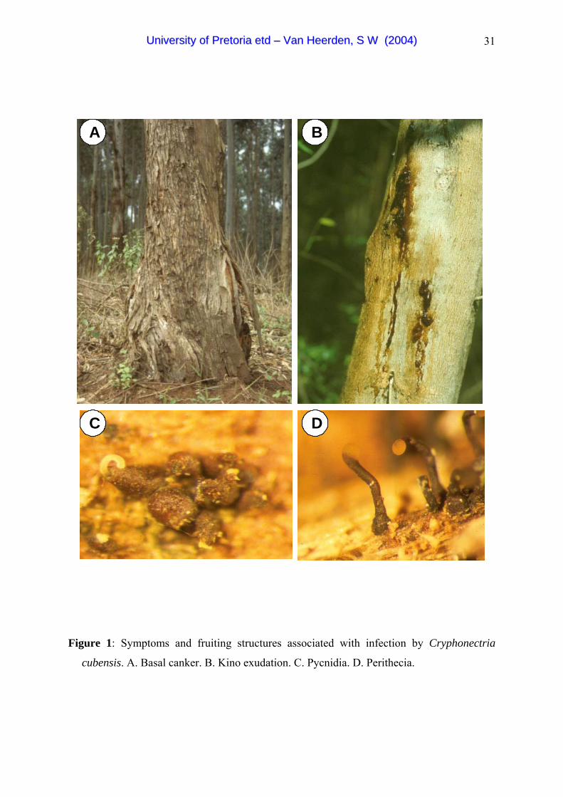
31
A B
C D
AA BB
CC DD
Figure 1: Symptoms and fruiting structures associated with infection by Cryphonectria
cubensis. A. Basal canker. B. Kino exudation. C. Pycnidia. D. Perithecia.
UUnniivveerrssiittyy ooff PPrreettoorriiaa eettdd –– VVaann HHeeeerrddeenn,, SS WW ((22000044))

32
p48
Pol UAA
AUG Hel
p29
9498 ntORF A
ORF B
1869 nt
p40
p69
AUTOCATALYTIC
Figure 2: Genome organisation of the Cryphonectria parasitica hypovirus (CHV1-EP713).
The genome is 12712 nucleotides in size and contain two open reading frames, ORF A
and ORF B, which encode the polypeptides p29, p40 and p48. The positions of the
polymerase (pol) and the helicase (hel) domain are indicated on ORF B. (The figure is
according to Dawe & Nuss 2001)
UUnniivveerrssiittyy ooff PPrreettoorriiaa eettdd –– VVaann HHeeeerrddeenn,, SS WW ((22000044))
33
Chapter 2:
Molecular characterisation of mitoviruses co-
infecting South African isolates of the Eucalyptus
canker pathogen Cryphonectria cubensis
ABSTRACT
Cryphonectria canker caused by Cryphonectria cubensis is an important disease of
Eucalyptus trees in South Africa and other parts of the world. The importance of this disease
has led to numerous studies on the biology, taxonomy and population biology of the causal
agent. In this study the complete nucleotide sequence of double stranded (ds) RNA segments
occurring in South African isolates of C. cubensis was determined. Sequence analysis led to
the identification of two distinct viruses that are related to mitochondrial RNA viruses in the
genus Mitovirus, family Narnaviridae. The viruses were thus provided with the names
Cryphonectria cubensis mitovirus 1 (CcMV1) and Cryphonectria cubensis mitovirus 2
(CcMV2). The RNA genomes of the two viruses are 2601 bp and 2639 bp in size
respectively and share a 22.6 % sequence identity at the protein level. Using the
mitochondrial genetic code where UGA codes for a tryptophan, the RNA of CcMV1 encodes
a putative protein of 744 amino acids (84255 Da) and CcMV2 a protein of 556 amino acids
(63552 Da). These proteins probably function as RNA-dependant RNA polymerase (RdRp)
since the conserved motifs for the RdRp could be identified. Northern blot hybridisations
indicate that the viruses occur predominantly in the positive single stranded RNA form, rather
than the dsRNA form. It was also shown that the viruses could be transmitted through the
asexual spores, which is the predominant form of reproduction for the fungus in South Africa.
Pathogenicity tests in this study suggest that the viruses have no significant impact on the
virulence of infected isolates. This diminishes opportunities for applying these viruses for
biological control of C. cubensis.
Genbank accession numbers of the sequences reported in this paper are: AY328476, AY328477, AY328478,
AY328479, AY328480, AY328481
UUnniivveerrssiittyy ooff PPrreettoorriiaa eettdd –– VVaann HHeeeerrddeenn,, SS WW ((22000044))

34
INTRODUCTION
Cryphonectria cubensis (Bruner) Hodges is an important pathogen of Eucalyptus spp. and
causes substantial losses to plantation forestry. High temperatures and rainfall favour this
disease, which is most serious in the tropics and sub-tropics (Hodges et al. 1979; Alfenas et
al. 1983; Sharma et al. 1985; Florence et al. 1986). Cryphonectria canker was first reported
in South Africa in 1989 and it has subsequently become important to implement effective
control measures against this disease (Wingfield et al. 1989). Currently, the most effective
means of reducing losses due to C. cubensis is through planting disease tolerant hybrid
Eucalyptus clones (Alfenas et al. 1983; Wingfield 1990). It has, however, also been
suggested that double stranded (ds) RNA linked to hypovirulence might form the basis of a
strategy to reduce the impact of Cryphonectria canker in South Africa (van Heerden et al.
2001).
Double stranded RNA viruses are known to occur in many fungi. The best studied of these is
the hypovirus that occurs in the chestnut blight pathogen Cryphonectria parasitica (Murr)
Barr (Day et al. 1977). Day et al. (1977) have shown that this virus residing in the genus
Hypovirus (Hypoviridae) causes a significant reduction in virulence of the fungus and could,
therefore, be a suitable agent for biological control (Hillman et al. 2000). The elucidation of
the complete nucleotide sequence of the C. parasitica hypovirus has enabled researchers to
expand the viral host range by transfecting other fungi in the genera Cryphonectria and
Endothia (Shapira et al. 1991; Chen et al. 1996). A South African C. cubensis isolate has
also been transfected with the C. parasitica hypovirus, leading to effective hypovirulence in
transfected strains (van Heerden et al. 2001).
Cryphonectria parasitica has been shown to harbour a second virus type, other than the
hypovirus. This ssRNA virus occurs in the mitochondria and belongs to the genus Mitovirus
(Mitochondrial) residing in the family Narnaviridae (Naked RNA virus) (Polashock &
Hillman 1994; Wickner et al. 2000). Mitoviruses are naked viruses without capsids and with
a genome containing one open reading frame (Wickner et al. 2000). The genome of the type
species, the Cryphonectria parasitica mitovirus 1-NB631 (CpMV1-NB631) is 2728 bp in size
and encodes for a RNA-dependant RNA polymerase (RdRp) (Polashock & Hillman 1994;
Wickner et al. 2000). This virus slightly reduces virulence in C. parasitica, but not to the
levels observed with the hypoviruses (Polashock & Hillman 1994). This virus can be
UUnniivveerrssiittyy ooff PPrreettoorriiaa eettdd –– VVaann HHeeeerrddeenn,, SS WW ((22000044))

35
transmitted through the fungal population via hyphal anastomosis, as well as through the
asexual conidia or the sexual ascospores (Polashock et al. 1997). Ascospore transmission has
been observed only when the donor strain was the female in a cross between isolates
(Polashock et al. 1997). This is consistent with the maternal inheritance of the mitochondria
in C. parasitica (Milgroom & Lipari 1993). Transmission both through asexual as well as
sexual spores would enhance opportunities of the establishment of mitoviruses in a fungal
population.
Double stranded RNA elements have previously been observed in the Eucalyptus pathogen
C. cubensis. Van Zyl et al. (1999) screened a large number of Brazilian C. cubensis isolates
for the presence of dsRNA, and discovered a 3 kilo base pair dsRNA element in some
isolates. The virus infected isolates showed altered colony morphology as well as a reduction
in virulence, suggesting hypovirulence (van Zyl et al. 1999). It is known that C. cubensis in
Brazil reproduces sexually and that the population diversity of this fungus in that country is
high (van Zyl et al. 1998). This would reduce the ease with which viruses could spread
through the pathogen population (Anagnostakis 1977). This low level of spread is also
consistent with the fact that low numbers of isolates were found to be virus infected (van Zyl
et al. 1999). In South Africa, C. cubensis has a low genetic diversity and sexual reproduction
is extremely rare (van Heerden & Wingfield 2001). This would enhance opportunities to
utilise hypovirulence to reduce the impact of Cryphonectria canker in South Africa.
The objectives of this study were, to screen the South African C. cubensis population for the
presence of dsRNA elements, using slow growth as a selection criterion. The complete
nucleotide sequence of the genomes was then determined for the two mitoviruses that were
subsequently discovered. Once these sequences have been deduced the open reading frames
were analysed to ascertain homologies to other viruses. Since mitoviruses are known to be
ssRNA, it was important to established whether the C. cubensis viral genome was single
stranded. Furthermore, relative abundance of the viruses in the fungal population,
quantification of the viruses in a single C. cubensis isolate, transmission of the viruses to
asexual spores and the pathogenicity of mitovirus – containing isolates were considered.
UUnniivveerrssiittyy ooff PPrreettoorriiaa eettdd –– VVaann HHeeeerrddeenn,, SS WW ((22000044))
36
MATERIALS AND METHODS
Isolate selection and growth studies
One hundred bark samples from trees infected with C. cubensis were randomly collected in
KwaZulu-Natal, South Africa. The fungus was induced to sporulate and isolations were
made using the method previously described by van Heerden & Wingfield (2001). All
isolates used in this study are maintained in the culture collection (CMW) of the Forestry and
Agricultural Biotechnology Institute (FABI), University of Pretoria.
One hundred C. cubensis isolates were inoculated onto 2% Malt extract agar (MEA) and
incubated at 25ºC for 7 days at normal illumination cycles (12 hours light and 12 hours
darkness). A 5 mm diam. mycelial plug from each of these isolates was inoculated in the
middle of two 9 cm diam. Petri dishes containing 2% MEA. The plates were incubated at
25ºC. Colony diameters were measured four days after inoculation. Seven slow growing
(CMW11325, CMW11332, CMW11341, CMW11353, CMW11354, CMW11342 and
CMW11329) and two rapidly growing (CMW11355 and CMW11351) South African C.
cubensis isolates were selected for dsRNA extraction (Table 1). These isolates were grown
in Erlenmeyer flasks containing 2% Malt extract broth at 25°C. The mycelium was harvested
after sufficient growth, freeze-dried and stored at -20°C before use.
Extraction and purification of dsRNA
Double stranded (ds) RNA was extracted using the method of Valverde et al. (1990) with
modifications outlined by Preisig et al. (1998). One gram lyophilised ground mycelium was
transferred to 40 ml centrifuge tubes into which 10 ml 2 x STE (0.2 M NaCl, 0.1 M Tris HCl,
2 mM EDTA pH 8, pH 6.8) and 1% SDS was added. Samples were mixed using a vortex
mixer and subsequently incubated at 60°C for 10 min. After incubation, 10 ml Phenol (pH
7.5) was added. The solution was shaken at room temperature for 30 min and centrifuged in
a Beckman JA25.50 rotor for 30 min at 15000 rpm. The aqueous phase was transferred to
clean centrifuge tubes and 10 ml of chloroform was added. The samples were mixed with a
vortex mixer before being centrifuged at 10000 rpm for 15 min. This step was repeated until
the inter phase was free of protein. After the last extraction step, the aqueous phase was
transferred to a clean centrifuge tube and 16% absolute ethanol was added and centrifuged at
5000 rpm for 5 min to remove the chromosomal DNA.
UUnniivveerrssiittyy ooff PPrreettoorriiaa eettdd –– VVaann HHeeeerrddeenn,, SS WW ((22000044))
37
The supernatant was passed through CF11 cellulose columns, to isolate the dsRNA. The
columns were prepared by packing 2 ml syringes (Promega) with 0.5 g CF11 cellulose
(Whatman). The columns were washed with 10 ml 2 x STE containing 16% ethanol after
which the dsRNA-containing samples were passed through. The cellulose bound dsRNA was
washed with 10 ml 2 x STE containing 16% ethanol. The dsRNA was eluted with 8 ml 2 X
STE in eight fractions collected in 1.5 ml Eppendorf tubes. The dsRNA was precipitated,
washed with 70% ethanol, dried and resuspended in 50 µl 0.1% (v/v) diethyl pyrocarbonate
(DEPC) treated double deionised water. The dsRNA found in isolates CMW11329 and
CMW11332 was then separated using an agarose gel in the presence of ethidium bromide and
visualised using UV light. The dsRNA segments were excised from the gel and purified
using an RNaid kit with spin (BIO101 Inc., QBiogene Inc.). The purified dsRNA was stored
at -20ºC until further use.
cDNA synthesis and RT-PCR
Purified dsRNA was used to determine the complete nucleotide sequence of the genomes of
virus infected isolate CMW11329. The dsRNA was initially denatured for 10 min at 99ºC.
cDNA was produced using the cDNA synthesis system (Roche Diagnostics). cDNA
fragments were cloned in the vector pGEM®-3Zf(+) and transformed into E. coli JM 109 high
efficiency competent cells (Promega).
Once a small number of cDNA clones had been sequenced, the remainder of the viral genome
sequences were obtained by reverse transcription polymerase chain reaction (RT-PCR)
experiments, using sequence specific 20 to 22-mer primers (MWG-Biotech, Inqaba Biotec)
produced from the cDNA fragments. RT-PCR was done using the Titan one tube RT-PCR
system (Roche Diagnostics). The reverse transcription was performed for 1 hour at 50ºC
followed by the PCR reaction with the amplification conditions of a single cycle at 99ºC for 2
min, 10 cycles at 94ºC for 30 s, 60ºC for 30 s and 68ºC for 2 min. This was followed by
another 35 cycles of 94ºC for 30 s, 60ºC for 30 s and 68ºC for 2 min with a cycle elongation
of 5 s per cycle. A final elongation step of 10 min at 68ºC was included.
Determination of the distal ends of the viral genomes
The sequences of the distal ends of the dsRNA elements were determined by the RACE
approach (Frohman 1994) using a 5'/3' RACE kit (Roche Diagnostics). cDNA was initially
synthesised from the dsRNA at 55ºC for 1 hour with 1 U AMV reverse transcriptase. The
UUnniivveerrssiittyy ooff PPrreettoorriiaa eettdd –– VVaann HHeeeerrddeenn,, SS WW ((22000044))
38
cDNA was purified using the High Pure PCR Product Purification Kit (Roche Diagnostics).
A poly (A) tail was enzymatically added to the purified cDNA by the addition of 2 mM
dATP in the presence of terminal transferase. The tailed cDNA was then used in a PCR
amplification with the mixture containing a nested internal sequence specific primer and an
oligo dT-anchor primer using the Expand High Fidelity PCR system (Roche Diagnostics).
Amplification conditions for the PCR were 1 cycle at 94ºC for 2 min, 10 cycles at 94ºC for
20 s, 60ºC for 45 s and 72ºC for 40 s. This was followed by 25 cycles at 94ºC for 20 s, 60ºC
for 45 s and 72ºC for 40 s with a time increase of 5 s per cycle. A final elongation step at
72ºC for 7 min was included. In some cases a secondary PCR was performed.
Cloning and sequencing of cDNA
The PCR products from both the RT-PCR and RACE-PCR protocols were gel-purified from
the agarose gel using the PCR product purification kit (Roche Diagnostics). The purified
products were ligated into the pGEM®-T Easy Vector (Promega), and incubated overnight at
4ºC. Two µl of the reaction mixture was transformed in 25 µl of JM 109 E. coli high
efficiency competent cells (Promega). Positive clones were selected on LB medium (10 g/l
Bacto®-Tryptone, 5 g/l Bacto®-Yeast extract, 5 g/l NaCl, 15 g/l Biolab agar) containing 0.5
mM IPTG, 80 µg/ml X-Gal and 120 µg/ml ampicillin. White colonies were selected and
used to inoculate 2 ml LB medium containing 100 µg/ml ampicillin. The plasmids were
isolated from the cells using alkaline lysis (Sambrook & Russell 2001). The insert size was
determined by cutting the plasmids with the restriction enzyme EcoRI (Roche Diagnostics).
The inserts were sequenced using universal primers SP6 (5' TATTTAGGTGACACTATAG
3') and T7 (5' TAATACGACTCACTATAGGG 3') to obtain both strands. The products were
sequenced using the ABI PRISM Big Dye Terminator cycle sequencing ready reaction kit
(Perkin Elmer). The sequenced products were analysed using an ABI 377 and ABI 3100
automated DNA sequencers (Applied Biosystems). The sequences were analysed using
Sequence Navigator version 1.0.1 (Applied Biosystems) and CLUSTAL X multiple sequence
alignment programs (Thompson et al. 1994). Analyses were also done with programs
available on the NCBI (http://www.ncbi.nlm.nih.gov/BLAST) and ExPASY
(http://www.expasy.ch) web pages. These included: BLAST, Translate, Protein machine and
SIM + LALNVIEW. Genbank/EMBL and the Swiss-Prot databases were used for the
homology searches.
UUnniivveerrssiittyy ooff PPrreettoorriiaa eettdd –– VVaann HHeeeerrddeenn,, SS WW ((22000044))

39
The conserved domains of the aligned sequence were analysed using Phylogenetic Analysis
Using Parsimony (PAUP) software (Swofford 1998). The heuristic search option based on
parsimony with random stepwise addition and tree bisection reconnection was used. Gaps
were treated as fifth character and confidence intervals using 1000 bootstrap replicates were
calculated. Saccharomyces cerevisiae Narnavirus sequence (Rodriguez-Cousino et al. 1991)
was used as an outgroup taxon.
Preparation of DIG labeled strand specific RNA probes
Plasmids containing cDNA, specific for the C. cubensis dsRNA elements were selected as
templates to generate digoxigenin (DIG) labeled probes. These plasmids were linearised with
SpeI and NcoI respectively, in the multiple cloning sites, before being subjected to a
phenol/chloroform purification step to eradicate the RNase in the mixture. The in vitro
production of positive and negative strand specific DIG labeled RNA probes was achieved by
preparing a reaction mixture containing 200 ng of the linearised plasmid DNA, 1 X DIG
RNA labeling mix (10 mM dATP, 10 mM dGTP, 10 mM dCTP, 3.5 mM DIG-11-UTP), 1 X
transcription buffer, 0.5 U T7 RNA polymerase or SP6 RNA polymerase with a total reaction
volume of 20 µl. The in vitro transcription reaction was performed for 2 hours at 30ºC. The
probes were stored at -20ºC.
Northern blot hybridisations and colorimetric detection
Northern blot hybridisations were performed to determine whether C. cubensis mitovirus
genomes are maintained in an ssRNA or dsRNA form. Total nucleic acid was extracted from
the virus infected isolate (CMW11329) and a Colombian isolate (CMW11348). The positive
and negative stranded RNA was produced in vitro with either SP6 RNA polymerase or T7
RNA polymerase as described previously. The isolated total nucleic acids and the in vitro
produced RNA was separated on a 1% Agarose gel in 1 x TAE (0.04 M Tris Base, 0.001 M
EDTA, 1.14ml/l Acetic acid) buffer at a current of 80 V for 45 min. The agarose gel was
denatured for 30 min in a 50 mM NaOH / 150 mM NaCl solution before being neutralised
twice for 15 min in a 1 M Tris-HCl (pH 7.5). The agarose gel was placed on a tray and
overlaid with a positively charged nylon membrane (Roche Diagnostics) and covered with
several Whatman filter papers (Whatman). The procedure was allowed to proceed for 3
hours to ensure the complete transfer of the nucleic acids to the membrane. Hybridisation
with the DIG labeled strand specific RNA probes was carried out according to
manufacturer’s guidelines in DIG Easy Hyb buffer at 68ºC. Detection was achieved using
UUnniivveerrssiittyy ooff PPrreettoorriiaa eettdd –– VVaann HHeeeerrddeenn,, SS WW ((22000044))
40
the DIG High Prime Labeling and Detection Kit I (Roche Diagnostics). Colorimetric-
detection was done by adding freshly prepared colour solution (5 µl NBT/BCIP (18.75 mg/ml
Blue tetrazolium chloride, 9.4 mg/ml 5-Bromo-4-Chloro-3-Indolyl Phosphate) added to 10 ml
detection buffer) to the membrane and incubated in the dark until colour formation. The
reaction was terminated by washing the membrane in 50 ml strerilised distilled H2O.
Abundance of the mitoviruses
To determine whether the two C. cubensis mitoviral genomes occur singly or together in
infected C. cubensis isolates and to determine their relative occurrence in a population of
isolates, two sequence specific primer pairs were designed for the two viruses. These were
ccmv62 (5' GCACCGATGTCTCGTAAGAG 3') and ccmv68 (5'
GCCCCACTTGTCAATCATCC 3') for viral genome 1 (amplification product of 215 bp)
and ccmv46 (5' CGTTGGGTCGCTAGCCCGTG 3') and ccmv59 (5'
GACTCTTCAAGTCACTTGAC 3') for genome 2 (amplification product of 126 bp).
Twenty C. cubensis isolates were randomly selected for this experiment. These isolates
included 18 from South Africa and two from Colombia (Table 2).
Total RNA was extracted from lyophilised mycelium of all isolates using the High Pure RNA
Isolation Kit (Roche Diagnostics). The RNA was stored at -70ºC until use. For detection of
the viruses, a one step RT-PCR reaction was performed using a LightCycler (Roche
Diagnostics) with the LightCycler- RNA Amplification Kit SYBR Green I (Roche
Diagnostics). A reaction mixture was prepared that contained 0.75 ng total RNA, 0.5 µM
primer ccmv 62 or ccmv 46, 0.5 µM primer ccmv 68 or ccmv 59, 1 x LightCycler-RT-PCR
reaction mix SYBR green I, 6 mM MgCl2, 0.4 µl LightCycler-RT-PCR enzyme mix and PCR
grade sterile H2O to a total volume of 20 µl. The cycle conditions were a reverse
transcription reaction at 50ºC for 10 min, followed by a single cycle at 95ºC for 10 sec, 35
cycles at 95ºC for 0 sec, 62ºC for 4 sec and 72ºC for 16 sec. At the end of each PCR cycle,
the amount of the amplified product was monitored by a single fluorescence reading of the ds
DNA binding dye, SYBR Green I. A melting curve and cooling program was also added.
The data were analysed based on the melting curves of the PCR products, which would
indicate whether sequence specific products were obtained. The RT-PCR products are
indicated by a sharp peak that is centered at the Tm of the product. Non-specific
amplification products such as dimers tend to melt at lower temperatures over a broader
UUnniivveerrssiittyy ooff PPrreettoorriiaa eettdd –– VVaann HHeeeerrddeenn,, SS WW ((22000044))

41
range. The final PCR products were also separated on a 1.5% agarose gel stained with
ethidium bromide and visualised under UV light.
Quantification of the two mitoviruses in a single isolate
Quantification of the two mitoviral genomes in a single virus-infected C. cubensis isolate
(CMW11329) was achieved by using a LightCycler (Roche Diagnostics). The same primer
pairs as those discussed above were used. Purified dsRNA of isolate CMW11329 was used
to produce cDNA using the cDNA Synthesis System (Roche Diagnostics) with AMV
Reverse Transcriptase. For quantification, the LightCycler – FastStart DNA Master SYBR
Green I kit was used (Roche Diagnostics). For the quantification reaction, a dilution series
was prepared with the cDNA (1:1, 1:10, 1:100 and 1:1000 dilutions). A reaction mixture was
prepared containing 5 mM MgCl2, 0.5 µM primer ccmv 46 or ccmv 62, 0.5 µM primer ccmv
59 or ccmv 68 and 2 µl LightCycler – FastStart DNA Master SYBR Green I. Four µl
template was added to this mixture, with the total volume being adjusted to 20 µl with sterile
PCR grade water. The templates used were the 1:1 cDNA dilution, 1:10 cDNA dilution,
1:100 cDNA dilution and a 1:1000 cDNA dilution. The cycle conditions were 10 min at
95ºC followed by 35 cycles at 95ºC for 10 sec, 60ºC for 5 sec and 72ºC for 16 sec with the
amount of product being determined after each cycle based on SYBR green I fluorescence. A
melting and cooling program followed. Quantification of the difference in concentration
between the two viruses was analysed using the LightCycler software (Roche Diagnostics).
This was based on a standard amplification curve using the second derivative maximum
analysis method. The standards used in this experiment were the dilution series of CcMV2.
The relative concentration of the viral amplicons could then be determined based on the
standard curve. The experiment was repeated with independently extracted dsRNA and new
cDNA synthesis.
Mitovirus transfer to conidia
To determine whether the mitoviruses were present in the asexual spores of the virus-infected
C. cubensis isolate (CMW11329), it was first necessary to produce asexual fruiting
structures. To achieve this, twenty freshly cut stem sections (15 cm in length) of a
Eucalyptus grandis clone (ZG14) were inoculated with the fungus as described by van
Heerden & Wingfield (2001). Mass conidial cultures were made and transferred to
Erlenmeyer flasks containing 100 ml 2% ME broth and incubated at 25ºC for 1 week. After
sufficient growth, the mycelium was harvested and lyophilised. Total RNA was isolated
UUnniivveerrssiittyy ooff PPrreettoorriiaa eettdd –– VVaann HHeeeerrddeenn,, SS WW ((22000044))

42
from the different isolates using the High Pure RNA Isolation Kit (Roche Diagnostics). The
presence of the viruses in the isolates was detected using specific primers for CcMV1 and
CcMV2 in a RT-PCR amplification using the LightCycler (Roche Diagnostics).
Amplification conditions were the same as those described above. The products were
separated on a 2% agarose gel stained with ethidium bromide and fragments visualised using
UV light.
Pathogenicity tests
A greenhouse inoculation trial was conducted to determine whether the mitoviruses present in
isolates of C. cubensis have an influence on pathogenicity of the fungus. Eucalyptus grandis
clone ZG14, which is known to be highly susceptible to C. cubensis infection (van Heerden
& Wingfield 2001) was used for the inoculations. The trees were planted in 2 litre planting
bags and maintained under shade nets until they were approximately 1 m high. The plants
were moved to a greenhouse and allowed to acclimatise prior to inoculation. Eleven South
African C. cubensis isolates including those known to harbour mitoviruses or to be free of
these mitoviruses were used for the inoculation, and sterile 2% MEA disks were used for the
control (Fig. 2). The C. cubensis isolates were grown on 2% MEA plates.
Trees were inoculated by removing a cambial disc from the main stem with an 8 mm diam.
cork borer. Similar sized discs from the actively growing margins of the fungus were placed
mycelium side facing towards the wounds and covered with Parafilm to reduce desiccation.
Ten trees were inoculated with each of the isolates and an additional ten trees were inoculated
with a sterile MEA disc to serve as controls. After 4 weeks, the Parafilm covering the
wounds was removed and the lesion lengths were measured. The differences in lesion length
associated with the test isolates were analysed using a one way ANOVA. A Bonferroni
pairwise comparison was used to determine differences between the isolates.
RESULTS
Growth studies
The average colony diameter after 4 days for all of the 100 isolates of C. cubensis initially
tested in this study was 35 mm (± 0.85 SEM). Of these, seven isolates (CMW11325,
CMW11332, CMW11341, CMW11353, CMW11354, CMW11342 and CMW11329) with
the slowest mean growth and two isolates (CMW11355 and CMW11351) that grew most
UUnniivveerrssiittyy ooff PPrreettoorriiaa eettdd –– VVaann HHeeeerrddeenn,, SS WW ((22000044))
43
rapidly, were selected for dsRNA isolation (Table 1). Two (CMW11329 and CMW11332) of
these seven isolates contained dsRNA elements and reached a mean diameter of 16 mm and
24 mm in 4 days respectively (Table 1). No dsRNA was detected in the remaining seven
isolates (CMW11325, CMW11355, CMW11341, CMW11353, CMW11354, CMW11342
and CMW11351), which in 4 days, attained a mean colony diameter of 22, 49, 22, 22, 23, 18
and 51 mm respectively.
Determination of the complete nucleotide sequence and sequence analysis of the genomes
The dsRNA element from the South African C. cubensis isolate (CMW11329) was
approximately 2600 base pairs in size (Fig. 1). Using the cDNA synthesis system, random
cDNA fragments of the viral dsRNA were produced. Cloning of the cDNA fragments
yielded plasmids with inserts ranging from 100 -500 bp in size. A BLAST search showed
homology of these fragments to the Cryphonectria parasitica mitovirus and other mitoviral
genomes.
The translation of the nucleotide sequence to an amino acid sequence and subsequent
alignment to the C. parasitica mitoviral RdRp indicated the probable positions and
orientation of the cDNA fragments in the genome. The entire genome sequence was obtained
and two distinct mitoviral genomes were detected within the single C. cubensis isolate.
These have been given the names CcMV1 and CcMV2 (Cryphonectria cubensis mitovirus)
respectively.
The terminal 5' and 3' end sequences of the genomes were determined by the 5'/3' RACE
approach. The sequence data showed a poly-A-tail at the 3' end and a poly-U-tail at the 5'
end for both the genome sequences (Appendix 1 & 2). It was further shown that different
possible ends exist for both mitoviruses (Appendix 1 & 2). This led to the establishment of
the C. cubensis mitovirus (CcMV) genomes namely CcMV1a (2555 nucleotides,
AY328476), CcMV1b (2601 nucleotides, AY328477), CcMV1c (2501 nucleotides,
AY328478), CcMV2a (2639 nucleotides, AY328479), CcMV2b (2257 nucleotides,
AY328480) and CcMV2c (2419 nucleotides, AY328481). For further analysis, the largest
fragment for each of these two viruses (CcMV1b and CcMV2a) were selected. These are,
hereafter, referred to as CcMV1 and CcMV2. These virus genomes are A-U rich with,
CcMV2 having a A-U content of 62.4% and CcMV1 having a A-U content of 51.8%.
UUnniivveerrssiittyy ooff PPrreettoorriiaa eettdd –– VVaann HHeeeerrddeenn,, SS WW ((22000044))

44
The homology of the C. cubensis mitoviruses to the C. parasitica mitoviral RdRp suggests
that the viruses are located in the mitochondria. The genetic code for the mitochondria was
used when the nucleotide sequence was translated to the amino acid sequence. In the
mitochondrial genetic code UGA codes for a tryptophan. Since there are various possible
initiation codons for mitochondrial open reading frames, we used the first methionine after
the stop codons in the amino acid sequence to locate the start codon. The data showed a
single large ORF for CcMV1 and CcMV2. The ORF of CcMV1 has the potential to encode a
protein of 744 amino acids (Molecular mass: 84255 Da) (Appendix 3, position 184 to 2418 in
Appendix 1) and CcMV2 has the potential to encode a protein of 556 amino acids (Molecular
mass: 63552 Da) respectively (Appendix 4, position 893 to 2563 in Appendix 2). Complete
amino acid sequence identity comparisons using the SIM alignment tool (www.expasy.ch)
indicated that CcMV1 and CcMV2 are 22.5% identical with CcMV2 having a 40.5% identity
to the C. parasitica mitovirus (CpMV1-NB63) (Table 3). The alignment of the amino acids
showed conserved domains as described by Hong et al. (1999) (Fig. 3). These are typical of
RdRp, suggesting that the ORF probably encodes for a functional RdRp (Poch et al. 1989).
Parsimony analysis using amino acid sequence within the conserved area of the viruses in the
Mitovirus genus showed that CcMV2 and CpMV1-NB63 grouped together in one clade.
CcMV1 grouped with the O. novo-ulmi mitoviral segment (OnuMV3A), but away from
CcMV2. All these viruses grouped within the genus, Mitovirus with the Narnavirus forming
an outgroup (Fig. 4).
Northern blot hybridisations
Hybridisation with the probe for the positive-stranded RNA of CcMV2 resulted in an intense
signal in the lane where the total nucleic acid for the virus infected (CMW11329) isolate was
loaded (Fig. 5 A). A fainter signal was observed in the lane where the dsRNA was loaded
(Fig. 5 A). This dsRNA was isolated using CF11 cellulose column chromatography. The
negative strand specific probe resulted only in a signal in the lane where the purified dsRNA
was loaded (Fig. 5 B). No signal was detected in the lane where the total nucleic acid of the
virus infected isolate was loaded (Fig. 5 B). No hybridisation occurred in the lanes where the
virus-free isolate was loaded, for either probe. For CcMV1 the positive strand specific probe
gave a signal for the lane where the total nucleic acid of the virus infected isolate was loaded
(Fig 5 C). No signal was observed for the negative strand specific probe in the lane where
the total nucleic acids of the virus infected isolate was loaded (Fig. 5 D). Both the probes
gave a signal in the lanes where the purified dsRNA was loaded but this was less intense
UUnniivveerrssiittyy ooff PPrreettoorriiaa eettdd –– VVaann HHeeeerrddeenn,, SS WW ((22000044))

45
where the negative stranded specific probe was used for CcMV1. No signal was observed in
the lanes where the virus-free isolate was loaded. These results showed that the positive
stranded RNA molecules are present in a much higher concentration than the dsRNA, in the
virus-infected isolate. This was true for both the C. cubensis mitoviruses, CcMV1 and
CcMV2.
Abundance of the mitoviruses
Separation of the PCR products using the viral specific primers for the two viral genomes on
an agarose gel confirmed the presence of either a 215 bp amplicon for CcMV1 or a 126 bp
amplicon for CcMV2 (Fig. 6). Of the 20 isolates tested, 9 were shown to contain CcMV1
and 11 isolates contained CcMV2 (Table 2). Some isolates had smaller peaks on the melting
curve as well as different intensity of the amplicons on the gel, suggesting that the viruses
occur in different concentrations in the different isolates (Fig. 6).
Quantification of the mitoviruses in a single C. cubensis isolate
cDNA was successfully produced from the viral RNA of isolate CMW11329. The Tm of the
CcMV1 amplicon produced using the primers ccmv 62 and ccmv 68 was 84.5ºC while the
Tm of the CcMV2 amplicon using primers ccmv 46 and ccmv 59 was 87ºC. From the
profiles on the real time PCR, it was possible to distinguish the presence of the different viral
amplicons as well as the relative abundance of these products in the fungus (Fig. 7). The
average ratio of the relative concentration for CcMV2 vs. CcMV1 was 1:4.9 (Table 4). This
experiment was repeated and the ratio was 1:3.9. The overall average ratio was thus 1:4.4.
Mitovirus transfer to conidia
Six weeks after inoculation of the E. grandis stem pieces, pycnidia of C. cubensis were
evident on the bark. RT-PCR reactions showed that viruses were present in most of the mass
conidial cultures derived from them (Fig. 8). This established that the viruses are transmitted
to the asexual spores of the fungus.
Pathogenicity tests
All trees inoculated with the C. cubensis isolates developed distinct lesions 4 weeks after
inoculation. No lesions were associated with the control inoculations. Lesion lengths
differed significantly between the different isolates inoculated (F=17.62; df=11; p<0.001).
UUnniivveerrssiittyy ooff PPrreettoorriiaa eettdd –– VVaann HHeeeerrddeenn,, SS WW ((22000044))

46
The lesion lengths of the virus-infected isolates (CMW11329, CMW11332 and CMW11326)
did not differ significantly from the other isolates used in the inoculation study (Fig 2).
DISCUSSION
In this study we have established that fungal viruses commonly occur in South African
isolates of C. cubensis. The complete nucleotide sequences produced from the dsRNA
elements have shown that these are mitochondrial viruses in the genus Mitovirus, family
Narnaviridae. Northern blot hybridisation showed that the genomes of these viruses occur
predominantly as positive single stranded RNA viruses. The names Cryphonectria cubensis
mitovirus 1 (CcMV1) and Cryphonectria cubensis mitovirus 2 (CcMV2) have been provided
for these two viruses.
Mitoviruses such as those found in C. cubensis have previously been found in other fungi
such as C. parasitica and O. novo-ulmi (Polashock & Hillman 1994; Rodgers et al. 1986,
1987). Our discovery of two different mitoviruses in a single isolate of C. cubensis is also
not unusual because 12 elements of virus-like dsRNA have been identified in O. novo-ulmi
(Rodgers et al. 1986, 1987; Cole et al. 1998). In this study it was shown that in some isolates
the C. cubensis mitoviruses do not occur together. For the O. novo-ulmi dsRNA elements, it
has been shown that some do not cause the degenerate disease in the O. novo-ulmi isolate
alone, but that they require other dsRNA elements to do so (Rodgers et al. 1986; Cole et al.
1998).
The C. cubensis mitovirus (CcMV) genomes range in size from 2257-2639 bases. This is in
comparison to the C. parasitica mitovirus genome (Polashock & Hillman 1994) which is
2728 bases, and is similar in size to the CcMV’s. In O. novo-ulmi, the virus like dsRNAs
range from 330 bases - 3500 bases in size (Rodgers et al. 1986, 1987; Cole et al. 1998). The
mitoviruses identified in this study are, therefore, similar in size to some other known
mitoviruses and especially to that in C. parasitica.
The CcMV2 genome was found to have an A-U content of 62.4%. This is consistent with
findings for other mitoviruses such as the O. novo ulmi mitoviruses which all have an A-U %
of higher than 60% (Hong et al. 1998a; Hong et al. 1999). The C. parasitica mitovirus has a
A-U % of 63.4% (Polashock & Hillman 1994). In contrast, CcMV1 has a lower A-U % of
UUnniivveerrssiittyy ooff PPrreettoorriiaa eettdd –– VVaann HHeeeerrddeenn,, SS WW ((22000044))

47
51.8%, which was unexpected and is similar to the A-U % of the Rhizoctonia solani dsRNA
virus which is 56.8 % (Hong et al. 1998a; Jian et al. 1997). The high A-U content of the
mitoviruses is comparable with the plant mitochondrial genomes (Unseld et al. 1997) and
confirms the mitochondrial localisation of the mitoviruses.
Using the mitochondrial genetic code where UGA codes for tryptophan, it was possible to
identify the open reading frames of the two mitoviruses discovered in this study. We could
thus show that CcMV1 possibly encodes for a single protein of 744 amino acids in size.
CcMV2 apparently encodes for a single protein of 556 amino acids in size. The alignment of
these amino acid sequences with protein sequences encoded by other mitoviruses showed the
presence of six conserved RNA dependant RNA polymerase (RdRp) motifs (motifs I-VI).
This is similar to motifs described by Poch et al. (1989). Similar motifs II, III and IV have
also been identified by Habili & Symons (1989) for positive stranded RNA plant viruses.
Motif VI is also similar to motif 7 described by Bruenn (1993). The motifs (II-VI) identified
in this study are all similar to those that have been identified for the O. novo-ulmi mitoviruses
(Hong et al. 1998a, 1999). Motif I is not found in all the RdRp’s of other RNA viruses, but it
is characteristic of the RdRp, which is encoded by the mitochondrial viruses (Hong et al.
1998a, 1999). This provides further evidence that the C. cubensis viral genomes are of
mitochondrial origin and have a single ORF that encodes for a RNA dependant RNA
polymerase.
The C. cubensis mitovirus genome contains a poly-A tail at the 3' end and a poly-U tail at the
5' end. This could promote secondary structure development that would protect the naked
single stranded RNA from degradation. It has been shown that the O. novo-ulmi mitovirus 7
can form a very stable stem loop structure since the 5' terminal sequence and the 3' sequence
contain complementary sequences, consequently a pan handle can also form (Hong et al.
1998b). These complementary 5' and 3' terminal sequences are a characteristic of negative
stranded RNA viruses such as the Myxoviridae (Desselberger et al. 1980), but stem loop
structures are known to occur for the positive single stranded RNA viruses (Buck 1996).
Sequence alignment and parsimony analysis of the ORF showed that CcMV2 groups together
with the C. parasitica mitovirus. CcMV1 grouped most closely with the O. novo-ulmi
mitovirus 3. Interestingly, CcMV1 did not group with CcMV2 from the same fungus but still
falls within the group accommodating mitoviruses. This low amino acid identity (22.6%) of
UUnniivveerrssiittyy ooff PPrreettoorriiaa eettdd –– VVaann HHeeeerrddeenn,, SS WW ((22000044))

48
the two C. cubensis mitoviruses suggests that they might have different origins. Polashock &
Hillman (1994) showed that the C. parasitica mitovirus is ancestrally related to the T (23S)
and the W (20S) Saccharomyces cerevisiae RNA replicons as well as to positive stranded
RNA bacteriophages of the Leviviridae. Hong et al. (1998a) used a large set of isolates to
show that mitochondrial viruses formed a well defined clade within this set of isolates based
on the relationship between the viral RdRp sequence. They could further distinguish between
two lineages in the RNA viruses based on RdRp sequence. One of these lineages includes
the mitoviruses, yeast RNA replicons and the Leviviridae and the other consists of all the
other viruses (Hong et al. 1998a).
Both of the C. cubensis mitoviruses could be transmitted via asexual spores. The majority of
the mass conidial cultures tested in this study contained the viral genomes. Transmission via
asexual spores suggests a reasonably easy route of transfer, which is consistent with our
finding that viral genomes were present in approximately half the isolates screened.
Mitoviruses have previously been reported to also be transmitted to ascospores of C.
parasitica (Pollashock et al. 1997) and of O. novo-ulmi (Rodgers et al. 1986). As the South
African C. cubensis population has a narrow genetic diversity and apparently only reproduces
asexually (van Heerden & Wingfield 2001), it was not possible to test whether transmission
to ascospores is possible for CcMV1 and CcMV2.
Greenhouse inoculations showed that isolates of C. cubensis infected with the C. cubensis
mitoviruses are equally pathogenic as virus-free isolates. This is in contrast to the C.
parasitica mitovirus that reduces levels of virulence slightly although its impact is not
equivalent to that of the C. parasitica hypovirus (Polashock & Hillman 1994; Polashock et al.
1997). The C. cubensis mitoviruses will, therefore, not be suitable for biological control of
Cryphonectria canker in South Africa
UUnniivveerrssiittyy ooff PPrreettoorriiaa eettdd –– VVaann HHeeeerrddeenn,, SS WW ((22000044))

49
REFERENCES
Alfenas, A. C., Jeng, R. & Hubbes, M. (1983) Virulence of Cryphonectria cubensis on
Eucalyptus species differing in resistance. European Journal of Forest Pathology 13:
179-205.
Anagnostakis, S. L. (1977) Vegetative incompatibility in Endothia parasitica. Experimental
Mycology 71: 213-215.
Bruenn, J. A. (1993) A closely related group of RNA-dependant RNA polymerases from
double-stranded RNA viruses. Nucleic Acids Research 21: 5667-5669.
Buck, K. W. (1996) Comparison of the replication of positive-stranded RNA viruses of plants
and animals. Advances in Virus Research 47: 159-251.
Chen, B., Chen, C.-H., Bowman, B.H., & Nuss, D.L. (1996) Phenotypic changes associated
with wild-type and mutant hypovirus RNA transfection of plant pathogenic fungi
phylogenetically related to Cryphonectria parasitica. Phytopathology 86:301-310.
Cole, T. E., Müller, B., Hong, Y., Brasier, C. M. & Buck, K. W. (1998) Complexity of virus-
like double-stranded RNA elements in a diseased isolate of the dutch elm disease fungus
fungus, Ophiostoma novo-ulmi. Journal of Phytopathology 146: 593-598.
Day, P. R., Dodds, J. A., Elliston, J. E., Jaynes, R. A. & Anagnostakis, S. L. (1977) Double-
stranded RNA in Endothia parasitica. Phytopathology 67: 1393-1396.
Desselberger, U., Racaniello, V. R., Zazra, J. J. & Palese, P. (1980) The 3’ and 5’ end
terminal sequences of influenza A, B and C virus RNA segments are highly conserved and
show partial inverted complementary. Gene 8: 315-328.
Florence, E. J. M., Sharma, J. K. & Mohanan, C. (1986) Stem canker disease of Eucalyptus
caused by Cryphonectria cubensis in Kerala. Kerala Forest Research Institute Scientific
Paper 66: 384-387.
UUnniivveerrssiittyy ooff PPrreettoorriiaa eettdd –– VVaann HHeeeerrddeenn,, SS WW ((22000044))

50
Frohman, M. A. (1994) On beyond classic RACE (rapid amplification of cDNA ends). PCR
Methods and Applications 4: S40-S58.
Habili, N. & Symons, R. H. (1989) Evolutionary relationship between Luteoviruses and other
RNA plant viruses based on sequence motifs in their putative RNA polymerases and
nucleic acid helicases. Nucleic Acids Research 17: 9543-9555.
Hillman, B. I., Fulbright, D. W., Nuss, D. L. & van Alfen, N. K. (2000) Family Hypoviridae.
In: Virus Taxonomy: Seventh report of the International Committee on Taxonomy of
Viruses. Van Regenmortel, M. H. V., Fauquet, C. M., Bishop, D. H. L., Carstens, E. B.,
Estes, M. K., Lemon, S. M., Maniloff, J., Mayo, M. A., McGeoch, D. J., Pringle, C. R. &
Wickner, R. B. (Eds.). Academic Press, California, USA. pp. 515-520.
Hodges, C. S., Geary, T. F. & Cordell, C. E. (1979) The occurrence of Diaporthe cubensis on
Eucalyptus in Florida, Hawaii and Puerto Rico. Plant Disease Reporter 63: 216-220.
Hong, Y., Cole, T. E., Brasier, C. M. & Buck, K. W. (1998a) Evolutionary relationships
among putative RNA dependant RNA polymerases encoded by a mitochondrial virus-like
RNA in the dutch elm disease fungus, Ophiostoma novo ulmi, by other viruses and virus
like RNAs and by the Arabidopsis mitochondrial genome. Virology 246: 158-169.
Hong, Y., Cole, T. E., Brasier, C. M. & Buck, K. W. (1998b) Novel structures of two virus-
like RNA elements from a disease isolate of the dutch elm disease fungus, Ophiostoma
novo-ulmi. Virology 242: 80-89.
Hong, Y., Dover, S. L., Cole, T. E., Brasier, C. M., Buck, K. W. (1999) Multiple
mitochondrial viruses in an isolate of the Dutch elm disease fungus Ophiostoma novo-
ulmi. Virology 258: 118-127.
Jian, J., Lakshman, D. K. & Tavantzis, S. M. (1997) Association of distinct double-stranded
RNAs with enhanced or diminished virulence in Rhizoctonia solani infecting potato.
Molecular Plant Microbe Interactions 10: 1002-1009.
UUnniivveerrssiittyy ooff PPrreettoorriiaa eettdd –– VVaann HHeeeerrddeenn,, SS WW ((22000044))
51
Milgroom, M. G. & Lipari, S. E. (1993) Maternal inheritance and diversity of mitochondrial
DNA in the chestnut blight fungus, Cryphonectria parasitica. Phytopathology 83: 563-
567.
Poch, O., Sauvaget, I., Delarue, M. & Tordo, N. (1989) Identification of four conserved
motifs among the RNA dependant polymerase encoding elements. The EMBO Journal 8:
3867-3874.
Polashock, J. J., Bedker, P. J. & Hillman, B. I. (1997) Movement of a small mitochondrial
double-stranded RNA element of Cryphonectria parasitica: ascospore inheritance and
implications for mitochondrial recombination. Molecular General Genetics 256: 566-571.
Polashock, J. J. & Hillman, B. I. (1994) A small mitochondrial double-stranded (ds) RNA
element associated with a hypovirulent strain of the chestnut blight fungus and ancestrally
related to yeast cytoplasmic T and W dsRNAs. Proceedings of the National Academy of
Sciences USA 91: 8680-8684.
Preisig, O., Wingfield, B. D. & Wingfield, M. J. (1998) Coinfection of a fungal pathogen
with two distinct double stranded RNA viruses. Virology 252: 399-406.
Rodgers, H. J., Buck, K. W. & Brasier, C. M. (1986) Transmission of double-stranded RNA
and a disease factor in Ophiostoma ulmi. Plant Pathology 35: 277-287.
Rodgers, H. J., Buck, K. W. & Brasier, C. M. (1987) A mitochondrial target for double-
stranded RNA in diseased isolates of the fungus that causes Dutch elm disease. Nature
329: 558-560.
Rodriguez-Cousino, N., Esteban, L. M. & Esteban, R. (1991) Molecular cloning and
characterization of w double-stranded RNA, a linear molecule present in Saccharomyces
cerevisiae. Journal of Biological Chemistry 266: 12772-12778.
Sambrook, J. & Russell, D. W. (2001) Molecular cloning- a laboratory manual 3rd edition.
Cold Spring Harbor Press, Cold Spring Harbor, New York, USA. pp. 1.31-1.42.
UUnniivveerrssiittyy ooff PPrreettoorriiaa eettdd –– VVaann HHeeeerrddeenn,, SS WW ((22000044))

52
Shapira, R., Choi, G. H. & Nuss, D. L. (1991) Virus-like genetic organization and expression
strategy for a double-stranded RNA genetic element associated with biological control of
chestnut blight. The EMBO Journal 10: 731-739.
Sharma, J. K., Mohanan, C. & Florence, E. J. M. (1985) Occurrence of Cryphonectria canker
disease of Eucalyptus in Kerala, India. Annals of Applied Biology 106: 265-276.
Swofford, D. L. (1998) PAUP: (Phylogenetic analysis using parsimony). Version 4. Sinauer
Associates, Sunderland, MA.
Thompson, J. D., Higgens, D. G. & Gibson, T. J. (1994) CLUSTALW: Improving the
sensitivity of progressive multiple sequence alignment through sequence weighting,
position-specific gap pealties and weight matrix choice. Nucleic Acid Research 22: 4673-
4680.
Unseld, M., Marienfeld, J. R., Brandt, P. & Brennicke, A. (1997) The mitochondrial genome
of Arabidopsis thaliana contains 57 genes in 399924 nucleotides. Nature Genetics 15: 57-
61.
Valverde, R. A., Nameth, S. T. & Jordan, R. L. (1990) Analysis of double-stranded RNA for
plant virus diagnosis. Plant Disease 74: 255-258.
van Heerden, S. W., Geletka, L. M., Preisig, O., Nuss, D. L., Wingfield, B. D. & Wingfield,
M.J. (2001) Characterization of South African Cryphonectria cubensis isolates infected
with a C. parasitica hypovirus. Phytopathology 91: 628-632.
van Heerden, S. W. & Wingfield, M. J. (2001) Genetic diversity of Cryphonectria cubensis
isolates in South Africa. Mycological Research 105: 94-99.
van Zyl, L. M., Wingfield, M. J., Alfenas, A. C. & Crous, P. W. (1998) Population diversity
among Brazilian isolates of Cryphonectria cubensis. Forest Ecology and Management
112: 41-47.
UUnniivveerrssiittyy ooff PPrreettoorriiaa eettdd –– VVaann HHeeeerrddeenn,, SS WW ((22000044))

53
van Zyl, L. M., Wingfield, M. J., Alfenas, A. C. & Crous, P. W. (1999) Hypovirulence
detected in Brazilian isolates of Cryphonectria cubensis. Plant Pathololgy 48: 267-272.
Wickner, R. B., Esteban, R. & Hillman, B. I. (2000) Family Narnaviridae. In: Virus
Taxonomy: Seventh report of the International Committee on Taxonomy of Viruses. Van
Regenmortel, M. H. V., Fauquet, C. M., Bishop, D. H. L., Carstens, E. B., Estes, M. K.,
Lemon, S. M., Maniloff, J., Mayo, M. A., McGeoch, D. J., Pringle, C. R. & Wickner, R.
B. (Eds.). Academic Press, California, USA. pp. 651-656.
Wingfield, M. J. (1990) Current status and future prospects of forest pathology in South
Africa. South African Journal of Science 86: 60-62.
Wingfield, M. J., Swart, W. J. & Abear, B. J. (1989) First record of Cryphonectria canker of
Eucalyptus in South Africa. Phytophylactica 21: 311-313.
UUnniivveerrssiittyy ooff PPrreettoorriiaa eettdd –– VVaann HHeeeerrddeenn,, SS WW ((22000044))
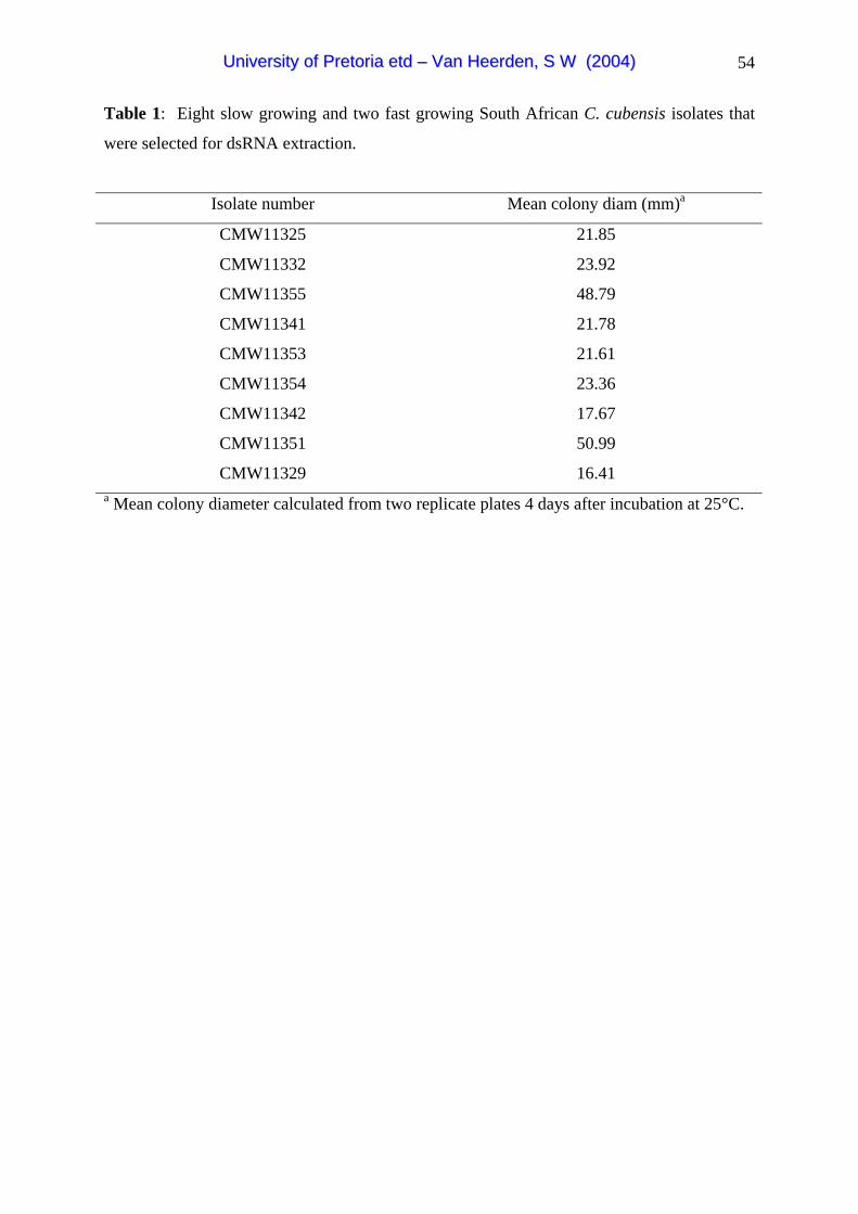
54
Table 1: Eight slow growing and two fast growing South African C. cubensis isolates that
were selected for dsRNA extraction.
Isolate number Mean colony diam (mm)a
CMW11325 21.85
CMW11332 23.92
CMW11355 48.79
CMW11341 21.78
CMW11353 21.61
CMW11354 23.36
CMW11342 17.67
CMW11351 50.99
CMW11329 16.41
a Mean colony diameter calculated from two replicate plates 4 days after incubation at 25°C.
UUnniivveerrssiittyy ooff PPrreettoorriiaa eettdd –– VVaann HHeeeerrddeenn,, SS WW ((22000044))
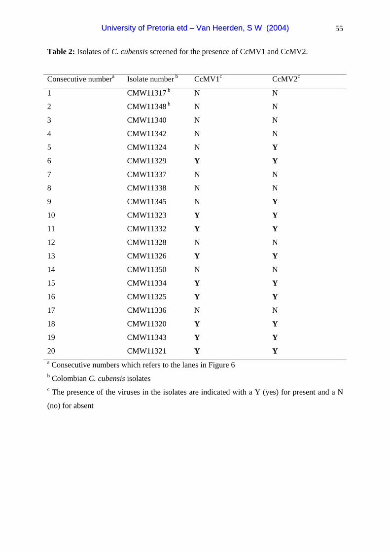
55
Table 2: Isolates of C. cubensis screened for the presence of CcMV1 and CcMV2.
Consecutive numbera Isolate number b CcMV1c CcMV2c
1 CMW11317 b N N
2 CMW11348 b N N
3 CMW11340 N N
4 CMW11342 N N
5 CMW11324 N Y
6 CMW11329 Y Y
7 CMW11337 N N
8 CMW11338 N N
9 CMW11345 N Y
10 CMW11323 Y Y
11 CMW11332 Y Y
12 CMW11328 N N
13 CMW11326 Y Y
14 CMW11350 N N
15 CMW11334 Y Y
16 CMW11325 Y Y
17 CMW11336 N N
18 CMW11320 Y Y
19 CMW11343 Y Y
20 CMW11321 Y Y a Consecutive numbers which refers to the lanes in Figure 6 b Colombian C. cubensis isolatesc The presence of the viruses in the isolates are indicated with a Y (yes) for present and a N
(no) for absent
UUnniivveerrssiittyy ooff PPrreettoorriiaa eettdd –– VVaann HHeeeerrddeenn,, SS WW ((22000044))

56
Table 3: Comparison of amino acid sequence identities (%) of some known mitochondrial
virusesa
CcMV2 CpMV1-
NB63
OnuMV3
A
OnuMV4
-LD
OnuMV5
-LD
OnuMV6
-LD
ScNV-
20S
CcMV1 22.6 21.7 25.3 25.1 23 23.9 19.2
CcMV2 100 40.5 25 26.7 26.2 24.7 17.8 a Ophiostoma novo-ulmi, OnuMV3A (Hong et al. 1998a), OnuMV4-LD, OnuMV5-LD,
OnuMV6-LD (Hong et al. 1999), Cryphonectria parasitica CpMV1-613 (Polashock &
Hillman 1994), Saccharomyces cerevisiae ScNV (Rodriguez-Cousino et al. 1991)
Table 4: Relative concentrations and ratios of concentration between CcMV2 and CcMV1
in isolate CMW11329.
Experiment
number
Dilution Relative concentration
CcMV2 (Units)
Relative concentration
CcMV1 (Units)
Ratio
CcMV2:CcMV1
1 1:10 100.6 311.9 1:3.1
1 1:100 9.9 49.4 1:4.9
1 1:1000 1 6.8 1:6.8
2 1:10 60.9 201 1:3.3
2 1:100 10 34.3 1:3.4
2 1:1000 1 5.1 1:5.1
UUnniivveerrssiittyy ooff PPrreettoorriiaa eettdd –– VVaann HHeeeerrddeenn,, SS WW ((22000044))

57
1 2
3530 bp2027 bp
dsRNA
1 2
3530 bp2027 bp
dsRNA
Figure 1: Double stranded RNA isolated from a South African Cryphonectria cubensis
isolate (CMW11329). Lane 1: Lambda DNA marker cut with HindIII was used as an
approximate size marker. Lane 2: dsRNA element isolated with a size of 2600 bp based on a
DNA size marker. A 1% agarose gel stained with ethidium bromide was used. The
fragments are visualised with UV light.
UUnniivveerrssiittyy ooff PPrreettoorriiaa eettdd –– VVaann HHeeeerrddeenn,, SS WW ((22000044))

58
Con
trol
CM
W11
335
CM
W11
346
CM
W11
332
CM
W11
329
CM
W11
318
CM
W11
344
CM
W 2
113
CM
W11
326
CM
W11
319
CM
W11
339
CM
W11
342
ISOLATES
0
50
100
150
200
LE
SIO
N L
EN
GT
H (M
M)
ab
c
Con
trol
CM
W11
335
CM
W11
346
CM
W11
332
CM
W11
329
CM
W11
318
CM
W11
344
CM
W 2
113
CM
W11
326
CM
W11
319
CM
W11
339
CM
W11
342
ISOLATES
0
50
100
150
200
LE
SIO
N L
EN
GT
H (M
M)
ab
c
Figure 2: Mean lesion length (mm) (±SEM), 4 weeks after inoculation on Eucalyptus
grandis clone (ZG14) with different South African Cryphonectria cubensis isolates. Isolate
CMW11329, CMW11332 and CMW11326 are co-infected with CcMV1 and CcMV2 and the
remainder of the isolates was free of virus. Columns marked with the same letter do not
differ significantly from each other
UUnniivveerrssiittyy ooff PPrreettoorriiaa eettdd –– VVaann HHeeeerrddeenn,, SS WW ((22000044))
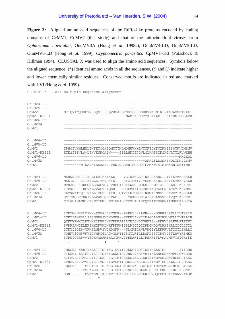
59
Figure 3: Aligned amino acid sequences of the RdRp-like proteins encoded by coding
domains of CcMV1, CcMV2 (this study) and that of the mitochondrial viruses from
Ophiostoma novo-ulmi, OnuMV3A (Hong et al. 1998a), OnuMV4-LD, OnuMV5-LD,
OnuMV6-LD (Hong et al. 1999), Cryphonectria parasitica CpMV1-613 (Polashock &
Hillman 1994). CLUSTAL X was used to align the amino acid sequences. Symbols below
the aligned sequence: (*) identical amino acids in all the sequences, (:) and (.) indicate higher
and lower chemically similar residues. Conserved motifs are indicated in red and marked
with I-VI (Hong et al. 1999). CLUSTAL X (1.81) multiple sequence alignment OnuMV4-LD ------------------------------------------------------------ OnuMV5-LD ------------------------------------------------------------ CcMV2 MTTQVTWQSXCTNPSQSTLPSQVRCAFGYNGTFHSDSRRVXWDNIKIRIARASDPTXXXI CpMV1-NB631 -----------------------------MAMIIHDPVYKLWYAX---RAKSNLPGLAPH OnuMV6-LD ------------------------------------------------------------ OnuMV3A ------------------------------------------------------------ CcMV1 ------------------------------------------------------------ OnuMV4-LD ------------------------------------------------------------ OnuMV5-LD ------------------------------------------------------------ CcMV2 TPNCIVKHLHXLINTKTQQHIQNYTTNLMQNWVXXEVTIFTCYFYXNHSSSSTKVLMSNV CpMV1-NB631 KTESITTVLS-LTNVKNKQATN----SILLNKITDLVLGLKAFSIKSKGVRTTLRVNKHW OnuMV6-LD ------------------------------------------------------MKLKQL OnuMV3A --------------------------------------MKRLTLSQNKSNQLTNNDLSNV CcMV1 --------MTKADHYSSASSDPPRWTDCCHNTRQHQPTFAMKWIMTKTMKRNTNKTYMHT OnuMV4-LD MKRNNLQIIIIKRLILHIFKINLS----VEIDKFLGFINHLRKSNGLLYTIKYMKAAKLH OnuMV5-LD MKKIN---KTIKILLSIYFNRKYS----SYGIRWIVTVERMRKINGLKFTIKYMKAVKLH CcMV2 MVRAGSPKRRFQNLSSWFVGVPVHSPIKDTLWRCRMDLSLSEMTTATHSSLSILKRXCVL CpMV1-NB631 ITPKEFP--RFVKLVVWCTRTQEH---EDSFMKIIGKCDHIWQTAGPNFLFKYLKEVMRL OnuMV6-LD KLMKNKTYQIIRILLIVFFPSIKR--QTVILNIFMSKINKMIKNNGTLFTVKYLKELRLH OnuMV3A GYITKQLFPHWIRLLVWSLQLSPAP-----YKKFGSRIAILWKANGVSFTVQYLKECTRI CcMV1 NTLRDIGAMKLSTVWFTHWSHTETGMASRTVSSAVARFQTTATTRGRNAAMSEFKASRTA : . .. :* OnuMV4-LD ITRYMCGKPLYSNN-ENVALDKTGFP--LRFWYLKRLVN----DNPRALLTLLTYTRRIV OnuMV5-LD ITKYIANERLLSISGSRVSVDKDGFP--TKFNYIKHIIDSGDIDGIRFVMTLLTYTRAIN CcMV2 QXEDWRMXTLFTVKKIFVKLNKYRFPAIIPTEICKDTSWFPG--RFHVLSRKVMATTTVI CpMV1-NB631 SVRRIANIELEPSKKIFVKLNKFRFPNIIPLPICDQIIRDQNDQVLWASKRLIICLLTIL OnuMV6-LD ITKYISGEP-YRNSLNRVSVDKDGFP---TLCKELKVLVNGTYLEKRFVLTIITLSKLLI OnuMV3A VQHFVSGHPVFVTDVMPIGLAG-GLPTIIPGTLRTLLRSKDSSTIRGVLSTLAVYRIMKM CcMV1 FTRWTCGRP--TSGKVGAPMTKAGTPKVIPREARTLLTRERPTYLVKAVMTVTSIGRYFK . : * OnuMV4-LD PNKSES-KARIVKLSTITDPYKG-KVYTIPKWFILDFISKYNLSSTKP------IYTDND OnuMV5-LD PTKKEY-LKIFPDYSTITNEFTGSNKIAIPNKIIKEFVDYYKLANPNNNNNPELQWSRDD CcMV2 SIFRVLPTKVLPDYTTISKPHSGTIETCSSESIALACKNTEIKRVDKVMKTRLKGSTKAG CpMV1-NB631 SVHRVLPTKVVPDYSTIVDPFTGVSKTIDQKLLRKAIHLLNIKRV-KQLKLKITGSMKAG OnuMV6-LD PQKSES---IPFSTKSITDHWSGIDNISNEELDKSCSELNISTREVQWDVKNFKLLTKAG OnuMV3A P--------CVLKLESITDPFKGISDTLPKSEIINGLASLG-FEIPKGRSKHLLTLSNPI CcMV1 GGN-------PVKWENITKPSTPTTPKDGEITFGLEKLNIDVGQFEPTDWKFRWVVTAGP . .* . .
UUnniivveerrssiittyy ooff PPrreettoorriiaa eettdd –– VVaann HHeeeerrddeenn,, SS WW ((22000044))

60
OnuMV4-LD HYLSIKGSPNGKASMSS---LYSIIS--FNSSNIRYLFNIVG--DYQLVLNKFYQDLSQF OnuMV5-LD HYLSFKSSPNGQSTLHSSYGLFSMIF--VGHTILENILKIVGEKQYHEIIGNHIKKLYHD CcMV2 PNGKMSLTTSLLDATAFWSDPLRVIH--FIWFNIRCYGYFWGTMWSMWLIFIMIISLPYY CpMV1-NB631 PNGKISLLTSSVDALSFITQPTKIFT--YLDFSVRVY-KFRGLLLWMWMMCILLITLPYA OnuMV6-LD PHGPQSLTWYHTIKLYDFNQWLGIIG--ILPKSVLDLFTETLSYASKLVLPEIKSNIKS- OnuMV3A IYLLSAGPNHSISMMGIWKDIYAWYVSPLFPTLLSFIGRMNRGNVLIDLLRAEVSYWEAT CcMV1 NGPSMSSCLQDTPKFNG-------------TFRSQVEVITPETTPIIDTTLTWEKSFKTS I OnuMV4-LD YTKYIN-RDKLGLGKLSIVHDPELKERVIAMVDYTTQFALRPIHNILLNNLSKLPCDRTF
FGKISIVKDPELKMRVIAMVDYHSQFVLKKIHNSLFNKLKLIKSDRTF MGQLATVYDQAGKARIVASTNSWIQCSTFGLHNKIFSILRSIPQDGTF MGKLSVVYDQAGKARIVAITNSWIQTAFYSLHLHVFKLLKNIDQDGTF IRRLSIVHDPECKERVIAIFDYGSQMVLKPIADVLFDLLRNIPSDRTF LGKLAIKEEAAGKARVFAMADSITQSVMAPLNSWVFSKLKDLPMDGTF TRKMAIKDDREGKSRPFAMFDYWSQTVLSPTHDWAYATTRSIPQDCTF
TQDP-- RYHSLDLSAATDRFPTQDP-- RFWSMDLSAATDRFPDQNKPF MLYGFDTSAATDRTPDQERPF KFYGFDLTAATDRLPTQSP-- KFWSIDLSSATDRFPNQQAPL EFYSYDLSSATDRLPNQAEG- YFYSYDTEAATDRFP
YSVGQPMGAYTSWAAFTLTHHLVVHWAAE FKDYIILG YKVGQPMGAYSSWAAFTLTHHLVVFYSAR FTNYILLG YAVGQPMGAYSSFAMTATTHHVLVQVAAQ FTDYCITG YAVGQPMGAYSSFAMLALTHHVIVQVAAL FTDYCILG YNCGQPMGAQSSWPMFTLAHHVIVRVAAN FDKYIILG YSVGQPMGALSSWAMLALSHHVIVQIAAM FTNYALLG WGAGQPTGAKSSWAIFTLCHHTVVHMAAV --YYVMTG
DDIVIKNNKVAQIYINLMTKWG-VDISLSKTHVSYD EFAKRWIDDIVINNDKVAKYYIRTMKRLG-VELSMNKTHVSKN EFAKRWFDDIVMANSLVAEAYKSLIFDLG-LEISESKSVISGT EFAKKLRDDIVIAHDTVASEYLKLMETLG-LSISSGKSVISSE EFAKKLKDDIVINNDNVALKYMEIMNDFK-VEISRNKTHVSND EFAKRWIDDIVIADKAVATSYHMIMTQILGVEINLSKSLVSNN EFAKRLVDDMVTRGSRTATVYKRMMSETG-VSMSETKSHVSKD EFAKMWM
OnuMV5-LD HRLFIPGKIDYLCcMV2 TMALCTGARAPVCpMV1-NB631 IVSFMLGALIPIOnuMV6-LD ---------TKLOnuMV3A GVKPSVSPLDLKCcMV1 TTMGLAGFKDDS ::: : * * .* : * : * ** II OnuMV4-LD -----FHKWNDDHKE IFLQQKLISLIFNDYEFGKNWRNL OnuMV5-LD -----IFTT-PTMGH IDLQERLLSYLYG-SEISSAWKQL CcMV2 ---DLLLEST-QPGY IAFQKDITNHLG---YPGGPWRRL CpMV1-NB631 ---KLLIKWLNEPTQ IDLQVDILNIIFK-NSPGSSWRSL OnuMV6-LD ----FFTHTDLDNKS IVFQKRVLQKILG-KQMTDSWERI OnuMV3A NRLVQLYQDGLLHDV MAFQKQIISVLFG-SKFAKDWATL CcMV1 ----TSKVTARPSQK MQFQKKVTSTIFN-TTYAQAWAEM * . * :**** *: :* : . : * : III IV OnuMV4-LD LVDRNYDY--QGISYR LAGLKN-OnuMV5-LD LIDRTYKTP-EGDELH MAGIKD-CcMV2 TGIKYNSP---CGFIS KAGFSDRCpMV1-NB631 LRIKYKSP---QGFLT NSGFTTROnuMV6-LD MIGSKFLAP-DGDTVS RCGLSN-OnuMV3A LVGRDWYL--KDIPYR RVGKLP-CcMV1 MTQEPFRVKGLSDPLR RTNSDP- : *** ** :*:. : ** :* :* . * : * V VI OnuMV4-LD TY K-NGKEISGISLKGI OnuMV5-LD TY K-NKKEITGLPLRGI CcMV2 FT G-PTMDISPIGAGLM CpMV1-NB631 FT GRNNFDIFYRSWFSI OnuMV6-LD TY K-NKMEFFPLPIRGI OnuMV3A SF T-MDGEVSAVGAKNL CcMV1 TF HQGRNASGFPVVGTA **:* . .* * : :.:. *: :* **** OnuMV4-LD LTNIRHIHVVYMNIFTYLQR--IPSLN-VDILTCVGKLYGYLLIRNRIKS-PNTIKRSLY OnuMV5-LD LNNLNNYGIVFQELFKFHYK--YPHLTNVKLTDIMFIIFKGLKIKGRIIT-NSQLRFNLM CcMV2 LYSLRNKYYICVLVFEITER--GLCMWYDVYPQLTSLLP---KIYRRYFK-TCDWFIATH CpMV1-NB631 IHFEKQILHLCT-VFELLRR--GVCELYDLYPQYINKLP---KIYLRYNL-LIDWVVVAF OnuMV6-LD VDNINNKYIIFNILYSFFVEKGNTFLNKDTLLVCVSKFIQLHSLTLKKPIGLNKVKGILY OnuMV3A LVALKSRWGISSVILDLYNK--GLALSEQDLRQRFSSIPTVSKQFGVDKLLWLVLGPFGF CcMV1 ETTRKPTEMAATFVFETPAKGYPVTITPRTVSQYFTTVARYNTIPPRTAVWTADKVVWYY
UUnniivveerrssiittyy ooff PPrreettoorriiaa eettdd –– VVaann HHeeeerrddeenn,, SS WW ((22000044))
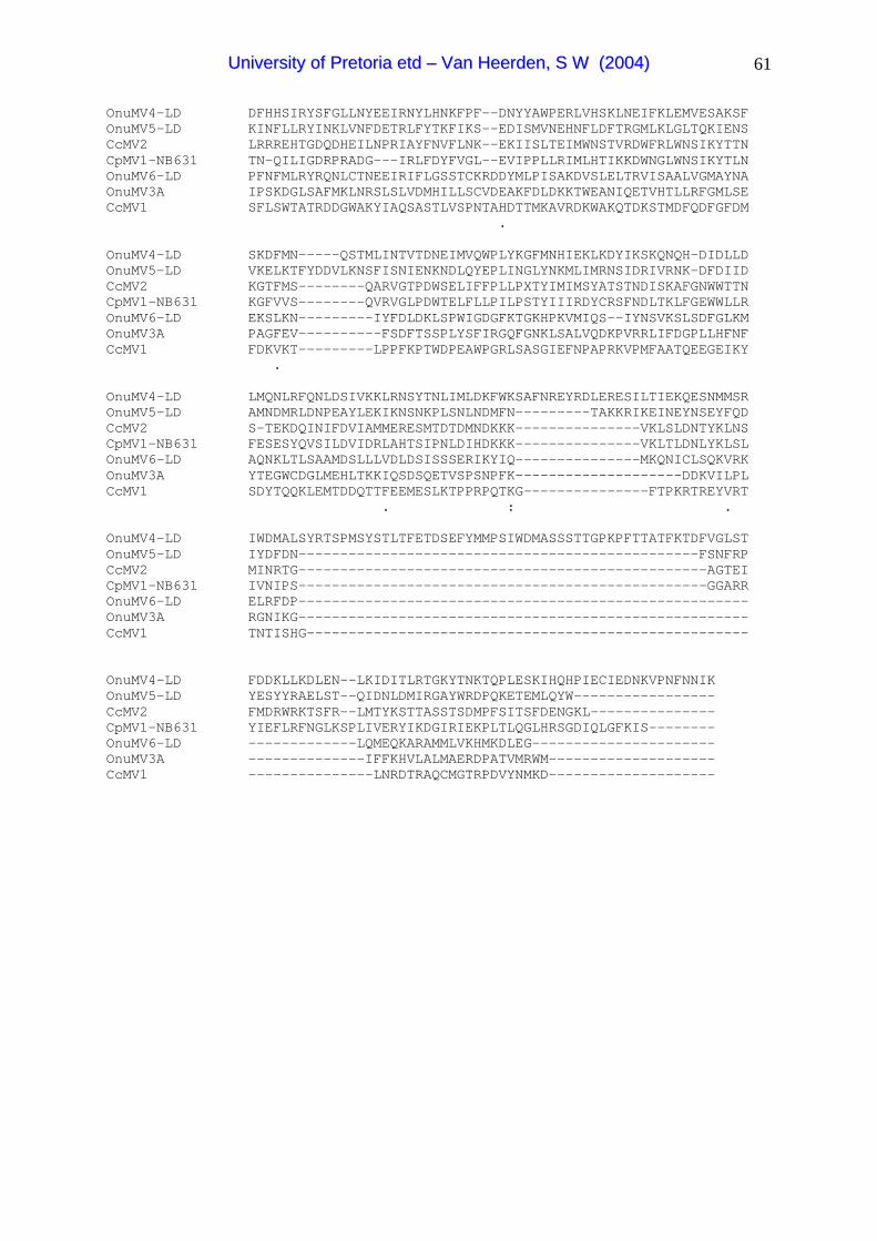
61
OnuMV4-LD DFHHSIRYSFGLLNYEEIRNYLHNKFPF--DNYYAWPERLVHSKLNEIFKLEMVESAKSF OnuMV5-LD KINFLLRYINKLVNFDETRLFYTKFIKS--EDISMVNEHNFLDFTRGMLKLGLTQKIENS CcMV2 LRRREHTGDQDHEILNPRIAYFNVFLNK--EKIISLTEIMWNSTVRDWFRLWNSIKYTTN CpMV1-NB631 TN-QILIGDRPRADG---IRLFDYFVGL--EVIPPLLRIMLHTIKKDWNGLWNSIKYTLN OnuMV6-LD PFNFMLRYRQNLCTNEEIRIFLGSSTCKRDDYMLPISAKDVSLELTRVISAALVGMAYNA OnuMV3A IPSKDGLSAFMKLNRSLSLVDMHILLSCVDEAKFDLDKKTWEANIQETVHTLLRFGMLSE CcMV1 SFLSWTATRDDGWAKYIAQSASTLVSPNTAHDTTMKAVRDKWAKQTDKSTMDFQDFGFDM . OnuMV4-LD SKDFMN-----QSTMLINTVTDNEIMVQWPLYKGFMNHIEKLKDYIKSKQNQH-DIDLLD OnuMV5-LD VKELKTFYDDVLKNSFISNIENKNDLQYEPLINGLYNKMLIMRNSIDRIVRNK-DFDIID CcMV2 KGTFMS--------QARVGTPDWSELIFFPLLPXTYIMIMSYATSTNDISKAFGNWWTTN CpMV1-NB631 KGFVVS--------QVRVGLPDWTELFLLPILPSTYIIIRDYCRSFNDLTKLFGEWWLLR OnuMV6-LD EKSLKN---------IYFDLDKLSPWIGDGFKTGKHPKVMIQS--IYNSVKSLSDFGLKM OnuMV3A PAGFEV----------FSDFTSSPLYSFIRGQFGNKLSALVQDKPVRRLIFDGPLLHFNF CcMV1 FDKVKT---------LPPFKPTWDPEAWPGRLSASGIEFNPAPRKVPMFAATQEEGEIKY . OnuMV4-LD LMQNLRFQNLDSIVKKLRNSYTNLIMLDKFWKSAFNREYRDLERESILTIEKQESNMMSR OnuMV5-LD AMNDMRLDNPEAYLEKIKNSNKPLSNLNDMFN---------TAKKRIKEINEYNSEYFQD CcMV2 S-TEKDQINIFDVIAMMERESMTDTDMNDKKK---------------VKLSLDNTYKLNS CpMV1-NB631 FESESYQVSILDVIDRLAHTSIPNLDIHDKKK---------------VKLTLDNLYKLSL OnuMV6-LD AQNKLTLSAAMDSLLLVDLDSISSSERIKYIQ---------------MKQNICLSQKVRK OnuMV3A YTEGWCDGLMEHLTKKIQSDSQETVSPSNPFK--------------------DDKVILPL CcMV1 SDYTQQKLEMTDDQTTFEEMESLKTPPRPQTKG---------------FTPKRTREYVRT . : . OnuMV4-LD IWDMALSYRTSPMSYSTLTFETDSEFYMMPSIWDMASSSTTGPKPFTTATFKTDFVGLST OnuMV5-LD IYDFDN------------------------------------------------FSNFRP CcMV2 MINRTG-------------------------------------------------AGTEI CpMV1-NB631 IVNIPS-------------------------------------------------GGARR OnuMV6-LD ELRFDP------------------------------------------------------ OnuMV3A RGNIKG------------------------------------------------------ CcMV1 TNTISHG----------------------------------------------------- OnuMV4-LD FDDKLLKDLEN--LKIDITLRTGKYTNKTQPLESKIHQHPIECIEDNKVPNFNNIK OnuMV5-LD YESYYRAELST--QIDNLDMIRGAYWRDPQKETEMLQYW----------------- CcMV2 FMDRWRKTSFR--LMTYKSTTASSTSDMPFSITSFDENGKL--------------- CpMV1-NB631 YIEFLRFNGLKSPLIVERYIKDGIRIEKPLTLQGLHRSGDIQLGFKIS-------- OnuMV6-LD -------------LQMEQKARAMMLVKHMKDLEG---------------------- OnuMV3A --------------IFFKHVLALMAERDPATVMRWM-------------------- CcMV1 ---------------LNRDTRAQCMGTRPDVYNMKD--------------------
UUnniivveerrssiittyy ooff PPrreettoorriiaa eettdd –– VVaann HHeeeerrddeenn,, SS WW ((22000044))

62
OnuMV4.LD
100
100
100
OnuMV5.LD
OnuMV6.LD
CcMV2
CpMV1.NB631
OnuMV3A
CcMV1
ScNV.20S
Figure 4: Phylogenetic relationships inferred from the conserved area of the aligned amino
acid sequences of the viruses in the genus Mitovirus. These viruses are: Ophiostoma novo-
ulmi mitoviruses, OnuMV3A (Hong et al. 1998a), OnuMV4-LD, OnuMV5-LD, OnuMV6-
LD (Hong et al. 1999), Cryphonectria parasitica mitovirus CpMV1-613 (Polashock &
Hillman 1994) as well as the C. cubensis mitoviruses CcMV1 and CcMV2 (current study).
Bootstrap values are indicated above the branches (1000 replicates). The Saccharomyces
cerevisiae Narnavirus (Rodriguez-Cousino et al. 1991) is used as the outgroup taxon.
UUnniivveerrssiittyy ooff PPrreettoorriiaa eettdd –– VVaann HHeeeerrddeenn,, SS WW ((22000044))
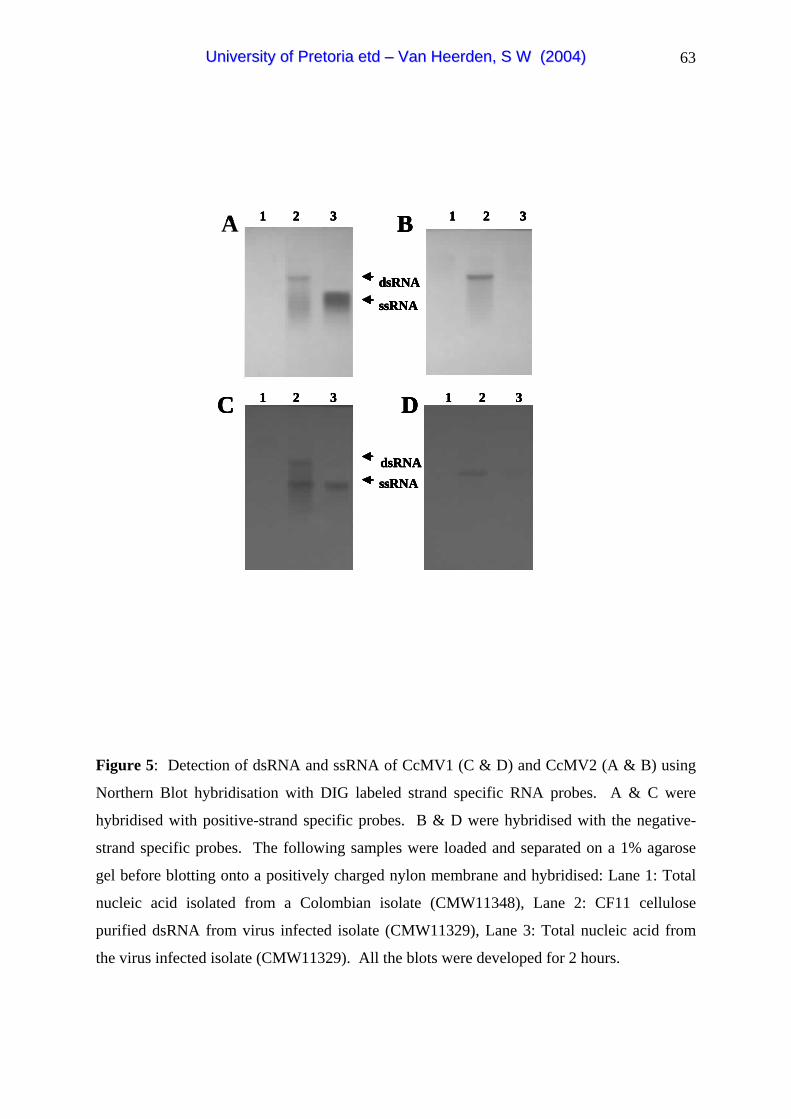
63
1 2 3
1 2 3
B1 2 3
1 2 3C D
dsRNA
ssRNA
dsRNAssRNA
A 1 2 3
1 2 3
B1 2 3
1 2 3C D
dsRNA
ssRNA
dsRNAssRNA
A 1 2 3
1 2 3
B1 2 3
1 2 3C D
dsRNA
ssRNA
dsRNAssRNA
A 1 2 3
1 2 3
B1 2 3
1 2 3C D
dsRNA
ssRNA
dsRNAssRNA
A
Figure 5: Detection of dsRNA and ssRNA of CcMV1 (C & D) and CcMV2 (A & B) using
Northern Blot hybridisation with DIG labeled strand specific RNA probes. A & C were
hybridised with positive-strand specific probes. B & D were hybridised with the negative-
strand specific probes. The following samples were loaded and separated on a 1% agarose
gel before blotting onto a positively charged nylon membrane and hybridised: Lane 1: Total
nucleic acid isolated from a Colombian isolate (CMW11348), Lane 2: CF11 cellulose
purified dsRNA from virus infected isolate (CMW11329), Lane 3: Total nucleic acid from
the virus infected isolate (CMW11329). All the blots were developed for 2 hours.
UUnniivveerrssiittyy ooff PPrreettoorriiaa eettdd –– VVaann HHeeeerrddeenn,, SS WW ((22000044))

64
C
V1 V2V1 V2V1 V2 V1 V2 V1 V2V1 V2V1 V2V1 V2V1 V21 2 3 4 5 6 7 8
V1 V2V1 V2V1 V2 V1 V2 V1 V2V1 V2V1 V2V1 V2V1 V29 10 11 12 13 14 15 16 17
V1 V2V1 V2V1 V218 19 20
M M
M M
M
215 bp126 bp
215 bp126 bp
215 bp126 bp
C V1 V2V1 V2V1 V2 V1 V2 V1 V2V1 V2V1 V2V1 V2V1 V2
1 2 3 4 5 6 7 8
V1 V2V1 V2V1 V2 V1 V2 V1 V2V1 V2V1 V2V1 V2V1 V29 10 11 12 13 14 15 16 17
V1 V2V1 V2V1 V218 19 20
M M
M M
M
C V1 V2V1 V2V1 V2 V1 V2 V1 V2V1 V2V1 V2V1 V2V1 V2
1 2 3 4 5 6 7 8C V1 V2V1 V2V1 V2 V1 V2 V1 V2V1 V2V1 V2V1 V2V1 V2 V1 V2V1 V2V1 V2V1 V2V1 V2 V1 V2V1 V2 V1 V2V1 V2V1 V2V1 V2V1 V2V1 V2V1 V2V1 V2V1 V2V1 V2
1 2 3 4 5 6 7 8
V1 V2V1 V2V1 V2 V1 V2 V1 V2V1 V2V1 V2V1 V2V1 V29 10 11 12 13 14 15 16 17
V1 V2V1 V2V1 V2 V1 V2 V1 V2V1 V2V1 V2V1 V2V1 V2 V1 V2V1 V2V1 V2V1 V2V1 V2 V1 V2V1 V2 V1 V2V1 V2V1 V2V1 V2V1 V2V1 V2V1 V2V1 V2V1 V2V1 V29 10 11 12 13 14 15 16 17
V1 V2V1 V2V1 V218 19 20
V1 V2V1 V2V1 V2V1 V2V1 V2V1 V218 19 20
M MM M
M MM M
M
215 bp126 bp215 bp126 bp
215 bp126 bp215 bp126 bp
215 bp126 bp
Figure 6: Confirmation of the presence of the two viruses CcMV1 (V1) and CcMV2 (V2) in
20 C. cubensis isolates. The RT-PCR products were separated on a 1.5% agarose gel stained
with ethidium bromide and visualised under UV light. Lanes M: 100 bp molecular weight
marker. Lane C: Negative control. Lanes 1-20: Isolates used in this study as indicated in
Table 2. The presence of the viruses is indicated by a 215 bp amplicon for CcMV1 and a 126
bp amplicon for CcMV2. Different band intensities indicate varying concentrations of the
virus present.
UUnniivveerrssiittyy ooff PPrreettoorriiaa eettdd –– VVaann HHeeeerrddeenn,, SS WW ((22000044))
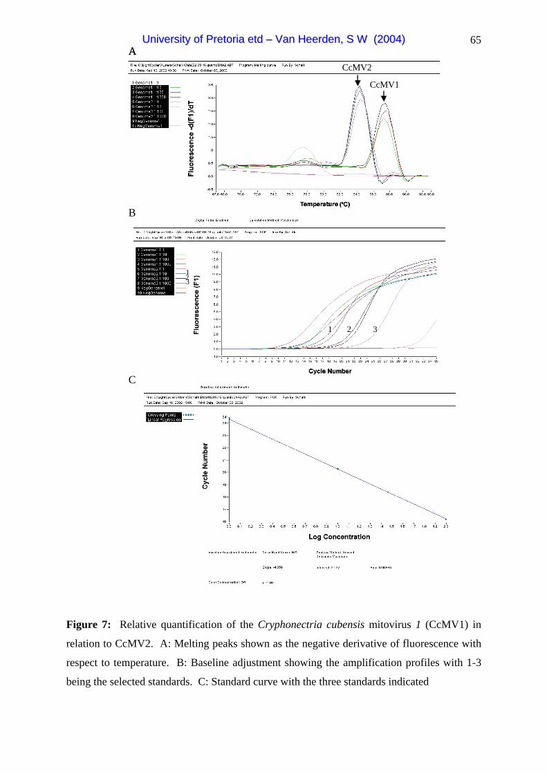
65
31 2
123
CcMV2
CcMV1
A
B
C
31 2
123
31 2
123
CcMV2
CcMV1
A
B
C
Figure 7: Relative quantification of the Cryphonectria cubensis mitovirus 1 (CcMV1) in
relation to CcMV2. A: Melting peaks shown as the negative derivative of fluorescence with
respect to temperature. B: Baseline adjustment showing the amplification profiles with 1-3
being the selected standards. C: Standard curve with the three standards indicated
UUnniivveerrssiittyy ooff PPrreettoorriiaa eettdd –– VVaann HHeeeerrddeenn,, SS WW ((22000044))

66
1 2 3 4 5 6 7 8 9 10 11 12 13 14 15
215 bp126 bp
1 2 3 4 5 6 7 8 9 10 11 12 13 14 151 2 3 4 5 6 7 8 9 10 11 12 13 14 15
215 bp126 bp
Figure 8: Confirmation of the transmission of the mitoviruses to the conidia using RT-PCR.
The PCR products were separated on a 1.5% agarose gel stained with ethidium bromide.
Lane 1: 100 bp molecular weight marker. Lanes 2-13: Mass conidial cultures from isolate
CMW11329. Lane 14: Wild type CRYCMW11329 isolate included as positive control.
Lane 15: Negative control. The presence of the viruses is indicated by a 215 bp amplicon for
CcMV1 and a 126 bp amplicon for CcMV2.
UUnniivveerrssiittyy ooff PPrreettoorriiaa eettdd –– VVaann HHeeeerrddeenn,, SS WW ((22000044))

67
Appendix 1: Aligned RNA sequence of the Cryphonectria cubensis mitovirus 1 using the
CLUSTAL X program. The direction is 5' to 3'. The virus has different sequences at the 5'
end. Symbols below the sequence alignment: (*) indicates identical nucleotides
CLUSUAL X (1.81) multiple sequence alignment 1 60 CcMV1b CUUUUUUUUUUUUUUUUUCUUUCUUCUUUUUUUUUUUUUUCUUUUUUUUCCCCCCUUGCC CcMV1c ------------------------------------------------------------ CcMV1a ----------------------------------------------CUUAGCCCCUUGCC 120CcMV1b CCGGUUUCCCGGGGAAGGCAACGGAGAAGGGGUGACAGGACCCGAAAGGCCCCACUUGUC CcMV1c ----------------------------------------CUUUUCCUUUUUCUUUUUUU CcMV1a CCGGUUUCCCGGGGAAGGCAACGGAGAAGGGGUGACAGGACCCGAAAGGCCCCACUUGUC * * ** * 180CcMV1b AAUCAUCCGUUCCGUGUCCACUAGCUGCUGACUGUUACUGACUAGCCUUGUAACAACAAA CcMV1c UUUUAUCCGUUCCGUGUCCACUAGCUGCUGACUGUUACUGACUAGCCUUGUAACAACAAA CcMV1a AAUCAUCCGUUCCGUGUCCACUAGCUGCUGACUGUUACUGACUAGCCUUGUAACAACAAA * ******************************************************** 240CcMV1b GGGAUAACCAAAGCUGACCACUACUCAAGUGCGAGCAGCGACCCGCCAAGGUGAACCGAU CcMV1c GGGAUAACCAAAGCUGACCACUACUCAAGUGCGAGCAGCGACCCGCCAAGGUGAACCGAU CcMV1a GGGAUAACCAAAGCUGACCACUACUCAAGUGCGAGCAGCGACCCGCCAAGGUGAACCGAU ************************************************************ 300CcMV1b UGCUGUCAUAAUACUAGGCAGCACCAGCCCACGUUUGCUAUAAAAUGAAUUAUAACAAAA CcMV1c UGCUGUCAUAAUACUAGGCAGCACCAGCCCACGUUUGCUAUAAAAUGAAUUAUAACAAAA CcMV1a UGCUGUCAUAAUACUAGGCAGCACCAGCCCACGUUUGCUAUAAAAUGAAUUAUAACAAAA ************************************************************ 360CcMV1b ACCAUGAAAAGAAAUCUCAAUAAACUCUAUAUACACCUAAAUCUCUUACGAGACAUCGGU CcMV1c ACCAUGAAAAGAAAUCUCAAUAAACUCUAUAUACACCUAAAUCUCUUACGAGACAUCGGU CcMV1a ACCAUGAAAAGAAAUCUCAAUAAACUCUAUAUACACCUAAAUCUCUUACGAGACAUCGGU ************************************************************ 420CcMV1b GCAAUAAAAUUGAGUCUAGUGUGAUUCCUUCACUGAUCCCACCUUGAGCUAGGUAUAGCG CcMV1c GCAAUAAAAUUGAGUCUAGUGUGAUUCCUUCACUGAUCCCACCUUGAGCUAGGUAUAGCG CcMV1a GCAAUAAAAUUGAGUCUAGUGUGAUUCCUUCACUGAUCCCACCUUGAGCUAGGUAUAGCG ************************************************************ 480CcMV1b UCACGUCUCGUCUCUUCCGCGGUAGCCAGAUUUCAACUUCUGGCCACCACGAGAGGGCGG CcMV1c UCACGUCUCGUCUCUUCCGCGGUAGCCAGAUUUCAACUUCUGGCCACCACGAGAGGGCGG CcMV1a UCACGUCUCGUCUCUUCCGCGGUAGCCAGAUUUCAACUUCUGGCCACCACGAGAGGGCGG ************************************************************ 540CcMV1b AACGCCGCUAUAUCCGAGUUUAAGGCGUCGCGGCUAGCGUUUACACGAUGACUCUGUGGG CcMV1c AACGCCGCUAUAUCCGAGUUUAAGGCGUCGCGGCUAGCGUUUACACGAUGACUCUGUGGG CcMV1a AACGCCGCUAUAUCCGAGUUUAAGGCGUCGCGGCUAGCGUUUACACGAUGACUCUGUGGG ************************************************************ 600CcMV1b CGGCCGCUCUCAGGUAAAGUUGGUGCCCCGAUAACAAAAGCUGGGCUUCCUAAGGUAAUC CcMV1c CGGCCGCUCUCAGGUAAAGUUGGUGCCCCGAUAACAAAAGCUGGGCUUCCUAAGGUAAUC CcMV1a CGGCCGCUCUCAGGUAAAGUUGGUGCCCCGAUAACAAAAGCUGGGCUUCCUAAGGUAAUC ************************************************************ 660CcMV1b CCUAGGGAGGCCAGACUUUUGUUGCUUCGGGAACGCCCUCUUUACUUGGUAAAGGCCGUC CcMV1c CCUAGGGAGGCCAGACUUUUGUUGCUUCGGGAACGCCCUCUUUACUUGGUAAAGGCCGUC CcMV1a CCUAGGGAGGCCAGACUUUUGUUGCUUCGGGAACGCCCUCUUUACUUGGUAAAGGCCGUC ************************************************************
UUnniivveerrssiittyy ooff PPrreettoorriiaa eettdd –– VVaann HHeeeerrddeenn,, SS WW ((22000044))
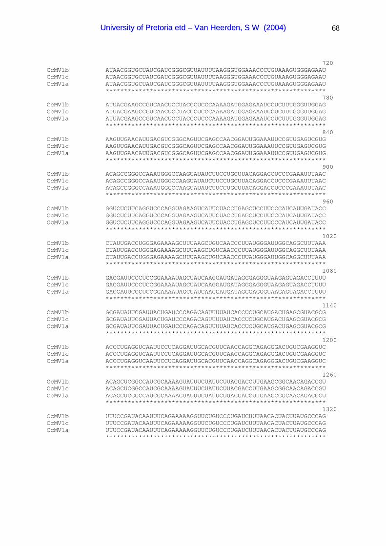
68
720CcMV1b AUAACGGUGCUAUCGAUCGGGCGUUAUUUUAAGGGUGGAAACCCUGUAAAGUGGGAGAAU CcMV1c AUAACGGUGCUAUCGAUCGGGCGUUAUUUUAAGGGUGGAAACCCUGUAAAGUGGGAGAAU CcMV1a AUAACGGUGCUAUCGAUCGGGCGUUAUUUUAAGGGUGGAAACCCUGUAAAGUGGGAGAAU ************************************************************ 780CcMV1b AUUACGAAGCCGUCAACUCCUACCCUCCCAAAAGAUGGAGAAAUCCUCUUUGGGUUGGAG CcMV1c AUUACGAAGCCGUCAACUCCUACCCUCCCAAAAGAUGGAGAAAUCCUCUUUGGGUUGGAG CcMV1a AUUACGAAGCCGUCAACUCCUACCCUCCCAAAAGAUGGAGAAAUCCUCUUUGGGUUGGAG ************************************************************ 840CcMV1b AAGUUGAACAUUGACGUCGGGCAGUUCGAGCCAACGGAUUGGAAAUUCCGUUGAGUCGUG CcMV1c AAGUUGAACAUUGACGUCGGGCAGUUCGAGCCAACGGAUUGGAAAUUCCGUUGAGUCGUG CcMV1a AAGUUGAACAUUGACGUCGGGCAGUUCGAGCCAACGGAUUGGAAAUUCCGUUGAGUCGUG ************************************************************ 900CcMV1b ACAGCCGGGCCAAAUGGGCCAAGUAUAUCUUCCUGCUUACAGGACCUCCCGAAAUUUAAC CcMV1c ACAGCCGGGCCAAAUGGGCCAAGUAUAUCUUCCUGCUUACAGGACCUCCCGAAAUUUAAC CcMV1a ACAGCCGGGCCAAAUGGGCCAAGUAUAUCUUCCUGCUUACAGGACCUCCCGAAAUUUAAC ************************************************************ 960CcMV1b GGUCUCUUCAGGUCCCAGGUAGAAGUCAUUCUACCUGAGCUCCUUCCCAUCAUUGAUACC CcMV1c GGUCUCUUCAGGUCCCAGGUAGAAGUCAUUCUACCUGAGCUCCUUCCCAUCAUUGAUACC CcMV1a GGUCUCUUCAGGUCCCAGGUAGAAGUCAUUCUACCUGAGCUCCUUCCCAUCAUUGAUACC ************************************************************ 102CcMV1b CUAUUGACCUGGGAGAAAAGCUUUAAGCUGUCAACCCUUAUGGGAUUGGCAGGCUUUAAA CcMV1c CUAUUGACCUGGGAGAAAAGCUUUAAGCUGUCAACCCUUAUGGGAUUGGCAGGCUUUAAA CcMV1a CUAUUGACCUGGGAGAAAAGCUUUAAGCUGUCAACCCUUAUGGGAUUGGCAGGCUUUAAA ************************************************************ 108CcMV1b GACGAUUCCCUCCGGAAAAUAGCUAUCAAGGAUGAUAGGGAGGGUAAGAGUAGACCUUUU CcMV1c GACGAUUCCCUCCGGAAAAUAGCUAUCAAGGAUGAUAGGGAGGGUAAGAGUAGACCUUUU CcMV1a GACGAUUCCCUCCGGAAAAUAGCUAUCAAGGAUGAUAGGGAGGGUAAGAGUAGACCUUUU ************************************************************ 114CcMV1b GCGAUAUUCGAUUACUGAUCCCAGACAGUUUUAUCACCUCUGCAUGACUGAGCGUACGCG CcMV1c GCGAUAUUCGAUUACUGAUCCCAGACAGUUUUAUCACCUCUGCAUGACUGAGCGUACGCG CcMV1a GCGAUAUUCGAUUACUGAUCCCAGACAGUUUUAUCACCUCUGCAUGACUGAGCGUACGCG ************************************************************ 120CcMV1b ACCCUGAGGUCAAUUCCUCAGGAUUGCACGUUCAACCAGGCAGAGGGACUGUCGAAGGUC CcMV1c ACCCUGAGGUCAAUUCCUCAGGAUUGCACGUUCAACCAGGCAGAGGGACUGUCGAAGGUC CcMV1a ACCCUGAGGUCAAUUCCUCAGGAUUGCACGUUCAACCAGGCAGAGGGACUGUCGAAGGUC ************************************************************ 126CcMV1b ACAGCUCGGCCAUCGCAAAAGUAUUUCUAUUCUUACGACCUUGAAGCGGCAACAGACCGU CcMV1c ACAGCUCGGCCAUCGCAAAAGUAUUUCUAUUCUUACGACCUUGAAGCGGCAACAGACCGU CcMV1a ACAGCUCGGCCAUCGCAAAAGUAUUUCUAUUCUUACGACCUUGAAGCGGCAACAGACCGU ************************************************************ 132CcMV1b UUUCCGAUACAAUUUCAGAAAAAGGUUCUGUCCCUGAUCUUUAACACUACUUAUGCCCAG CcMV1c UUUCCGAUACAAUUUCAGAAAAAGGUUCUGUCCCUGAUCUUUAACACUACUUAUGCCCAG CcMV1a UUUCCGAUACAAUUUCAGAAAAAGGUUCUGUCCCUGAUCUUUAACACUACUUAUGCCCAG ************************************************************
0
0
0
0
0
0
UUnniivveerrssiittyy ooff PPrreettoorriiaa eettdd –– VVaann HHeeeerrddeenn,, SS WW ((22000044))

69
138CcMV1b GCGUGAGCUGAGAUAAUGACUCAAGAGCCUUUUAGAGUCAAGGGAUUGUCCGACCCCUUA CcMV1c GCGUGAGCUGAGAUAAUGACUCAAGAGCCUUUUAGAGUCAAGGGAUUGUCCGACCCCUUA CcMV1a GCGUGAGCUGAGAUAAUGACUCAAGAGCCUUUUAGAGUCAAGGGAUUGUCCGACCCCUUA ************************************************************ 144CcMV1b AGAUGAGGAGCCGGGCAGCCUCUUGGAGCUAAAAGUUCCUGAGCCAUUUUCACAUUAUGC CcMV1c AGAUGAGGAGCCGGGCAGCCUCUUGGAGCUAAAAGUUCCUGAGCCAUUUUCACAUUAUGC CcMV1a AGAUGAGGAGCCGGGCAGCCUCUUGGAGCUAAAAGUUCCUGAGCCAUUUUCACAUUAUGC ************************************************************ 150CcMV1b CACCACCUAGUAGUUCAUAUAGCAGCGGUACGGACUAACUCCGACCCCUACUACGUGAUA CcMV1c CACCACCUAGUAGUUCAUAUAGCAGCGGUACGGACUAACUCCGACCCCUACUACGUGAUA CcMV1a CACCACCUAGUAGUUCAUAUAGCAGCGGUACGGACUAACUCCGACCCCUACUACGUGAUA ************************************************************ 156CcMV1b CUAGGCGAUGACAUAGUGCUCCGUGGCUCACGGCUGGCGACAGUGUACAAACGGAUAAUG CcMV1c CUAGGCGAUGACAUAGUGCUCCGUGGCUCACGGCUGGCGACAGUGUACAAACGGAUAAUG CcMV1a CUAGGCGAUGACAUAGUGCUCCGUGGCUCACGGCUGGCGACAGUGUACAAACGGAUAAUG ************************************************************ 162CcMV1b UCCGAACUUGGAGUAUCCAUAUCCGAAACGAAAUCGCACGUGUCAAAAGACACGUUCGAA CcMV1c UCCGAACUUGGAGUAUCCAUAUCCGAAACGAAAUCGCACGUGUCAAAAGACACGUUCGAA CcMV1a UCCGAACUUGGAGUAUCCAUAUCCGAAACGAAAUCGCACGUGUCAAAAGACACGUUCGAA ************************************************************ 168CcMV1b UUCGCUAAGAUGUGAAUGCACCAAGGUAGGAACGCGAGUGGGUUCCCUGUAGUGGGACUA CcMV1c UUCGCUAAGAUGUGAAUGCACCAAGGUAGGAACGCGAGUGGGUUCCCUGUAGUGGGACUA CcMV1a UUCGCUAAGAUGUGAAUGCACCAAGGUAGGAACGCGAGUGGGUUCCCUGUAGUGGGACUA ************************************************************ 174CcMV1b GCUGAGACACUCAGAAAGCCACUAGAAAUGGCGGCUCUCUUUGUGUUUGAGCUCCCUGCU CcMV1c GCUGAGACACUCAGAAAGCCACUAGAAAUGGCGGCUCUCUUUGUGUUUGAGCUCCCUGCU CcMV1a GCUGAGACACUCAGAAAGCCACUAGAAAUGGCGGCUCUCUUUGUGUUUGAGCUCCCUGCU ************************************************************ 180CcMV1b AAAGGGUAUCCAGUCACUAUUACUCCGCGCACCGUGUCGCAGUACUUCCUUCUAGUAGCA CcMV1c AAAGGGUAUCCAGUCACUAUUACUCCGCGCACCGUGUCGCAGUACUUCCUUCUAGUAGCA CcMV1a AAAGGGUAUCCAGUCACUAUUACUCCGCGCACCGUGUCGCAGUACUUCCUUCUAGUAGCA ************************************************************ 186CcMV1b CGUUAUAAUACCAUUCCACCUCGUCUGGCGGUAUGAACUGCCGACAAAGUGGUAUGGUAC CcMV1c CGUUAUAAUACCAUUCCACCUCGUCUGGCGGUAUGAACUGCCGACAAAGUGGUAUGGUAC CcMV1a CGUUAUAAUACCAUUCCACCUCGUCUGGCGGUAUGAACUGCCGACAAAGUGGUAUGGUAC ************************************************************ 192CcMV1b UAUAGUUUCUUGUCAUGACUGGCCACCCGGGAUGACGGAUGGGCGAAAUAUAUCGCCCAG CcMV1c UAUAGUUUCUUGUCAUGACUGGCCACCCGGGAUGACGGAUGGGCGAAAUAUAUCGCCCAG CcMV1a UAUAGUUUCUUGUCAUGACUGGCCACCCGGGAUGACGGAUGGGCGAAAUAUAUCGCCCAG ************************************************************ 198CcMV1b UCGGCCUCCCUAUUGGUGAGCCCCAAUACCGCCCACGACCUCCUUAUGAAGGCCGUGAGA CcMV1c UCGGCCUCCCUAUUGGUGAGCCCCAAUACCGCCCACGACCUCCUUAUGAAGGCCGUGAGA CcMV1a UCGGCCUCCCUAUUGGUGAGCCCCAAUACCGCCCACGACCUCCUUAUGAAGGCCGUGAGA ************************************************************
0
0
0
0
0
0
0
0
0
0
0
UUnniivveerrssiittyy ooff PPrreettoorriiaa eettdd –– VVaann HHeeeerrddeenn,, SS WW ((22000044))

70
204CcMV1b GAUAAAUGGGCUAAACAGCUUGACAAGUCUCUAAUGGAUUUCCAGGAUUUUGGAUUUGAU CcMV1c GAUAAAUGGGCUAAACAGCUUGACAAGUCUCUAAUGGAUUUCCAGGAUUUUGGAUUUGAU CcMV1a GAUAAAUGGGCUAAACAGCUUGACAAGUCUCUAAUGGAUUUCCAGGAUUUUGGAUUUGAU ************************************************************ 210CcMV1b AUGUUCGACAAAGUGAAGACCUUGCCGCCAUUCAAGCCUACCUGGGAUCCAGAAGCGUGA CcMV1c AUGUUCGACAAAGUGAAGACCUUGCCGCCAUUCAAGCCUACCUGGGAUCCAGAAGCGUGA CcMV1a AUGUUCGACAAAGUGAAGACCUUGCCGCCAUUCAAGCCUACCUGGGAUCCAGAAGCGUGA ************************************************************ 216CcMV1b CCUGGUCGGUUGUCUGCGAGUGGAAUCGAAUUCAAUCCAGCCCCUAGGAAGGUACCAAUA CcMV1c CCUGGUCGGUUGUCUGCGAGUGGAAUCGAAUUCAAUCCAGCCCCUAGGAAGGUACCAAUA CcMV1a CCUGGUCGGUUGUCUGCGAGUGGAAUCGAAUUCAAUCCAGCCCCUAGGAAGGUACCAAUA ************************************************************ 222CcMV1b UUUGCUGCUCUCCAAGAAGAAGGAGAGAUCAAGUAUUCCGAUUAUCUCCAACAGAAGUUG CcMV1c UUUGCUGCUCUCCAAGAAGAAGGAGAGAUCAAGUAUUCCGAUUAUCUCCAACAGAAGUUG CcMV1a UUUGCUGCUCUCCAAGAAGAAGGAGAGAUCAAGUAUUCCGAUUAUCUCCAACAGAAGUUG ************************************************************ 228CcMV1b GAGAUGACCGACGAUCAGCUGACGUUCGAGGAGAUAGAAUCUUUGAAACUACCUCCUCGU CcMV1c GAGAUGACCGACGAUCAGCUGACGUUCGAGGAGAUAGAAUCUUUGAAACUACCUCCUCGU CcMV1a GAGAUGACCGACGAUCAGCUGACGUUCGAGGAGAUAGAAUCUUUGAAACUACCUCCUCGU ************************************************************ 234CcMV1b CCGCAACUGAAAGGGUUCCUUCCGAAAAGGACUAGAGAGUAUGUUCGUACACUUAACCUA CcMV1c CCGCAACUGAAAGGGUUCCUUCCGAAAAGGACUAGAGAGUAUGUUCGUACACUUAACCUA CcMV1a CCGCAACUGAAAGGGUUCCUUCCGAAAAGGACUAGAGAGUAUGUUCGUACACUUAACCUA ************************************************************ 240CcMV1b AUUAGUCACGGCUUAAACCGGGAUCUAAGGGCUCAGUGCAUAGGAACCAGACCAGACGUA CcMV1c AUUAGUCACGGCUUAAACCGGGAUCUAAGGGCUCAGUGCAUAGGAACCAGACCAGACGUA CcMV1a AUUAGUCACGGCUUAAACCGGGAUCUAAGGGCUCAGUGCAUAGGAACCAGACCAGACGUA ************************************************************ 246CcMV1b UACAAUAUGAAAGACUAAGAGGUUCUCCCUCCCACCUUGAGAAAGUGGUGAAAGCUCUCC CcMV1c UACAAUAUGAAAGACUAAGAGGUUCUCCCUCCCACCUUGAGAAAGUGGUGAAAGCUCUCC CcMV1a UACAAUAUGAAAGACUAAGAGGUUCUCCCUCCCACCUUGAGAAAGUGGUGAAAGCUCUCC ************************************************************ 252CcMV1b UUAGCCCCUACACAUUGUGUACCCCCAUUCCCCUUUCGGGGUAGGCAGAAGUCAACUGCU CcMV1c UUAGCCCCUACACAUUGUGUACCCCCAUUCCCCUUUCGGGGUAGGCAGAAGUCAACUGCU CcMV1a UUAGCCCCUACACAUUGUGUACCCCCAUUCCCCUUUCGGGGUAGGCAGAAGUCAACUGCU ************************************************************ 258CcMV1b UGCUCCGGCAGGGAUGCCGGAUGGGAAGUCGACAGCGCUGGGGCAAAAAAAAAAAAAAAA CcMV1c UGCUCCGGCAGGGAUGCCGGAUGGGAAGUCGACAGCGCUGGGGCAAAAAAAAAAAAAAAA CcMV1a UGCUCCGGCAGGGAUGCCGGAUGGGAAGUCGACAGCGCUGGGGCAAAAAAAAAAAAAAAA ************************************************************ 2601 CcMV1b AAAAGAAAAAAAAAAGAAAAA CcMV1c AAAAGAAAAAAAAAAGAAAAA CcMV1a AAAAGAAAAAAAAAAGAAAAA *********************
0
0
0
0
0
0
0
0
0
0
UUnniivveerrssiittyy ooff PPrreettoorriiaa eettdd –– VVaann HHeeeerrddeenn,, SS WW ((22000044))

71
Appendix 2: Aligned RNA sequence of the Cryphonectria cubensis mitovirus 2 using the
CLUSTAL X program. The direction is 5' to 3'. The virus has different ends at the 3' end.
Symbol below the sequence alignment where (*) indicates identical nucleotides.
CLUSTAL X (1.81) multiple sequence alignment 1 60 CcMV2b CUUUUUUUGUUUUUUUUUUCUUUUCUUUGGGGUAAUGACUCUUCAAGUCACUUGACAGUC CcMV2a CUUUUUUUGUUUUUUUUUUCUUUUCUUUGGGGUAAUGACUCUUCAAGUCACUUGACAGUC CcMV2c CUUUUUUUGUUUUUUUUUUCUUUUCUUUGGGGUAAUGACUCUUCAAGUCACUUGACAGUC ************************************************************ 120CcMV2b AUAAUGCCUAAACCCCAGUCAGUCUCUCUUGCCUUCACAGGUGAGGUGUGCUUUCGGCUA CcMV2a AUAAUGCCUAAACCCCAGUCAGUCUCUCUUGCCUUCACAGGUGAGGUGUGCUUUCGGCUA CcMV2c AUAAUGCCUAAACCCCAGUCAGUCUCUCUUGCCUUCACAGGUGAGGUGUGCUUUCGGCUA ************************************************************ 180CcMV2b UAAUGGUACUUUCCAUUCGGACAGUAGACGAGUGUAAUGAGAUAACAUCAAAAUUCGAAU CcMV2a UAAUGGUACUUUCCAUUCGGACAGUAGACGAGUGUAAUGAGAUAACAUCAAAAUUCGAAU CcMV2c UAAUGGUACUUUCCAUUCGGACAGUAGACGAGUGUAAUGAGAUAACAUCAAAAUUCGAAU ************************************************************ 240CcMV2b UGCACGGGCUAGCGACCCAACGUAAUAGUAAAUCACUCCAAACUGUAUCGUGAAACAUUU CcMV2a UGCACGGGCUAGCGACCCAACGUAAUAGUAAAUCACUCCAAACUGUAUCGUGAAACAUUU CcMV2c UGCACGGGCUAGCGACCCAACGUAAUAGUAAAUCACUCCAAACUGUAUCGUGAAACAUUU ************************************************************ 300CcMV2b GCAUUAAUUAAUUAACACAAAGCUACAACAACACAUUCAAAAUUAUACUCUAAACUUAAU CcMV2a GCAUUAAUUAAUUAACACAAAGCUACAACAACACAUUCAAAAUUAUACUCUAAACUUAAU CcMV2c GCAUUAAUUAAUUAACACAAAGCUACAACAACACAUUCAAAAUUAUACUCUAAACUUAAU ************************************************************ 360CcMV2b GCAAAAUUGAGUCUAAUAAGAAGUAACUAUUUUUCUUUGUUACUUUUAUUAAAAUCAUUC CcMV2a GCAAAAUUGAGUCUAAUAAGAAGUAACUAUUUUUCUUUGUUACUUUUAUUAAAAUCAUUC CcMV2c GCAAAAUUGAGUCUAAUAAGAAGUAACUAUUUUUCUUUGUUACUUUUAUUAAAAUCAUUC ************************************************************ 420CcMV2b UUCAUCAAGCCUAAAAGUGUUAAUGUCAAACGUGAUGGUAAGAGCUGGAUCUCCCAAAAG CcMV2a UUCAUCAAGCCUAAAAGUGUUAAUGUCAAACGUGAUGGUAAGAGCUGGAUCUCCCAAAAG CcMV2c UUCAUCAAGCCUAAAAGUGUUAAUGUCAAACGUGAUGGUAAGAGCUGGAUCUCCCAAAAG ************************************************************ 480CcMV2b GAGAUUCCAAAAUUUAUCAUCAUGGUUUGUUGGUGUACCGGUACACAGUCCUAUCAAAGA CcMV2a GAGAUUCCAAAAUUUAUCAUCAUGGUUUGUUGGUGUACCGGUACACAGUCCUAUCAAAGA CcMV2c GAGAUUCCAAAAUUUAUCAUCAUGGUUUGUUGGUGUACCGGUACACAGUCCUAUCAAAGA ************************************************************ 540CcMV2b UCUUUUAUGAAGAUGCAGGAUAGAUUUGUCUUUAUCUGAAAUACUGCUGGCUCUACAUUC CcMV2a UCUUUUAUGAAGAUGCAGGAUAGAUUUGUCUUUAUCUGAAAUACUGCUGGCUCUACAUUC CcMV2c UCUUUUAUGAAGAUGCAGGAUAGAUUUGUCUUUAUCUGAAAUACUGCUGGCUCUACAUUC ************************************************************ 600CcMV2b CUCUUUAAGUAUCUUAAAGAGGUAAUGCGUCUUACAGUAAGAAGAUUGGCGAAUAUAGAC CcMV2a CUCUUUAAGUAUCUUAAAGAGGUAAUGCGUCUUACAGUAAGAAGAUUGGCGAAUAUAGAC CcMV2c CUCUUUAAGUAUCUUAAAGAGGUAAUGCGUCUUACAGUAAGAAGAUUGGCGAAUAUAGAC ************************************************************ 660CcMV2b CUUAUUCCUAGUAAAGAAAAUCUUUGUUAAAUUAAACAAAUAUAGAUUUCCUGCUAUUAU CcMV2a CUUAUUCCUAGUAAAGAAAAUCUUUGUUAAAUUAAACAAAUAUAGAUUUCCUGCUAUUAU CcMV2c CUUAUUCCUAGUAAAGAAAAUCUUUGUUAAAUUAAACAAAUAUAGAUUUCCUGCUAUUAU ************************************************************
UUnniivveerrssiittyy ooff PPrreettoorriiaa eettdd –– VVaann HHeeeerrddeenn,, SS WW ((22000044))

72
720CcMV2b UCCUCUAGAGAUUUGCAAGGAUCUUUCCUGGUUUCCAGGGAGAUUCCAUGUUUUGUCUCG CcMV2a UCCUCUAGAGAUUUGCAAGGAUCUUUCCUGGUUUCCAGGGAGAUUCCAUGUUUUGUCUCG CcMV2c UCCUCUAGAGAUUUGCAAGGAUCUUUCCUGGUUUCCAGGGAGAUUCCAUGUUUUGUCUCG ************************************************************ 780CcMV2b AAAGGUAAUAGCUCUCCUUACCGUUAUUUCGAUUUUCAGGGUUUUACCUACUAAGGUAUU CcMV2a AAAGGUAAUAGCUCUCCUUACCGUUAUUUCGAUUUUCAGGGUUUUACCUACUAAGGUAUU CcMV2c AAAGGUAAUAGCUCUCCUUACCGUUAUUUCGAUUUUCAGGGUUUUACCUACUAAGGUAUU ************************************************************ 840CcMV2b GCCUGAUUACACGACUAUCAGUAAGCCCCAUUCGGGACUUAUUGAAACGUGUUCAUCAGA CcMV2a GCCUGAUUACACGACUAUCAGUAAGCCCCAUUCGGGACUUAUUGAAACGUGUUCAUCAGA CcMV2c GCCUGAUUACACGACUAUCAGUAAGCCCCAUUCGGGACUUAUUGAAACGUGUUCAUCAGA ************************************************************ 900CcMV2b GUCAAUUGCUUUAGCCUGUAAAAAUCUUGAGAUCAAAAGAGUUGAUAAGGUGAUGAAGCU CcMV2a GUCAAUUGCUUUAGCCUGUAAAAAUCUUGAGAUCAAAAGAGUUGAUAAGGUGAUGAAGCU CcMV2c GUCAAUUGCUUUAGCCUGUAAAAAUCUUGAGAUCAAAAGAGUUGAUAAGGUGAUGAAGCU ************************************************************ 960CcMV2b AAGAUUGAAAGGUUCUCUAAAAGCCGGACCGAAUGGUAAAAUAUCAUUACUUACUUCGUU CcMV2a AAGAUUGAAAGGUUCUCUAAAAGCCGGACCGAAUGGUAAAAUAUCAUUACUUACUUCGUU CcMV2c AAGAUUGAAAGGUUCUCUAAAAGCCGGACCGAAUGGUAAAAUAUCAUUACUUACUUCGUU ************************************************************ 102CcMV2b AUUAGAUGCUCUGGCUUUUUGGUCAGAUCCUUUAAGGGUAAUCCACUUCAUCUGAUUUAA CcMV2a AUUAGAUGCUCUGGCUUUUUGGUCAGAUCCUUUAAGGGUAAUCCACUUCAUCUGAUUUAA CcMV2c AUUAGAUGCUCUGGCUUUUUGGUCAGAUCCUUUAAGGGUAAUCCACUUCAUCUGAUUUAA ************************************************************ 108CcMV2b UAUCAGGUGUUAUGGUUACUUUUGGGGACUAAUAUGAAGUAUGUGAUUGAUUUUUAUCAU CcMV2a UAUCAGGUGUUAUGGUUACUUUUGGGGACUAAUAUGAAGUAUGUGAUUGAUUUUUAUCAU CcMV2c UAUCAGGUGUUAUGGUUACUUUUGGGGACUAAUAUGAAGUAUGUGAUUGAUUUUUAUCAU ************************************************************ 114CcMV2b GAUCAUUUCCUUACCGUAUUACCUUAUAGCAUUGUGUCUUGGUGCGAGAGCCCCAGUAAU CcMV2a GAUCAUUUCCUUACCGUAUUACCUUAUAGCAUUGUGUCUUGGUGCGAGAGCCCCAGUAAU CcMV2c GAUCAUUUCCUUACCGUAUUACCUUAUAGCAUUGUGUCUUGGUGCGAGAGCCCCAGUAAU ************************************************************ 120CcMV2b GGGUCAAUUGGCAACUGUUUAUGAUCAAGCUGGAAAAGCGAGAAUUGUAGCUUCUACAAA CcMV2a GGGUCAAUUGGCAACUGUUUAUGAUCAAGCUGGAAAAGCGAGAAUUGUAGCUUCUACAAA CcMV2c GGGUCAAUUGGCAACUGUUUAUGAUCAAGCUGGAAAAGCGAGAAUUGUAGCUUCUACAAA ************************************************************ 126CcMV2b CUCGUGGAUUCAGUGUUCUCUCUUUGGUUUACACAAUAAGAUUUUUUCUAUCUUACGGAG CcMV2a CUCGUGGAUUCAGUGUUCUCUCUUUGGUUUACACAAUAAGAUUUUUUCUAUCUUACGGAG CcMV2c CUCGUGGAUUCAGUGUUCUCUCUUUGGUUUACACAAUAAGAUUUUUUCUAUCUUACGGAG ************************************************************ 132CcMV2b UAUUCCUCAAGAUGGAACUUUUGAUCAAAACAAGCCUUUUGAUUUAUUAUUGGAGUCUCU CcMV2a UAUUCCUCAAGAUGGAACUUUUGAUCAAAACAAGCCUUUUGAUUUAUUAUUGGAGUCUCU CcMV2c UAUUCCUCAAGAUGGAACUUUUGAUCAAAACAAGCCUUUUGAUUUAUUAUUGGAGUCUCU ************************************************************
0
0
0
0
0
0
UUnniivveerrssiittyy ooff PPrreettoorriiaa eettdd –– VVaann HHeeeerrddeenn,, SS WW ((22000044))

73
138CcMV2b UCAGCCAGGGUACAUGUUAUAUGGUUUCGACCUGAGUGCAGCGACAGAUAGACUUCCUAU CcMV2a UCAGCCAGGGUACAUGUUAUAUGGUUUCGACCUGAGUGCAGCGACAGAUAGACUUCCUAU CcMV2c UCAGCCAGGGUACAUGUUAUAUGGUUUCGACCUGAGUGCAGCGACAGAUAGACUUCCUAU ************************************************************ 144CcMV2b UGCAUUCCAAAAGGAUAUCCUAAAUCAUUUAGGGUAUCCCGGAGGUCCUUGAAGAAGGUU CcMV2a UGCAUUCCAAAAGGAUAUCCUAAAUCAUUUAGGGUAUCCCGGAGGUCCUUGAAGAAGGUU CcMV2c UGCAUUCCAAAAGGAUAUCCUAAAUCAUUUAGGGUAUCCCGGAGGUCCUUGAAGAAGGUU ************************************************************ 150CcMV2b ACUGGGUAUCAAAUAUAAUUCACCUUGUGGAUUUAUUUCUUACGCAGUUGGCCAACCAAU CcMV2a ACUGGGUAUCAAAUAUAAUUCACCUUGUGGAUUUAUUUCUUACGCAGUUGGCCAACCAAU CcMV2c ACUGGGUAUCAAAUAUAAUUCACCUUGUGGAUUUAUUUCUUACGCAGUUGGCCAACCAAU ************************************************************ 156CcMV2b GGGUGCAUAUUCUUCCUUUGCAAUGCUUGCUCUUACACAUCACGUGUUGGUUCAAGUAGC CcMV2a GGGUGCAUAUUCUUCCUUUGCAAUGCUUGCUCUUACACAUCACGUGUUGGUUCAAGUAGC CcMV2c GGGUGCAUAUUCUUCCUUUGCAAUGCUUGCUCUUACACAUCACGUGUUGGUUCAAGUAGC ************************************************************ 162CcMV2b CGCCCAAAAGGCGGGUUUCUCAGACCGUUUCACGGACUACUGUAUUCUUGGUGACGAUAU CcMV2a CGCCCAAAAGGCGGGUUUCUCAGACCGUUUCACGGACUACUGUAUUCUUGGUGACGAUAU CcMV2c CGCCCAAAAGGCGGGUUUCUCAGACCGUUUCACGGACUACUGUAUUCUUGGUGACGAUAU ************************************************************ 168CcMV2b CGUCAUAGCCAACUCUUUAGUUGCUGAAGCUUAUAAGUCCUUAAUCUUUGAUUUAGGCUU CcMV2a CGUCAUAGCCAACUCUUUAGUUGCUGAAGCUUAUAAGUCCUUAAUCUUUGAUUUAGGCUU CcMV2c CGUCAUAGCCAACUCUUUAGUUGCUGAAGCUUAUAAGUCCUUAAUCUUUGAUUUAGGCUU ************************************************************ 174CcMV2b AGAAAUUUCAGAAUCUAAGAGUGUUAUUUCCGGAACAUUUACCGAAUUCGCAAAGAAGUU CcMV2a AGAAAUUUCAGAAUCUAAGAGUGUUAUUUCCGGAACAUUUACCGAAUUCGCAAAGAAGUU CcMV2c AGAAAUUUCAGAAUCUAAGAGUGUUAUUUCCGGAACAUUUACCGAAUUCGCAAAGAAGUU ************************************************************ 180CcMV2b GAGAGGUCCACUUAUGGAUAUCUCACCUAUCGGAGCGGGUUUGAUAUUAUAUUCCUUACG CcMV2a GAGAGGUCCACUUAUGGAUAUCUCACCUAUCGGAGCGGGUUUGAUAUUAUAUUCCUUACG CcMV2c GAGAGGUCCACUUAUGGAUAUCUCACCUAUCGGAGCGGGUUUGAUAUUAUAUUCCUUACG ************************************************************ 186CcMV2b UAACAAGUACUACAUCUGUGUGUUGGUUUUUGAGAUCCUGGAAAGGGGAUUAUGCAUGUG CcMV2a UAACAAGUACUACAUCUGUGUGUUGGUUUUUGAGAUCCUGGAAAGGGGAUUAUGCAUGUG CcMV2c UAACAAGUACUACAUCUGUGUGUUGGUUUUUGAGAUCCUGGAAAGGGGAUUAUGCAUGUG ************************************************************ 192CcMV2b GUAUGACGUCUACCCCCAAUUACUCAGCUUGUUACCUAAGAUUUAUCGUAGGUAUUUCAA CcMV2a GUAUGACGUCUACCCCCAAUUACUCAGCUUGUUACCUAAGAUUUAUCGUAGGUAUUUCAA CcMV2c GUAUGACGUCUACCCCCAAUUACUCAGCUUGUUACCUAAGAUUUAUCGUAGGUAUUUCAA ************************************************************ 198CcMV2b GCUUUGUGAUUGGUUUAUUGCGCUUCACUUGCGUCGGAGAGAGCAUCUGGGUGACCAAGA CcMV2a GCUUUGUGAUUGGUUUAUUGCGCUUCACUUGCGUCGGAGAGAGCAUCUGGGUGACCAAGA CcMV2c GCUUUGUGAUUGGUUUAUUGCGCUUCACUUGCGUCGGAGAGAGCAUCUGGGUGACCAAGA ************************************************************
0
0
0
0
0
0
0
0
0
0
0
UUnniivveerrssiittyy ooff PPrreettoorriiaa eettdd –– VVaann HHeeeerrddeenn,, SS WW ((22000044))

74
204CcMV2b UCAUGAGAUCUUGAAUCCUAGAAUUGCUUACUUCAACGUAUUCUUAAAUAAAGAGAAGAU CcMV2a UCAUGAGAUCUUGAAUCCUAGAAUUGCUUACUUCAACGUAUUCUUAAAUAAAGAGAAGAU CcMV2c UCAUGAGAUCUUGAAUCCUAGAAUUGCUUACUUCAACGUAUUCUUAAAUAAAGAGAAGAU ************************************************************ 210CcMV2b UAUCUCUUUACUUGAGAUUAUGUGAAACUCUACUGUAAGGGAUUGGUUCCGAUUAUGGAA CcMV2a UAUCUCUUUACUUGAGAUUAUGUGAAACUCUACUGUAAGGGAUUGGUUCCGAUUAUGGAA CcMV2c UAUCUCUUUACUUGAGAUUAUGUGAAACUCUACUGUAAGGGAUUGGUUCCGAUUAUGGAA ************************************************************ 216CcMV2b UUCGAUUAAGUAUACUCUAAAUAAGGGUCUAUUUAUAUCUCAAGCCAGAGUCGGUCUUCC CcMV2a UUCGAUUAAGUAUACUCUAAAUAAGGGUCUAUUUAUAUCUCAAGCCAGAGUCGGUCUUCC CcMV2c UUCGAUUAAGUAUACUCUAAAUAAGGGUCUAUUUAUAUCUCAAGCCAGAGUCGGUCUUCC ************************************************************ 222CcMV2b GGACUGAAGUGAAUUGAUCUUCUCAAAAAAAAAAA----AAAAAAAGAGAAAAGGAAAAA CcMV2a GGACUGAAGUGAAUUGAUCUUCUUUCCUUUGUUACCCUCNACUUAUAUUAUGAUCAUGUC CcMV2c GGACUGAAGUGAAUUGAUCUUCUUUCCUUUGUUACCCUCNACUUAUAUUAUGAUCAUGUC *********************** * * * * * * 228CcMV2b AAAAGGAAAGAAAAAAAAAGACAAAAAAAAAAAAACUAAAA------------------- CcMV2a CUACGCAACGUCCCUGAAUGACAUUUCCAAAGCUUUUGGAAAUUGAUGACUUCUCAAUUC CcMV2c CUACGCAACGUCCCUGAAUGACAUUUCCAAAGCUUUUGGAAAUUGAUGACUUCUCAAUUC * * ** * ** **** *** * ** 234CcMV2b ------------------------------------------------------------ CcMV2a UCUCGAGAAGGACCAGAUUAACAUCUUCGAUGUAAUCGCCAUGAUGGAACGUGAGUCAAU CcMV2c UCUCGAGAAGGACCAGAUUAACAUCUUCGAUGUAAUCGCCAUGAUGGAACGUGAGUCAAU 240CcMV2b ------------------------------------------------------------ CcMV2a ACUUGAUCUUGAUAUAAAUGAUAAGAAGAAGGUUAAAUUAUCUUUGGAUAAUCUAUAUAA CcMV2c ACUUGAUCUUGAUAUAAAUGAUAAGAAGAAGGUUAAAUUAUCUUUGGAUAAUCUAUAAAA 246CcMV2b ------------------------------------------------------------ CcMV2a GUUAAAUUCUAUAAUCAAUAGGACUGGAGCAGGGCUGGAAAUCUUUAUAGAUCGUUGGCG CcMV2c AAAACAAAAAAAAAAGAGA----------------------------------------- 252CcMV2b ------------------------------------------------------------ CcMV2a GAAAACUUCAUUCCGUUUAAUGACCUAUAAAAGCCUAACAGCUUCUUCUCUGUCCGAUAU CcMV2c ------------------------------------------------------------ 258CcMV2b ------------------------------------------------------------ CcMV2a ACCCUUUAGCAUUCUUAGUUUUGACGAGAAUGGCAAAUUGUAAUAGAAUCUAACCAACAU CcMV2c ------------------------------------------------------------ 264CcMV2b ----------------------------------------------------------- CcMV2a GGGAUCGAGGUCUACACACUUCAGUGUGCCUUGUGUACCUGAGCUGAUGUUGCUUAGGA CcMV2c -----------------------------------------------------------
0
0
0
0
0
0
0
0
0
0
0
UUnniivveerrssiittyy ooff PPrreettoorriiaa eettdd –– VVaann HHeeeerrddeenn,, SS WW ((22000044))
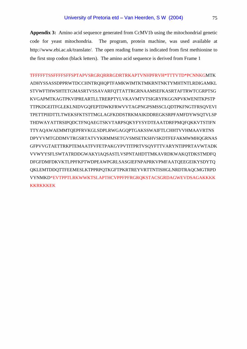
75
Appendix 3: Amino acid sequence generated from CcMV1b using the mitochondrial genetic
code for yeast mitochondria. The program, protein machine, was used available at
http://www.ebi.ac.uk/translate/. The open reading frame is indicated from first methionine to
the first stop codon (black letters). The amino acid sequence is derived from Frame 1
TFFFFFTSSFFFFSFFSPTAPVSRGRQRRRGDRTRKAPTVNHPFRVH*TTTVTD*PCNNKGMTK
ADHYSSASSDPPRWTDCCHNTRQHQPTFAMKWIMTKTMKRNTNKTYMHTNTLRDIGAMKL
STVWFTHWSHTETGMASRTVSSAVARFQTTATTRGRNAAMSEFKASRTAFTRWTCGRPTSG
KVGAPMTKAGTPKVIPREARTLLTRERPTYLVKAVMTVTSIGRYFKGGNPVKWENITKPSTP
TTPKDGEITFGLEKLNIDVGQFEPTDWKFRWVVTAGPNGPSMSSCLQDTPKFNGTFRSQVEVI
TPETTPIIDTTLTWEKSFKTSTTMGLAGFKDDSTRKMAIKDDREGKSRPFAMFDYWSQTVLSP
THDWAYATTRSIPQDCTFNQAEGTSKVTARPSQKYFYSYDTEAATDRFPMQFQKKVTSTIFN
TTYAQAWAEMMTQEPFRVKGLSDPLRWGAGQPTGAKSSWAIFTLCHHTVVHMAAVRTNS
DPYYVMTGDDMVTRGSRTATVYKRMMSETGVSMSETKSHVSKDTFEFAKMWMHQGRNAS
GFPVVGTAETTRKPTEMAATFVFETPAKGYPVTITPRTVSQYFTTVARYNTIPPRTAVWTADK
VVWYYSFLSWTATRDDGWAKYIAQSASTLVSPNTAHDTTMKAVRDKWAKQTDKSTMDFQ
DFGFDMFDKVKTLPPFKPTWDPEAWPGRLSASGIEFNPAPRKVPMFAATQEEGEIKYSDYTQ
QKLEMTDDQTTFEEMESLKTPPRPQTKGFTPKRTREYVRTTNTISHGLNRDTRAQCMGTRPD
VYNMKD*EVTPPTLRKWWKTSLAPTHCVPPFPFRGRQKSTACSGRDAGWEVDSAGAKKKK
KKRKKKEK
UUnniivveerrssiittyy ooff PPrreettoorriiaa eettdd –– VVaann HHeeeerrddeenn,, SS WW ((22000044))

76
Appendix 4: Amino acid sequence generated from CcMV2a using the mitochondrial genetic
code for yeast mitochondria. The coding region is indicated form the first methionine. The
program, protein machine, was used available at http://www.ebi.ac.uk/translate/. The open
reading frame is indicated from first methionine after the stop codons to the first stop codon
(black letters). The amino acid sequence is derived from Frame 2
FFCFFFTFFGVMTTQVTWQS*CTNPSQSTLPSQVRCAFGYNGTFHSDSRRV*WDNIKIRIARAS
DPT***ITPNCIVKHLH*LINTKTQQHIQNYTTNLMQNWV**EVTIFTCYFY*NHSSSSTKVLMS
NVMVRAGSPKRRFQNLSSWFVGVPVHSPIKDTLWRCRMDLSLSEMTTATHSSLSILKR*CVL
Q*EDWRM*TLFTVKKIFVKLNKYRFPAIIPTEICKDTSWFPGRFHVLSRKVMATTTVISIFRVLP
TKVLPDYTTISKPHSGTIETCSSESIALACKNTEIKRVDKVMKTRLKGSTKAGPNGKMSLTTSL
LDATAFWSDPLRVIHFIWFNIRCYGYFWGTMWSMWLIFIMIISLPYYTMALCTGARAPVMGQ
LATVYDQAGKARIVASTNSWIQCSTFGLHNKIFSILRSIPQDGTFDQNKPFDLLLESTQPGYML
YGFDTSAATDRTPIAFQKDITNHLGYPGGPWRRLTGIKYNSPCGFISYAVGQPMGAYSSFAM
TATTHHVLVQVAAQKAGFSDRFTDYCITGDDIVMANSLVAEAYKSLIFDLGLEISESKSVISG
TFTEFAKKLRGPTMDISPIGAGLMLYSLRNKYYICVLVFEITERGLCMWYDVYPQLTSLLPKI
YRRYFKTCDWFIATHLRRREHTGDQDHEILNPRIAYFNVFLNKEKIISLTEIMWNSTVRDWFR
LWNSIKYTTNKGTFMSQARVGTPDWSELIFFPLLPXTYIMIMSYATSTNDISKAFGNWWTTNS
TEKDQINIFDVIAMMERESMTDTDMNDKKKVKLSLDNTYKLNSMINRTGAGTEIFMDRWRK
TSFRLMTYKSTTASSTSDMPFSITSFDENGKL**NTTNMGSRSTHFSVPCVPETMLTRX
UUnniivveerrssiittyy ooff PPrreettoorriiaa eettdd –– VVaann HHeeeerrddeenn,, SS WW ((22000044))
77
Chapter 3:
Relative pathogenicity of Cryphonectria cubensis on
Eucalyptus clones differing in their tolerance to C.
cubensis
ABSTRACT
Cryphonectria cubensis causes a destructive canker disease of Eucalyptus. Management of
this disease is primarily through breeding and selection of disease tolerant trees. One means
of selecting such trees is by artificial inoculation with the pathogen. In routine screening
trials in South Africa, a highly pathogenic isolate of C. cubensis is used for such inoculations.
Although the most tolerant clones under natural conditions are the same as those detected in
inoculation trials, a question has arisen whether all clones respond similarly to different C.
cubensis isolates. Thus, a trial consisting of five clones, known to differ in susceptibility to
infection by C. cubensis was established. These trees were inoculated with nine South
African C. cubensis isolates previously shown to differ in pathogenicity. Inoculations
showed a significant isolate x clone interaction as well as evidence for vertical resistance.
Based on these results disease screening should not be done with a single isolate.
UUnniivveerrssiittyy ooff PPrreettoorriiaa eettdd –– VVaann HHeeeerrddeenn,, SS WW ((22000044))

78
INTRODUCTION
Cryphonectria canker caused by Cryphonectria cubensis (Bruner) Hodges causes a serious
canker disease on Eucalyptus in many tropical and sub-tropical areas of the world (Boerboom
& Maas 1970; Hodges & Reis 1974; Hodges et al. 1979; Gibson 1981; Florence et al. 1986).
Cryphonectria canker was first reported in South Africa in 1989 (Wingfield et al. 1989). This
disease is characterised by swollen basal cankers in South Africa and is also favoured by high
rainfall (2000-2400 mm/ annum) and temperatures above 23ºC (Sharma et al. 1985; Florence
et al. 1986; Wingfield et al. 1989). Since Eucalyptus is one of the major plantation trees in
the country, it has been important to develop effective management to ensure minimal losses
due to Cryphonectria canker.
Various options exist to reduce the impact of Cryphonectria canker. Chemical control has
been considered but due to the low value of individual Eucalyptus trees this is not
economically viable (Sharma et al. 1985). Biological control using hypovirulent strains of
the pathogen is also attractive but more a longer term option (van Heerden et al. 2001).
However, the most commonly used approach is to breed and select disease tolerant
Eucalyptus trees (Alfenas et al. 1983; Wingfield 1990).
Deployment of naturally selected disease tolerant Eucalyptus spp. has reduced losses in
plantations due to C. cubensis (Campinhos & Ikemori 1983; Conradie et al. 1992).
Monoclonal plantations are attractive to forestry companies because of the uniformity of
selected clones and their higher productivity over shorter periods of time. However, the
combination of favorable environmental conditions and the genetic uniformity of these
plantations might lead to substantial losses due to C. cubensis, if clones with poor tolerance
are inadvertently planted. Virtually nothing is known regarding the genetics of susceptibility
of Eucalyptus spp. to infection by C. cubensis. It has, however, been assumed that resistance
to this pathogen is a quantitative trait. This is due to the broad range of susceptibility in
inoculation trials on progeny resulting from a cross between a resistant and susceptible
Eucalyptus clone (van Heerden unpublished). One means to screen trees for disease
tolerance is through artificial inoculation, which reduces the confusion relating to disease
escape in natural infection trials. Thus artificial inoculation has been effective in screening
Eucalyptus trees for tolerance to Cryphonectria canker (Alfenas et al. 1983; van Heerden &
Wingfield 2002).
UUnniivveerrssiittyy ooff PPrreettoorriiaa eettdd –– VVaann HHeeeerrddeenn,, SS WW ((22000044))

79
Routine screening of Eucalyptus grandis clones and hybrids with C. cubensis to identify
disease tolerant planting stock has been conducted in South Africa for several years.
Associated trials such as those assessing the capacity of Eucalyptus clones to heal wounds
after mechanical damage have shown a positive correlation with tolerance to disease caused
by C. cubensis (van Zyl et al. 1999). Likewise, a strong genotype by environmental effect
has been shown using inoculation trials with the fungus in different areas of South Africa
(van Heerden & Wingfield 2002). These trials have all been conducted using a single
genotype of C. cubensis, which was selected from a large collection of isolates to represent
an isolate with a high level of pathogenicity. However, the question has arisen as to whether
different Eucalyptus clones might show differential tolerance to infection by different isolates
of C. cubensis. The aim of this study was thus to resolve this question by inoculating a
selection of clones with a suite of isolates chosen to have different levels of pathogenicity.
MATERIALS AND METHODS
Isolates
Nine South African C. cubensis isolates were selected for this study. In a previous trial (van
Heerden & Wingfield 2001) eight of these isolates were shown to differ in pathogenicity.
Four isolates with low levels of pathogenicity and four highly pathogenic isolates were
specifically selected. All isolates had also previously been shown to belong to different
vegetative compatibility groups (VCGs) of C. cubensis (van Heerden & Wingfield 2001). As
a positive control, C. cubensis isolate CMW 2113, which has been shown to be highly
pathogenic (van Heerden & Wingfield 2001), and has been used in annual disease screening
trials was included. All the isolates used in this study are stored in the culture collection
(CMW) of the Forestry and Agricultural Biotechnology Institute (FABI), University of
Pretoria, Pretoria, South Africa.
Eucalyptus clones
Five Eucalyptus clones were selected for this experiment and these were planted in a
specifically designed field trial. These clones were selected based on differences in their
level of disease tolerance when challenged with the C. cubensis isolate, CMW2113 (Table 1),
as determined in a previous inoculation trial (Wingfield, unpublished data). Each of the
Eucalyptus clones was vegetatively propagated by making cuttings from parent hedge plants.
UUnniivveerrssiittyy ooff PPrreettoorriiaa eettdd –– VVaann HHeeeerrddeenn,, SS WW ((22000044))

80
These cuttings were rooted and hardened before being planted in a fully randomised block
design.
The trail was established in the Canewoods plantation, Kwambonambi area, Kwazulu-Natal,
South Africa (28º38' S; 32º06' E) (1998). The trial consisted of 20 rows of trees each with
five blocks made up of ten trees. The five clones were planted in the five blocks and they
were randomised between the rows. Thus, a total of 200 trees were planted per clone. The
trees were planted with a spacing of 3 x 2.5m and the trial was surrounded by buffer rows of
E. grandis trees and allowed to grow for 24 months before treatment.
Inoculation procedure and evaluation
The inoculum was prepared by growing each of the nine C. cubensis isolates on 90 mm diam.
Petri dishes containing 2% Malt Extract Agar (MEA) (Biolab). The plates were incubated at
25 °C for seven days prior to inoculation. The trees were inoculated by removing a cambial
disc about 140 cm from the ground with a 20 mm diam. cork borer. A similar sized disc
taken from an actively growing fungal culture (one of the nine isolates) was placed in the
wound with the mycelium side facing the cambium. The wounds were sealed with masking
tape to reduce desiccation. All the trees were inoculated on the shadow side of the tree. The
trial was inoculated in such a way that there were 20 replicates for each of the nine isolates on
all five clones. Twenty trees were also inoculated in a similar manner on the five clones,
with a sterile MEA plug, which acted as the negative control. Thus, a total of 1000 trees
were tested in this experiment.
Lesion length, width and the circumference of the tree at the point of inoculation were
measured six months after inoculation. Differences in lesion width among tree genotypes and
the isolates were analysed using a two way ANOVA (Systat version 7.0), with lesion width
as the dependant and the clones and isolates as the trial factors. The tree circumference was
included as a covariate. A simple effects analysis was done with data for the nine different
isolates to determine the individual effects of the isolates on the clones. The data were also
re-analysed with the exclusion of the most disease intolerant E. grandis clone, ZG14. This
was done to ensure that the interactions observed were not unjustifiably influenced by one
clone.
UUnniivveerrssiittyy ooff PPrreettoorriiaa eettdd –– VVaann HHeeeerrddeenn,, SS WW ((22000044))

81
RESULTS
Six months after inoculation most of the inoculated trees had developed obvious cankers in
the cambium (Fig. 1). No lesion development was associated with the control inoculations,
which were grown over by callus tissue. This was indicated by a lesion width of 20 mm and
thus the same as that of the cork borer. The trees used in this inoculation study tended to
have relatively small lesions (mean lesion width 60.8 mm) and thus exhibited a high level of
tolerance to infection. Lesion widths differed significantly between the isolates (F=13.6;
df=9; p<0.001) and between the clones (F=96.2; df=4; p<0.001) (Table 2a). Since the clones
used in this study were known to differ in tolerance to C. cubensis and the isolates to differ in
pathogenicity, this result was expected. There was also a significant Isolate x Clone
interaction (Table 2a) indicating that not all the clones responded in the same way, to all the
isolates. There was also a significant difference observed for the tree circumference which
was used as a co-variat in the analysis of variance (Table 2a).
Simple effects analysis on the inoculation data for C. cubensis isolate CMW2113 showed that
clone ZG14 was the least tolerant and lesions on this clone were significantly larger than
those on the other clones (Table 3; Fig. 2). Inoculations with isolate CMW2113 also showed
that clone TAG 5 had obviously larger lesions than those on clones GT529, GC121 and
GU21, although they were not significantly different from each other (Table 3; Fig. 2)
In all the inoculations clone ZG14 was the most diseased. Typically followed by clone
TAG5 while clones GT529, GC121 and GU21 for disease severity fluctuated slightly from
isolate to isolate (Fig. 2). The only significant difference, which existed, was between clone
ZG14 and the other clones. This was true for the inoculation with all the isolates except
isolate CMW11346 where clones ZG14 and TAG5 did not differ significantly from each
other (Table 3; Fig. 2).
The exclusion of ZG14 from the data and the subsequent analysis of variance also indicated
that lesion widths differed significantly between the isolates (F=9.4; df=9; p<0.001) and
between the clones (F=11.5; df=3; p<0.001) (Table 2b). However, the difference between the
previous analysis and this was that there was a significant Isolate x Clone interaction at the
95% confidence interval and not at the 99% confidence interval in the analysis with ZG14
UUnniivveerrssiittyy ooff PPrreettoorriiaa eettdd –– VVaann HHeeeerrddeenn,, SS WW ((22000044))
82
(Table 2b). There was also a significant difference observed for the circumference which
was used as a co-variat in the analysis of variance as previously observed (Table 2b).
DISCUSSION
The current breeding strategy for Eucalyptus in South Africa involves disease resistance
screening of possible planting stock in the field. Up until the present time, all the
inoculations for disease screening have been done with a single highly pathogenic C.
cubensis isolate. Results of this study have indicated that inoculation data for this isolate are
not similar to that of the other C. cubensis isolates. Results have also shown a significant
clone x isolate interaction as well as a case of clone GC121 showing immunity to isolate
CMW11335. It can, therefore, be concluded that the disease screening protocol for disease
resistance in Eucalyptus should not be done using a single isolate, and that more than one
genotype of the fungus should be taken into consideration.
Van der Plank (1984) has indicated that virulence and vertical resistance are indicated by an
interaction in the analysis of virulence. He further suggested that aggressiveness and
horizontal resistance are indicated by main effects between pathogen isolates and host
varieties. The overall conclusion here was that both these types of host pathogen response
can be present in a host-pathogen system (Van der Plank 1984). Results of the present study
have shown a significant isolate by clone interaction. Based on these results we can,
therefore, speculate that disease resistance to Cryphonectria canker of Eucalyptus in South
Africa follows a probable vertical resistance model. This also provides further evidence that
a single isolate is not sufficient for appropriate disease screening, in the case of this pathogen.
It is suggested that polygenic disease resistance (Horizontal resistance) is a durable resistance
and a gene-for-gene resistance (vertical resistance) represents temporary resistance (Robinson
1996). The inheritance of disease resistance in forest trees has mostly been explained by
polygenic models (von Weissenberg 1990). However, a possible gene for gene model has
also been proposed for resistance to fusiform rust caused by Cronartium quercuum (Berk.)
Miyabe ex. Shirai f. sp. fusiforme in Pinus teada L. (Loblolly pine) (Kinloch & Walkinshaw
1991). Further studies have indicated that long term resistance to fusiform rust could be
obtained from a single qualitative resistance gene in Loblolly pine (Wilcox et al. 1996). This
indicated that resistance in this system was not exclusively polygenic. It is, therefore,
UUnniivveerrssiittyy ooff PPrreettoorriiaa eettdd –– VVaann HHeeeerrddeenn,, SS WW ((22000044))
83
important in any breeding model to take the host-pathogen interaction pertaining disease
resistance into consideration.
Results of this study provide further support for the reliability of the artificial inoculation
protocol used to screen for trees tolerant towards Cryphonectria canker. Van Zyl et al.
(1999) have shown that the capacity of Eucalyptus clones to heal wounds, caused by
mechanical damage, can be directly correlated with the susceptibility of the trees. Molecular
markers might also be used in the near future to select disease tolerant trees. However, for an
effective disease screening strategy, which will select clones of Eucalyptus that will have a
durable resistance, it is important to understand the genetics of plant-pathogen interactions.
Our preliminary data suggesting a vertical resistance will need to be confirmed using a
detailed genetic analysis of the host pathogen interaction of Eucalyptus.
UUnniivveerrssiittyy ooff PPrreettoorriiaa eettdd –– VVaann HHeeeerrddeenn,, SS WW ((22000044))

84
REFERENCES
Alfenas, A. C., Jeng, R. & Hubbes, M. (1983) Virulence of Cryphonectria cubensis on
Eucalyptus species differing in resistance. European Journal of Forest Pathology 13:
179-205.
Boerboom, J. H. A. & Maas, P. W. T. (1970) Canker of Eucalyptus grandis and E. saligna in
Surinam caused by Endothia havanensis. Turialba 20: 94-99.
Campinhos, E. & Ikemori, Y. K. (1983) Mass production of Eucalyptus spp. by rooting
cuttings. Silvicultura 8: 770-775.
Conradie, E., Swart, W. J. & Wingfield, M. J. (1992) Susceptibility of Eucalyptus grandis to
Cryphonectria cubensis. European Journal of Forest Pathology 22: 312-315.
Florence, E. J. M., Sharma, J. K. & Mohanan, C. (1986) Stem canker disease of Eucalyptus
caused by Cryphonectria cubensis in Kerala. Kerala Forest Research Institute Scientific
Paper 66: 384-387.
Gibson, I. A. S. (1981) A canker disease new to Africa. FAO, Forest Genetic Resources
Information 10: 23-24.
Hodges, C. S., Geary, T. F. & Cordell, C. E. (1979) The occurrence of Diaporthe cubensis on
Eucalyptus in Florida, Hawaii and Puerto Rico. Plant Disease Reporter 63: 216-220.
Hodges, C. S. & Reis, M. S. (1974) Identificacao do fungo causador de cancro de Eucalyptus
spp. no Brazil. Brazil Florestal 5: 19.
Kinloch, B. B. & Walkinshaw, C. H. (1991) Resistance to fusiform rust in Southern Pines:
How is it inherited? In: Proceedings of the IUFRO Rusts of Pine Working Party
Conference September 18-22 1989. Hiratsuka, Y., Samoil, J. K., Blenis, P. V., Crane, P.
E., Lainshley, B. L. (Eds.). pp 219-228.
UUnniivveerrssiittyy ooff PPrreettoorriiaa eettdd –– VVaann HHeeeerrddeenn,, SS WW ((22000044))

85
Robinson, R. A. (1996) Return to resistance: Breeding crops to reduce pesticide dependence.
agAccess, Davis, California, USA.
Sharma, J. K., Mohanan, C. & Florence, E. J. M. (1985) Occurrence of Cryphonectria canker
disease of Eucalyptus in Kerala, India. Annals of Applied Biology 106: 265-276.
van der Plank, J. E. (1984) Host-Pathogen interactions in plant disease. Academic Press, Inc,
New York, U.S.A.
van Heerden, S. W., Geletka, L. M., Preisig, O., Nuss, D. L., Wingfield, B. D. & Wingfield,
M.J. (2001) Characterization of South African Cryphonectria cubensis isolates infected
with a C. parasitica hypovirus. Phytopathology 91: 628-632.
van Heerden, S. W. & Wingfield, M. J. (2001) Genetic diversity of Cryphonectria cubensis
isolates in South Africa. Mycological Research 105: 94-99.
van Heerden, S. W. & Wingfield, M. J. (2002) Effect of environment on the response of
Eucalyptus clones to inoculation with Cryphonectria cubensis. Forest Pathology 32: 395-
402.
van Zyl, L. M. & Wingfield, M. J. (1999) Wound response of Eucalyptus clones after
inoculation with Cryphonectria cubensis. European Journal of Forest Pathology 29: 161-
167.
von Weissenberg, K. (1990) Värd-parasitförhållanden i skogliga ecosystem en
litteraturstudie. Silva Fennica 24: 129-139.
Wilcox, P. L., Amerson, H. V., Kuhlman, E. G., Liu, B-H., O’Malley, D. M. & Sederoff, R.
R. (1996) Detection of a major gene for resistance to fusiform rust diseasein loblolly pine
by genomic mapping. Proceedings of the National Academy of Sciences USA. 93: 3859-
3864.
Wingfield, M. J. (1990) Current status and future prospects of forest pathology in South
Africa. South African Journal of Science 86: 60-62.
UUnniivveerrssiittyy ooff PPrreettoorriiaa eettdd –– VVaann HHeeeerrddeenn,, SS WW ((22000044))

86
Wingfield, M. J., Swart, W. J. & Abear, B. J. (1989) First record of Cryphonectria canker of
Eucalyptus in South Africa. Phytophylactica 21: 311-313.
UUnniivveerrssiittyy ooff PPrreettoorriiaa eettdd –– VVaann HHeeeerrddeenn,, SS WW ((22000044))

87
Table 1: Eucalyptus clones selected from previous field inoculations with C. cubensis isolate
(CMW2113) with the difference in disease susceptibility indicated. These clones were used
in the current study.
Clone number Clone Disease susceptibility
ZG14 E. grandis Highly susceptible
TAG5 E. grandis Moderately susceptible
GU21 E. grandis x E. urophylla Tolerant
GC121 E. grandis x E. camaldulensis Tolerant
GT529 E. grandis x E teretericornis Moderately susceptible
Table 2: Two way analysis of variance table with all the clones (a) and without ZG14 (b).
A
SS df Mean Square F-Ratio P
Isolates 464056.3 9 51561.8 13.6 0.0001
Clones 1457067.3 4 364266.8 96.2 0.0001
Isolate x Clone 397320.9 36 11036.7 2.9 0.0001
Circumference 53726.9 1 53726.9 14.2 0.0001
Error 3453295.6 912 3786.5
B
SS df Mean Square F-Ratio P
Isolates 158820.1 9 17645 9.4 0.0001
Clones 64825.3 3 21608 11.5 0.0001
Isolate x Clone 84650.7 27 3135 1.7 0.018
Circumference 23756.7 1 23756 12.7 0.0001
Error 1370493 732 1872
UUnniivveerrssiittyy ooff PPrreettoorriiaa eettdd –– VVaann HHeeeerrddeenn,, SS WW ((22000044))

88
Table 3: Mean lesion width (mm) ± SEM caused by South African Cryphonectria cubensis isolates after inoculation on five different Eucalyptus
clones. Significant differences within the single isolates and within the single clones were determined by the Tukey’s test (p= 0.05). 5 Eucalyptus clones
xw w x w x w x w x
Isolates GC121 GT529 GU21 TAG5 ZG14 yMean within
isolates
CONTROL 20 ± 0 a 20 ± 0 a 20 ± 0 a 20 ± 0 a 20 ± 0 a 20
CMW2113 34.2 ± 6.7 ab a 23.9 ± 3.9 ab a 24.5 ± 2.3 a a 72.9 ± 11.4 ab a 144.7 ± 30.5 abc b 60.04
CMW11345 58.9 ± 10.6 b a 61.3 ± 7 b a 52.8 ± 15.0 a a 90.5 ± 10.2 b a 228.9 ± 27.4 c b 98.48
CMW11344 31.9 ± 6.2 ab a 23.3 ± 3.2 ab a 35 ± 13.9 a a 37 ± 9.3 a a 144.7 ± 28.9 abc b 54.38
CMW11319 37.5 ± 9.1 ab a 48.3 ± 16.1 ab a 61.8 ± 18.6 a a 107.6 ± 17.9 b a 210.3 ± 23.7 bc b 93.10
CMW11326 28.2 ± 5.3 a a 21.0 ± 1.0 a a 38.2 ± 11.9 a a 37.2 ±8.1 a a 125.8 ± 29.0 ab b 50.08
CMW11339 21 ± 1.0 a a 23.5 ± 3.5 ab a 38 ± 13.0 a a 36.6 ± 7.9 a a 94 ± 22.8 a b 42.62
CMW11335 20 ± 0 a a 38.4 ± 18.4 ab a 54.2 ± 20.3 a a 30.5 ± 5.3 a a 151.3 ± 25.7 abc b 58.88
CMW11346 35.5 ±3.1 ab a 37.3 ± 4.4 ab a 45.5 ± 13.7 a a 71.7 ± 10.1 ab ab 119.3 ± 22.9 abc b 61.86
CMW11318 41.8 ± 5.8 ab a 32.9 ± 5.1 ab a 36.1 ± 13.1 a a 75.5 ± 10.8 ab a 185.3 ± 25.8 abc b 74.32 zMean within
clone
32.90 32.99 40.61 57.95 142.43
w Significant differences between the different C. cubensis isolates within a single Eucalyptus clone. Isolates with the same letter do not differ
significantly from each other x Significant differences between the different Eucalyptus clones within a single C. cubensis isolate. Clones with the same letter do not differ
significantly from each other y Mean lesion width (mm) within the isolates z Mean lesion width within the clones
UUnniivveerrssiittyy ooff PPrreettoorriiaa eettdd –– VVaann HHeeeerrddeenn,, SS WW ((22000044))
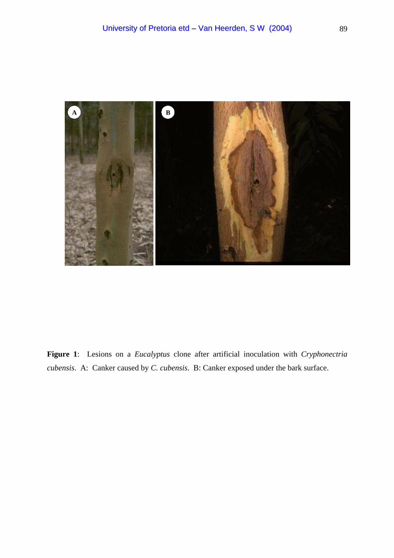
89
A BA B
Figure 1: Lesions on a Eucalyptus clone after artificial inoculation with Cryphonectria
cubensis. A: Canker caused by C. cubensis. B: Canker exposed under the bark surface.
UUnniivveerrssiittyy ooff PPrreettoorriiaa eettdd –– VVaann HHeeeerrddeenn,, SS WW ((22000044))

90
CMW2113
CMW11318
CMW11319
CMW11326
CMW11335
CMW11339
CMW11344
CMW11345
CMW11346
Control
ISOLATE
0
100
200
300
400
LE
SIO
N W
IDT
H (M
M)
GC121
GT529
GU21
TAG5
ZG14
CMW2113
CMW11318
CMW11319
CMW11326
CMW11335
CMW11339
CMW11344
CMW11345
CMW11346
Control
ISOLATE
0
100
200
300
400
LE
SIO
N W
IDT
H (M
M)
GC121
GT529
GU21
TAG5
ZG14
Figure 2: Mean lesion width (mm) ±SEM of the South African Cryphonectria cubensis isolates inoculated on the five Eucalyptus clones
UUnniivveerrssiittyy ooff PPrreettoorriiaa eettdd –– VVaann HHeeeerrddeenn,, SS WW ((22000044))

91
Chapter 4:
Transfection studies with the Diaporthe RNA virus
(DaRV) and other Cryphonectria cubensis isolates
A portion of this work forms part of a larger study and is published as:
Characterization of Diaporthe perjuncta transfected with Diaporthe RNA virus (DaRV).
MOLELEKI, N., VAN HEERDEN, S. W., WINGFIELD, M. J., WINGFIELD B.D. and
PREISIG O. (2003). Transfection of Diaporthe perjuncta with Diaporthe RNA virus
(DaRV). Applied and Environmental Microbiology 6(7) 3952-3956.
ABSTRACT
Cryphonectria cubensis and Diaporthe perjuncta are important pathogens of Eucalyptus and
grapevine respectively. A positive stranded RNA virus, Diaporthe RNA virus (DaRV) from
a South African D. perjuncta isolate has been fully characterised. RNA transcripts from a
cDNA clone of DaRV have also been transfected into an isolate of the peach pathogen D.
ambigua. Due to the lack of the availability of D. perjuncta isolates that are free of viruses,
transfection of the natural host could not be attempted at that time. The aim of this study was
to expand the transfection studies using the DaRV RNA transcripts. Isolates chosen in this
study included, a virus free D. perjuncta isolate, a virulent C. cubensis isolate and a
hypovirulent C. cubensis isolate containing the hypovirus CHV1-EP713. Using
electroporation, we were able to infect virus-free D. perjuncta isolate with DaRV. The
transfectant exhibited altered colony morphology in comparison to the original isolate.
Furthermore, the vector derived nucleotides which were present in the RNA used for the
transfection were not present in the RNA isolated from the transfected isolate. Pathogenicity
tests showed that the transfection did not result in a reduction of virulence of the fungus. We
were unable to transfect either of the C. cubensis isolates included in this study. This
suggests that DaRV does not replicate in C. cubensis isolates. The successful transfection of
D. perjuncta for the first time with the virus from this fungus extends the transfection range
for this virus.
UUnniivveerrssiittyy ooff PPrreettoorriiaa eettdd –– VVaann HHeeeerrddeenn,, SS WW ((22000044))
92
INTRODUCTION
Mycoviruses are known to occur in a wide range of fungi. Many of these viruses have been
well characterised and infect phytopathogens such as Sphaeropsis sapinea (Fr.:Fr.) Dyko &
Sutton (Preisig et al. 1998; Steenkamp et al. 1998), Diaporthe perjuncta Niessl. (Smit et al.
1996; Moleleki et al. 2002), Leucostoma persoonii (Nits.) Hoehn (Hammar et al. 1989),
Ophiostoma novo-ulmi (Rodgers 1986, 1987), Cryphonectria cubensis (Bruner) Hodges (van
Heerden, Chapter 2) and Cryphonectria parasitica (Murr.) Barr (Day et al. 1977; Shapira et
al. 1991). Many of these viruses cause hypovirulence in the host fungi and are, therefore, of
interest as potential biological control agents.
Cryphonectria cubensis is a serious Eucalyptus pathogen in numerous parts of the world
including South Africa where it was first reported in 1989 (Wingfield et al. 1989). This is a
well-studied fungus and could serve as a model organism for various similar tree pathogens.
Diaporthe perjuncta is a pathogen of Vitis vinifera (Melanson et al. 2002). A double
stranded RNA virus has been found in a slow growing hypovirulent isolate of this fungus
(Smit et al. 1996). The nucleotide sequence of the dsRNA element has been determined and
characterised as the replicating stage of the Diaporthe RNA virus (DaRV) genome (Preisig et
al. 2000; Moleleki et al. 2002). Moleleki (2002) used the DaRV in transfection studies and
successfully transfected D. ambigua a related pathogen of pome and stone fruit, as well as a
Phomopsis isolate from peach with this virus. Due to a taxonomic confusion at the time, D.
perjuncta, original host of DaRV (D. perjuncta) was never transfected.
The virus (CHV1-EP713) associated with the chestnut blight pathogen C. parasitica is one of
the dsRNA viruses that confers hypovirulence to its host (Day et al. 1977). Other
characteristics include altered colony morphology (Anagnostakis 1982; Elliston 1985a,
1985b), reduced or lost sporulation (Anagnostakis 1982; Elliston 1985a), reduced
pigmentation (Anagnostakis 1982), reduced oxalate accumulation (Havir & Anagnostakis
1983) and reduced laccase production (Rigling et al. 1989). These characteristics are
promising for its possible use in biological control. Since the completion of the nucleotide
sequence of this hypovirus, it has been used in various transfection and transformation studies
(Dawe & Nuss 2001).
UUnniivveerrssiittyy ooff PPrreettoorriiaa eettdd –– VVaann HHeeeerrddeenn,, SS WW ((22000044))

93
Transfection of spheroplasts produced from virus free C. parasitica with the full length in
vitro produced CHV1-EP713 transcripts using electroporation have been successful (Chen et
al. 1994a). The host range for CHV1-EP713 has been expanded to three species of
Cryphonectria namely C. cubensis, C. havanenesis (Bruner) Barr. and C. radicalis
(Schw.:Fries) Barr., and one species of Endothia namely E. gyrosa (Schw.: Fries) Fries (Chen
et al. 1994a). All of these species have been successfully transfected with the C. parasitca
hypovirus RNA (Chen et al. 1994a). A virulent South African C. cubensis isolate
(CMW2113) has also been transfected with CHV1-EP713, resulting in hypovirulence as well
as the production of a bright yellow-orange pigment in culture (van Heerden et al. 2001).
The aim of this study was to use the Diaporthe RNA virus (DaRV) in transfection studies.
We attempted to transfect a C. cubensis isolate containing CHV1-EP713, a wild type virus-
free C. cubensis isolate, and a D. perjuncta isolate. This would enable us to determine
whether D. perjuncta could be transfected by DaRV and to assess the possibility of using
such as transfectant isolate as a biological control agent. By including the CHV1-EP713
infected C. cubensis isolate, we considered the possibility of co-infecting an isolate with
DaRV.
MATERIALS AND METHODS
Isolates and cultural conditions
The isolates selected for this study were the C. cubensis isolate (CMW2113-T) transfected
with the C. parasitica hypovirus CHV1-EP713 (Van Heerden et al. 2001), a virus infected D.
perjuncta isolate (CMW3407) (Smit et al. 1996; Moleleki et al. 2002) and a virus free D.
perjuncta isolate (CMW8597). The isolates were all maintained on 2% MEA (Malt extract
agar) and are stored in the culture collection (CMW) of the Forestry and Agricultural
Biotechnology Institute (FABI), University of Pretoria.
Preparation of fungal sheroplasts
Fungal spheroplasts were prepared using the method of Royer & Yamashiro (1999) and
Moleleki (2002) with minor modifications. The fungal isolates CMW2113-T, CMW2113
and CMW8597 were all grown in 20 ml McCartney bottles containing 5 ml 2% ME (Malt
extract) broth for 6 days. The mycelium was removed from the medium and placed in a 6
mm diam. Petri dish. The excess medium was removed with a pipette. Chitinase (0.5% w/v)
UUnniivveerrssiittyy ooff PPrreettoorriiaa eettdd –– VVaann HHeeeerrddeenn,, SS WW ((22000044))
94
(Fluka) and cellulose (1% w/v) (Sigma) were dissolved in 6 ml of 1M Magnesium Sulphate
(MgSO4.7H2O), added to the mycelium and incubated overnight at room temperature. The
mixture was then sieved through 120 micron flour gauze (Swiss Milling Company). An
equal volume of ice cold 1M sorbitol (Sigma) was added to the resulting spheroplasts. The
spheroplast solution was placed in 1.5 ml Eppendorf tubes and centrifuged at 5000 rpm for 5
min at 4ºC. The pellet was then washed with 500 µl ice cold 1M sorbitol before being
centrifuged at 5000 rpm for 5 min at 4ºC. The pellet was resuspended in 500 µl STC (1M
Sorbitol, 50 mM CaCl2, 50 mM Tris-HCl pH 8) and centrifuged at 5000 rpm for 5 min at
4ºC. The spheroplasts were resuspended in 85 µl sorbitol and stored on ice for a short period
before electroporation.
Transfection of the fungal spheroplasts with the in vitro produced DaRV RNA
A full length cDNA copy of the D. ambigua RNA virus (DaRV) was cloned in the pGEM®-T
Easy Vector (plasmid pDV3) (Preisig et al. 2000). The RNA was synthesized from the Sal I
linearised plasmid. A reaction mixture was prepared containing 150 ng linearised plasmid, 1
mM of each NTP’s (ATP, CTP, GTP, UTP), 1 x transcription buffer, 0.5 U T7 RNA
polymerase, 1 U RNase inhibitor (Roche Diagnostics). The volume was adjusted to 20 µl.
The reaction was allowed to proceed at 30ºC for 2 hours. The transcription products were
subsequently analysed on a 1% agarose gel stained with ethidium bromide and viewed under
UV light.
Transfections were done by electroporation using a multiporator (Eppendorf) as described by
Chen et al. (1993) with minor modifications. A mixture was made that contained 85 µl of
freshly prepared spheroplasts, 0.12 U RNase inhibitor, 15 µl in vitro produced RNA and was
incubated for 5 min on ice. This mixture was then added to a pre chilled 100 µl cuvette (1
mm gap width) (Eppendorf). The spheroplast RNA solution was pulsed 10 times at 1500-
2500 Volts with a 5 sec interval between the pulses. For each set of fungi, a negative control
transfection was done where the RNA was replaced with water. A volume of 500 µl 1M
sorbitol was added to the cuvette and placed on ice for 10 min. Two hundred microlitres of
the transfected spheroplasts were placed on the middle of a 90 mm diam. Petri dish.
Regeneration medium (48ºC) (0.1% casein hydrolyslate, 0.1% yeast extract, 34.2% sucrose,
1.6% agar) was then slowly added to the Petri dish until the transfected spheroplasts were
covered with medium. The plates were allowed to solidify and incubated in the dark at room
temperature for 1-2 weeks. After sufficient growth, small pieces of agar were arbitrarily cut
UUnniivveerrssiittyy ooff PPrreettoorriiaa eettdd –– VVaann HHeeeerrddeenn,, SS WW ((22000044))

95
from the culture and placed in the centre of a sterile MEA plate. The plates were sealed with
parafilm and incubated at 25ºC. After the incubation period, Erlenmeyer flasks containing
200 ml ME broth were inoculated with putatively transfected fungi and incubated at 25ºC for
1 week. The mycelium was then harvested and lyophilised.
Detection of DaRV RNA
Total RNA was extracted from all the isolates used in the transfection process as well as from
the naturally DaRV-infected D. ambigua isolate (CMW3407) using the High Pure RNA
isolation kit (Roche Diagnostics). The detection of DaRV RNA was done by a one step RT-
PCR amplification using DaRV specific primers (Preisig et al. 2000) and the LightCycler
instrument (Roche) with the LightCycler- RNA Amplification Kit SYBR Green I (Roche
Diagnostics). A reaction mixture was prepared which contained 0.75 ng total RNA, 0.5 µM
primer DaRV5' (5' GGGAAATTTGTGAGATTATCGCC3'), 0.5 µM primer Oli 78 (5'
CCTGGGTGACGGTTGTTACAC 3'), 1 x LightCycler-RT-PCR reaction mix SYBR green I,
6 mM MgCl2, 0.4 µl LightCycler-RT-PCR enzyme mix and PCR grade sterile ddH2O to a
total volume of 20 µl. The cycle conditions were a reverse transcription reaction at 50ºC for
10 min, followed by a denaturation step at 95ºC for 10 sec, 35 cycles at 95ºC for 0 sec, 62ºC
for 4 sec and 72ºC for 16 sec. The final PCR products were separated on a 1.5% agarose gel
stained with ethidium bromide and visualized under UV light.
Sequence of ends of the viral genomes from the transfectants
To determine whether the complete virus was transfected, the distal ends were determined
using the RACE approach as described by Frohman (1994). For this procedure a 5'/3' RACE
kit was used (Roche Diagnostics). Double-stranded RNA was extracted from the
successfully transfected D. perjuncta isolate (CMW8597-DaRV) and purified using the BIO
101 RNaid kit (BIO 101; Qbiogene Inc.) (Valverde et al. 1990; Preisig et al. 1998). The 5'
and 3' end were reverse transcribed with primer Oli 73 (5' GTGCCCTGCACAAACAACTC
3') and Oli 75 (5' TCCATCTCACCGGGAGCGGCAG 3') respectively. A poly-A tail was
added to the cDNA by the addition of terminal transferase and 2 mM dATP. The tailed
cDNA was used in the PCR amplification to determine the 5' and 3' terminal sequence. PCR
was performed with an oligo-dT anchor primer and the nested primers Oli 78 (5'
CCTGGGTGACGGTTGTTACAC 3') and Oli 81 (5' TTGAACGATGGGTGTAGGTGG 3')
for the 5' and 3' ends, respectively. The PCR products were cloned in the pGEM®-T-Easy
UUnniivveerrssiittyy ooff PPrreettoorriiaa eettdd –– VVaann HHeeeerrddeenn,, SS WW ((22000044))

96
vector (Promega). The inserts were sequenced using the ABI PRISM Big Dye Terminator
cycle sequencing ready reaction kit (Perkin Elmer). The sequenced products were analysed
using an ABI PRISM 3100 automated DNA sequencer (Applied Biosystems).
Pathogenicity tests on apples
The pathogenicity of the D. perjuncta isolates CMW3407, CMW8597-DaRV and the non-
transfected isolate CMW8597-WT were compared using an apple based test as described by
De Lange et al. (1998). Prior to inoculation the Golden delicious apples were surface
sterilised with 70% ethanol. Discs (15 mm in depth) were removed from the sides of the
apples using a 5 mm diam. cork borer. Similar sized mycelial plugs were placed mycelial
side down into the wounds and sealed with masking tape to reduce desiccation. Ten apples
were each inoculated with the fungal isolates. In a similar manner 10 apples were inoculated
with sterile PDA discs which served as negative controls. The apples were incubated at room
temperature for ten days whereafter the masking tape was removed and the lesion area
measured. The data were analysed using a one way ANOVA (SYSTAT version 7.0.1). The
entire trial was repeated once and a Pearson correlation test was performed to compare the
two trials
RESULTS
Transfection of the fungal spheroplasts with the in vitro produced DaRV RNA
Diaporthe perjucta isolate (CMW8597) was successfully transfected with the Diaporthe RNA
virus (DaRV). The transfectant was named CMW8597-DaRV. The successful transfection
with DaRV in this study was accomplished using 10 pulses at 2000V during the
electroporation. Transfection also occurred at a lower voltage of 1500 V but no transfection
occurred at higher voltages. Reverse transcription PCR using the LightCycler with the DaRV
specific primer pair, DaRV5' and Oli 78, resulted in an amplicon of ≈350 bp in size in
CMW8597-DaRV as well as in the naturally virus infected isolate (CMW3407) (Fig. 1).
Cryphonectria cubensis isolates, CMW 2113 (virulent) and CMW 2113-T (CHV1-EP713
infected), were not transfected with the Diaporthe RNA virus (DaRV) at any of the conditions
used in the electroporation (Fig. 1) and no PCR amplification was observed.
UUnniivveerrssiittyy ooff PPrreettoorriiaa eettdd –– VVaann HHeeeerrddeenn,, SS WW ((22000044))

97
Sequence of ends of the viral genomes from the transfectants
Transcription from the Sal I linearised plasmid pDV3 resulted in the introduction of 35 vector
derived nucleotides at the 3' end of the viral genome. The 5' end of the virus had 61 vector
derived nucleotides since T7 RNA polymerase initiates transcription 61 bases before the
cDNA insert. Sequence analysis of the 5'/3' RACE PCR products for the 3' and 5' ends of the
transfected Diaporthe RNA virus isolate (CMW8597-DaRV) showed that the vector-derived
nucleotides had not been replicated in the transfected isolate.
Pathogenicity tests on apples
Lesions were observed when the D. perjuncta isolates CMW3407, CMW8597-WT and
CMW8597-DaRV were inoculated into the apples. No lesions were observed when the
inoculation was performed with the sterile PDA discs. Significant differences were observed
between the isolates for both the first (F=45; df=3; P<0.001) and the second repeat (F=33;
df=3; p<0.001) of this experiment. The Bonferroni pairwise comparison indicated that there
were no significant differences between the mean lesion area for CMW8597-DaRV and
CMW8597-WT. The Pearson correlation test showed a good correlation of 96% between the
two experiments.
Phenotypic changes
When the transfected isolate CMW8597-DaRV was inoculated onto PDA the mycelial
growth was slightly changed from the untransfected isolate. The transfected CMW8597-
DaRV isolates resulted in slight morphological changes such as the enhanced aerial growth, a
fluffy appearance with the production of a slight yellowish pigment when compared to the
naturally virus infected and virus free isolate (Fig. 2).
DISCUSSION
In this study we were able to transfect an isolate of D. perjuncta (CMW8597) with the
Diaporthe RNA virus (DaRV). This is the first time that the natural host of DaRV has been
transfected with this virus. Although other fungi have been transfected with hypoviruses
(Chen et al. 1994a; Chen et al. 1996, Chen & Nuss 1999; van Heerden et al. 2001), this is the
first time that D. perjuncta has been transfected with a mycovirus and, therefore, extends the
range of transfection for spheroplasts of a filamentous fungus
UUnniivveerrssiittyy ooff PPrreettoorriiaa eettdd –– VVaann HHeeeerrddeenn,, SS WW ((22000044))
98
Reverse transcription PCR from the transfected Diaporthe RNA virus isolate (CMW8597-
DaRV), showed that the dsRNA was derived from DaRV. The determination of the 3' and 5'
ends of CMW8597-DaRV showed that there were no vector-derived nucleotides. A similar
observation has been made for transformed C. parasitica strains, where it was shown that the
cDNA derived hypoviral RNA are trimmed of vector derived nucleotides (Chen et al. 1994b).
It is, therefore, possible that a similar mechanism must be involved in the trimming effect
although the virus replication was initiated in this case from a plasmid construct. It is also
possible that virus replication only commences exactly at the start of DaRV sequences with
the recognition of replication initiation motifs.
The transfected CMW8597-DaRV isolates exhibited slight morphological changes when
compared with the virus free isolate. These differences included increased aerial growth,
fluffy appearance and the production of a light yellow pigment. Transfection of C. cubensis
and C. parasitica isolates with CHV1-EP713 has resulted in morphological changes in the
host (Chen et al. 1994a; Chen et al. 1996; van Heerden et al. 2001). However, the
phenotypic changes observed for our study were not as striking as those for the hypovirus C.
parasitica transfected isolates.
Cryphonectria cubensis isolates, CMW 2113 (virulent) and CMW 2113-T (CHV1-EP713
infected), could not be transfected with the Diaporthe RNA virus (DaRV). This may be due
to large differences between C. cubensis and D. perjuncta. Although these fungi reside in the
same taxonomic order, they may be insufficiently similar to enable the virus from D.
perjuncta to replicate in C. cubensis. Effective transfection relies on the ability of the fungal
recipient to undergo anastomosis after cell wall regeneration to permit the spread of the viral
RNA (Nuss et al. 2002). However, many fungi can exhibit a filamentous or yeast-like
morphology and anastomosis occurs only in the filamentous phase (Nuss et al. 2002) The
regeneration medium used in this study enhances yeast-like growth (Chen & Nuss,
unpublished), which might influence the successful anastomosis and thus the spread of the
virus from transfected cells. Further studies will need to be conducted to understand why the
transfection was not possible in C. cubensis.
No significant differences in the mean lesion area were observed on apples inoculated with
virus free and transfected isolates of D. perjuncta. Since the naturally virus infected isolates
are known to be hypovirulent, this result was surprising. We had expected that the
UUnniivveerrssiittyy ooff PPrreettoorriiaa eettdd –– VVaann HHeeeerrddeenn,, SS WW ((22000044))
99
transfected isolate would exhibit hypovirulence similar to that found in naturally infected
isolates (Smit et al. 1996). Smit et al. (1996) further observed that the hypovirulence factor
could spread via hyphal anastomosis to other isolates. For the C. parasitica genomes it was
shown that the relative contribution of the hypovirus to hypovirulence associated symptoms
should be interpreted with caution since the movement of hypoviruses via hyphal anastomisis
might lead to the potential transmission of organelles or nuclear genetic information (Chen &
Nuss 1999). The other possibility for lack of hypovirulence in transfected isolates might be
that the D. perjuncta isolate used in this study was relatively non-pathogenic and that its
transfection with the virus did not have a significant impact on the virulence.
The slight effect on the morphology as well as the inability of the transfected strain to induce
hypovirulence, reduces the potential usefulness of DaRV as a biological control agent.
However, results of this study have shown the potential of expanding the transfection system
to other viruses. Further, the transfection of other fungal viruses for which full genome
sequences are available might lead to the identification of a suitable biological control agents
for other pathogens.
UUnniivveerrssiittyy ooff PPrreettoorriiaa eettdd –– VVaann HHeeeerrddeenn,, SS WW ((22000044))

100
REFERENCES
Anagnostakis, S. L. (1982) Biological control of chestnut blight. Science 215: 466-471.
Chen, B., Chen, C.-H., Bowman, B. H., & Nuss, D. L. (1996) Phenotypic changes associated
with wild-type and mutant hypovirus RNA transfection of plant pathogenic fungi
phylogenetically related to Cryphonectria parasitica. Phytopathology 86: 301-310.
Chen, B., Choi, G.H., & Nuss, D.L. (1993) Mitotic stability and nuclear inheritance of
integrated viral cDNA in engineered hypovirulent strains of the chestnut blight fungus.
EMBO Journal 12: 2991-2998.
Chen, B., Choi, G. H. & Nuss, D. L. (1994a) Attenuation of fungal virulence by synthetic
infectious hypovirus transcripts. Science 264: 1762-1764.
Chen, B., Craven, Choi, G. H. & Nuss, D. L. (1994b) cDNA derived hypovirus RNA in
transformed chestnut blight fungus is spliced and trimmed of vector nucleotides. Virology
202: 441-448.
Chen, B. & Nuss, D. L. (1999) Infectious cDNA clone of hypovirus CHV1-Euro7: a
comparative virology approach to investigate virus mediated hypovirulence of the chestnut
blight fungus Cryphonectria parasitica. Journal of Virology 73: 985-992.
Dawe, A. L. & Nuss, D. L. (2001) Hypoviruses and chestnut blight: Exploiting viruses to
understand and modulate fungal pathogenesis. Annual Review of Genetics 35: 1-29.
Day, P. R., Dodds, J. A., Elliston, J. E., Jaynes, R. A. & Anagnostakis, S. L. (1977) Double-
stranded RNA in Endothia parasitica. Phytopathology 67: 1393-1396.
De Lange, W. J., Wingfield, B. D. & Wingfield, M. J. (1998) A rapid, apple-based test for
virulence in Cryphonectria cubensis isolates. European Journal of Forest Pathology 28:
409-412.
UUnniivveerrssiittyy ooff PPrreettoorriiaa eettdd –– VVaann HHeeeerrddeenn,, SS WW ((22000044))

101
Elliston, J. E. (1985a) Characteristics of dsRNA-free and dsRNA-containing strains of
Endothia parasitica in relation to hypovirulence. Phytopathology 75: 151-158.
Elliston, J. E. (1985b) Preliminary evidence for two debilitating cytoplasmic agents in a strain
of Endothia parasitica from Western Michigan. Phytopathology 75: 170-173.
Frohman, M. A. (1994) On beyond classic RACE (rapid amplification of cDNA ends). PCR
Methods and Applications 4: S40-S58.
Hammar, S., Fulbright, D. W. & Adams, G. C. (1989) Association of double-stranded RNA
with low virulence in an isolate of Leucostoma persoonii. Phytopathology 79: 568-572.
Havir, E.A. & Anagnostakis, S.L. (1983) Oxalate production by virulent but not hypovirulent
strains of Endothia parasitica. Physiological Plant Pathology 23: 369-376.
Melanson, D. L., Rawnsley, B. & Scheper, R. W. A. (2002) Molecular detection of
Phomopsis taxa 1 and 2 in grapevine canes and buds. Australasian Plant Pathology 31:
67-73.
Moleleki, N. (2002) RNA viruses of Sphaeropsis sapinea and Diaporthe ambigua and their
possible use as biological control agents. (PhD Thesis), University of Pretoria, Pretoria.
South Africa.
Moleleki, N., Preisig, O., Wingfield, M. J., Cous, P. W. & Wingfield, B. D. (2002) PCR-
RFLP and sequence data delineate three Diaporthe species associated with stone and
pome fruit trees in South Africa. Europrean Journal of Plant Pathology 108: 909-912.
Nuss, D. L., Chen, B., Geletka, L. M., Parsley, T. B. & Suzuki, N. (2002) Engineering
hypoviruses for fundamental and practical applications. In: dsRNA genetic elements:
Concepts and applications in agriculture, forestry and medicine. Tavantzis, S. M. (Ed).
CRC Press LCC, Florida, USA. pp. 145-163.
Preisig, O., Wingfield, B. D. & Wingfield, M. J. (1998) Coinfection of a fungal pathogen by
two distinct double stranded RNA viruses. Virology 252: 399-406.
UUnniivveerrssiittyy ooff PPrreettoorriiaa eettdd –– VVaann HHeeeerrddeenn,, SS WW ((22000044))
102
Preisig, O., Moleleki, N., Smit, W. A., Wingfield, B. D. & Wingfield, M. J. (2000) A novel
RNA mycovirus in a hypovirulent isolate of the plant pathogen Diaporthe ambigua.
Journal of General Virology 81: 3107-3114.
Rigling, D., Heiniger, U. & Hohl, H. R. (1989) Reduction of laccase activity in dsRNA-
containing hypovirulent strains of Cryphonectria (Endothia) parasitica. Phytopathology
79: 219-223.
Rodgers, H. J., Buck, K. W. & Brasier, C. M. (1986) Transmission of double-stranded RNA
and a disease factor in Ophiostoma ulmi. Plant Pathology 35: 277-287.
Rodgers, H. J., Buck, K. W. & Brasier, C. M. (1987) A mitochondrial target for double-
stranded RNA in diseased isolates of the fungus that causes Dutch elm disease. Nature
329: 558-560.
Royer, J. C. & Yamashiro, C. T. (1999) Generation of transformable spheroplasts from
mycelia, macroconidia, microconidia and germinating ascospores of Neurospora crassa
(online) http://www.hgmp.mrc.ac.uk.
Shapira, R., Choi, G. H. & Nuss, D. L. (1991) Virus-like genetic organization and expression
strategy for a double-stranded RNA genetic element associated with biological control of
chestnut blight. The EMBO Journal 10: 731-739.
Smit, W. A., Wingfield, B. D. & Wingfield, M. J. (1996) Reduction of laccase activity and
other hypovirulence associated triats in dsRNA-containing strains of Diaporthe ambigua.
Phytopathology 86: 1311-1316.
Steenkamp, E. T., Wingfield, B. D., Swart, W. J. & Wingfield, M. J. (1998) Double-stranded
RNA and associated virulence in South African isolates of Sphaeropsis sapinea.
Canadian Journal of Botany 76: 1412-1417.
Valverde, R. A., Nameth, S. T. & Jordan, R. L. (1990) Analysis of double-stranded RNA for
plant virus diagnosis. Plant Disease 74: 255-258.
UUnniivveerrssiittyy ooff PPrreettoorriiaa eettdd –– VVaann HHeeeerrddeenn,, SS WW ((22000044))

103
van Heerden, S. W., Geletka, L. M., Preisig, O., Nuss, D. L., Wingfield, B. D. & Wingfield,
M.J. (2001) Characterization of South African Cryphonectria cubensis isolates infected
with a C. parasitica hypovirus. Phytopathology 91: 628-632.
Wingfield, M. J., Swart, W. J. & Abear, B. J. (1989) First record of Cryphonectria canker of
Eucalyptus in South Africa. Phytophylactica 21: 311-313.
UUnniivveerrssiittyy ooff PPrreettoorriiaa eettdd –– VVaann HHeeeerrddeenn,, SS WW ((22000044))

104
Table 1: Mean lesion area on golden delicious apples ten days after inoculation with the
natural infected (CMW3407) isolate, DaRV transfected (CMW8597-DaRV) isolate, and the
virus free isolate (CMW8597) of Diaporthe perjuncta.
Isolate Mean lesion areaa Significant differencesb
Repeat 1 Repeat 2
Agar 113±0 113±0 a
CMW3407 355±33 369±11 b
CMW8597-DaRV 629±41 1024±80 c
CMW8597-WT 611±49 891±126 c
aMean lesion area (mm2) were determine by calculating the lesion area with the formula
area=πr2 with the radius being the (horizontal diameter + vertical diameter) / 4. bIsolates with different letters differed significantly from each other (p=0.001) as indicated
for the Bonferroni pairwise comparison.
UUnniivveerrssiittyy ooff PPrreettoorriiaa eettdd –– VVaann HHeeeerrddeenn,, SS WW ((22000044))

105
2 3 541
350bp
2 3 541
350bp
Figure 1: Confirmation of successful transfection with the Diaporthe RNA virus (DaRV)
using reverse transcription (RT) PCR. The products were separated on a 1.5% agarose gel,
stained with ethidium bromide and visualised under UV light. Lane 1: 100 bp molecular
weight marker, Lane 2: D. perjuncta isolate, CMW8597-DaRV, Lane 3: CMW8597 water
transfected, Lane 4: D. perjuncta natural virus infected isolate, CMW3407- positive control
Lane 5: C. cubensis isolate, CMW2113-T after transfection,
UUnniivveerrssiittyy ooff PPrreettoorriiaa eettdd –– VVaann HHeeeerrddeenn,, SS WW ((22000044))
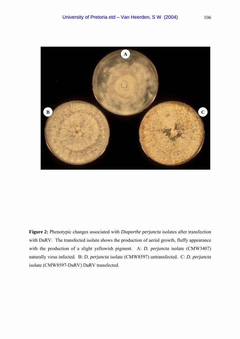
106
A
B C
A
B C
Figure 2: Phenotypic changes associated with Diaporthe perjuncta isolates after transfection
with DaRV. The transfected isolate shows the production of aerial growth, fluffy appearance
with the production of a slight yellowish pigment. A: D. perjuncta isolate (CMW3407)
naturally virus infected. B: D. perjuncta isolate (CMW8597) untransfected. C: D. perjuncta
isolate (CMW8597-DaRV) DaRV transfected.
UUnniivveerrssiittyy ooff PPrreettoorriiaa eettdd –– VVaann HHeeeerrddeenn,, SS WW ((22000044))

107
Chapter 5:
Biological control of Cryphonectria canker of
Eucalyptus using an isolate transfected with the C.
parasitica hypovirus
ABSTRACT
Cryphonectria cubensis is one of the most serious Eucalyptus pathogens in South Africa.
Previously, a virulent South African C. cubensis isolate was transfected with the full-length
coding strand of the C. parasitica hypovirus (CHV1-EP713). The transfectant isolate was
shown in a limited laboratory study to impart hypovirulence. Here, we report on the use of
the transfectant isolate in the first field trial evaluating its potential as a biological control
agent. The field trial was established using one-year-old Eucalyptus grandis clones ZG14
and TAG5, known to be susceptible to Cryphonectria canker. Inoculations with the
transfected C. cubensis isolate were characterised by significantly smaller lesions than those
associated with the virulent, virus-free isolate. Co-inoculation on single trees with both the
virulent and virus-containing isolate resulted in the significant reduction in the size of the
lesions. Treatment of already established Cryphonectria cankers with the transfectant isolate
at four points around the periphery of cankers did not lead to a significant reduction in canker
size, but did alter the morphology of the cankers. The virus was also shown to be transmitted
via hyphal anastomosis to the virulent isolates causing the cankers.
UUnniivveerrssiittyy ooff PPrreettoorriiaa eettdd –– VVaann HHeeeerrddeenn,, SS WW ((22000044))
108
INTRODUCTION
Cryphonectria cubensis (Bruner) Hodges is an ascomycetous fungus that causes a severe
canker disease on Eucalyptus. Since the discovery of C. cubensis in South Africa (Wingfield
et al. 1989) the need to reduce the impact of this disease has grown. Currently, the most
effective means to manage the impact of Cryphonectria canker is through breeding and
selection of disease-tolerant planting stock (Alfenas et al. 1983; Wingfield 1990; van
Heerden & Wingfield 2002). Another exciting, but possibly longer term approach could be
through biological control using double stranded (ds) RNA viruses linked to hypovirulence
(van Heerden et al. 2001).
The possibility of using dsRNA viruses as biological control agents to reduce the impact of
plant diseases has been considered for various pathogens. The best studied system is that of
the causal agent of chestnut blight, Cryphonectria parasitica (Murr) Barr. This emerged after
Biraghi (1950) observed healing chestnut blight cankers in Italy, and the subsequent
identification of hypovirulent C. parasitca strains (Grente 1965). The causal agents of this
hypovirulence were later shown to be double stranded RNA elements (Day et al. 1977).
These viruses were later assigned to the genus Hypovirus in the virus family Hypoviridae
(Hillman et al. 2000). The hypoviruses are transmitted either vertically to the conidia
(asexual spores) or via cytoplasmic exchange, after hyphal anastomosis (Nuss 1996).
Various field experiments have been conducted in an effort to establish whether the impact of
chestnut blight can be reduced using hypoviruses. One of the first of these trials was in
France between 1966 and 1974, where Grente & Berthelay-Sauret (1978) developed an
inoculation technique for blight affected chestnut trees. In this procedure, growers were
supplied with a mixture of hypovirulent C. parasitca isolates, which they then inoculated at
the edges of existing cankers (Grente & Berthelay-Sauret 1978; Heiniger & Rigling 1994).
These cankers started to heal and mortality decreased. Biological control has also been
successfully applied in Italy where similar application techniques have been used (Heiniger &
Rigling 1994). In North America, cankers on trees have been treated successfully but spread
of naturally occurring hypoviruses has not occurred (Anagnostakis 1982). In contrast,
transgenic hypovirulent strains have shown effective transmission of the virus in field trials
(Nuss 2000).
UUnniivveerrssiittyy ooff PPrreettoorriiaa eettdd –– VVaann HHeeeerrddeenn,, SS WW ((22000044))

109
Recently, the full length coding strand of the C. parasitica hypovirus (CHV1-EP713) was
transfected into a virulent South African C. cubensis isolate and the transfectant was shown
to be stable (van Heerden et al. 2001). This resulted in pronounced morphological changes in
the recipient fungus, such as the production of a bright yellow-orange pigment, reduced
sporulation and growth reduction. Preliminary greenhouse inoculations showed that the
transfected isolate was significantly less virulent than the wild type virus free isolate.
The aim of this study was to use the CHV1-EP713 transfected C. cubensis isolate in a first
field experiment and to test whether it shows bio-control characteristics on established trees
in the field. The trial was also designed to test whether the transfected isolate could slow the
expansion of already established cankers.
MATERIALS AND METHODS
Isolates used
A virulent South African C. cubensis isolate (CMW2113) was selected for this study. This
isolate has been used in a suite of previous studies on Cryphonectria canker in South Africa
and is known to represent the higher order of pathogenicity in the local C. cubensis
population (van Heerden & Wingfield 2001). In addition, another South African C. cubensis
isolate CMW11336 was selected to consider the potential of the biocontrol isolate to reduce
the expansion of cankers caused by a different isolate of C. cubensis. The isolate
(CMW2113-T), previously transfected with the C. parasitica hypovirus CHV1-EP713 (van
Heerden et al. 2001), was used in this study as the potential biological control agent. All
three isolates are maintained in the culture collection (CMW) of the Forestry and Agricultural
Biotechnology Institute (FABI), University of Pretoria, Pretoria, South Africa.
Evaluation of field bio-control characteristics
One-year-old stems of the Eucalyptus grandis clones ZG14 and TAG 5 were used for this
inoculation study. Trees were inoculated by removing a cambial disc with a 20 mm diam.
cork borer, and approximately 150 cm from the ground. Similar sized agar discs from the
edges of actively growing cultures on 2% malt extract agar (MEA) were placed in the
wounds with the mycelium side towards the wound. The wounds were sealed with masking
tape to reduce desiccation. Twenty trees of each clone were inoculated with each of the
isolates CMW2113 and CMW2113-T. In addition to inoculation of trees with single isolates,
UUnniivveerrssiittyy ooff PPrreettoorriiaa eettdd –– VVaann HHeeeerrddeenn,, SS WW ((22000044))

110
the two isolates CMW2113 (virulent) and CMW2113-T (transfected) were co-inoculated into
trees. Here, isolates were inoculated on the same trees, alongside each other, with inoculation
points 5 mm apart from each other. Ten trees of each clone were inoculated with a sterile
MEA plug that acted as a control.
Six weeks after inoculation, the masking tape was removed from the inoculation sites and
lesion lengths, lesion widths and tree circumferences were measured. Lesion areas were
computed from lesion lengths and widths. These values were analysed using a one way
ANOVA (SYSTAT version 7.0.1) to test for significant differences in lesion size associated
with the various inoculations. Tree circumference was used a co-variat in the analysis. A
post-hoc pairwise Bonferroni comparison was also performed to determine isolate differences
based on their lesion areas.
Inhibition of canker expansion
To determine whether the expansion of actively growing cankers can be halted, newly
established cankers were treated with the virus transfected isolate. The cankers were
established on trees of the E. grandis clone ZG 14. Thirty trees were each inoculated with
the South African C. cubensis isolates CMW2113 and CMW11336. These trees were left for
six weeks for the cankers to become well established.
In order to treat the cankers, wounds (20 mm in diam.) were made in healthy tissue at the
periphery of the cankers. The wounds were made at four sites at the edges of the actively
growing six-week-old canker (Fig. 1). Two of these were placed at the canker periphery in
horizontal positions across the centers and at opposite sides of the canker. The other two
inoculation points were also placed at the canker periphery but vertically across the centre
and at opposite ends of the canker. Wounds were made in the healthy tissue at the periphery
of the cankers. Similar sized discs of the actively growing transfectant isolate CMW2113-T
on MEA agar were placed into the wounds (Fig. 1). The wounds were sealed with masking
tape to prevent desiccation. Fifteen trees of each isolate were inoculated in this manner. The
remaining 15 cankered trees were left untreated and acted as controls. After six weeks,
cankers were inspected and canker lengths, widths and tree circumferences were measured.
The data were analysed using a one way ANOVA (SYSTAT version 7.0.1). The data were
found to be inordinately skewed, not meeting the assumptions of parametric testing and were
UUnniivveerrssiittyy ooff PPrreettoorriiaa eettdd –– VVaann HHeeeerrddeenn,, SS WW ((22000044))

111
thus log transformed to normalise the distribution. A post-hoc pairwise Bonferroni
comparison was performed on the data.
Re-isolations were made from each of three cankers caused by the two C. cubensis isolates
(CMW2113 and CMW11336), which were subsequently treated with the virus infected
isolate. The re-isolations were made from different positions at the canker periphery (Fig. 4).
Some of these positions were selected to be as far away as possible from the point at which
the virus infected isolate had been inoculated. Others were specifically from points adjacent
to the areas where the virus infected isolate had been placed. Four-five re-isolations were
made per canker (Fig. 4) and these were to determine whether the virus could be retrieved
from parts of the cankers distant from the sites where the virus transfected isolates had been
placed. This would make it possible to determine whether the virus had been transferred
from the virus containing isolate to the virus free virulent isolates
To confirm that the retrieved isolates from the treated cankers caused by isolate CMW11336
represented the original fungus and not overgrowth from the virus-infected isolate
(CMW2113-T), vegetative compatibility tests (VCG’s) were done. These were with all the
retrieved isolates from the cankers caused by CMW11336 and subsequently treated with the
transfectant isolate. The procedure for testing VCG's was the same as that described by van
Heerden and Wingfield (2001), using a medium containing Bromcresol green.
In order to screen the isolates retrieved from the cankers for the presence of the virus they
were grown in 2% Malt extract broth. Total RNA was extracted from freeze dried mycelium
using the High Pure RNA Isolation Kit (Roche Diagnostics). For detection of the virus, a one
step RT-PCR reaction was performed using a LightCycler (Roche Diagnostics) with the
LightCycler- RNA Amplification Kit SYBR Green I (Roche Diagnostics). The primer pair
RSDS10 and BR43 was selected to determine the presence of CHV1-EP713. Primer RSDS
10 (5'-GCCTATGGGTGGTCTACATAGG-3') corresponds to the 5´-terminal sequence of
CHV1-EP713 coding strand and primer BR43 (5'-GGATCCACTGTAGTAGGATCAA-3') is
complementary to nucleotide positions 566-545 of the CHV1-EP713 coding strand (van
Heerden et al. 2001). A reaction mixture was prepared containing 0.75 ng total RNA, 0.5
µM primer RSDS10, 0.5 µM primer BR43, 1 x LightCycler-RT-PCR reaction mix SYBR
green I, 6 mM MgCl2, 0.4 µl LightCycler-RT-PCR enzyme mix and PCR grade sterile ddH2O
to a total volume of 20 µl. The cycle conditions were a reverse transcription reaction at 50ºC
UUnniivveerrssiittyy ooff PPrreettoorriiaa eettdd –– VVaann HHeeeerrddeenn,, SS WW ((22000044))

112
for 10 min, followed by a single cycle at 95ºC for 10 sec, 35 cycles at 95ºC for 0 sec, 62ºC
for 4 sec and 72ºC for 16 sec. The data were analysed based on the melting curves of the
PCR products. A melting temperature of 87ºC would indicate that a virus specific product
had been obtained. The PCR products were also separated on a 1.5% agarose gel stained
with ethidium bromide and visualised under UV light.
RESULTS
Evaluation of bio-control characteristics
Distinct lesions were observed six weeks after inoculation with the virulent C. cubensis
isolates. No lesion development was observed for the control inoculations. Lesion size
differed significantly for the two C. cubensis isolates, and the control isolate used in this
inoculation on both the ZG14 E. grandis clone (F=22.9; df=3; p<0.001) and the E. grandis
clone, TAG5 (F=113.3; df=3; p<0.001). The post-hoc Bonferroni pairwise comparisons
showed that isolate CMW2113 gave rise to cankers significantly larger to those associated
with the CMW2113-T and the control inoculations on both clones (Fig. 2). For example on
clone ZG14, CMW2113 gave rise to lesions with a mean length of 123±11 mm and for clone
TAG 5 these were a mean of 135±4 mm (Table 1). Lesions caused by the hypovirus-
transfected isolate CMW2113-T did not differ significantly from inoculations using a sterile
MEA plug on either of the tree clones (Fig. 2; Table 1).
Where the transfected isolate CMW2113-T was inoculated alongside the virulent isolate
(CMW2113), lesion size associated with the virulent isolate was significantly reduced. This
combined inoculation resulted in significant smaller lesions than those associated with
CMW2113 on both clones (Fig. 2). The resulting mean lesion lengths for clone ZG14 and
clone TAG5 were 69±8 mm and 72±2.5 mm respectively (Table 1).
Inhibition of canker development
There were no significant difference in the size of cankers for the hypovirus treated and the
virulent CMW2113 cankers (F=0.005; df=1; p=0.903) (Fig. 3). The mean lesion length for
the cankers caused by the untreated CMW2113 canker was 222±15 mm and the mean lesion
width was 107±8 mm. Cankers caused by this fungus, which were subsequently treated with
the transfected isolate, had a mean lesion length of 238±19 mm and a mean lesion width of
104±10 mm. There were also no significant difference in the size of cankers caused by the
UUnniivveerrssiittyy ooff PPrreettoorriiaa eettdd –– VVaann HHeeeerrddeenn,, SS WW ((22000044))

113
hypovirus-treated and the untreated CMW11336 cankers (F=0.008; df=1; p=0.859) (Fig. 3).
The mean length for the untreated canker caused by CMW11336 was 185 ±11 mm and the
mean width was 59± 8mm. The hypovirus treated canker had a mean lesion length of
133±19 mm and a mean lesion width of 72±4 mm.
The canker morphology of the treated cankers were irregular in shape. Cryphonectria
cubensis was reisolated from selected sections of the cankers that had been treated with the
virus containing isolate, CMW2113-T. The RT-PCR with the total RNA extracted to
determine the presence of the virus resulted in a 600 bp amplicon (Fig. 4 & 5). These results
show that the virus was transferred from the hypovirulent isolate CMW2113-T to both
isolates CMW2113 and CMW11336. The VCG tests showed that the fungi reisolated from
the different parts of the cankers caused by isolate CMW11336, belong to the same VCG as
CMW 11336 and not the transfected isolate (CMW2113-T).
DISCUSSION
In this study we have provided the first illustration of the impact of the C. parasitica
hypovirus (CHV1-EP713) on Cryphonectria cankers caused by C. cubensis under field
conditions. Inoculation of Eucalyptus trees with the hypovirus-transfected C. cubensis isolate
(CMW2113-T) resulted in lesions significantly smaller than those caused by the virulent
isolate CMW2113. These results are similar to those of van Heerden et al. (2001) in which
the transfected isolate (CMW2113-T) reduced virulence under greenhouse conditions. The
combination inoculation, in which the virulent and virus transfected isolates were co-
inoculated on the same tree at the same time, has shown that the virus infected isolate
CMW2113-T reduces the development of the virulent isolate. These results, therefore, show
that CMW2113-T is able to reduce canker expansion if it develops together with the virulent
isolate.
Where the transfectant isolate was inoculated at the periphery of expanding cankers, canker
development was not significantly reduced. A similar method was applied in the biological
control program instituted by the French Ministry of Agriculture in an effort to reduce the
impact of chestnut blight. In that protocol, blighted chestnut trees were treated annually with
mixtures of hypovirulent compatible C. parasitica strains by placing them in holes around
cankers caused by C. parasitica (Grente & Berthelay-Sauret 1979; van Alfen 1982;
UUnniivveerrssiittyy ooff PPrreettoorriiaa eettdd –– VVaann HHeeeerrddeenn,, SS WW ((22000044))
114
MacDonald & Fulbright 1991). This method was shown to be effective. However, C.
cubensis is a very aggressive pathogen on Eucalyptus and will generally kill susceptible trees
such as those inoculated in this study in less than six months after inoculation (van Heerden,
unpublished). It was, therefore, necessary to assess results in the early stages of
development. The method used in this study to reduce Cryphonectria cankers was not
effective and other methods using the transfectant isolate as a potential bio-control agent for
the reduction of the cankers will be required.
Our study has further shown that the virus CHV1-EP713 is transmitted via hyphal
anastomosis to the virulent virus-free isolates. This was observed for both the isolates
inoculated in this study and confirmed using VCG tests to identify the isolate genotypes, and
RT PCR to determine the presence of the virus, thus indicating the effective movement and
establishment of the virus in two different isolates. The virus was also observed in all the
sections of the cankers that were the furthest away from the point of inoculation of the virus-
infected isolate. Van Heerden et al. (2001) have shown in a laboratory experiment that
CHV1-EP713 is transmitted via hyphal anastomosis to many individuals of a population of
isolates of the fungus from South Africa. Since it is known that the efficacy of any biological
control strategy relies on the effective dissemination of the virus, our observation was
encouraging.
Inoculation of the CHV1-EP713 transfected isolate in all the experiments resulted in the
formation of very small lesions. This suggests that the transfectant strain is ecologically unfit
and would probably not survive under natural conditions. A similar observation was made
for other CHV1-EP713 transgenic isolates released in a field experiment (Anagnostakis et al.
1998). Although that virus was shown to be transmitted to the ascospores of C. parasitica, it
failed to persist at the release site for more than two years (Anagnostakis et al. 1998).
MacDonald and Fulbright (1991) have also noted that hypovirulent strains used in North
America were highly curative, but they had a poor capacity to colonise and produce spores.
This resulted in limited persistence and poor prospects for biological control under natural
conditions. One means of overcoming this impediment will be to use an alternative virus for
biological control. CHV1-Euro 7 has suitable characteristics such as enhanced colonisation
and spore production (Chen & Nuss 1999; Dawe & Nuss 2001). Using a strain such as this
could balance the ecological fitness of the biological control agent.
UUnniivveerrssiittyy ooff PPrreettoorriiaa eettdd –– VVaann HHeeeerrddeenn,, SS WW ((22000044))
115
REFERENCES
Alfenas, A. C., Jeng, R. & Hubbes, M. (1983) Virulence of Cryphonectria cubensis on
Eucalyptus species differing in resistance. European Journal of Forest Pathology 13:
179-205.
Anagnostakis, S. L. (1982) Biological control of chestnut blight. Science 215: 466-471.
Anagnostakis, S. L., Chen, B. C., Geletka, L. M. & Nuss, D. L. (1998) Hypovirus
transmission to ascosopre progeny by field released transgenic hypovirulent strains of
Cryphonectria parasitica. Phytopathology 88: 598-604.
Biraghi, A. (1950). Caratteri di resistenza in Castanea sativa nei confronti di Endothia
parasitica. Bollettino Stazione di Patologia Vegetale di Roma 7: 161-171.
Chen, B. & Nuss, D. L. (1999) Infectious cDNA clone of hypovirus CHV1-Euro7: a
Comparitive virology approach to investigate virus mediated hypovirulence of the
chestnut blight fungus Cryphonectria parasitica. Journal of Virology 73: 985-992.
Dawe, A. L. & Nuss, D. L. (2001) Hypoviruses and chestnut blight: Exploiting viruses to
understand and modulate fungal pathogenesis. Annual Review of Genetics 35: 1-29.
Day, P. R., Dodds, J. A., Elliston, J. E., Jaynes, R. A. & Anagnostakis, S. L. (1977) Double-
stranded RNA in Endothia parasitica. Phytopathology 67: 1393-1396.
Grente, J. & Berthelay-Sauret, S. (1978) Biological control of chestnut blight in France. In:
Proceedings of the American Chestnut Symposium. MacDonald, W. L., Cech, F. C.,
Luchok J. & Smith H. C. (Eds.). West Virginia University Press: Morgantown, West
Virginia, USA. pp. 30-34.
Grente, J. (1965) Les formes hypovirulentes d’Endothia parasitica et les espoirs de lutte
contre le chancre du chataignier. Compte Rendu Hebdomadaire des Seances de
I’Academie des Agriculture de France 51: 1033-1037.
UUnniivveerrssiittyy ooff PPrreettoorriiaa eettdd –– VVaann HHeeeerrddeenn,, SS WW ((22000044))
116
Heiniger, U. & Rigling, D. (1994) Biological control of chestnut blight in Europe. Annual
Review of Phytopathology 32: 581-599.
Hillman, B. I., Fulbright, D. W., Nuss, D. L. & van Alfen, N. K. (2000) Family Hypoviridae.
In: Virus Taxonomy: Seventh report of the International Committee on Taxonomy of
Viruses. Van Regenmortel, M. H. V., Fauquet, C. M., Bishop, D. H. L., Carstens, E. B.,
Estes, M. K., Lemon, S. M., Maniloff, J., Mayo, M. A., McGeoch, D. J., Pringle, C. R. &
Wickner, R. B. (Eds.). Academic Press, California, USA. pp. 515-520.
MacDonald, W. L. & Fulbright, D. W. (1991) Biological control of chestnut blight: use and
limitation of transmissible hypovirulence. Plant Disease 75: 656-661.
Nuss, D. L. (1996) Using hypoviruses to probe and perturb signal transduction processes
underlying fungal pathogenisis. The Plant Cell 8: 1845-1853.
Nuss, D. L. (2000) Hypovirulence and chestnut blight: From the field to the laboratory and
back. In: Fungal Pathology. Kronstad, J. W. (Ed). Kluwer Academic Publishers,
Netherlands. pp. 149-170.
van Alfen, N. K. (1982) Biology and potential for disease control of hypovirulence of
Endothia parasitica. Annual Review of Phytopathology 20: 349-362.
van Heerden, S. W., Geletka, L. M., Preisig, O., Nuss, D. L., Wingfield, B. D. & Wingfield,
M.J. (2001) Characterization of South African Cryphonectria cubensis isolates infected
with a C. parasitica hypovirus. Phytopathology 91: 628-632.
van Heerden, S. W. & Wingfield, M. J. (2001) Genetic diversity of Cryphonectria cubensis
isolates in South Africa. Mycological Research 105: 94-99.
van Heerden, S. W. & Wingfield, M. J. (2002) Effect of environment on the response of
Eucalyptus clones to inoculation with Cryphonectria cubensis. Forest Pathology 32: 395-
402.
UUnniivveerrssiittyy ooff PPrreettoorriiaa eettdd –– VVaann HHeeeerrddeenn,, SS WW ((22000044))

117
Wingfield, M. J. (1990) Current status and future prospects of forest pathology in South
Africa. South African Journal of Science 86: 60-62.
Wingfield, M. J., Swart, W. J. & Abear, B. J. (1989) First record of Cryphonectria canker of
Eucalyptus in South Africa. Phytophylactica 21: 311-313.
UUnniivveerrssiittyy ooff PPrreettoorriiaa eettdd –– VVaann HHeeeerrddeenn,, SS WW ((22000044))

118
Table 1: Mean lesion size for the virulent and virus transfected South African Cryphonectria
cubensis isolates inoculated on Eucalyptus grandis clones ZG14 and TAG5.
Isolate a Clone Mean lesion length
(L) (mm) ±SEM
Mean lesion width
(W) (mm) ±SEM
LxW b
CONTROL ZG14 20 ± 0.0 20 ± 0.0 400±0.0 a
CMW2113-T ZG14 20 ± 0.0 20 ± 0.0 400±0.0 ab
COMB ZG14 69±8 35±2 2635±422 ac
CMW2113 ZG14 123±11 47±4 6186±793 d
CONTROL TAG5 20 ± 0.0 20 ± 0.0 400±0.0 a
CMW2113-T TAG5 20 ± 0.0 20 ± 0.0 400±0.0 ab
COMB TAG5 72±2.5 32±0.8 2312±111 c
CMW2113 TAG5 135±4 40±2 5351±340 d a Isolates: CMW2113-T is the hypovirus (CHV1-EP713) transfected South African C.
cubensis isolate. CMW2113 virulent South African C. cubensis isolate. COMB is the
combination inoculation where CMW2113 and CMW2113-T were inoculated into trees
together, alongside each other with inoculation points 5 mm apart. b Isolates with the same letter do not differ significantly from each other.
UUnniivveerrssiittyy ooff PPrreettoorriiaa eettdd –– VVaann HHeeeerrddeenn,, SS WW ((22000044))

119
Figure 1: Lesions on the Eucalyptus grandis clone (ZG14) 12 weeks after inoculation with
C. cubensis isolate CMW2113. A: Untreated canker. B: Canker that was inoculated on four
sides with virus containing isolate, CMW2113-T, with the inoculation sites indicated by the
arrows.
UUnniivveerrssiittyy ooff PPrreettoorriiaa eettdd –– VVaann HHeeeerrddeenn,, SS WW ((22000044))

120
Figure 2: Lesions on the Eucalyptus grandis clone ZG14, after artificial inoculations with
virulent and a virus transfected Cryphonectria cubensis isolates. A: Lesion caused by the
hypovirus infected isolate CMW2113-T. B: Lesion caused by the wild type virulent virus
free isolate CMW2113. C: Lesion caused by the combination inoculation with both
CMW2113-T on the left and CMW2113 on the right.
UUnniivveerrssiittyy ooff PPrreettoorriiaa eettdd –– VVaann HHeeeerrddeenn,, SS WW ((22000044))

121
Untreated
CMW2113
Virus treat
ed CMW2113
Untreated
CMW11336
Virus treat
ed CMW11336
Treatment
0
10000
20000
30000
40000
50000
60000
70000
Les
ion
leng
th (m
m) x
Les
ion
wid
th (m
m)
a
a
b b
Untreated
CMW2113
Virus treat
ed CMW2113
Untreated
CMW11336
Virus treat
ed CMW11336
Treatment
0
10000
20000
30000
40000
50000
60000
70000
Les
ion
leng
th (m
m) x
Les
ion
wid
th (m
m)
Untreated
CMW2113
Virus treat
ed CMW2113
Untreated
CMW11336
Virus treat
ed CMW11336
Treatment
0
10000
20000
30000
40000
50000
60000
70000
Les
ion
leng
th (m
m) x
Les
ion
wid
th (m
m)
a
a
b b
Figure 3: Mean lesion length (mm) x mean lesion width (mm) (± SEM) of virus treated and
the untreated Cryphonectria cubensis cankers in an investigation to determine whether the
hypovirus (CHV1-EP713) transfected C. cubensis isolate (CMW2113-T) can reduce actively
growing cankers from expanding. The cankers were caused by two virulent C. cubensis
isolates CMW2113 and CMW11336. Columns with the same letter do not differ
significantly from each other (P<0.001) indicating that the virus infected isolate did not cause
a reduction in canker expansion.
UUnniivveerrssiittyy ooff PPrreettoorriiaa eettdd –– VVaann HHeeeerrddeenn,, SS WW ((22000044))

122
BA
Figure 4: Cankers caused by two virulent South African Cryphonectria cubensis isolates. A. Canker
caused by isolate CMW11336. B: Canker caused by isolate CMW2113. Both these cankers were
treated with the hypovirus (CHV1-EP713) transfected isolate on four corners on the canker periphery.
These positions are indicate by a . Reisolations of the fungus with the purpose to determine the
presence of the virus at different positions on the canker were also done and are indicated by the
numbers 4-12.
*
**
*
**
*
*10
7 9
8
*4
11
5
6
12
*
UUnniivveerrssiittyy ooff PPrreettoorriiaa eettdd –– VVaann HHeeeerrddeenn,, SS WW ((22000044))

123
M 2 3 4 5 6 7 8 9 10 11 12
500bp
MM 2 3 4 5 6 7 8 9 10 11 12
500bp
M
Figure 5: Confirmation of the presence of the hypovirus (CHV1-EP713) using reverse
transcription PCR. The PCR products were separated on a 1.5% agarose gel stained with
ethidium bromide and visualised under UV light. Lanes M: 100 base pair (bp) molecular
weight marker. Lanes 2: Virus free isolate which was used as a negative control. Lane 3:
Hypovirus transfected isolate CMW2113-T as positive control. Lanes 4-6 and 8-12: RT-
PCR amplicons derived from fungi re-isolated from the canker and corresponds to the
positions on the cankers indicated in Figure 4. Presence of the virus is indicated by a 600 bp
amplicon.
UUnniivveerrssiittyy ooff PPrreettoorriiaa eettdd –– VVaann HHeeeerrddeenn,, SS WW ((22000044))

124
SUMMARY
Cryphonectria cubensis is an ascomycetous fungus that causes a serious canker disease on
Eucalyptus trees in many parts of the world. The importance of the disease has led to
numerous studies involving the taxonomy, genetic diversity and the control of Cryphonectria
canker. However, there remain many questions pertaining to the disease that have not been
considered. The objectives of the studies presented in this thesis were, therefore, to
investigate the possibility of biological control of Cryphonectria canker, to evaluate the
currently used disease screening strategy in South Africa and to establish a transfection
system with dsRNA elements in Diaporthe, which is closely related to Cryphonectria.
The introductory chapter of this thesis provides a review of the literature pertaining to
Cryphonectria cubensis. In addition literature on hypovirulence in fungi is also extensively
reviewed, with a special emphasis on the genus Cryphonectria.
The aim of study in the second chapter of the thesis was to screen the South African C.
cubensis population for the presence of dsRNA viruses. Two viruses were identified and the
full sequence of these elements showed a strong homology to the mitochondrial viruses
(mitoviruses) within the family Narnaviridae. We, therefore, named the viruses
Cryphonectria cubensis mitovirus 1 (CcMV1) and Cryphonectria cubensis mitovirus 2
(CcMV2). The two viral genomes are 2601 nucleotides and 2639 nucleotides in size
respectively and encode for a protein that probably functions as an RNA-dependant RNA
polymerase (RdRp). Pathogenicity studies indicated that the viruses do not result in a
significant reduction in pathogenicity of C. cubensis.
In the third chapter, results of a study to consider whether different Eucalyptus clones
responded similarly to various South African C. cubensis isolates, are presented. The aim
was, therefore, to evaluate the current C. cubensis resistant screening method used on
Eucalyptus spp. in South Africa. The statistical analysis of the inoculation data showed a
significant isolate x clone interaction. This data also suggest the possibility of vertical
resistance, which is different to previous assumptions.
Transfection studies (Chapter 4) involving a positive stranded RNA virus, Diaporthe RNA
virus (DaRV) from a South African D. perjuncta isolate are presented here. In this study, a
UUnniivveerrssiittyy ooff PPrreettoorriiaa eettdd –– VVaann HHeeeerrddeenn,, SS WW ((22000044))

125
virus free D. perjuncta isolate, a virulent C. cubensis isolate and a hypovirulent C. cubensis
isolate containing the hypovirus CHV1-EP713 were chosen to be transfected with DaRV. By
using electroporation, it was possible to infect a virus free D. perjuncta isolate with the
Diaporthe RNA virus, thus extending the transfection range of this virus. The resulting
transfection led to altered colony morphology but did not lead to a reduction in pathogenicity.
We were also not successful in attempts to transfect isolates of C. cubensis with DaRV,
indicating that the virus does not replicate in this host.
In a previous study a virulent South African C. cubensis isolate was transfected with the
Cryphonectria parasitica hypovirus CHV1-EP713. This resulted in the fungus becoming
hypovirulent. Chapter five of this thesis presents the results of a study to evaluate the
potential use of this virus in the biological control of Cryphonectria canker in South Africa.
A field trial was established and existing cankers were treated with the transfected isolate.
The treatment of the cankers did not lead to a significant reduction in canker size, but did
alter the morphology of the cankers. The virus was also shown to be transmitted via hyphal
anastomosis to the virulent canker causing isolates. In addition the co-inoculation on single
trees with both the virulent and virus-containing isolate, resulted in a significant reduction in
the size of the lesions. This study also showed that the transfected C. cubensis isolate are
characterised by significantly smaller lesions than those associated with the virulent, virus-
free isolate.
Cryphonectria cubensis and the associated canker disease of Eucalyptus threaten the forestry
industry in South Africa. The overall aims of the studies presented in this thesis were to gain
a more complete understanding of this fungus and to evaluate potential control strategies.
Each of these chapters should contribute towards a better understanding of the viruses
associated with C. cubensis and other important aspects of Cryphonectria canker, which will
hopefully lead to enhanced control strategies of the disease in South Africa.
UUnniivveerrssiittyy ooff PPrreettoorriiaa eettdd –– VVaann HHeeeerrddeenn,, SS WW ((22000044))

![[Psilocybin]How to Grow Magic Mushrooms a Simple Psilocybe Cubensis Growing Technique](https://static.fdocuments.us/doc/165x107/546f1e76b4af9f29368b45c7/psilocybinhow-to-grow-magic-mushrooms-a-simple-psilocybe-cubensis-growing-technique-558457b1a604e.jpg)