Chemical Structures of Streptococcus pneumoniae Capsular ...

Structure of the Type IX Group B Streptococcus Capsular Polysaccharide and its Evolutionary Relationship
with Types V and VII
1
Structure of the Type IX Group B Streptococcus Capsular Polysaccharide and its
Evolutionary Relationship with Types V and VII
Francesco Berti1, Edmondo Campisi
1, Chiara Toniolo
1, Laura Morelli
1, Stefano Crotti
1, Roberto
Rosini1, Maria Rosaria Romano
1, Vittoria Pinto
1, Barbara Brogioni
1, Giulia Torricelli
1, Robert
Janulczyk1, Guido Grandi
1, Immaculada Margarit
1
1From Novartis Vaccines Research, Siena, Italy
*Running title: Structural and Genetic Characterization of GBS Type IX polysaccharide
Keywords: Group B Streptococcus; capsular polysaccharide.
Background:
The capsule of Group B Streptococcus is an
important virulence factor and vaccine target.
Results: The analysis of type IX polysaccharide
revealed a similar structure to type V and VII.
Conclusion: The structural and phylogenetic
basis for the differentiation between types V, VII
and IX was elucidated.
Significance: The determination of the type IX
structure can be instrumental for the development
of a carbohydrate-based vaccine.
SUMMARY
The Group B Streptococcus capsular
polysaccharide type IX was isolated and purified, and the structure of its repeating unit
determined. Type IX capsule {4)[NeupNAc-α-
(23)-Galp-β-(14)-GlcpNAc-β-(16)]-β-
GlcpNAc-(14)-β-Galp-(14)-β-Glcp-(1} appears most similar to types VII and V,
although it contains two GlcpNAc residues.
Genetic analysis identified differences in cpsM,
cpsO and cpsI gene sequences as responsible for
the differentiation between the three capsular
polysaccharide types, leading us to hypothesize
that type V emerged from a recombination
event in a type IX background.
Streptococcus agalactiae (Group B
Streptococcus) is a β-hemolytic, encapsulated
Gram positive microorganism that colonizes the
anogenital tract of 25-30% of healthy women. It
is a major cause of neonatal sepsis and
meningitis, particularly in infants born to mothers
carrying the bacteria (1). The pathogen can also
infect adults with underlying disease, particularly
the elderly, and cause bovine mastitis.
The GBS capsule is a major virulence factor
enabling the bacterium to evade human innate
immune defenses (2, 3). It is constituted by high
molecular weight polysaccharides, of which ten
variants exist (Ia, Ib, II, III, IV, V, VI, VII, VIII,
IX) differing in chemical composition. Like for
other Gram positive and Gram negative bacteria,
individual polysaccharide repeating units are
assembled on a carrier lipid (undecaprenyl
phosphate) by the sequential activities of
glycosyltransferase enzymes. Repeat units are
transferred across the plasma membrane by a
‘flippase’ protein. Polymerization occurs at the
periplasmic face of the plasma membrane and is
catalyzed by a polymerase enzyme.
All the genes responsible for the synthesis and
http://www.jbc.org/cgi/doi/10.1074/jbc.M114.567974The latest version is at JBC Papers in Press. Published on July 2, 2014 as Manuscript M114.567974
Copyright 2014 by The American Society for Biochemistry and Molecular Biology, Inc.
by guest on March 4, 2020
http://ww
w.jbc.org/
Dow
nloaded from

Structure of the Type IX Group B Streptococcus Capsular Polysaccharide and its Evolutionary Relationship
with Types V and VII
2
cell wall attachment of the GBS capsular
polysaccharides (CPS) are clustered in the cps
operon. This operon is composed of 16 to 18 genes,
the sequences of which differ among serotypes (4).
High titers of maternal antibodies to each CPS
variant correlate with serotype specific reduction of
disease risk in the newborn (5). Hence, maternal
vaccination with CPS conjugates shows great
promise for the prevention of GBS neonatal disease
(6).
The most common serotypes among GBS infective
isolates are Ia, Ib, II, III and V, although there are
some geographical and historical variations (7). For
instance, the frequency of type V has increased in
the last few years, while serotype IV has recently
emerged as a cause of adult and neonatal invasive
disease (8). Therefore, accurately monitoring the
variation of this important target antigen is essential
for predicting vaccine coverage.
The serotype IX was identified only in 2007 during
the analysis of three human isolates that failed to
react with antisera against the nine GBS types
described at that time. Preparation of specific
antisera allowed the identification of additional type
IX strains among formerly non-typeable isolates (9).
The identity of GBS type IX was also confirmed by
a multiplex PCR assay containing a mix of primers
specific for the ten different allelic variants of the
cps genes (10). Interestingly, type IX strains can
also cause neonatal infections (11, 12).
The precise chemical structures of GBS types Ia, Ib,
II, III, IV, V, VI, VII and VIII CPS are well
described (13-20). They are composed of repeating
units of four to seven monosaccharides with a
backbone and one or two side chains. Four
monosaccharides (e.g. Glcp, Galp, GlcpNAc and
NeupNAc) are present in all nine described
serotypes, and the NeupNAc residue is always
found at the terminus of one of their side chains.
However, the pattern of glycosidic linkages is
unique to each serotype.
Structural comparison of the nine GBS
polysaccharide variants and alignment of their cps
gene sequences indicated that their differential
evolution proceeded through en bloc replacement of
individual glycosyltransferase genes with DNA
sequences encoding enzymes with new linkage
specificities (22).
The chemical structure of CPS type IX and the
genetic composition of its cps operon have not yet
been delineated. Here we report the isolation,
purification, structural characterization and
immunochemical analysis of type IX CPS and the
genetic analysis of the cps genes encoding the
enzymes responsible for its synthesis.
EXPERIMENTAL PROCEDURES
Bacterial strains
GBS type IX strains IT-NI-016 (isolated
from a neonatal Early Onset Disease case), IT-
PW-62 and IT-PW-64 (from colonized pregnant
women) were kindly provided by Alberto Berardi
(Policlinico di Modena, Italy), Roberta Creti,
Lucilla Baldassarri and Graziella Orefici (ISS
Rome, Italy). The three strains, as well as CZ-
PW-119 (VI), CZ-PW-045 (VII) and CZ-NI-016
(serotype IV) were isolated and typed by Latex
and molecular approaches in the frame of the
DEVANI study (10, 11, 23). Capsular genotype
of type IX isolates was confirmed by genome
analysis (see below).
Strains 2603 V/R (serotype V), COH1
(serotype III), 18RS21 (serotype II) and
JM9130013 (serotype VIII) were obtained from
Dennis Kasper (Harvard Medical School, Boston,
MA). Strains 515 (serotype Ia), and H36B
(serotype Ib) were obtained from Carol Baker
(Baylor College of Medicine, Houston, TX).
Isolation and purification of the type IX capsular
polysaccharide
The GBS strain IT-NI-016 was used for
preparation of CPS IX from 1 liter of bacterial
culture grown to exponential phase in Todd
Hewitt Broth (THB). The purification process
was based on previously described procedures
(24). Briefly, the bacterial pellet was recovered by
centrifugation at 4,000 rpm for 20 min and
incubated with 0.8 N NaOH at 37°C for 36 hours.
After centrifugation at 4,000 rpm for 20 min, 1 M
TRIS buffer (1:9 v/v) was added to the
supernatant and diluted 1:1 (v/v) HCl to reach a
neutral pH.
To further purify type IX CPS, 2 M CaCl2
(0.1 M as final concentration) and ethanol (30%
v/v as final concentration) were added to the
solution. After centrifugation at 4,000 g for 20
min, the supernatant was subjected to a tangential
by guest on March 4, 2020
http://ww
w.jbc.org/
Dow
nloaded from

Structure of the Type IX Group B Streptococcus Capsular Polysaccharide and its Evolutionary Relationship
with Types V and VII
3
flow filtration on a 10 kDa MW cut-off (Hydrosart
Sartorius, 0.1 m2 surface) against 14 volumes of 50
mM TRIS / 500 mM NaCl pH 8.8, and 7 volumes of
10 mM sodium phosphate pH 7.2.
To reconstitute full N-acetylation of possibly
present GlcpNAc and NeupNAc residues, 1:5 (v/v)
of 400 mM sodium acetate buffer pH 4.4 (final
concentration 80 mM) was added. The material was
stored at room temperature for 15 min and
centrifuged at 4,000 g for 20 min to remove any
minor particles. The supernatant was filtrated on 10
kDa MW cut-off (Vivaspin Sartorius, 20 mL device)
against 300 mM Na2CO3 / 300 mM NaCl pH 8.8. A
1:1 diluted solution of 4.15 µL/mL acetic anhydride
in ethanol was added, and the reaction was
incubated at room temperature for 2 hrs. The sample
was filtered on 10 kDa MW cut-off (Vivaspin
Sartorius, 20 mL device) against MilliQ water,
CaCl2 (0.1 M final concentration) and ethanol (30%
v/v final concentration) were added, and centrifuged
at 4,000 g for 20 min. The purified CPS IX was
finally recovered by precipitation with ethanol (80%
v/v) and the pellet was dried under vacuum
(SpeedVac, Thermo).
The purity of the polysaccharide preparation
was assessed by colorimetric assays, which
indicated a content of residual proteins and nucleic
acids below 1% w/w. Endotoxin content measured
by LAL assay was <10 EU/µg of saccharide.
Highly purified GBS type Ia, Ib, II, III, V and
VII CPS were obtained by applying the same
purification process as described above for type IX.
Analysis of monosaccharide composition in CPS IX
The monosaccharides constituting the GBS
type IX repeating unit were obtained by acidic
hydrolysis. Two different series of standard samples
with five increasing concentrations, the first
containing Glcp, Galp and GlcpNAc (Sigma)
ranging between 0.2 and 4.0 µg/mL (as saccharide
powder/mL) and the second containing NeupNAc
(Sigma) between 1.0 and 10.0 µg/mL (as saccharide
powder/mL), were prepared for building individual
calibration curves for each monosaccharide. Two
CPS IX samples were prepared targeting final
concentrations in the calibration curve range. All the
reference and analytical samples for
Glcp/Galp/GlcpNAc were prepared in 2 M
trifluoroacetic acid (Sigma), incubated at 100°C for
2 hrs, dried under vacuum (SpeedVac Thermo), and
suspended in water.
Reference and analytical samples for
NeupNAc were prepared in 0.05 M HCl (Merck),
incubated at 75°C for 1 hr, and neutralized by
addition of NaOH.
All analytical samples were filtered with
0.45 µm Acrodisc (Pall) filters before analysis.
HPAEC-PAD analysis was performed with a
Dionex ICS3000 equipped with a CarboPac PA1
column (4 x 250 mm; Dionex) coupled with PA1
guard column (4 x 50 mm; Dionex). Samples (20
µL injection volume) were run at 1 mL/min,
using either an isocratic elution with 24 mM
NaOH (Carlo Erba), followed by a washing step
with 0.5 M NaOH, or 30 mM Na Acetate (Sigma)
in 100 mM NaOH, followed by a washing step
with NaNO3 (gradient from 50 to 200 mM) in 100
mM NaOH, respectively. The effluent was
monitored using an electrochemical detector in
the pulse amperometric mode with a gold
working electrode and an Ag/AgCl reference
electrode. A quadruple-potential waveform for
carbohydrates was applied. The resulting
chromatographic data were processed using
Chromeleon software 6.8 (Dionex).
Detection of monosaccharide configurations
The configurations of the monosaccharides
were determined by methylation according to the
procedure of Ciucanu and Kerek (25), followed
by hydrolysis, reduction, acetylation, and GC
analysis.
A 1 mg solution of polysaccharide sample in
DMSO/water 95:5 (0.3-0.5 mL) was added to 3-5
mg of finely powdered NaOH and, after 10
minutes of incubation at room temperature, 0.1 ml
of methyl iodide was added. The reaction was
stirred at room temperature for 3 hrs. Water (2
mL) and dichloromethane (10 mL) were then
added, and the organic layer was washed with
water (2 mL for three times) and then dried with
Na2SO4. The dried sample was then hydrolyzed
by the addition of 0.8 mL of 4 M trifluoroacetic
acid and incubation at 100°C for 2 hrs with
stirring. After complete evaporation under
vacuum (SpeedVac Thermo at 45°C for 4 hrs),
the dried sample was reduced with 1 M aqueous
sodium borohydride (0.4 mL) and stirred for 5 hrs
at room temperature. The sample was then
acetylated with a mixture of pyridine (2 mL) and
by guest on March 4, 2020
http://ww
w.jbc.org/
Dow
nloaded from

Structure of the Type IX Group B Streptococcus Capsular Polysaccharide and its Evolutionary Relationship
with Types V and VII
4
acetic anhydride (1 mL) and the reaction was stirred
overnight. The excess of reagent was removed by
evaporation and alditole acetates were extracted
with a mixture of dichloromethane/water. The
generated monosaccharides were analyzed by
capillary gas-liquid chromatography using a Thermo
Scientific Trace GC Ultra / TSQ Quantum XLS
equipped with Restek Rxi-17Sil MS Column 30m,
0.25mm ID, 0.25um in the isothermally program at
190°C for 80 min.
NMR Spectroscopy 1H and
13C NMR experiments were recorded on
Bruker Avance III 400 or 500 MHz spectrometer,
equipped with a high precision temperature
controller, and using 5-mm broadband probe
(Bruker). TopSpin version 2.6 software (Bruker)
was used for data acquisition and processing. 1H NMR spectra were collected at 25 or 35 +/-
0.1°C with 32k data points over a 10 ppm spectral
width, accumulating 128 scans. The spectra were
weighted with 0.2 Hz line broadening and Fourier-
transformed. The transmitter was set at the water
frequency which was used as the reference signal
(4.79 ppm). 13
C NMR spectra were recorded at 100.6 or
125.7 MHz and 25 ± 0.1°C, with 32k data points
over a 200 ppm spectral width, accumulating 4k
scans. The spectra were weighted with 0.2 Hz line
broadening and Fourier-transformed. The
transmitter was set at the ethanol frequency which
was used as the reference signal (17.47 ppm).
All mono-dimensional proton NMR spectra
were obtained in quantitative manner using a total
recycle time to ensure a full recovery of each signal
(5 x Longitudinal Relaxation Time T1).
Bi-dimensional 1H-
1H homo- and
1H-
13C
hetero-correlated experiments (COSY, TOCSY,
NOESY, HSQC, HMQC, HMBC) were acquired
with standard pulse-programs. Typically, 2048 and
256 data points were collected in F2 and F1
dimension respectively, 128 scans were
accumulated prior to Fourier transformation to yield
a digital resolution of 0.2 Hz and 3.0 Hz per point in
F2 and F1 respectively.
Selective 1H mono-dimensional TOCSY and
NOESY experiments were collected at 35 +/- 0.1°C
with 32k data points over a 10 ppm spectral width,
accumulating 1024 scans. A gaussian shape was
used for selective excitation. For NOESY, a mixing
time of 300 ms was used. The spectra were
weighted with 0.2 Hz line broadening and
Fourier-transformed.
The NMR analytical samples of CPS type
IX, Ia, Ib, II, III, V, VII CPS, fully hydrolyzed IX
CPS (prepared by acidic treatment in 1 M HCl at
80°C for 16 hours and dried under vacuum),
Glcp, Galp and GlcpN (Sigma) samples used as
standards for comparison, were prepared by
solubilizing approximately 0.5-2 mg of dried
saccharide in 0.75 mL of deuterium oxide (99.9%
atom D - Aldrich). The solutions were mixed to
obtain a uniform concentration and subsequently
transferred to 5 mm NMR tube (Wilmad).
Estimation of the Molecular Weight of CPS
IX by size exclusion chromatography
The average molecular weight of CPS IX
was estimated by an Ultimate 3000 system
(Dionex) on PolySep GFC-P 6000 and PolySep
GFC-P 5000 analytical columns with GFC-Guard
column (Phenomenex) connected in series and
calibrated with a series of defined pullulan
standards (Polymer) of average molecular weights
ranging from 20,000 to 800,000 Da. The void and
total volumes were determined with dextran and
sodium azide, respectively.
The polysaccharide samples were analyzed
at a concentration of 1 mg/mL, using 10 mM
sodium phosphate buffer pH 7.2 as mobile phase,
at a flow rate of 0.5 mL/min.
Sera and monoclonal antibody reagents
Polyclonal rabbit sera were obtained from
the Statens Serum Institute (Denmark). CPS type
IX specific mouse antiserum was obtained by
immunizing CD1 mice with purified CRM197-CPS
type IX conjugate prepared as previously
described (25). Animal treatments were
performed in compliance with the Italian laws and
approved by the institutional review board
(Animal Ethical Committee) of Novartis Vaccines
and Diagnostics, Siena, Italy.
CPS serotyping and flow cytometry analysis
GBS serotyping by latex agglutination assay
was conducted using the Strep-B-Latex kit
(Statens Serum Institut, Copenhagen, Denmark)
according to manufacturer’s instructions.
For flow cytometry analysis using lectins,
by guest on March 4, 2020
http://ww
w.jbc.org/
Dow
nloaded from

Structure of the Type IX Group B Streptococcus Capsular Polysaccharide and its Evolutionary Relationship
with Types V and VII
5
bacteria were grown overnight on tryptic soy agar
plates at 37°C, harvested using a sterile loop and
diluted in Phosphate Buffer Saline (PBS) to OD600 =
0.3. The bacterial suspension (400 µL) was
centrifuged at 3,000 g, suspended in 200 µL of PBS
containing 1% BSA, and incubated for 20 min at 25°C with shaking (blocking). Bacteria were then
diluted 1:10 in in PBS containing 0.1% BSA, 0.05%
Tween 20 (PBSAT) and 5 µg/ml biotinylated-MAL
I (biotinylated Maackia Amurensis Lectin I, Vector
Laboratories), specific for NeupNAc-α-(23)-
Galp-β-(14)-GlcpNAc-β-(1 trisaccharide). The
solution was incubated at 4°C for 1 h. Samples were
washed twice in PBSAT, suspended in
phycoreythrin (PE)-conjugated streptavidin
(Southern Biotech) diluted 1:200 in PBSAT, and
incubated for 30 minutes at 4°C. Bacteria were
washed twice in PBS, fixed in PBS containing 2%
(w/v) paraformaldehyde (PFA) for 20 min at room
temperature, centrifuged and suspended in 150 µl
PBS.
Flow cytometry using specific anti-capsular
polysaccharide antibodies was performed as
described elsewhere (26) with minor differences.
Briefly, bacteria grown in THB to exponential phase
were harvested and fixed in PBS containing 0.1%
(w/v) PFA. The fixed cells were washed with PBS
and incubated for 1 h at room temperature with
immune mouse sera raised against type V or type IX
purified polysaccharides, diluted 1:200 in PBS
containing 0.1% BSA. The cells were incubated for
1 hour at 23°C with R-phycoerythrin-conjugated
F(ab)2 goat anti-mouse immunoglobulin G, diluted
1:100 in PBS containing 0.1% BSA.
All data were collected using a Calibur and a
BD FACS CANTO II (BD Bioscience) by acquiring
10,000 events, and data analysis was performed with
Flow-Jo software (v.8.6, TreeStar Inc.).
DNA sequence analysis of cps capsule
biosynthetic clusters
For high-throughput full genome deep
sequencing of type IX strains IX IT-NI-016, IT-PW-
62, IT-PW-64, 5 µg of genomic DNA were
randomly sheared by a Covaris S2 instrument with
set target size of 300 bp. DNA libraries were
automatically generated by mixing 2.5 µg of sheared
DNA with 2.5 µl of indexed Illumina adapter from
TruSeq DNA Sample Preparation Kits (Illumina).
SPRIworks Fragment Library Kit I cartridges
(Beckman Coulter) and size selection of 300-600
bp were used for library preparation. Library
enrichment was performed using a TruSeq
Sample Preparation Kit (Illumina) and amplified
products were purified by Ampure XP Magnetic
Beads (Beckman Coulter) according to
manufacturer’s protocol. Adapted libraries were
quantified for optimal cluster density with the
7900 HT Fast RT-PCR System (Life
Technologies) using the “KAPA SYBR FAST
ABI Prism qPCR Kit” (Kapa). Libraries were
then pooled, denatured and diluted to a final
concentration of 6 pM, and loaded on the
cBOTTM System (Illumina) for the cluster
generation step. Sequencing was performed on a
HiSeq2000 in a 100 bp paired-end run, using
TruSeq SBS chemistry (Illumina).
For alignment comparisons, nucleotide
sequences of the cps serotype-specific regions
from reference GBS strains were retrieved from
the NCBI database (type V reference strain
2603V/R, accession number NC_004116; type
VII reference, accession number AY376403).
Multiple and pairwise sequence alignments were
performed with MUSCLE using Geneious version
7.05 (Biomatters, http://www.geneious.com/).
Genetic engineering of GBS types IX, V and VII
Two different plasmids were designed to
obtain the chimeric-CPS expressing strains. First,
DNA fragments, consisting of the cps typeV-
specific (cps5M, cps5O and cps5I) or the type IX-
specific genes (cps9M and cps9I) were amplified
by PCR from S. agalactiae CJB111 or from IT-
NI-016 genomic DNA, respectively, using
specifically designed primers (Tab. 2) and the
following reaction cycle: 1’ at 98°C; 10’’ at 98°C,
20’’ at 55°C, 3’ at 72°C (30 cycles); 7’ at 72 °C.
The resulting fragments were alternatively cloned
into the expression vector pAM-p80 (26) to obtain
plasmids pAM-cps5MOI (containing cps5M,
cps5O and cps5I; pAM-V) and pAM-cps9MI,
(cps9M and cps9I; pAM-IX). Plasmids were
purified and used to transform GBS by
electroporation. Strain IT-NI-016 (serotype IX)
was transformed with pAM-cps5MOI, and pAM-
cps9MI was used to transform strain 2603 V/R.
Both plasmids were also used to transform
serotype VII strain CZ-PW-045.
by guest on March 4, 2020
http://ww
w.jbc.org/
Dow
nloaded from
Structure of the Type IX Group B Streptococcus Capsular Polysaccharide and its Evolutionary Relationship
with Types V and VII
6
RESULTS
The type IX CPS chemical structure is unique among
GBS and contains 2 GlcpNAc residues per
hexasaccharide repeating unit
In a first attempt to investigate the chemical structure of the type IX capsular polysaccharide, we performed flow cytometry on different GBS strains representing the ten serotypes. Bacteria were labeled with biotinylated MAL I lectin, specific for NeupNAc-α-(23)-Galp-β-(14)-GlcpNAc-β-(1. As expected, MAL I reacted with serotypes Ia, III, IV, V and VII bacteria that contain the trisaccharide in their side chain but not with types Ib, II, VI or VIII (Fig. 1). The type IX strain IT-NI-016 was clearly labeled by the lectin, indicating that this trisaccharide is also present on the surface of type IX bacteria.
Type IX CPS was then isolated by alkaline treatment of GBS IT-NI-016 and highly purified following the procedures described in the Material and Methods section. The purified material was spotted onto nitrocellulose and showed good reactivity with a commercial rabbit serum anti-type IX in dot blot analysis (data not shown).
The CPS IX preparation was structurally characterized by evaluating its molecular size, monosaccharide composition and structural identity. The average Molecular Weight estimated by SEC-HPLC was approximately 100 kDa. HPAEC-PAD compositional analysis revealed Glc, Gal, GlcNAc and NeuNAc in a molar ratio of 1.0 : 1.9 : 2.0 : 1.3. The data indicated that CPS IX is composed of the same monosaccharide residues as Ia, Ib, II, III, IV, V, V and VII although, differently from the other CPS types, it contains 2 moles of GlcNAc per repeating unit.
To preliminarily assess the configuration of the monosaccharides composing a type IX repeating unit, a sugar linkage-type analysis was performed by GC-MS. Several variably linked monosaccharide units were revealed, including 3-monosubstituted Galp [3)-Galp-(1], and 4-monosubstituted Galp [4)-Galp-(1], Glcp [4)-Glcp-(1] and GlcpNAc [4)-GlcpNAc-(1].
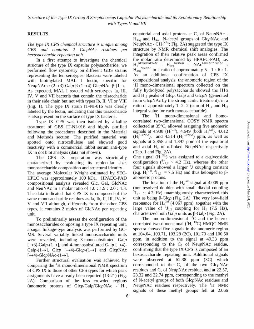
Further structural evaluation was achieved by comparing the
1H mono-dimensional NMR spectrum
of CPS IX to those of other CPS types for which peak assignments have already been reported (13-21) (Fig. 2A). Comparison of the less crowded regions (anomeric protons of Glcp/Galp/GlcpNAc - H1,
equatorial and axial protons at C2 of NeupNAc - H3eq and H3ax, N-acetyl groups of GlcpNAc and NeupNAc - CH3
NAc; Fig. 2A) suggested the type IX
structure by NMR chemical shift analogies. The integration of their relative peak areas confirmed the molar ratio determined by HPAEC-PAD, i.e. H1
Glc/Gal/GlcNAc : H3eq
NeuNAc : NAc
GlcNAc/NeuNAc :
H3eqNeuNAc
in a ratio of approximately 5 : 1 : 6 : 1. As an additional confirmation of CPS IX compositional analysis, the anomeric region of the 1H mono-dimensional spectrum collected on the
fully hydrolyzed polysaccharide showed the H1α and H1β peaks of Glcp, Galp and GlcpN (generated from GlcpNAc by the strong acidic treatment), in a ratio of approximately 1: 2: 2 (sum of H1α and H1β integral value for each monosaccharide).
The 1H mono-dimensional and homo-
correlated two-dimensional COSY NMR spectra collected at 35°C, allowed assigning five anomeric signals at 4.938 (H1
Glc), 4.649 (both H1
Gal), 4.612
(H1GlcNAc
), and 4.514 (H1GlcNAc
) ppm, as well as signals at 2.858 and 1.897 ppm of the equatorial and axial H3 of α-linked NeupNAc respectively (Tab. 1 and Fig. 2A). One signal (H1
Glc) was assigned to a α-glycosidic
configuration (3J1,2 ~ 4.2 Hz), whereas the other
four signals showed a larger 3J coupling constant
(e.g. H1Gal
, 3J1,2 = 7.5 Hz) and thus belonged to β-
anomeric protons. The location of the H2
Glc signal at 4.009 ppm
(not resolved doublet with small diaxial coupling 3J1,2 ~ 4.2 Hz) unambiguously characterized this
unit as being β-Glcp (Fig. 2A). The very low-field resonance for H4
Gal (4.067 ppm), together with the
large value of 3J1,2 coupling for H1 (7.5 Hz),
characterized both Galp units as β-Galp (Fig. 2A). The mono-dimensional
13C and the hetero-
correlated two-dimensional (1H,
13C) HMQC NMR
spectra showed five signals in the anomeric region at 104.04, 103.71, 103.28 (2C), 101.70 and 100.50 ppm, in addition to the signal at 40.33 ppm corresponding to the C3 of NeupNAc residue, confirming that the type IX CPS is composed of an hexasaccharide repeating unit. Additional signals were observed at 52.38 ppm (3C) which corresponded to the C2 of the two GlcpNAc residues and C5 of NeupNAc residue, and at 22.57, 23.32 and 22.74 ppm, corresponding to the methyl of N-acetyl groups of both GlcpNAc residues and NeupNAc residues respectively. The
1H NMR
signals of these methyl groups fell at 2.066
by guest on March 4, 2020
http://ww
w.jbc.org/
Dow
nloaded from

Structure of the Type IX Group B Streptococcus Capsular Polysaccharide and its Evolutionary Relationship
with Types V and VII
7
(GlcpNAc, 6H) and 2.015 ppm (NeupNAc, 3H). All protons at C2 of Glcp, Galp and GlcpNAc
residues were assigned using COSY, as well as the TOCSY method. The two GlcpNAc and the two Galp residues present in the CPS IX repeating unit were designated a and b to facilitate their differentiation.
To identify the O-glycosidic linkages, selective TOCSY and NOESY experiments were collected by irradiating the four anomeric protons (H1
Glc, H1
Gal a
and H1Gal b
together, H1GlcNac a
and H1GlcNac b
) and the equatorial/axial protons at C3 of the NeupNAc residue (H3eq
NeuNAc and H3ax
NeuNAc). The same chemical shift
and J coupling values revealed for both H1Gal
signals, which fell at the same resonance of other already assigned GBS CPS types, unambiguously characterized these units as being Galp β-linked to the position C4 of GlcpNAc residues [Galp-(14)-GlcpNAc].
In accordance with flow cytometry results using the MAL I lectin, interglycosidic NOEs from H3eq
NeuNAc and H3ax
NeuNAc to H3
Gal a, and from H1
Gal a to
H4GlcNAc a
were revealed, confirming the presence of the NeupNAc-α-(23)-Galp-β-(14)-GlcpNAc-β-(1 trisaccharide branch. In this regard, the NMR represented a validation and confirmation of the previous data and inferences. Therefore, by the time we carried out NMR experiments, only the configuration of a second β-GlcpNAc residue, named GlcNAc b, and the anchoring site of branched trisaccharide to the backbone were unknown. Inter-residual NOE couplings H1
Glc-H4
GlcNAc b, H1
GlcNAc a-
H6GlcNAc b
, H1GlcNAc b
-H4Gal b
were revealed by selective irradiation of the anomeric protons of Glc, GlcNAc a and GlcNAc b residues (Fig. S1), indicating that the 4)-β-GlcpNAc-(14)-β-Galp-(14)-α-Glcp-(1 trisaccharide constitutes the backbone of CPS IX repeating unit.
Low intensity NOE correlations were also revealed between H1
Glc, H1
GlcNAc and H1
Gal b and
protons of the own saccharide ring and the vicinal residue.
Overall, NOE data were consistent with the structure of the CPS type IX repeating unit illustrated in Fig. 3, assembled by combining the NMR results with data obtained from other methodologies previously described. To further confirm the uniqueness of the type IX CPS structure, its collected 1H NMR profile was compared to those obtained for
its most similar polysaccharides, belonging to types V and VII (Fig. 2B). The different fingerprints of the anomeric region supported the previously elucidated
structural diversity. Closer scrutiny of the N-acetyl groups region (NeupNAc and GlcpNAc signals) showed a higher intensity of the GlcpNAc peak in CPS IX due to the presence of two GlcpNAc residues, compared to the other two capsular polysaccharides. CPS type IX mostly resembles types V and VII both at structural and genetic levels
As indicated above and illustrated in Fig. 4A, the type IX polysaccharide repeating unit most closely resembles that of type VII (19) and of type V (17). The type V structure is identical to type VII, except for the presence of a second side chain containing a Glcp residue β-linked to the C3 of the backbone Galp residue. Type IX contains only one lateral chain, just like type VII, but the Glcp residue present in the backbone of types V and VII is here replaced by GlcpNAc.
The machinery responsible for the synthesis of GBS capsular polysaccharides is encoded in the cps operon. The 5’ cpsABCD genes, possibly involved in the regulation of capsular synthesis, and the 3’ neuBCDA, responsible for the synthesis of sialic acid, are highly conserved (22). The remaining cpsE-L genes are more variable between the different serotypes, and encode the enzymes responsible for the synthesis of the repeating units, transport to the outside leaflet of the cell membrane, and repeating unit polymerization.
To investigate the evolutionary relationship between the synthesis machineries of type IX, V and VII capsular polysaccharides, we sought to compare the DNA sequences of their corresponding cps operons (herein cps9, cps5 and cps7).
Firstly, three independent type IX isolates were subjected to deep-sequencing, and their cps9 sequences were compared. The overall DNA sequence identity exceeded 99% in pairwise alignment, similar to the high intra-serotype homology observed for the cps operons of all other GBS serotypes downloaded from public databases.
Secondly, cps9 genes were aligned to their homologs in all other serotypes (Tab. 3). This analysis revealed two well conserved regions spanning from cpsA to cpsF and from neuB to neuA, flanking a more variable central region from cpsG to cpsL. In this central region, type IX showed a higher similarity to types V and VII compared to the other serotypes.
by guest on March 4, 2020
http://ww
w.jbc.org/
Dow
nloaded from

Structure of the Type IX Group B Streptococcus Capsular Polysaccharide and its Evolutionary Relationship
with Types V and VII
8
The putative function of each of the glycosyl transferases and polymerases involved in the assembly of the type IX, V and VII hexasaccharide repeating unit was predicted based on literature data and on comparisons with other serotypes (Fig. 4A). A schematic pairwise alignment of the DNA segment comprising genes cpsF to cpsL from serotypes IX, V and VII is illustrated in Fig. 4B. The highest sequence identity in this region (>95%) was observed between types V and IX from the full cpsF up to nucleotide 326 of cpsM and from cpsJ to cpsL. Sequence identity decreased dramatically (<60%) for the second half of cpsM and the full cpsI gene sequence, a region that also included the unique type V cpsO gene (see also Tab. 3). The data suggested a recombination event as the possible cause for the differentiation between the two serotypes.
Comparison between the variable region of types IX and VII revealed 99% identity for cpsG up to nucleotide 59, and lower sequence identity (between 84 and 92%) from that position up to nucleotide 246 of neuB. The degree of similarity between types VII and IX matches that observed when comparing type VII to type V, if one excludes the hyper-variable cps5MOI region. Also in this case, the sequence identity decreased from nucleotide 178 of cpsG up to nucleotide 82 of neuB. This observation is in contrast with the previously reported high conservation of cpsL among all GBS serotypes (22).
Overall, these results indicate that, at the genetic level, type IX is more related to type V than to type VII. Sequence polymorphisms in cpsMOI genes determine capsular polysaccharide specificity between serotypes V, VII and IX
As reported above, the cpsMOI genes showed the highest degree of variability in the otherwise highly similar cps5, cps7 and cps9 operons. This observation led us to hypothesize that differences in these three genes could account for the structural diversity between serotypes IX, V and VII.
To test this hypothesis, we investigated whether episomal transfer of cpsMOI genes between the three serotypes could be sufficient to drive the synthesis of heterologous GBS polysaccharides.
To this end, we first designed the plasmid construct pAM-IX containing cps9MI genes under the GBS p80 promoter (27). The plasmid was subsequently used to transform a type V strain. Expression of different types of CPS in the recipient
and recombinant strains was analyzed by latex agglutination and flow cytometry, using mouse sera raised against type V and rabbit sera against type IX polysaccharides.
As shown in Fig. 5A, the recipient strain showed positive latex agglutination only with anti-type V, while recombinant GBS V harboring pAM-IX showed double agglutination with both type V and type IX antibodies. Similar results were obtained by flow cytometry experiments (Fig. 5B), where the recipient strain showed a fluorescence shift only with anti-CPS V, while positive shifts were observed in the recombinant strain for both anti-CPS V and anti-CPS IX antibodies.
The data indicated that anti-type IX antibodies are highly specific for the type IX CPS and do not cross-react with the similar but not identical type V structure. Furthermore, we confirmed that transfer of type IX cps9MI genes in a type V background is sufficient to drive the synthesis of both type V and IX CPS. Fluorescence intensities with anti-CPS V antibodies decreased in the recombinant strain compared to the wild-type recipient strain, indicating decreased synthesis of CPS V when cps5MOI and cps9MI were co-expressed by recombinant bacteria.
We then constructed the plasmid pAM-V containing the cps5MOI genes under the GBS p80 promoter, and this construct was transferred into a GBS type IX isolate. As shown in Fig. 5C-D, latex serotyping and flow cytometry using anti-CPS V and anti-CPS IX rabbit and mouse sera respectively revealed the presence of polysaccharide IX only in the recipient strain, and of both type IX and type V CPS in recombinant bacteria. Again, type IX CPS decreased in GBS co-synthesizing type V. Again the data confirmed specificity of anti-type V antibodies and the possibility of co-expressing type V and IX CPS in a type IX background by transfer of the cps5MOI genes.
Finally, we introduced in a GBS type VII strain either pAM-IX or pAM-V plasmids. Latex serotyping showed type VII reactivity in the recipient strain, double type VII and IX reactivity for the clone harboring pAM-IX, and double type VII and V reactivity for pAM-V transformed bacteria (Fig. 6). As in former experiments, latex agglutination for the endogenous type VII decreased in transformed recipient strains also expressing heterologous polysaccharides.
The obtained results confirmed that
by guest on March 4, 2020
http://ww
w.jbc.org/
Dow
nloaded from

Structure of the Type IX Group B Streptococcus Capsular Polysaccharide and its Evolutionary Relationship
with Types V and VII
9
differences in cpsMOI gene can fully explain the diversity in the polysaccharide structures and serological characteristics between CPS IX, V and VII.
DISCUSSION
The CPS repeating unit of the recently described GBS serotype IX was confirmed to contain a unique structural motif differing from all previously identified GBS CPSs. Similar to CPS types Ia, Ib, II, III, IV, V, VI, and VII, type IX CPS is exclusively composed of Glcp, Galp, GlcpNAc and NeupNAc residues. The NeupNAc is always positioned at the terminal end of the repeating unit side chain, and its involvement in the immuno-dominant CPS epitope has been demonstrated for type III, Ia, II, V and VI antigens (28-34).
The overall CPS type IX structure most closely resembles that of type VII (19) except that the tri-linked Glcp residue in its backbone is replaced by GlcpNAc, resulting in 2 moles of GlcNAc per mol repeating unit. In addition to this monosaccharide substitution, type IX differs from type V for the presence in the latter of a second side chain of Glcp linked β-(13) to the backbone Galp residue.
Recent comparisons of GBS latex agglutination serotyping results and PCR-based capsular gene typing revealed some inconsistencies between the two methods, mostly associated with type IX (23). Serotyping difficulties could in part be due to weak serum cross-reactivity, requiring pre-absorption with the different antigens for correct serological discrimination between the different types.
We further investigated immunological cross-reactivity between types IX, V and VII by FACS analysis of GBS belonging to the three related serotypes using both, commercial rabbit sera and mouse sera obtained by immunizing animals with purified CPS-V and CPS-IX conjugated to CRM197. The obtained data indicate higher cross-reactivity between types VII and IX compared to type V, and confirmed a higher specificity of mouse antibodies raised against purified polysaccharides compared to commercial rabbit antisera in the highly sensitive FACS setting (Fig. 8). The difficulties in obtaining highly specific antisera confirm the strong similarity between immuno- dominant epitopes of these structurally similar polysaccharides, and could also
explain why type IX was identified only recently. One consequence of this similarity is the possibility that the three polysaccharide types could induce cross-reactive functional antibodies. The high sensitivity of the available type VII and IX strains to complement did not allow testing this hypothesis by functional opsonophagocitic or in vivo mouse protection assays.
The observation that type IX is genetically closer to type V than to type VII was unexpected, given the closer structural similarity and higher serological cross-reactivity between types IX and VII compared to type V. However, genetic evidence seems to indicate that the type V cps operon originated from a pre-existing type IX cps by a horizontal transfer-recombination event targeting positions 326 of cps9M and 10 of cps9J. This event resulted in the formation of a functional cpsM chimera catalyzing the incorporation of a Glcp residue in position 3 in place of GlcpNAc, and in a new variant of cpsI, attaching the first GlcpNAc of the lateral chain to this 3
rd backbone
residue. Finally, the generation of a new Glcp lateral branch resulted from the acquisition of the cps5O gene.
A proposed phylogenetic model for the evolution of type VII, IX and V capsular types in the GBS population is shown in Fig. 7. According to this model, CPS VII and IX derived from a common ancestor by sequential addition of point mutations, leading to divergent structures containing either Glcp or GlcpNAc in position 3 of the backbone of the repeating unit. A subsequent recombination event in a type IX background generated the type V CPS, where the 3
rd backbone
residue is again Glcp. Unfortunately, the mining of publicly available genome databases has not yet provided any clues on the exact identity of the donor strain/species. However, we were able to identify cpsO homologs in S. suis and S. infantarius, thus suggesting a possible involvement of these related streptococci in the arise of type V CPS.
The phylogenetic relationship between serotype V and serotype IX GBS strains remains to be investigated in detail. Type V strains are highly diverse in that, based on MLST analysis, they have been assigned to different GBS Clonal Complexes, including the prevalent CC1, but also CC19, CC23 and CC10 (35). The three type IX isolates sequenced in this study belonged to CC130 that, to
by guest on March 4, 2020
http://ww
w.jbc.org/
Dow
nloaded from

Structure of the Type IX Group B Streptococcus Capsular Polysaccharide and its Evolutionary Relationship
with Types V and VII
10
our knowledge, has not been ascribed to any type V strain. The genetic distance between the two serotypes will need to be confirmed by genome wide analyses on multiple strains, and would imply that in addition to the proposed lateral transfer involving part of the cps operon that led to the appearance of CPS type V, capsular switching events have occurred in ancestor strains belonging to one of the two serotypes.
Interestingly, while the relative frequency of type IX isolates in the GBS population seems to be very low, although awaiting more comprehensive molecular or sero-epidemiological studies, serotype V recently emerged as one of the major serotypes causing adult disease and is now increasing in neonatal infections (7).
The data showing that an anti-CPS V reactive polysaccharide could be synthesized by a recombinant type IX strain expressing cps5MOI in trans, support the proposed model for the emergence of type V from a genetic exchange limited to the three genes. Similarly, an anti-CPS IX reactive polysaccharide was synthesized in a type V strain carrying cps9MI in a plasmid vector. Interestingly, the sole addition of the same plasmid to a type VII strain was sufficient to drive the synthesis of an anti-CPS IX-reactive CPS. The data indicate that the cps7H polymerase, which differs from cps9H by 29 amino acids, can still operate on a repeating unit substrate containing a terminal GlcpNAc instead of Glcp.
The above results are in agreement with previous data from Chaffin et al. reporting that a single gene can confer GBS serotype specificity (36). In that case, the authors reported that cpsH differences were the sole responsible for the structural diversity between types Ia and III. In the present study, differences in the cpsMOI genes accounts for the diversity between types IX, V and VII, while the cpsH enzyme shows more relaxed substrate specificity among the three serotypes. Similar to what observed by Chaffin et al., synthesis of a heterologous CPS reduced the synthesis of the homologous immuno-reactive polysaccharide. This is possibly due to a competition between the two alternative repeating units as substrates for the enzymes catalyzing the downstream steps of CPS production, also taking into account that multicopy expression from a strong promoter might favor the synthesis of the heterologous RU.
Like for other pathogens, the CPSs are excellent antigen targets for the prevention of GBS infections,
and conjugate vaccines have been developed against the major serotypes Ia, Ib, II, III and V. Elucidation of the CPS type IX motif provides additional knowledge that can be instrumental for the development of carbohydrate-based vaccines covering all GBS serotypes.
by guest on March 4, 2020
http://ww
w.jbc.org/
Dow
nloaded from

Structure of the Type IX Group B Streptococcus Capsular Polysaccharide and its Evolutionary Relationship
with Types V and VII
11
REFERENCES
1. Schuchat A. (1998) Epidemiology of Group B Streptococcal Disease in the United States: Shifting
Paradigms. Clin. Microb. Rev. 11, 497-513.
2. Rubens C. E., Wessels M. R., Heggen L. M., Kasper D. L. (1987) Transposon mutagenesis of type
III group B Streptococcus: correlation of capsule expression with virulence. Proc. Natl. Acad. Sci.
U S A 84, 7208-7212.
3. Wessels, M. R., Rubens, C.E., Benedi, V. Kasper, D.L. (1989) Definition of a bacterial virulence
factor: Sialylation of the group B streptococcal capsule. Proc. Natl. Acad. Sci. U S A 86, 8983-
8987.
4. Cieslewicz M.J., Kasper D.L., Wang Y., Wessels M.R. (2001) Functional analysis in type Ia group
B Streptococcus of a cluster of genes involved in extracellular polysaccharide production by
diverse species of streptococci. J. Biol. Chem. 276, 139-146.
5. Baker, C., J. Kasper, D. L. (1976) Correlation of maternal antibody deficiency with susceptibility
to neonatal group B streptococcal infection. N. Engl. J. Med.294:753-756.
6. Edwards, M. S. (2008) Group B streptococcal conjugate vaccine. A timely concept for which the
time has come. Human Vaccines 4, 444-448.
7. Le Doare K., Heath P. T. (2013) An overview of global GBS epidemiology. Vaccine 31 Suppl
4:D7-12.
8. Ferrieri P., Lynfield R., Creti R., Flores A. E. (2013) Serotype IV and invasive group B
Streptococcus disease in neonates, Minnesota, USA, 2000-2010. Emerg. Infect. Dis. 19, 551-558.
9. Slotved, H.C., Kong, F., Lambertsen, L., Sauer, S., and Gilbert, G.L. (2007) Serotype IX, a
proposed new Streptococcus agalactie serotype. J. Clin. Microbiol. 45, 2929-2936.
10. Imperi, M., Pataracchia, M., Alfarone, G., Baldassarri, L., Orefici, G., and Creti, R. (2010) A
multiplex PCR assay for the direct identification of the capsular type (Ia to IX) of Streptococcus
agalactiae. J. Microbiol. Methods 80, 212-214.
11. Melin, P., Efstratiou, A., (2013). Group B streptococcal epidemiology and vaccine needs in
developed countries. Vaccine. Suppl 4, D31-42.
12. Lamagni, T. M., Keshishian, C., Efstratiou, A., Guy, R., Henderson, K.L., Broughton, K.,
Sheridan, E. (2013). Emerging trends in the epidemiology of invasive group B streptococcal
disease in England and Wales, 1991-2010. Clin Infect Dis. 57:682-688.
13. Jennings, H. J., Katzenellenbogen, E., Lugowski, C., and Kasper, D. L. (1983) Structure of native
polysaccharide antigens of type Ia and type Ib group B Streptococcus. Biochemistry 22, 1258-
1264.
14. Jennings, H. J., Rosell, K.-G., Katzenellenbogen, E., and Kasper, D. L. (1983) Structural
determination of the capsular polysaccharide antigen of type II group B streptococci. J. Biol.
Chem. 258, 1793-1798.
15. Jennings, H. J., Lugowski, C., and Kasper D. L. (1981) Conformational aspects critical to the
immunospecificity of the type III group B streptococcal polysaccharide. Biochemistry 20, 4511-
4518.
16. Di Fabio, J. L., Michon, F., Brisson, J.-R., and Jennings, H. J. (1989) Structure of the capsular
polysaccharide antigen of type IV group B Streptococcus. Can. J. Chem. 67, 877-882.
17. Wessels, M. R., DiFabio, J. L., Benedi, V.-J., Kasper, D. L., Michon, F., Brisson, J.-R., Jelinkova,
J., and Jennings, H. J. (1991) Structural determination and immunochemical characterization of
the type V group B Streptococcus agalactiae capsular polysaccharide. J. Biol. Chem. 266, 6714-
6719.
18. von Hunolstein, C., D’Ascenzi, S., Wagner, B., Jelinkova, J., Alfarone, G., Recchia, S., Waner,
M., and Orefici, G. (1993) Immunochemistry of capsular type polysaccharide and virulence
by guest on March 4, 2020
http://ww
w.jbc.org/
Dow
nloaded from

Structure of the Type IX Group B Streptococcus Capsular Polysaccharide and its Evolutionary Relationship
with Types V and VII
12
properties of type VI Streptococcus agalactiae (group B Streptococcus). Infect. Immun. 61, 1272-
1280.
19. Kogan, G., Brisson, J.-R., Kasper, D. L., von Hunolstein, C., Orefici, G., and Jennings, H. J.
(1995) Structural elucidation of the novel type VII group B Streptococcus capsular
polysaccharide by high resolution NMR spectroscopy. Carbohydr. Res. 277, 1-9.
20. Kogan, G., Uhrín, D., Brisson, J.-R., Paoletti, L. C., Blodgett, A. E., Kasper, D. L., and Jennings
H. J. (1996) Structural and immunochemical characterization of the type VIII group B
Streptococcus capsular polysaccharide. J. Biol. Chem. 271, 8786-8790.
21. Pinto, V., Berti, F. (2014) Exploring the Group B Streptococcus capsular polysaccharides: The
structural diversity provides the basis for development of NMR-based identity assays. J. Pharm
Biomed. Anal. 98C, 9-15.
22. Cieslewicz, M.J., Chaffin, D., Glusman, G., Kasper, D. L., Madan, A., Rodrigues, S., Fahey, J.,
Wessels, M.R., and Rubens, C.E. (2005) Structural and genetic diversity of group B
Streptococcus capsular polysaccharides. Infect. Immun. 73, 3096-3103. 23. Yao, K., Poulsen, K., Maione, D., Rinaudo, C.D., Baldassarri, L., Telford, J.L., Sørensen, U.B.,
Members of the DEVANI Study Group, Kilian, M. (2013) Capsular gene typing of Streptococcus
agalactiae compared to serotyping by latex agglutination. J. Clin. Microbiol. 51:503-507.
24. Wessels, M.R., Paoletti, L.C., Kasper, D.L., DiFabio, J.L., Michon, F., Holme, K., and Jennings
H.J. (1990) Immunogenicity in animals of a polysaccharide-protein conjugate vaccine against
type III group B Streptococcus. J. Clin. Invest. 86, 1428-1433.
25. Ciucanu, I., Kerek, F. (1984) A simple and rapid method for the permethylation of carbohydrates.
Carbohydr. Res. 131, 209-217.
26. Maione D., Margarit I., Rinaudo C. D., Masignani V., Mora M., Scarselli M., Tettelin H., Brettoni
C., Iacobini E. T., Rosini R., D'Agostino N., Miorin L., Buccato S., Mariani M., Galli G.,
Nogarotto R., Nardi-Dei V., Vegni F., Fraser C., Mancuso G., Teti G., Madoff L. C., Paoletti L.
C., Rappuoli R., Kasper D. L., Telford J. L., Grandi G. (2005) Identification of a universal Group
B streptococcus vaccine by multiple genome screen. Science 309, 148-150.
27. Buccato S., Maione D., Rinaudo C. D., Volpini G., Taddei A. R., Rosini R., Telford J. L., Grandi
G., Margarit I. (2006) Use of Lactococcus lactis expressing pili from group B Streptococcus as a
broad-coverage vaccine against streptococcal disease. J. Infect. Dis. 194, 331-340.
28. Brisson, J.R., Uhrinova, S., Woods R.J., van der Zwan, M., Jarrell, H.C., Paoletti, L.C., Kasper,
D.L., Jennings, H.J. (1997) NMR and molecular dynamics studies of the conformational epitope of
the type III group B Streptococcus capsular polysaccharide and derivatives. Biochemistry 36,
3278-3292.
29. González-Outeiriño, J., Kadirvelraj, R., Woods, R.J. (2005) Structural elucidation of type III group
B Streptococcus capsular polysaccharide using molecular dynamics simulations: the role of sialic
acid. Carbohydr Res. 340, 1007-1018.
30. Jennings, H.J., Katzenellenbogen, E., Lugowski, C., Michon, F., Roy, R., Kasper, D.L. (1984)
Structure, conformation, and immunology of sialic acid-containing polysaccharides of human
pathogenic bacteria. Pure Appl. Chem. 56, 893-905.
31. Zou, W., Mackenzie, R., Thérien, L., Hirama, T., Yang, Q., Gidney, M.A., Jennings, H.J. (1999)
Conformational epitope of the type III group B Streptococcus capsular polysaccharide. J. Immunol.
163, 820-825.
32. Zou, W., Jennings H.J. (2001) The conformational epitope of type III group B Streptococcus
capsular polysaccharide. Adv. Exp. Med. Biol. 491, 473-84.
by guest on March 4, 2020
http://ww
w.jbc.org/
Dow
nloaded from

Structure of the Type IX Group B Streptococcus Capsular Polysaccharide and its Evolutionary Relationship
with Types V and VII
13
33. Safari, D., Dekker, H.A., Rijkers, G.T., van der Ende, A., Kamerling, J.P., Snippe, H. (2011) The
immune response to group B streptococcus type III capsular polysaccharide is directed to the -Glc-
GlcNAc-Gal- backbone epitope. Glycoconj. J. 28, 557-62.
34. Guttormsen, H.K., Paoletti, L.C., Mansfield, K.G., Jachymek, W., Jennings, H.J., Kasper, D.L.
(2008) Rational chemical design of the carbohydrate in a glycoconjugate vaccine enhances IgM-to-
IgG switching. Proc. Natl. Acad. Sci. U S A. 105, 5903-5908.
35. Honsa E., Fricke T., Stephens A. J., Ko D., Kong F., Gilbert G. L., Huygens F., Giffard P. M.
(2008) Assignment of Streptococcus agalactiae isolates to clonal complexes using a small set of
single nucleotide polymorphisms. BMC Microbiol. 8, 1-10.
36. Chaffin D. O., Beres S. B., Yim H. H., Rubens C. E. (2000) The serotype of type Ia and III group
B streptococci is determined by the polymerase gene within the polycistronic capsule operon. J.
Bacteriol. 182, 4466-4477.
by guest on March 4, 2020
http://ww
w.jbc.org/
Dow
nloaded from

Structure of the Type IX Group B Streptococcus Capsular Polysaccharide and its Evolutionary
Relationship with Types V and VII
14
Acknowledgments - We are grateful to Alberto Berardi, Roberta Creti, Lucilla Baldassarri, Graziella
Orefici and the DEVANI consortium for providing type IX GBS strains. We thank Stefano Censini and
Nicola Pacchiani of Novartis Vaccines (Siena, Italy) for support with genome sequence analysis.
FOOTNOTES
1To whom correspondence should be addressed: Francesco Berti, Novartis Vaccines Research, Via
Fiorentina 1, 53100 Siena, Italy, Tel.: +39 0577 243895; Fax: +39 0577 243564; E- mail:
2 Abbreviations: GBS, Group B Streptococcus; Glcp, glucose; CPS, capsular polysaccharide; Galp,
galactose; GlcpNAc, N-acetyl-glucosamine; GlcpN, glucosamine; NeupNAc, N-acetyl-neuraminic acid; Rhap, rhamsose; NMR, Nuclear Magnetic Resonance; SEC-HPLC, Size Exclusion Chromatography-High Pressure Liquid Chromatogrphy; HPAEC-PAD, High Performance Anionic Exchange Chromatography-Pulsed Amperometric Detection; GC-MS, Gas Chromatography-Mass Spectrometry; WB, Western Blot; FACS, Flow Cytometry.
by guest on March 4, 2020
http://ww
w.jbc.org/
Dow
nloaded from

Structure of the Type IX Group B Streptococcus Capsular Polysaccharide and its Evolutionary
Relationship with Types V and VII
15
TABLES LEGENDS
Table 1. NMR chemical shift of protons at C1 and C2 for GBS type IX CPS recorded at 35 ± 0.1°C. Other
signals: H3eqNeuNAc
2.858 ppm, H3axNeuNAc
1.897 ppm, NAcGlcNAc a/b
2.179 ppm, NAcNeuNAc
2.132
ppm.
Table 2. List of oligonucleotides used for plasmid construction.
Table 3. Nucleotide sequence identity between type IX cps genes (strain IT-NI-016) and types V
(2603/VR), VII (7271, AY376403), Ia (A909), Ib (H36B), II (18RS21), III (NEM316), IV
(CNTC 1/82, AF355776.1), and VI (NT6 type VI, AF337958). Identity levels are underlined
by a grey color scale. (n.a. not available).
by guest on March 4, 2020
http://ww
w.jbc.org/
Dow
nloaded from

Structure of the Type IX Group B Streptococcus Capsular Polysaccharide and its Evolutionary
Relationship with Types V and VII
16
Table 1
Sugar residue H1 H2
Glcp 4.938 4.009
Galp a 4.649 3.668
Galp b 4.649 3.862
GlcpNAc a 4.514 3.479
GlcpNAc b 4.612 3.722
by guest on March 4, 2020
http://ww
w.jbc.org/
Dow
nloaded from

Structure of the Type IX Group B Streptococcus Capsular Polysaccharide and its Evolutionary
Relationship with Types V and VII
17
Table 2
Primer name Sequence (5’→3’) Length (bp)
pAM-IXMI-F GCGGCGCGGCCGCGACATATTTGCTCTGATATGGCAG 37
pAM-IXMI-R GCGGCAGATCTGGGATAATGATACTAATCATCTTC 35
pAM-VMOI-F GCGGCGCGGCCGCGCTCTGATATGGCAGGAGGTAAGG 37
pAM-VMOI-R GCGGCAGATCTGGGATAATGATACTAACTTTATCC 35
by guest on March 4, 2020
http://ww
w.jbc.org/
Dow
nloaded from

Structure of the Type IX Group B Streptococcus Capsular Polysaccharide and its Evolutionary
Relationship with Types V and VII
18
Table 3
cpsA cpsB cpsC cpsD cpsE cpsF cpsG cpsH cpsM cpsO cpsI cpsJ cpsK cpsL neuB neuC neuD neuA
V 99.2 99.7 99.3 98.4 99 99.3 98.2 96.8 83.6 - 55.6 97.7 99.4 99 99.9 99.6 99.5 99
VII n.a. n.a. 99.3 99.7 98.8 99.1 92.5 92 87.2 - 84.4 85.8 87.5 91.7 98.1 99.2 99.5 98.8
Ia 99.4 99.6 99.1 99.6 98.9 99.8 79.8 36.8 - - 45.2 51.8 97.6 96.7 99.6 99 99.4 98.9
Ib 99.5 99.6 99.3 98.3 98.7 99.1 79.2 37 - - 45.1 44.4 62 98 99.4 99 99.2 98.7
II 99.5 99.7 99.1 99.6 99 99.8 98.6 42.8 - - 43.6 51.1 48.7 97.6 99.8 99.2 99.5 99
III 99.6 99.6 99.1 99.7 99.3 99.8 73.1 37.6 - - 45.9 52 97.6 96.7 99.6 99.1 99.4 99.3
IV 99.5 99.9 99.1 99.3 99.1 99.8 99.2 87 64.6 - 39.8 54.2 82.1 97.5 99.5 99.1 99.2 99.1
VI 99.9 99.9 99.3 97.9 98.6 99.3 72.7 37.8 - - 45.6 39.2 62.2 98.8 99.5 99.5 99.2 98.6
by guest on March 4, 2020
http://ww
w.jbc.org/
Dow
nloaded from

Structure of the Type IX Group B Streptococcus Capsular Polysaccharide and its Evolutionary
Relationship with Types V and VII
19
FIGURES LEGENDS
Figure 1. Flow cytometry analysis of strains belonging to the ten GBS serotypes after incubation with
MAL I lectin, specific for NeupNAc-α-(23)-Galp-β-(14)-GlcpNAc-β-(1.
Figure 2. A. 1H NMR spectrum of GBS type IX capsular polysaccharide recorded at 35 0.1°C. B.
Comparison of 1H NMR spectra obtained for capsular polysaccharides IX, VII and V recorded
at 25 0.1°C.
Type V: H1Glc a
: anomeric proton of Glcp residues in the branch; H1Glc b
and H1Glc c
: anomeric
protons of Glcp residue in the backbone; H1Gal a/b
: anomeric protons of Galp residues in the
branch and backbone; H1GlcNAc
: anomeric proton of GlcpNAc residue in the branch; H3eqNeuNAc
and H3axNeuNAc
: protons at position C3equatorial and axial of NeupNAc residue in the branch.
Type VII: H1Glc a
and H1Glc b
: anomeric protons of Glcp residue in the backbone; H1Gal a/b
:
anomeric protons of Galp residues in the branch and backbone; H1GlcNAc
: anomeric proton of
GlcpNAc residue in the branch; H3eqNeuNAc
and H3axNeuNAc
: protons at position C3 equatorial and
axial of NeupNAc residue in the branch.
Type IX: H1Glc
anomeric proton of Glcp residues in the backbone; H1Gal a/b
: anomeric protons of
Galp residues in the branch and backbone; H1GlcNAc a
: anomeric proton of GlcNAcp residue in
the branch; H1GlcNAc a
: anomeric proton of GlcpNAc residue in the backbone; H4Gal a/b
: protons
at position C4 of Galp residues in the branch and backbone; H2Glc
: proton at position C2 of
Glcp residue in the backbone; H3eqNeuNAc
and H3axNeuNAc
: protons at position C3 equatorial and
axial of NeupNAc residue in the branch.
Figure 3. Repeating unit structure of CPS IX with inter-residue NOEs revealed by selective
NOESY experiments on anomeric protons and H3eqNeuNAc
. The double GlcpNAc and
Galp residues are labeled a and b.
Figure 4. Comparison of CPS type IX with types VII and V. A. Chemical structure of the repeating
units of type V, VII and IX CPS. The putative function of the enzymes involved in the
assembly of the type IX CPS is indicated by arrows. cpsM, cpsO and cpsI
by guest on March 4, 2020
http://ww
w.jbc.org/
Dow
nloaded from

Structure of the Type IX Group B Streptococcus Capsular Polysaccharide and its Evolutionary
Relationship with Types V and VII
20
glycosyltransferases, likely catalyzing the addition of the differing monosaccharides, are
labeled in red for the three CPS types. B. Schematic illustration of the cps9 operon and
pairwise alignments between the central regions comprising genes cpsG to neuB of cps9, cps7
and cps5. The cps5O gene present only in type V is indicated in red. A gap was introduced
between cpsM and cpsI in the alignment of type IX and VII (top) to compensate for the lack of
cpsO. Green areas indicate regions with 100% identity and variable regions are indicated in
yellow.
Figure 5. Concomitant synthesis of CPS IX and CPS V in recombinant type V and type IX strains
transformed with pAM-cps5MOI and pAM-cps9MI, respectively. A and C: Serotyping of wild-
type and recombinant bacteria by latex agglutination. B and D: Flow cytometry analysis of the
same strains using anti CPS-V and anti-CPS IX mouse polyclonal antibodies. The red peaks
represent negative controls only labeled with secondary antibodies; the blue peaks represent
bacteria labeled with the anti-CPS antibodies.
Figure 6. Concomitant synthesis of CPS VII and CPS IX and of CPS VII and CPSV in recombinant type
VII strains transformed with plasmids harboring type IX cps9MI or type V cps5MOI genes.
Latex agglutination of GBS wild-type and recombinant bacteria using anti-type V, type VII
and type IX antisera, as well as anti-type Ia as negative control.
Figure 7. Proposed model for the genetic relationship between serotypes IX, VII and V. According to
this model, types VII and IX derived from a common ancestor by sequential addition of point
mutations, while type V emerged from a subsequent recombination event in a type IX
background. The boxed region highlights the genetic segment involved in the recombination
event resulting in a type V CPS.
by guest on March 4, 2020
http://ww
w.jbc.org/
Dow
nloaded from

Structure of the Type IX Group B Streptococcus Capsular Polysaccharide and its Evolutionary
Relationship with Types V and VII
21
Figure 8. Flow cytometry analysis of type V, VII and IX strains using anti- CPS-V, CPS-VII and CPS-IX
rabbit commercial sera (r, Statens Institut - Denmark) and anti- CPS-V and CPS-IX mouse
sera (m) obtained by immunizing animals with the purified polysaccharides conjugated to
CRM197. The red peaks represent negative controls labeled with secondary antibodies; the blue
peaks represent bacteria incubated with the anti-CPS sera and labeled secondary antibodies.
by guest on March 4, 2020
http://ww
w.jbc.org/
Dow
nloaded from

Structure of the Type IX Group B Streptococcus Capsular Polysaccharide and its Evolutionary
Relationship with Types V and VII
22
Figure 1
by guest on March 4, 2020
http://ww
w.jbc.org/
Dow
nloaded from
Structure of the Type IX Group B Streptococcus Capsular Polysaccharide and its Evolutionary
Relationship with Types V and VII
23
Figure 2
by guest on March 4, 2020
http://ww
w.jbc.org/
Dow
nloaded from

Structure of the Type IX Group B Streptococcus Capsular Polysaccharide and its Evolutionary
Relationship with Types V and VII
24
Figure 3
by guest on March 4, 2020
http://ww
w.jbc.org/
Dow
nloaded from

Structure of the Type IX Group B Streptococcus Capsular Polysaccharide and its Evolutionary
Relationship with Types V and VII
25
Figure 4
by guest on March 4, 2020
http://ww
w.jbc.org/
Dow
nloaded from

Structure of the Type IX Group B Streptococcus Capsular Polysaccharide and its Evolutionary
Relationship with Types V and VII
26
Figure 5
by guest on March 4, 2020
http://ww
w.jbc.org/
Dow
nloaded from

Structure of the Type IX Group B Streptococcus Capsular Polysaccharide and its Evolutionary
Relationship with Types V and VII
27
Figure 6
by guest on March 4, 2020
http://ww
w.jbc.org/
Dow
nloaded from

Structure of the Type IX Group B Streptococcus Capsular Polysaccharide and its Evolutionary
Relationship with Types V and VII
28
Figure 7
by guest on March 4, 2020
http://ww
w.jbc.org/
Dow
nloaded from

Structure of the Type IX Group B Streptococcus Capsular Polysaccharide and its Evolutionary
Relationship with Types V and VII
29
Figure 8
by guest on March 4, 2020
http://ww
w.jbc.org/
Dow
nloaded from

Janulczyk, Guido Grandi and Immaculada MargaritRosini, Maria Rosaria Romano, Vittoria Pinto, Barbara Brogioni, Giulia Torricelli, Robert
Francesco Berti, Edmondo Campisi, Chiara Toniolo, Laura Morelli, Stefano Crotti, Robertoevolutionary relationship with types V and VII
capsular polysaccharide and itsStreptococcusStructure of the type IX Group B
published online July 2, 2014J. Biol. Chem.
10.1074/jbc.M114.567974Access the most updated version of this article at doi:
Alerts:
When a correction for this article is posted•
When this article is cited•
to choose from all of JBC's e-mail alertsClick here
by guest on March 4, 2020
http://ww
w.jbc.org/
Dow
nloaded from