Structure of the twin-arginine signal-binding protein...
5
structural communications 746 doi:10.1107/S1744309109023811 Acta Cryst. (2009). F65, 746–750 Acta Crystallographica Section F Structural Biology and Crystallization Communications ISSN 1744-3091 Structure of the twin-arginine signal-binding protein DmsD from Escherichia coli Suresh Kumar Ramasamy and William M. Clemons Jr* Division of Chemistry and Chemical Engineering, California Institute of Technology, 1200 East California Boulevard, Pasadena, CA 91125, USA Correspondence e-mail: [email protected] Received 28 May 2009 Accepted 21 June 2009 PDB Reference: DmsD, 3cw0, r3cw0sf. The translocation of folded proteins via the twin-arginine translocation (Tat) pathway is regulated to prevent the futile export of inactive substrate. DmsD is part of a class of cytoplasmic chaperones that play a role in preventing certain redox proteins from premature transport. DmsD from Escherichia coli has been crystallized in space group P4 1 2 1 2, with unit-cell parameters a = b = 97.45, c = 210.04 A ˚ , in the presence of a small peptide. The structure has been solved by molecular replacement to a resolution of 2.4 A ˚ and refined to an R factor of 19.4%. There are four molecules in the asymmetric unit that may mimic a higher order structure in vivo. There appears to be density for the peptide in a predicted binding pocket, which lends support to its role as the signal- recognition surface for this class of proteins. 1. Introduction The twin-arginine translocation (Tat) pathway of bacteria is respon- sible for translocation across the cytoplasmic membrane of folded proteins. Typical Tat substrates are complexes and proteins that contain cofactors that are assembled in the cytoplasm. The minimal Tat system is composed of the integral membrane proteins TatA, TatB and TatC. The current model for translocation is that TatB and TatC form a receptor complex and bind the Tat signal motif, subse- quently targeting the preprotein for translocation across the mem- brane through a TatA complex (reviewed in Berks et al. , 2000; Mu ¨ ller & Klo ¨ sgen, 2005; Lee et al. , 2006). During the process, it is essential to prevent the futile export of immature protein. The dimethylsulfoxide reductase complex (DmsABC) is targeted to the bacterial periplasm via the Tat pathway and is involved in anaerobic respiration using dimethylsulfoxide (DMSO) as an elec- tron acceptor. It is a member of a group of Tat substrates that have a targeting chaperone, which is usually found in their operon. These chaperones, termed redox-enzyme maturation proteins (REMPs; Turner et al., 2004), bind to the Tat signal sequence and presumably regulate folding, substrate addition and complex assembly prior to targeting to the translocation machinery (Berks et al. , 2000; Sargent, 2007). DmsD is the REMP for DmsABC. It is a cytoplasmic protein and its size ranges between 20 and 30 kDa across species. DmsD null mutants fail to grow under anaerobic conditions, implying an essen- tial role in anaerobic respiration (Oresnik et al., 2001). The DmsA preprotein contains the Tat motif (S/TRRxLVK) in its N-terminal signal sequence and this region has been shown to bind directly to DmsD (Oresnik et al., 2001). The remainder of DmsA is a large globular domain with a catalytic molybdopterin (MoPt) cofactor. DmsA is synthesized in the cytoplasm and loaded with cofactor and the accessory protein DmsB prior to translocation. The best studied REMP, TorD, for the trimethylamine N-oxide (TMAO) reductase TorA, performs a parallel function to the DmsA/ DmsD system (Ilbert et al., 2004), and TorD and DmsD fall into a class of REMPs known by the first characterized structure TorD. The REMP motifs (Y/F/W)xxLF and E(Px or xP)D(H/Y) are conserved among all members of this family and mutations in these regions # 2009 International Union of Crystallography All rights reserved
Transcript of Structure of the twin-arginine signal-binding protein...
structural communications
746 doi:10.1107/S1744309109023811 Acta Cryst. (2009). F65, 746–750
Acta Crystallographica Section F
Structural Biologyand CrystallizationCommunications
ISSN 1744-3091
Structure of the twin-arginine signal-bindingprotein DmsD from Escherichia coli
Suresh Kumar Ramasamy and
William M. Clemons Jr*
Division of Chemistry and Chemical
Engineering, California Institute of Technology,
1200 East California Boulevard, Pasadena,
CA 91125, USA
Correspondence e-mail: [email protected]
Received 28 May 2009
Accepted 21 June 2009
PDB Reference: DmsD, 3cw0, r3cw0sf.
The translocation of folded proteins via the twin-arginine translocation (Tat)
pathway is regulated to prevent the futile export of inactive substrate. DmsD is
part of a class of cytoplasmic chaperones that play a role in preventing certain
redox proteins from premature transport. DmsD from Escherichia coli has been
crystallized in space group P41212, with unit-cell parameters a = b = 97.45,
c = 210.04 A, in the presence of a small peptide. The structure has been solved
by molecular replacement to a resolution of 2.4 A and refined to an R factor of
19.4%. There are four molecules in the asymmetric unit that may mimic a higher
order structure in vivo. There appears to be density for the peptide in a
predicted binding pocket, which lends support to its role as the signal-
recognition surface for this class of proteins.
1. Introduction
The twin-arginine translocation (Tat) pathway of bacteria is respon-
sible for translocation across the cytoplasmic membrane of folded
proteins. Typical Tat substrates are complexes and proteins that
contain cofactors that are assembled in the cytoplasm. The minimal
Tat system is composed of the integral membrane proteins TatA,
TatB and TatC. The current model for translocation is that TatB and
TatC form a receptor complex and bind the Tat signal motif, subse-
quently targeting the preprotein for translocation across the mem-
brane through a TatA complex (reviewed in Berks et al., 2000; Muller
& Klosgen, 2005; Lee et al., 2006). During the process, it is essential to
prevent the futile export of immature protein.
The dimethylsulfoxide reductase complex (DmsABC) is targeted
to the bacterial periplasm via the Tat pathway and is involved in
anaerobic respiration using dimethylsulfoxide (DMSO) as an elec-
tron acceptor. It is a member of a group of Tat substrates that have a
targeting chaperone, which is usually found in their operon. These
chaperones, termed redox-enzyme maturation proteins (REMPs;
Turner et al., 2004), bind to the Tat signal sequence and presumably
regulate folding, substrate addition and complex assembly prior to
targeting to the translocation machinery (Berks et al., 2000; Sargent,
2007).
DmsD is the REMP for DmsABC. It is a cytoplasmic protein and
its size ranges between 20 and 30 kDa across species. DmsD null
mutants fail to grow under anaerobic conditions, implying an essen-
tial role in anaerobic respiration (Oresnik et al., 2001). The DmsA
preprotein contains the Tat motif (S/TRRxLVK) in its N-terminal
signal sequence and this region has been shown to bind directly to
DmsD (Oresnik et al., 2001). The remainder of DmsA is a large
globular domain with a catalytic molybdopterin (MoPt) cofactor.
DmsA is synthesized in the cytoplasm and loaded with cofactor and
the accessory protein DmsB prior to translocation.
The best studied REMP, TorD, for the trimethylamine N-oxide
(TMAO) reductase TorA, performs a parallel function to the DmsA/
DmsD system (Ilbert et al., 2004), and TorD and DmsD fall into a
class of REMPs known by the first characterized structure TorD. The
REMP motifs (Y/F/W)xxLF and E(Px or xP)D(H/Y) are conserved
among all members of this family and mutations in these regions# 2009 International Union of Crystallography
All rights reserved

lower the binding affinity to substrates (Chan et al., 2008). Although
physical interaction data between REMPs and partners is limited,
several studies have looked directly at the problem. DmsD binds to
the twin-arginine signal peptide from DmsA via a hydrophobic
interaction with micromolar affinity in an equimolar ratio (Winstone
et al., 2006). DmsD can form a complex with the N-terminus of DmsA
independently of the globular domain of the protein (Oresnik et al.,
2001) and has been shown in vivo to bind to the TatBC complex
under anaerobic conditions, implying a direct role in targeting
(Papish et al., 2003). In vitro, TorD is able to form a complex with the
unfolded mature portions of TorA (Pommier et al., 1998). DmsD/
TorD REMPs are not essential for substrate translocation; instead,
they appear to be important for transport of the correctly folded
complex (Ray et al., 2003).
Many mechanistic questions about REMPs remain. The coordi-
nation between cofactor loading and the subsequent release of the
protein after folding is unknown. There is no direct evidence for
interaction between the Tat machinery, the Tat substrate and the
corresponding REMP protein. The Tat translocase itself has been
shown to accept or reject protein for transport through a quality-
control mechanism (Matos et al., 2008), so the exact role of REMPs is
unclear. This paper reports an additional crystal structure of DmsD
from Escherichia coli (EcDmsD) and provides further evidence for
functional interactions.
2. Methods
2.1. Cloning, overexpression and protein purification
The gene encoding EcDmsD was amplified from E. coli genomic
DNA using the primers 50-GGGCGGGTCGACATGACCCATTT-
TTCACAG-30 and 50-GGGGCGGAGATCTCTATCGAAACAGC-
GG-30, which incorporate the restriction-endonuclease sites SalI and
BglII (highlighted in bold), respectively. Digested PCR product was
ligated into a modified T7 promotor-based pET33b expression vector
(Novagen) that contained an N-terminal 6�His tag followed by a
thrombin cleavage site and a modified multiple cloning site and was
then transformed into E. coli BL21 (DE3) host strain. The plated cells
were directly inoculated into 1 l LB medium containing 35 mg ml�1
kanamycin. The culture was grown at 310 K to an OD600 of 0.6,
followed by induction with 300 mM isopropyl �-d-1-thiogalacto-
pyranoside (IPTG from Anatrace) for 3 h. The cells were harvested
by centrifugation and the cell pellet was resuspended in 100 ml buffer
structural communications
Acta Cryst. (2009). F65, 746–750 Ramasamy & Clemons � DmsD 747
Table 1Data-collection and refinement statistics.
Values in parentheses are for the highest resolution shell.
Data-collection statisticsSpace group P41212Wavelength used (A) 0.98Unit-cell parameters (A) a = b = 97.45, c = 210.04Resolution range (A) 30.0–2.4 (2.49–2.40)Total no. of observations 75194 (7411)Total no. of unique observations 40654Redundancy 3.8 (3.5)Mean I/�(I) 10.6 (2.2)Completeness (%) 99.2 (97.3)Rmerge (%) 13.3 (58.8)Wilson B factor (A2) 33.9
Refinement statisticsAsymmetric unit content 4 moleculesR factor (%) 19.4Rfree factor (%) 24.0No. of reflections in working set 40574No. of reflections in test set 2028Protein atoms 6588Water atoms 509Mean temperature factor (A2) 13.8Matthews coefficient (A3 Da�1) 2.8R.m.s.d. bond lengths (A) 0.014R.m.s.d. bond angles (�) 1.41Ramachandran plot, residues in
Most favored region (%) 97.3Additionally allowed region (%) 2.7
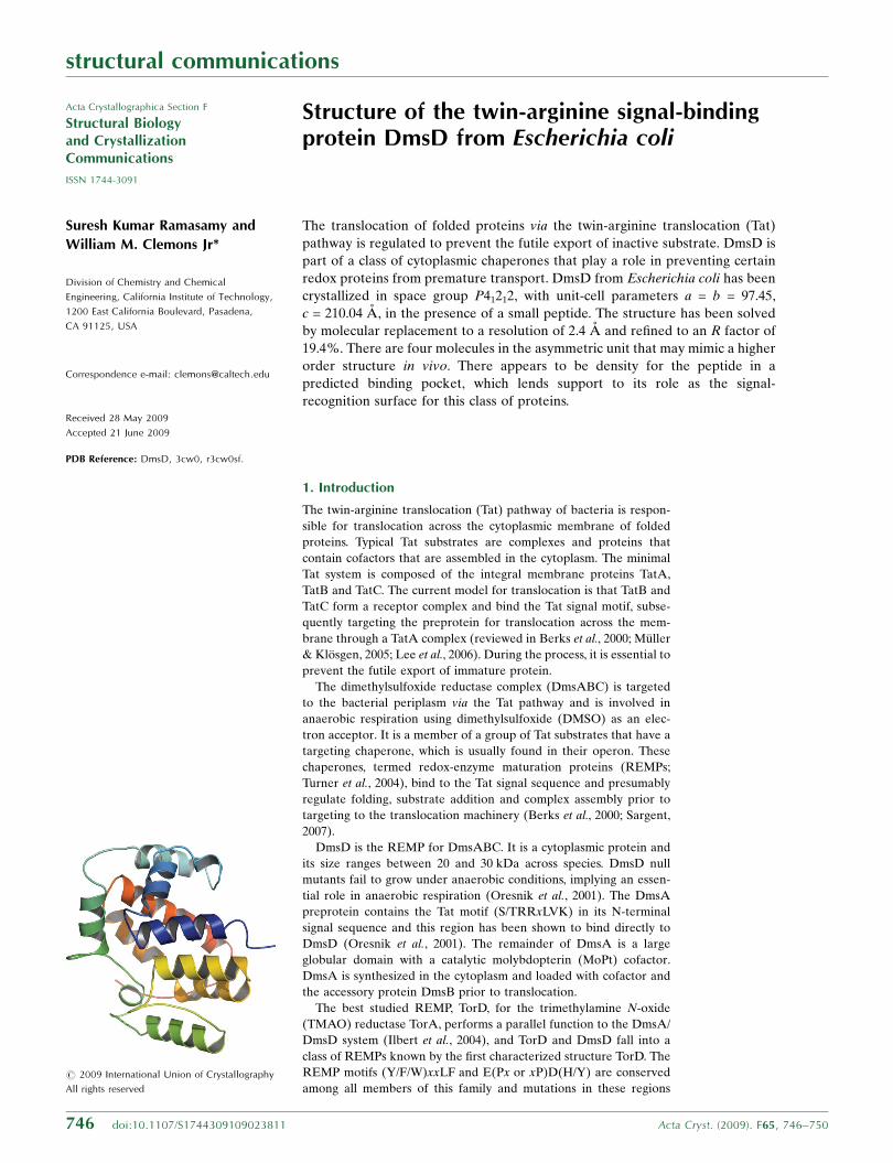
Figure 1(a) Ribbon diagram of a monomer of EcDmsD color-ramped blue to red from the N-terminus to the C-terminus. (b) The tetramer in the asymmetric unit of EcDmsD, witheach monomer colored differently. Monomer A ribbons are colored in a similar way to those in (a) and monomers B–D are colored by chain and shown as spheres. Exceptwhere indicated otherwise, structural figures were produced using PyMOL (DeLano, 2002).

A (100 mM NaCl, 10 mM �-mercaptoethanol, 30 mM imidazole and
50 mM Tris–HCl pH 7.5) and then lysed by passing it three times
though a microfluidizer (Microfluidics). Cell debris was removed by
centrifugation at 18 000g for 20 min at 277 K. The supernatant was
loaded onto a 5 ml Ni–NTA affinity resin (Qiagen) pre-equilibrated
and washed with 30 ml buffer A and then eluted with buffer A
containing 300 mM imidazole. The protein was diluted into PBS
buffer and the 6�His tag was cleaved with 20 units of thrombin
incubated overnight at 295 K, resulting in an additional four residues
at the native N-terminus (GSVD). The remaining uncleaved protein
was removed by passage over a second Ni–NTA affinity resin. The
flowthrough was concentrated and run on a Superdex 200 10/300 GL
column (GE Healthcare) in buffer A without imidazole. The purified
protein was concentrated to 20 mg ml�1 using a centrifugal concen-
tration device (Amicon Ultra, Millipore) before storage at 193 K. The
98.7% pure peptide (SRRDFLK) was ordered from Genescript,
USA.
2.2. Multi-angle light-scattering analysis
Purified EcDmsD (20 mg ml�1) was loaded onto a Shodex Protein
KW-803 size-exclusion column equilibrated with 50 mM Tris–HCl,
150 mM NaCl, 10 mM �-mercaptoethanol and connected in-line with
a Dawn 18-angle light-scattering detector coupled to an Optilab
interferometric refractometer and a WyattQELS Quasi-Elastic Light-
Scattering instrument (Wyatt Technologies). Data analysis was
performed with the ASTRA v5.3.4.14 software (Wyatt Technologies)
and molecular weights were calculated using the Zimm fit method.
2.3. Crystallization, data collection and processing
The purified DmsD was incubated for 2 h with a 1:1 molar ratio of
peptide to protein before crystallization. A Mosquito pipetting robot
was used to set up 200 nl drops, which were equilibrated against 50 ml
reservoir solution using sitting-drop vapor diffusion in a 96-well plate
format using standard crystal screening kits (Hampton Research and
Qiagen). Large single crystals appeared in drops made up from 1 ml
1.5 M ammonium tartrate, 0.1 M Tris pH 7.5 and 1 ml protein solution
and equilibrated against 500 ml reservoir solution using a 24-well
sitting-drop plate (Hampton Research). A single crystal was briefly
transferred to mother liquor supplemented with 30%(v/v) glycerol,
mounted in a cryoloop and flash-cooled in liquid nitrogen.
Diffraction data were collected on beamline BL9-2 at the Stanford
Synchrotron Radiation Laboratory (SSRL). The crystal was main-
tained at a constant 100 K throughout the experiment. A total of 180
images were collected with an oscillation range of 0.5� per image, an
exposure time of 8.1 s and a crystal-to-detector distance of 275 mm.
Images were collected on a MAR Mosaic 325 CCD detector and
indexed using HKL-2000 (Otwinowski & Minor, 1997). The crystals
belonged to space group P41212, with unit-cell parameters a = b = 97.45,
c = 210.04 A and a solvent content of 56% (Matthews, 1968) with four
molecules in the asymmetric unit. Scaling of the data was performed
using SCALA (Evans, 2006). Data statistics are presented in Table 1.
2.4. Structure determination and refinement
The structure of EcDmsD was solved by molecular replacement
using the program Phaser (McCoy, 2007) with a single chain of the
DmsD structure from Salmonella typhimurium (68% sequence
identity; PDB code 1s9u) as a search model. The initial model was
built using a combination of automatic building and refinement with
ARP/wARP (Langer et al., 2008). Rigid-body and TLS refinement
were performed using REFMAC (Vagin et al., 2004). Structure
refinement continued with successive rounds of model building in
Coot (Emsley & Cowtan, 2004) followed by REFMAC refinement. At
the end of the analysis, waters were added to the model using as
criteria a 3� peak height in the difference maps and hydrogen-
bonding distances to protein atoms of 2.0–3.5 A. The structure was
validated using MolProbity (Lovell et al., 2003). The final coordinates
and structure factors have been deposited in the Protein Data Bank
(http://www.rcsb.org/pdb) with PDB code 3cw0.
3. Results
The preparation of recombinant protein resulted in a reliable supply
of pure protein for crystallization, with a final yield of 15–18 mg pure
protein per litre of cell culture. The purity of the protein was con-
firmed using SDS–PAGE. Similar to previous results (Sarfo et al.,
2004), native gels showed many bands, possibly indicating multiple
oligomeric forms of the protein; however, gel-filtration data, along
with multi-angle light scattering, only showed the presence of a
monomer in solution with or without peptide (data not shown).
There are four molecules of EcDmsD in the asymmetric unit and
we were able to build a continuous chain trace for residues 2–204 that
only lacked the residues left from the thrombin cleavage site (Fig. 1a).
The most likely biological unit is a monomer, as evidenced by gel-
filtration chromatography. A total of 6588 protein atoms and 509
water molecules were included in the structural model. The structure
was successfully refined to an R factor of 19.6% and a free R factor of
23.9% at 2.4 A resolution. Further refinement statistics are presented
in Table 1.
DmsD has an all �-helical architecture composed of ten helices
with dimensions of approximately 52� 43� 33 A (Fig. 1a). The root-
mean-square deviation (r.m.s.d.) between the C� atoms in this
structure and the search model 1s9u is 0.9 A for all residues. The
average solvent-accessible surface area per monomer is 9979.6 A2
and the four monomers in the asymmetric unit superimpose with an
average r.m.s.d. of 0.29 A, burying an average of 624 A2 of surface at
each interface (values calculated using PISA; Krissinel & Henrick,
2007; Fig. 1b).
structural communications
748 Ramasamy & Clemons � DmsD Acta Cryst. (2009). F65, 746–750
Figure 2A view from inside the tetramer looking at monomer A. Monomer A is shown as anaccessible surface and monomers B and D are shown as ribbons. Subunits arecolored based on surface conservation calculated by the ConSurf server (http://consurf.tau.ac.il/) with ramping from unconserved (blue) to 25% conserved (grey)and then to 100% conserved (red). The pocket formed by the REMP motif is shownby a yellow asterisk. Density not accounted for by protein is shown in greencontoured at 2�. This figure was produced using Chimera (Pettersen et al., 2004).

The crystallization conditions included a small peptide, SRRD-
FLK, in an attempt to generate a complex between a minimal DmsA
Tat signal and DmsD. Addition of the peptide led to a marked
improvement in crystal size and quality (data not shown); however,
there was no density in the final maps that could conclusively be built
as peptide. We did observe several blobs of density that could not be
accounted for as waters or components of the mother liquor that may
be consistent with partially ordered peptide (Fig. 2).
4. Discussion
4.1. Structural comparison with other REMPs
Several REMP structures of the DmsD/TorD class have previously
been solved, including those of TorD from Shewanella massilia
(SmTorD; PDB code 1n1c), DmsD from Salmonella typhimurium
(StDmsD; PDB code 1s9u) and a putative REMP from Archaeglobus
fulgidus (AfREMP; PDB code 2o9x) (Qiu et al., 2008; Tranier et al.,
2003; Kirillova et al., 2007). The overall tertiary structure of each of
these REMPs is similar. When superimposing relevant regions of
monomers on EcDmsD, the r.m.s.d. of each is 3.0, 0.9 and 2.9 A,
respectively (Fig. 3).
Many of the salient differences in the various REMP structures
have been mentioned previously, so we will only highlight those that
pertain to EcDmsD (Qiu et al., 2008; Tranier et al., 2003; Kirillova et
al., 2007). In StDmsD the loop between helix 6 and helix 7 (117–122)
is missing owing to disorder and we were able to clearly build this in
EcDmsD (Fig. 3a). The structure of SmTorD is a dimer containing
monomers formed by swapped domains, with the last �-helix (resi-
dues 202–210) extending out of the globular domain (Fig. 3c). In both
DmsD structures the C-terminus forms a long extended region that
packs tightly with the globular domain (Figs. 3a and 3b). The orien-
tation of helix 7 of TorD, located before the hinge region (121–135), is
considerably different in both DmsD structures (Qiu et al., 2008). The
N-termini of both SmTorD and StDmsD are solvent-exposed, while
in EcDmsD this hydrophobic stretch packs against the rest of the
protein in a hydrophobic pocket.
structural communications
Acta Cryst. (2009). F65, 746–750 Ramasamy & Clemons � DmsD 749
Figure 3(a) Superposition of EcDmsD (rainbow) with other REMPs (magenta): (a) StDmsD (magenta), with the missing loop visible in EcDmsD shown with side-chain sticks, (b)AfREMP (magenta) and (c) SmTorD, with one monomer formed by swapped domains from the crystal dimer colored with the N-terminus in light blue and the C-terminus inlight red. All are oriented relative to Fig. 1(a). (d) A sequence alignment between the REMP structures discussed. Secondary-structure elements are displayed for EcDmsD(top) and for StDmsD (bottom) with helices numbered as in Fig. 1(a). Semi-conserved residues are shown in red and completely conserved residues are shown in white withred boxes. The REMP motif is highlighted by magenta asterisks and boxes.
The tetrameric packing in the asymmetric unit is a pseudo-fourfold
presenting a repeating interface between monomers (Fig. 1b). The
interface contains three hydrogen-bond interactions; however, a hole
formed at this interface is solvent-filled and contained density which
we were unable to fit (Fig. 2). Although the calculated interface
implies that the packing would not constitute a tight interface, many
of the conserved DmsD/TorD residues cluster at this interface. The
packing in the StDmsD and AfREMP crystals clearly suggests that
they exist as monomers.
In this REMP structural family the conserved sequence motifs
contain two residues that have been shown to be crucial for function:
Asp126 and His127 in EcDmsD (Fig. 3d; Chan et al., 2008). The
motifs occur in helix 4 and a long loop (EcDmsD 112–129) forming a
mainly hydrophilic groove. These regions in StDmsD and SmTorD
have been predicted to be involved in signal peptide binding (Qiu et
al., 2008). In AfREMP, this groove opens to form a more open funnel-
shaped cavity connected to a slightly hydrophobic surface that is
predicted to be a region that recognizes partially folded substrate
(Kirillova et al., 2007). This groove in EcDmsD contains the extra
density that is unaccounted for in our crystal structure (Fig. 2). We
could not fit the peptide we cocrystallized owing to poor density and
it is unclear whether this is representative of low occupancy. Further
investigations are necessary to confirm the peptide-binding region.
During the writing of this manuscript the coordinates for another
structure of EcDmsD in a different space group (P3121) were
released (PDB code 3efp; Stevens et al., 2009). Despite the different
space groups and crystallization conditions, the two E. coli monomers
are virtually identical, with an r.m.s.d. of 0.5 A. Interestingly, the 3efp
crystal form contains a dimer in the asymmetric unit; however, in the
crystal packing the same tetramer is formed as seen in our crystal
form. In the subsequent manuscript, the authors also conclude that
the EcDmsD structure is representative of the TorD REMP family
and the biochemical evidence points toward a specific binding pocket
for signal peptide (Stevens et al., 2009). The authors note that several
small molecules from their crystallization conditions occupy the
putative binding pocket, similar to our unfitted density. We had
considered this possibility for our crystals but were unable to fit any
of the compounds from our crystallization conditions into our density
(Fig. 2).
5. Conclusion
Despite the many crystal structures that are now available, it is still
difficult to interpret the functional interactions between the REMP
proteins and their substrates. Clearly, the consensus points toward a
binding groove for the Tat recognition motif and this report lends
support to this model. Curiously, although DmsD/TorD REMPs
appear to function as monomers, they behave anomalously in solu-
tion and in native gels, with the formation of dimers and multimers.
The crystallographic packing of EcDmsD perhaps sheds light on this
and may indicate a possible functional interface.
We thank A. Muller and D. Rees for discussion and comments on
the manuscript. We thank Gordon and Betty Moore for support of
the Molecular Observatory at Caltech. All data collection was
performed on beamline 9-2 at SSRL. Operations at SSRL are
supported by the US DOE and NIH. WMC is supported by awards
from the Searle Scholar program and the Burroughs–Wellcome fund
career award for biological sciences.
References
Berks, B. C., Sargent, F. & Palmer, T. (2000). Mol. Microbiol. 35, 260–274.Chan, C. S., Winstone, T. M., Chang, L., Stevens, C. M., Workentine, M. L., Li,
H., Wei, Y., Ondrechen, M. J., Paetzel, M. & Turner, R. J. (2008).Biochemistry, 47, 2749–2759.
Delano, W. L. (2002). The PyMOL Molecular Graphics System. http://www.pymol.org.
Emsley, P. & Cowtan, K. (2004). Acta Cryst. D60, 2126–2132.Evans, P. (2006). Acta Cryst. D62, 72–82.Ilbert, M., Mejean, V. & Iobbi-Nivol, C. (2004). Microbiology, 150, 935–943.Kirillova, O., Chruszcz, M., Shumilin, I. A., Skarina, T., Gorodichtchenskaia,
E., Cymborowski, M., Savchenko, A., Edwards, A. & Minor, W. (2007). ActaCryst. D63, 348–354.
Krissinel, E. & Henrick, K. (2007). J. Mol. Biol. 372, 774–797.Langer, G., Cohen, S. X., Lamzin, V. S. & Perrakis, A. (2008). Nature Protoc. 3,
1171–1179.Lee, P. A., Tullman-Ercek, D. & Georgiou, G. (2006). Annu. Rev. Microbiol.
60, 373–395.Lovell, S. C., Davis, I. W., Arendall, W. B. III, de Bakker, P. I., Word, J. M.,
Prisant, M. G., Richardson, J. S. & Richardson, D. C. (2003). Proteins, 50,437–450.
Matos, C. F., Robinson, C. & Di Cola, A. (2008). EMBO J. 27, 2055–2063.Matthews, B. W. (1968). J. Mol. Biol. 33, 491–497.McCoy, A. J. (2007). Acta Cryst. D63, 32–41.Muller, M. & Klosgen, R. B. (2005). Mol. Membr. Biol. 22, 113–121.Oresnik, I. J., Ladner, C. L. & Turner, R. J. (2001). Mol. Microbiol. 40, 323–331.Otwinowski, Z. & Minor, W. (1997). Methods Enzymol. 276, 307–326.Papish, A. L., Ladner, C. L. & Turner, R. J. (2003). J. Biol. Chem. 278, 32501–
32506.Pettersen, E., Goddard, T., Huang, C., Couch, G., Greenblatt, D., Meng, E. &
Ferrin, T. (2004). J. Comput. Chem. 25, 1605–1612.Pommier, J., Mejean, V., Giordano, G. & Iobbi-Nivol, C. (1998). J. Biol. Chem.
273, 16615–16620.Qiu, Y., Zhang, R., Binkowski, T. A., Tereshko, V., Joachimiak, A. &
Kossiakoff, A. (2008). Proteins, 71, 525–533.Ray, N., Oates, J., Turner, R. J. & Robinson, C. (2003). FEBS Lett. 534,
156–160.Sarfo, K. J., Winstone, T. L., Papish, A. L., Howell, J. M., Kadir, H., Vogel, H. J.
& Turner, R. J. (2004). Biochem. Biophys. Res. Commun. 315, 397–403.Sargent, F. (2007). Biochem. Soc. Trans. 35, 835–847.Stevens, C. M., Winstone, T. M., Turner, R. J. & Paetzel, M. (2009). J. Mol. Biol.
389, 124–133.Tranier, S., Iobbi-Nivol, C., Birck, C., Ilbert, M., Mortier-Barriere, I., Mejean,
V. & Samama, J.-P. (2003). Structure, 11, 165–174.Turner, R. J., Papish, A. L. & Sargent, F. (2004). Can. J. Microbiol. 50, 225–238.Vagin, A. A., Steiner, R. A., Lebedev, A. A., Potterton, L., McNicholas, S.,
Long, F. & Murshudov, G. N. (2004). Acta Cryst. D60, 2184–2195.Winstone, T. L., Workentine, M. L., Sarfo, K. J., Binding, A. J., Haslam, B. D. &
Turner, R. J. (2006). Arch. Biochem. Biophys. 455, 89–97.
structural communications
750 Ramasamy & Clemons � DmsD Acta Cryst. (2009). F65, 746–750


![Arginine...Arginine vasotocin ([8-arginine]-oxytocin) (AVT), the primary antidiuretic principle in submammalian vertebrates, has been reported to be present in mammalian pituitary](https://static.fdocuments.us/doc/165x107/5e81a7e1761a1c6f5832a8ca/arginine-arginine-vasotocin-8-arginine-oxytocin-avt-the-primary-antidiuretic.jpg)