STRUCTURE OF THE HEART - Homepage | Wiley€¦ · · 2004-03-17EXERCISE 23 STRUCTURE OF THE...
20
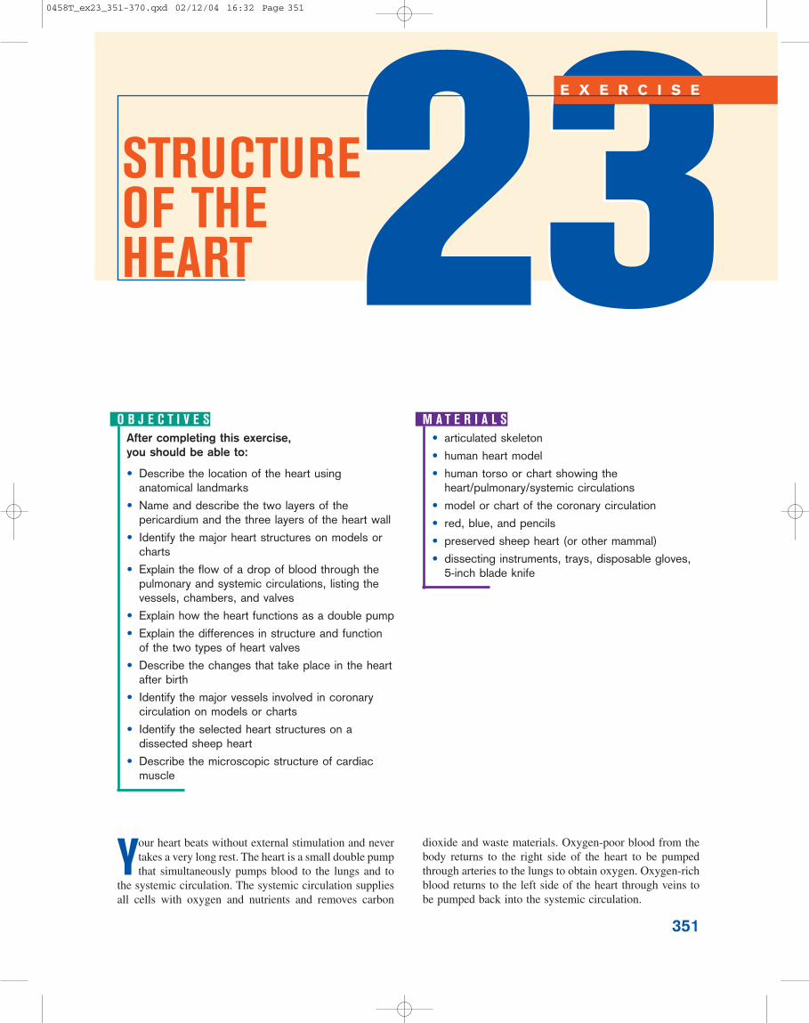
351 STRUCTURE OF THE HEART 23 23 EXERCISE After completing this exercise, you should be able to: • Describe the location of the heart using anatomical landmarks • Name and describe the two layers of the pericardium and the three layers of the heart wall • Identify the major heart structures on models or charts • Explain the flow of a drop of blood through the pulmonary and systemic circulations, listing the vessels, chambers, and valves • Explain how the heart functions as a double pump • Explain the differences in structure and function of the two types of heart valves • Describe the changes that take place in the heart after birth • Identify the major vessels involved in coronary circulation on models or charts • Identify the selected heart structures on a dissected sheep heart • Describe the microscopic structure of cardiac muscle OBJECTIVES • articulated skeleton • human heart model • human torso or chart showing the heart/pulmonary/systemic circulations • model or chart of the coronary circulation • red, blue, and pencils • preserved sheep heart (or other mammal) • dissecting instruments, trays, disposable gloves, 5-inch blade knife MATERIALS Y our heart beats without external stimulation and never takes a very long rest. The heart is a small double pump that simultaneously pumps blood to the lungs and to the systemic circulation. The systemic circulation supplies all cells with oxygen and nutrients and removes carbon dioxide and waste materials. Oxygen-poor blood from the body returns to the right side of the heart to be pumped through arteries to the lungs to obtain oxygen. Oxygen-rich blood returns to the left side of the heart through veins to be pumped back into the systemic circulation. 0458T_ex23_351-370.qxd 02/12/04 16:32 Page 351
Transcript of STRUCTURE OF THE HEART - Homepage | Wiley€¦ · · 2004-03-17EXERCISE 23 STRUCTURE OF THE...
351
STRUCTURE OF THE HEART 2323
E X E R C I S E
After completing this exercise, you should be able to:
• Describe the location of the heart usinganatomical landmarks
• Name and describe the two layers of thepericardium and the three layers of the heart wall
• Identify the major heart structures on models orcharts
• Explain the flow of a drop of blood through thepulmonary and systemic circulations, listing thevessels, chambers, and valves
• Explain how the heart functions as a double pump
• Explain the differences in structure and functionof the two types of heart valves
• Describe the changes that take place in the heartafter birth
• Identify the major vessels involved in coronarycirculation on models or charts
• Identify the selected heart structures on adissected sheep heart
• Describe the microscopic structure of cardiacmuscle
O B J E C T I V E S• articulated skeleton
• human heart model
• human torso or chart showing theheart/pulmonary/systemic circulations
• model or chart of the coronary circulation
• red, blue, and pencils
• preserved sheep heart (or other mammal)
• dissecting instruments, trays, disposable gloves,5-inch blade knife
M A T E R I A L S
Your heart beats without external stimulation and nevertakes a very long rest. The heart is a small double pumpthat simultaneously pumps blood to the lungs and to
the systemic circulation. The systemic circulation suppliesall cells with oxygen and nutrients and removes carbon
dioxide and waste materials. Oxygen-poor blood from thebody returns to the right side of the heart to be pumpedthrough arteries to the lungs to obtain oxygen. Oxygen-richblood returns to the left side of the heart through veins tobe pumped back into the systemic circulation.
0458T_ex23_351-370.qxd 02/12/04 16:32 Page 351

352 E X E R C I S E 2 3 S T R U C T U R E O F T H E H E A R T
A . L O C A T I O N O FT H E H E A R TThe heart is about the size of a fist and lies in the thoraciccavity within the mediastinum (mediastinus � midway),an area bounded by the lungs laterally, the sternum ante-riorly, and the thoracic vertebrae posteriorly. The base ofthe heart is the wide superior portion of the heart fromwhich the great vessels emerge, and the apex of the heartis the inferior pointed end.
The location of the heart in the thorax can be furtherdefined by finding the superior and inferior right and leftpoints of the heart. The superior right point of the heartis located at the superior border of the 3rd right costal car-tilage at its attachment to the rib. The superior left pointis located at the inferior border of the 2nd left costal car-tilage at its attachment to the rib. The inferior left pointis located in the 5th intercostal space, inferior to the fifth
A C T I V I T Y 1 L o c a t i o n o f t h e H e a r t
1 In Figure 23.1, identify the superior and inferior rightand left points of the heart. Mark dots at these pointsand connect these four dots to outline the heart location.
2 Identify the superior and inferior right and left pointsfor the heart location on an articulated skeleton and/ora torso model.
3 Palpate the superior and inferior right and left points onyourself.
• inferior border of the 2nd leftcostal cartilage
• superior border of the 3rdright costal cartilage
• 5th left intercostal space(midclavicular)
• 5th right costal cartilage
F I G U R E 2 3 . 1 Location of the heart.
costal cartilage at its attachment to the rib. The inferiorright point is at the inferior border of the 5th right costalcartilage, a little to the right of the border of the sternum.The heart is tilted at an angle so that its inferior surfacelies against the diaphragm, with two-thirds of the heart tothe left side of the sternum.
0458T_ex23_351-370.qxd 02/12/04 16:32 Page 352
E X E R C I S E 2 3 S T R U C T U R E O F T H E H E A R T 353
B . M A J O RH E A R T S T R U C T U R E S
1. The Pericardium and theLayers of the Heart Wall
In the mediastinum, the heart is surrounded and protectedby the pericardium (peri- � around). The pericardium con-sists of an outer, tough fibrous pericardium and an inner,delicate serous pericardium. The fibrous pericardium at-taches to the diaphragm and also to the great vessels of theheart. Like all serous membranes, the serous pericardiumis a double membrane composed of an outer parietal layerand an inner visceral layer. Between these two layers is thepericardial cavity filled with serous fluid. The outerparietal (paries � wall) pericardium is attached to thefibrous pericardium, and the inner visceral (viscera �internal organs) pericardium covers the cardiac muscle.
A C T I V I T Y 2 The Pe r i c a rd i um and L a y e r so f t h e H e a r t Wa l l
1 Label the structures in Figure 23.2.
2 Pronounce each term as you write the answer in theblank.
The wall of the heart has three layers: the outer epi-cardium (epi- � on, upon; cardia � heart), the middle my-ocardium (myo- � muscle), and the inner endocardium(endo- � within, inward). The epicardium is the viscerallayer of the pericardium. The majority of the heart isthe myocardium or cardiac muscle tissue. The endoc-ardium is a thin layer of endothelium deep to themyocardium that lines the inside chambers of the heartand the valves.
0458T_ex23_351-370.qxd 02/12/04 16:32 Page 353
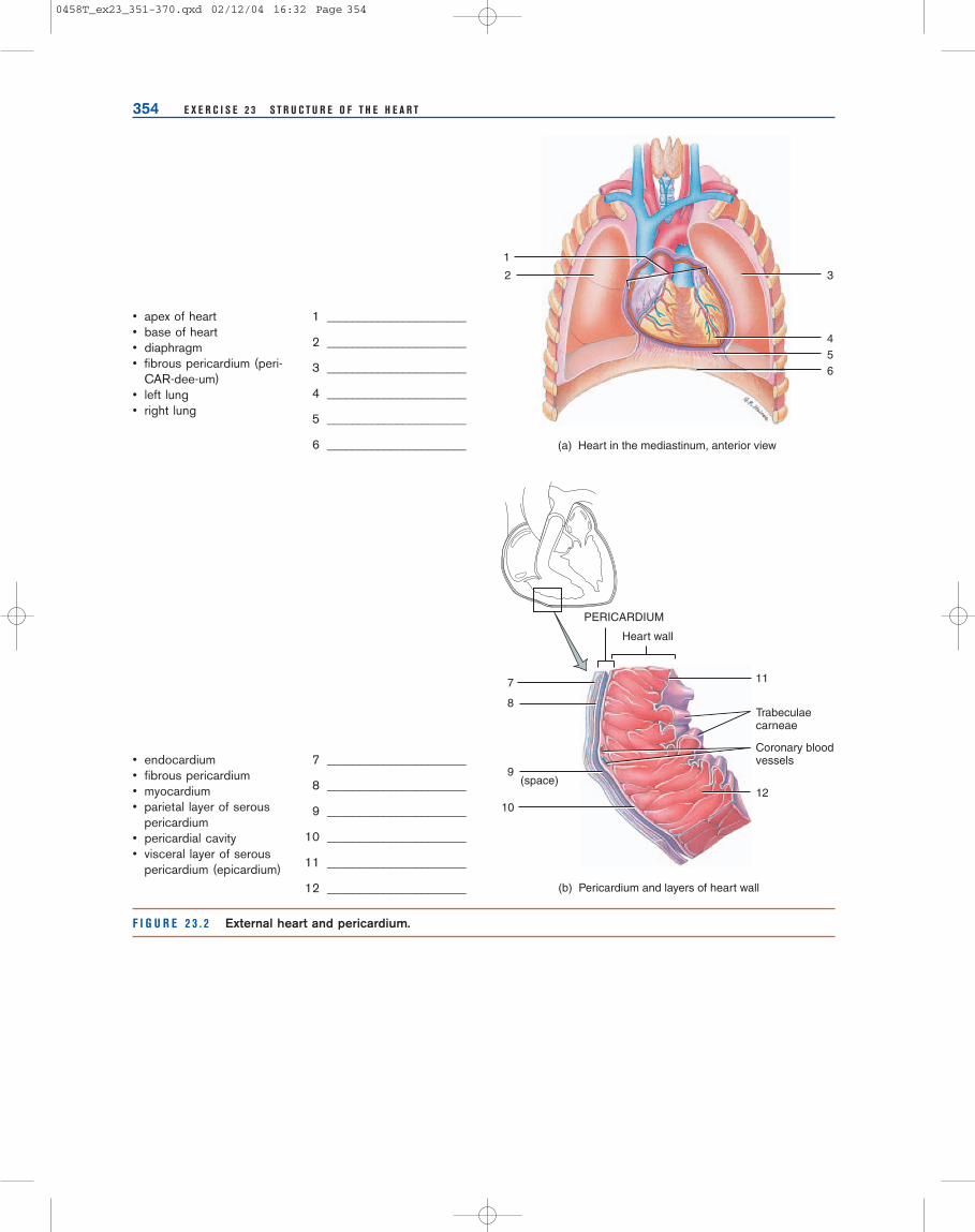
354 E X E R C I S E 2 3 S T R U C T U R E O F T H E H E A R T
• apex of heart• base of heart• diaphragm• fibrous pericardium (peri-
CAR-dee-um)• left lung• right lung
1 ______________________
2 ______________________
3 ______________________
4 ______________________
5 ______________________
6 ______________________
• endocardium• fibrous pericardium• myocardium• parietal layer of serous
pericardium• pericardial cavity• visceral layer of serous
pericardium (epicardium)
7 ______________________
8 ______________________
9 ______________________
10 ______________________
11 ______________________
12 ______________________
F I G U R E 2 3 . 2 External heart and pericardium.
11
3
456
7
1
2
8
10
9
12
Trabeculaecarneae
PERICARDIUM
Heart wall
Coronary bloodvessels
(a) Heart in the mediastinum, anterior view
(b) Pericardium and layers of heart wall
(space)
0458T_ex23_351-370.qxd 02/12/04 16:32 Page 354
E X E R C I S E 2 3 S T R U C T U R E O F T H E H E A R T 355
2. Surface Featuresof the Heart
Like all mammalian hearts, the human heart has four cham-bers and is divided into right and left sides. Each side hasan upper chamber called an atrium and a lower chambercalled a ventricle. The two atria form the base of the heart,and the tip of the left ventricle forms the apex. Auricles(auricle � little ear) are pouch-like extensions of the atria.From the exterior, the auricles look like flaps with wrin-kled edges.
Coronary blood vessels and adipose tissue are found inthe sulci or grooves that externally mark the boundariesbetween the four heart chambers. Although a considerableamount of external adipose tissue is present on the heartsurface for protection and padding, most heart models do
A C T I V I T Y 3 Su r f a c e F e a t u re s o f t h e H e a r t
1 Label the structures in Figure 23.3.
2 Identify each term on a model or chart.
3 Pronounce each term as you point to it.
not show this. The coronary sulcus is a deep sulcus thatexternally shows the separation of the atria and theventricles. The anterior interventricular sulcus and theposterior interventricular sulcus are shallow grooves thatdepict the surface boundaries between the two ventricles.
0458T_ex23_351-370.qxd 02/12/04 16:32 Page 355

356 E X E R C I S E 2 3 S T R U C T U R E O F T H E H E A R T
• anterior interventricular sulcus• auricle of left atrium• auricle of right atrium• coronary sulcus• left ventricle• right ventricle
1 ______________________________
2 ______________________________
3 ______________________________
4 ______________________________
5 ______________________________
6 ______________________________
• adipose tissue• coronary sulcus• left auricle• left ventricle
• posterior interventricularsulcus
• right auricle• right ventricle
7 _________________________________
8 _________________________________
9 _________________________________
10 _________________________________
11 _________________________________
12 _________________________________
13 _________________________________
F I G U R E 2 3 . 3 Surface features of the heart.
(b) Posterior view
8 (groove)
11 (groove)
7
9
10
12
13
4
6
5 (groove)
12 (groove)
(a) Anterior view
3
0458T_ex23_351-370.qxd 02/12/04 16:32 Page 356

E X E R C I S E 2 3 S T R U C T U R E O F T H E H E A R T 357
3. Great Vessels of the Heart
The great vessels of the heart either return blood to theatria or carry blood away from the ventricles. The superiorvena cava, inferior vena cava, and coronary sinus returnoxygen-poor blood to the right atrium. The superior venacava returns blood from the head, neck, and arms; the in-ferior vena cava returns blood from the body inferior tothe heart; and the coronary sinus is a smaller vein that re-turns blood from the coronary circulation. Blood leaves theright atrium to enter the right ventricle. From here, bloodpasses out the pulmonary trunk, the only vessel that re-moves blood from the right ventricle. This large artery di-vides into the right and left pulmonary arteries that carryblood to the lungs where it is oxygenated. Oxygen-richblood returns to the left atrium through two right and twoleft pulmonary veins. The blood then passes into the leftventricle that pumps blood into the large aorta. The aortadistributes blood to the systemic circulation. The aorta be-gins as a short ascending aorta, curves to the left to formthe aortic arch, descends posteriorly, and continues as thedescending aorta.
The fetal heart contains a short, temporary vascularchannel, the ductus arteriosus (ductus � duct; arteriosus� artery), which connects the pulmonary trunk and the aorta.This right heart to left heart shunt re-routes some of theblood destined for the lungs to the systemic circulation viathe aorta. In fetal life, oxygen is obtained through the pla-centa from the mother and not from the lungs. Therefore, itis not detrimental to the baby’s health for blood to bypassthe lungs. The ductus arteriosus changes into a ligament afterbirth and remains as the ligamentum arteriosum.
A C T I V I T Y 4 G re a t Ve s s e l s o f t h e H e a r t
1 Label the great vessels of the heart in Figure 23.4. Bloodvessels carrying oxygen-rich blood are red, and thosecarrying oxygen-poor blood are blue.
2 Identify each great vessel on a model or chart.
3 Pronounce each term as you point to it.
0458T_ex23_351-370.qxd 02/12/04 16:32 Page 357

358 E X E R C I S E 2 3 S T R U C T U R E O F T H E H E A R T
• aortic arch• ascending aorta• descending aorta• inferior vena cava• left pulmonary artery• left pulmonary veins
• ligamentum arteriosum• pulmonary trunk• right pulmonary artery• right pulmonary veins• superior vena cava
1 ______________________________
2 ______________________________
3 ______________________________
4 ______________________________
5 ______________________________
6 ______________________________
7 ______________________________
8 ______________________________
9 ______________________________
10 ______________________________
11 ______________________________
• aortic arch• ascending aorta• coronary sinus• inferior vena cava• left pulmonary artery• left pulmonary veins
• ligamentum arteriosum• right pulmonary artery• right pulmonary veins• superior vena cava
12 _________________________________
13 _________________________________
14 _________________________________
15 _________________________________
16 _________________________________
17 _________________________________
18 _________________________________
19 _________________________________
20 _________________________________
21 _________________________________
F I G U R E 2 3 . 4 Great vessels of the heart.
78
9
1011
1
2
3
4
5
(a) Anterior view
6
(b) Posterior view
12
13
14
15
16
17
18
19
20
21
0458T_ex23_351-370.qxd 02/12/04 16:33 Page 358

E X E R C I S E 2 3 S T R U C T U R E O F T H E H E A R T 359
4. Internal Featuresof the Heart
The myocardium of the anterior wall of the right atriumhas a honeycombed appearance, and these myocardialridges called pectinate muscles (pecten � comb-like) con-tinue into the auricles. The walls of the right and left atriaare separated by the thin interatrial septum. In the fetus,there is a hole in the interatrial septum called the foramenovale. The foramen ovale allows blood to bypass the lungsand go from the right atrium to the left atrium, forminganother right heart to left heart shunt. The fossa ovalis, aconnective tissue membrane, forms over and closes thefetal foramen ovale.
The ventricles have ridges of muscles called trabeculaecarneae (trabecula � little beam; carnea � flesh). Thelarger of these muscles, the papillary muscles, have string-like cords attached to them called the chordae tendineae(tendinous strands). The opposite ends of these cords areattached to the AV valves. The interventricular septumis a thin wall that separates the right and left ventricles.
The heart has four valves that control the one-way flowof blood: two AV valves and two semilunar valves(semi- � half; lunar � moon). Blood passing between theright atrium and the right ventricle goes through the rightAV valve, the tricuspid valve (tri- � three; cusp � flap).The left AV valve, the bicuspid valve, is between the leftatrium and the left ventricle. This valve clinically is calledthe mitral valve (miter � tall, liturgical headdress)because the open valve resembles a bishop’s headdress.The two AV valves are structurally similar, except the
tricuspid valve has three cusps or flaps and the bicuspidvalve has two cusps or flaps that prevent blood from flow-ing back into the atria.
Blood in the right ventricle goes through the pulm-onary (semilunar) valve to enter the pulmonary trunk (ar-tery). The aortic (semilunar) valve is located between theleft ventricle and the aorta. These two semilunar valves areidentical, with each having three pockets that fill withblood, preventing blood from flowing back into the ven-tricles.
The thinner walled atria receive the blood returning tothe heart from veins. The pressure of blood in the atriaopens the atrioventricular (AV) valves, and most of theblood flows into the ventricles. Both atria then contract si-multaneously to pump the remaining blood into the ven-tricles. The larger, thick ventricular walls are double pumpsthat contract simultaneously to send the blood from theright ventricle to the lungs and from the left ventricle tothe systemic circulation. The wall of the left ventricle isthicker than the right because the left side requires moreforce to pump blood through the systemic circulation.
A C T I V I T Y 5 I n t e r n a l F e a t u re s o f t h e H e a r t
1 Label the structures in Figure 23.5.
2 Identify each term on a model or chart.
3 Pronounce each term as you point to it.
0458T_ex23_351-370.qxd 02/12/04 16:33 Page 359

Frontal plane
1
7
8
9
10
11
12
13
2
Fossa ovalis
3
4
5
6
360 E X E R C I S E 2 3 S T R U C T U R E O F T H E H E A R T
• aortic (semilunar) valve• bicuspid valve (mitral)• chordae tendineae (CHOR-dee
ten-DIN-ee)• coronary sinus opening• interventricular septum (inter-ven-
TRIC-u-lar)• left atrium• left ventricle• papillary muscle (PAP-ih-lary)• pulmonary (semilunar) valve• right atrium• right ventricle• trabeculae carneae (tra-BEC-u-lee
CAR-nee)• tricuspid valve
1 ______________________________
2 ______________________________
3 ______________________________
4 ______________________________
5 ______________________________
6 ______________________________
7 ______________________________
8 ______________________________
9 ______________________________
10 ______________________________
11 ______________________________
12 ______________________________
13 ______________________________
F I G U R E 2 3 . 5 Internal features of the heart, frontal section.
0458T_ex23_351-370.qxd 02/12/04 16:33 Page 360
E X E R C I S E 2 3 S T R U C T U R E O F T H E H E A R T 361
4 In the blanks below, trace a drop of blood through theheart and lung by listing in order all vessels, heartchambers, and valves through which the blood passes,starting with the right atrium.
• aorta
• aortic (semilunar) valve
• bicuspid valve (mitral)
• left atrium
• left ventricle
• pulmonary arteries
• pulmonary capillaries
• pulmonary (semilunar) valve
• pulmonary trunk
• pulmonary veins
• right atrium
• right ventricle
• systemic arteries
• systemic capillaries
• systemic veins
• tricuspid valve
• venae cavae
1. __________________________________________
2. __________________________________________
3. __________________________________________
4. __________________________________________
5. __________________________________________
6. __________________________________________
7. __________________________________________
8. __________________________________________
9. __________________________________________
10. __________________________________________
11. __________________________________________
12. __________________________________________
13. __________________________________________
14. __________________________________________
15. __________________________________________
16. __________________________________________
17. __________________________________________
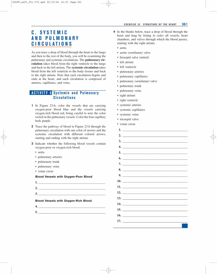
C . S Y S T E M I CA N D P U L M O N A R YC I R C U L A T I O N SAs you trace a drop of blood through the heart to the lungsand then to the rest of the body, you will be examining thepulmonary and systemic circulations. The pulmonary cir-culation takes blood from the right ventricle to the lungsand back to the left atrium. The systemic circulation takesblood from the left ventricle to the body tissues and backto the right atrium. Note that each circulation begins andends at the heart, and each circulation is composed ofarteries, capillaries, and veins.
A C T I V I T Y 6 Sy s t em i c a nd Pu lmona r yC i rc u l a t i o n s
1 In Figure 23.6, color the vessels that are carryingoxygen-poor blood blue and the vessels carryingoxygen-rich blood red, being careful to note the colorswitch in the pulmonary vessels. Color the four capillarybeds purple.
2 Trace the pathway of blood in Figure 23.6 through thepulmonary circulation with one color of arrows and thesystemic circulation with different colored arrows,starting and ending with the right atrium.
3 Indicate whether the following blood vessels containoxygen-poor or oxygen-rich blood.
• aorta
• pulmonary arteries
• pulmonary trunk
• pulmonary veins
• venae cavae
Blood Vessels with Oxygen-Poor Blood
1. __________________________________________
2. __________________________________________
3. __________________________________________
Blood Vessels with Oxygen-Rich Blood
4. __________________________________________
5. __________________________________________
0458T_ex23_351-370.qxd 02/12/04 16:33 Page 361

362 E X E R C I S E 2 3 S T R U C T U R E O F T H E H E A R T
F I G U R E 2 3 . 6 Systemic and pulmonary circulations.
Systemic capillariesof upper body
Pulmonary capillariesof left lung
Systemic capillariesof lower body
Pulmonary capillariesof right lung
Right pulmonary artery
Systemic arteriesto upper body
Systemic veinsfrom upper body
Systemic arteriesto lower body
Systemic veins fromlower body
Left pulmonary veins
0458T_ex23_351-370.qxd 02/12/04 16:33 Page 362

E X E R C I S E 2 3 S T R U C T U R E O F T H E H E A R T 363
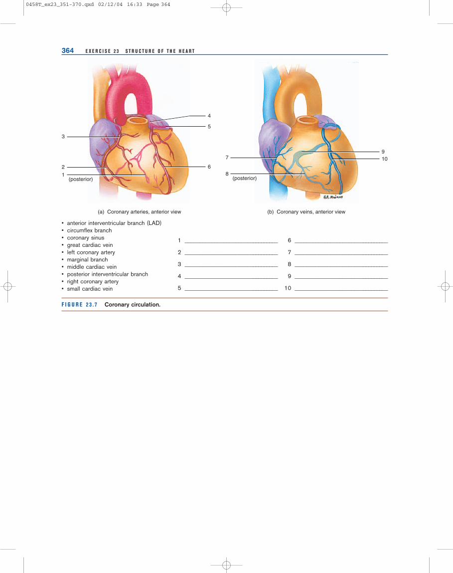
D . C O R O N A R YC I R C U L A T I O NThe walls of the heart have their own blood supply andcirculation, the coronary (corona � crown) circulation.These vessels encompass the heart similar to a crown. Theendothelium lining the heart chambers is too thick forblood in the chambers to supply nutrients to cardiac mus-cle tissue. Coronary blood vessels supply blood to cardiacmuscle tissue.
On the anterior surface of the heart, the right and leftcoronary arteries branch off the base of the ascendingaorta just superior to the aortic semilunar valve. Thesesmall arteries are supplied with blood when the ventriclesare resting. When the ventricles contract, the cusps of theaortic valve open to cover the openings to the coronary ar-teries. If the coronary arteries were not covered, they wouldnot be able to withstand the blood pressure and would burstlike an overinflated balloon.
As the left coronary artery passes the base of the leftauricle, it branches into the:
• anterior interventricular branch (left anterior de-scending branch, or LAD)
• circumflex branch
The anterior interventricular branch (LAD) suppliesboth ventricles with oxygen-rich blood and lies within theanterior interventricular sulcus, a shallow depression be-tween the two ventricles. The anterior interventricularbranch is commonly occluded and can result in a myocar-dial infarct and at times death. The circumflex branchcontinues around the left side of the heart, lying within thecoronary sulcus (atrioventricular sulcus), and suppliesblood to the left ventricle and left atrium. The circumflexbranch forms anastomoses (connections) with the poste-rior interventricular branch near the posterior interventric-ular sulcus.
The right coronary artery passes the right auricle,supplying it with blood, and continues inferiorly to theauricle in the coronary sulcus. The right coronary arterybranches into the:
• marginal branch
• posterior interventricular branch
The marginal branch supplies the anterior right sideof the right ventricle. The posterior interventricularbranch lies in the posterior interventricular sulcus on theposterior surface of the heart, supplying oxygen-rich bloodto both ventricles.
Arteries branch into smaller vessels, arterioles, whichpenetrate the heart muscle and divide into narrower ves-sels called capillaries which deliver oxygen to the cardiacmuscle. Capillaries drain into venules which exit the heartmuscle and connect to veins that receive oxygen-poorblood, returning it to the heart. The great cardiac vein isthe principal vein of the coronary circulation, draining theleft anterior portion of the heart. It lies near the anteriorinterventricular branch in the interventricular sulcus. Thesmall cardiac vein drains the right anterior portion of theheart. The middle cardiac vein that lies next to the pos-terior interventricular branch in the posterior interventric-ular sulcus drains the posterior portion of the heart. Bothof these veins empty into a large, thin-walled venous sinuscalled the coronary sinus located on the posterior surfaceof the heart. The coronary sinus empties its oxygen-poorblood into the right atrium.
A C T I V I T Y 7 Co ro n a r y C i rc u l a t i o n
1 Label the structures in Figure 23.7.
2 Identify each vessel on a model or chart.
3 Pronounce each term as you point to it.
0458T_ex23_351-370.qxd 02/12/04 16:33 Page 363
364 E X E R C I S E 2 3 S T R U C T U R E O F T H E H E A R T
• anterior interventricular branch (LAD)• circumflex branch• coronary sinus• great cardiac vein• left coronary artery• marginal branch• middle cardiac vein• posterior interventricular branch• right coronary artery• small cardiac vein
1 ________________________________
2 ________________________________
3 ________________________________
4 ________________________________
5 ________________________________
6 ________________________________
7 ________________________________
8 ________________________________
9 ________________________________
10 ________________________________
F I G U R E 2 3 . 7 Coronary circulation.
3
68
2
1
910
5
4
7
(a) Coronary arteries, anterior view (b) Coronary veins, anterior view
(posterior) (posterior)
0458T_ex23_351-370.qxd 02/12/04 16:33 Page 364
E X E R C I S E 2 3 S T R U C T U R E O F T H E H E A R T 365
E . D I S S E C T I O NO F A S H E E P H E A R TThe sheep heart is similar to the human heart in both struc-ture and size. It provides students the opportunity to ob-serve the flexibility of the valves and tissues.
A C T I V I T Y 8 D i s s e c t i o n o f aSheep Hea r t
1 Obtain a dissecting tray, tools, disposable gloves, anda sheep heart.
2 Examine the anterior surface of the heart. Greatvessels are often cut close to the base of the heart andmay be difficult to find. Refer to Figure 23.8(a) and aheart model to identify the following structures:
• pericardium (if present)
• epicardium
• base
• apex
• right auricle
• left auricle
• right ventricle
• left ventricle
• pulmonary trunk
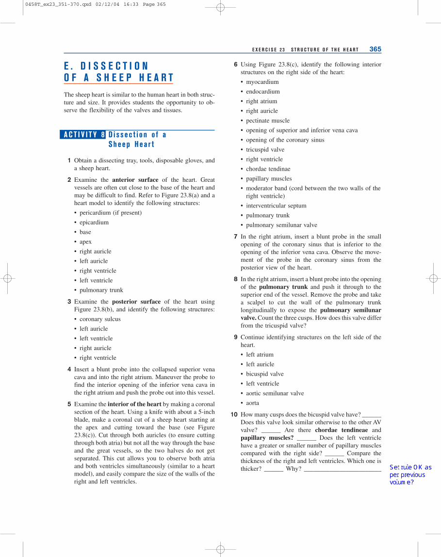
3 Examine the posterior surface of the heart usingFigure 23.8(b), and identify the following structures:
• coronary sulcus
• left auricle
• left ventricle
• right auricle
• right ventricle
4 Insert a blunt probe into the collapsed superior venacava and into the right atrium. Maneuver the probe tofind the interior opening of the inferior vena cava inthe right atrium and push the probe out into this vessel.
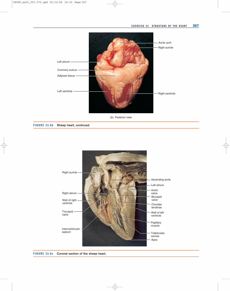
5 Examine the interior of the heart by making a coronalsection of the heart. Using a knife with about a 5-inchblade, make a coronal cut of a sheep heart starting atthe apex and cutting toward the base (see Figure23.8(c)). Cut through both auricles (to ensure cuttingthrough both atria) but not all the way through the baseand the great vessels, so the two halves do not getseparated. This cut allows you to observe both atriaand both ventricles simultaneously (similar to a heartmodel), and easily compare the size of the walls of theright and left ventricles.
6 Using Figure 23.8(c), identify the following interiorstructures on the right side of the heart:
• myocardium
• endocardium
• right atrium
• right auricle
• pectinate muscle
• opening of superior and inferior vena cava
• opening of the coronary sinus
• tricuspid valve
• right ventricle
• chordae tendinae
• papillary muscles
• moderator band (cord between the two walls of theright ventricle)
• interventricular septum
• pulmonary trunk
• pulmonary semilunar valve
7 In the right atrium, insert a blunt probe in the smallopening of the coronary sinus that is inferior to theopening of the inferior vena cava. Observe the move-ment of the probe in the coronary sinus from theposterior view of the heart.
8 In the right atrium, insert a blunt probe into the openingof the pulmonary trunk and push it through to thesuperior end of the vessel. Remove the probe and takea scalpel to cut the wall of the pulmonary trunklongitudinally to expose the pulmonary semilunarvalve. Count the three cusps. How does this valve differfrom the tricuspid valve?
9 Continue identifying structures on the left side of theheart.
• left atrium
• left auricle
• bicuspid valve
• left ventricle
• aortic semilunar valve
• aorta
10 How many cusps does the bicuspid valve have? ______Does this valve look similar otherwise to the other AVvalve? ______ Are there chordae tendineae andpapillary muscles? ______ Does the left ventriclehave a greater or smaller number of papillary musclescompared with the right side? ______ Compare thethickness of the right and left ventricles. Which one isthicker? ______ Why? ________________________
0458T_ex23_351-370.qxd 02/12/04 16:33 Page 365
SSEN 21
Set rule OK as per previous volume?
SSEN 21
Set rule OK as per previous volume?

366 E X E R C I S E 2 3 S T R U C T U R E O F T H E H E A R T
F I G U R E 2 3 . 8 a Sheep heart.
Apex
Left ventricle
Left auricle
Left pulmonary artery
Left pulmonary veins
Pulmonary trunk
Aorta
Right ventricle
Right auricle
Openingof superiorvena cava
Superiorvena cava
Openingof inferiorvena cava
Right ventricle
Coronary vessels inanterior interventricularsulcus
Right auricle
Ligamentumarteriosum
POSTERIORANTERIOR
Right auricle
Adipose tissue
Right ventricle
Pulmonary trunk
Left auricle
Anterior interventricularsulcus
Left ventricle
Apex of heart
(a) Anterior view
11 Look just above the cusps of the aortic valve for theopenings to the right and left coronary arteries. Usethe blunt probe to push into these small vessels.
12 Dispose of any removed dissection material in theproper container (NOT the sink!).
13 Wash the dissection pan, instruments, and hands withsoap and water when finished.
14 Clean up your lab space and wash the countertops withdisinfectant.
0458T_ex23_351-370.qxd 02/12/04 16:33 Page 366
E X E R C I S E 2 3 S T R U C T U R E O F T H E H E A R T 367
F I G U R E 2 3 . 8 b Sheep heart, continued.
(b) Posterior view
Aortic arch
Right auricle
Right ventricle
Left atrium
Coronary sulcus
Adipose tissue
Left ventricle
F I G U R E 2 3 . 8 c Coronal section of the sheep heart.
Right atrium
Left atrium
Tricuspidvalve
Wall of leftventricle
Wall of rightventricle
Right auricle
Ascending aorta
AorticvalveBicuspidvalve
Chordaetendinae
Papillarymuscle
Interventricularseptum Trabeculae
carnaeApex
0458T_ex23_351-370.qxd 02/12/04 16:33 Page 367
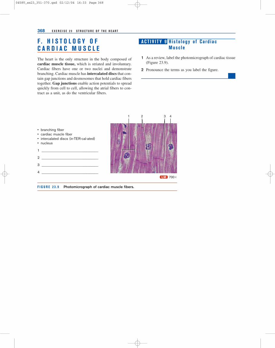
F . H I S T O L O G Y O FC A R D I A C M U S C L EThe heart is the only structure in the body composed ofcardiac muscle tissue, which is striated and involuntary.Cardiac fibers have one or two nuclei and demonstratebranching. Cardiac muscle has intercalated discs that con-tain gap junctions and desmosomes that hold cardiac fiberstogether. Gap junctions enable action potentials to spreadquickly from cell to cell, allowing the atrial fibers to con-tract as a unit, as do the ventricular fibers.
A C T I V I T Y 9 H i s t o l o g y o f C a rd i a cMusc l e
1 As a review, label the photomicrograph of cardiac tissue(Figure 23.9).
2 Pronounce the terms as you label the figure.
368 E X E R C I S E 2 3 S T R U C T U R E O F T H E H E A R T
F I G U R E 2 3 . 9 Photomicrograph of cardiac muscle fibers.
• branching fiber• cardiac muscle fiber• intercalated discs (in-TER-cal-ated)• nucleus
1 ________________________________
2 ________________________________
3 ________________________________
4 ________________________________LM 700�
1 2 3 4
0458T_ex23_351-370.qxd 02/12/04 16:33 Page 368

369
A. Location of the Heart
1. Describe the location of the heart using the lungs, rib cartilages, and intercostal spaces as landmarks.
B. Major Heart Structures
Completion: Fill in the blank with the word that fits the description.
__________________________ 1. Arteries that supply blood to cardiac muscle.
__________________________ 2. Layer of heart wall containing cardiac muscle.
__________________________ 3. Extensions of the atria.
__________________________ 4. Heart is located here (area between the lungs).
__________________________ 5. Lines the heart chambers.
__________________________ 6. Pointed inferior part of the heart.
__________________________ 7. Two heart pumps; lower heart chambers.
__________________________ 8. Superior heart chambers.
__________________________ 9. Another name for visceral pericardium.
__________________________ 10. Wide superior part of the heart.
__________________________ 11. Blood pumped by right ventricle (oxygen-rich or oxygen-poor).
__________________________ 12. Blood pumped by left ventricle (oxygen-rich or oxygen-poor).
__________________________ 13. Enlarged muscles in ventricles attached to chordae tendinae.
__________________________ 14. Muscle ridges in ventricles.
__________________________ 15. Ridges in anterior wall right atrium.
__________________________ 16. Strings attached to AV cusps.
R E V I E W I N G YO U R KN O W L E D G E 23E X E R C I S E
Name ___________________________________ Section _________________ Date ____________________
0458T_ex23_351-370.qxd 02/12/04 16:33 Page 369

370 E X E R C I S E 2 3 S T R U C T U R E O F T H E H E A R T
C. Coronary Circulation—Blood Vessels
Completion: After reviewing the coronary circulation in Figure 23.7, fill in the blank with the word that fits thedescription.
______________________ 1. Anterior branch of the left coronary artery.
______________________ 2. Posterior branch of the right coronary artery.
______________________ 3. Coronary artery that lies in anterior coronary sulcus.
______________________ 4. Curving branch of the left coronary artery.
______________________ 5. Main artery supplying anterior part of ventricles.
______________________ 6. Shorter coronary artery that divides at the base of an icle.
______________________ 7. Vein that drains coronary circulation into right atrium.
______________________ 8. Vein that drains most of anterior ventricles.
______________________ 9. Vein that drains the posterior ventricles.
______________________ 10. Vein that drains the right anterior side.
D. The Heart and Pulmonary Circulation
Place the following structures in order, tracing the blood flow from the superior vena cava to the heart, to the lungs, andout of the heart to the systemic circulation.
• aorta
• aortic valve
• bicuspid valve
• left atrium
• left ventricle
• pulmonary capillaries
• right atrium
• right ventricle
• pulmonary arteries
• pulmonary trunk
• pulmonary valve
• pulmonary veins
• superior vena cava
• tricuspid valve
1. ____________________________________________
2. ____________________________________________
3. ____________________________________________
4. ____________________________________________
5. ____________________________________________
6. ____________________________________________
7. ____________________________________________
8. ____________________________________________
9. ____________________________________________
10. ____________________________________________
11. ____________________________________________
12. ____________________________________________
13. ____________________________________________
14. ____________________________________________
0458T_ex23_351-370.qxd 02/12/04 16:33 Page 370
SSEN 21
Three columns OK?
SSEN 21
Three columns OK?