Structure of the digestive tract of tornaria larva in ...
15
HELGOLANDER MEERESUNTERSUCHUNGEN HelgolSnder Meeresunters. 48, 107-121 (1994) Structure of the digestive tract of tornaria larva in Enteropneusta (Hemichordata) S. Sh. Dautov 1, L.P. Nezlin 2 & V.V. Yushin 1 1Institute of Marine Biology, Far Hast Branch of Russian Academy of Sciences; 17 Paltchevsky Str., Vladivostok 690041, Russia 2Institute of Developmental Biology, Russian Academy of Sciences; 26 Vavilova Str., Moscow 117808, Russia ABSTRACT: The ultrastructure of the digestive tract of tornaria larva of enteropneusts was investi- gated. It showed that the digestive tract consists of three parts: esophagus, stomach, and intestine. The esophagus epithelium consists of two types of multiciliated epithelial ceils and solitary muscle cells. Axonal tracts and neurons were found in the ventral wall of the esophagus. The cardiac sphincter contains an anterior band of strongly ciliated ceils and a posterior band of cells with long vacuohzed processes which partition the sphincter lumen. The stomach consists of three cell types: (1) ceils with electron-opaque cytoplasm, bearing a fringed border on their apical sides; (2, 3) sparse cells with electron-light cytoplasm and different patterns of apical microviili. Cells of the pyloric sphincter bear numerous cilia and almost no microvilli. The intestine consists of three parts. The anterior part is formed of multiciliated cells which bear the fringed border. The middle part consists of flattened cells bearing rare cilia and vast numbers of mace-like microvilli. The posterior part of the intestine is formed of cells bearing numerous cilia and few microviUL Muscle cells were not found in either stomach or intestine epithelium. One noticed that the structure of the digestive tract of enteropneust tornaria larva differs from that of echinoid pluteus larva. INTRODUCTION Various species of enteropneusts have similar planktonic larva-tornariae. After the free-swimming period, the tdrnariae settle at the bottom and metamorphose. Tornariae are morphologically similar to the echinoderm larvae and are united with them as dipleurula-type larvae (Ivanova-Kazas, 1978). The feeding process of tornariae has much in common with echinoderm larvae and is accomplished through the use of ciliary bands (Strathmann, 1975; Strathmann & Bonar, 1976). As for the ultrastructure of the digestive tract of echinoderm larvae, it was shown to be tripartite (muscular esopha- gus, spherical stomach and small tubular intestine) and consists of various types of speciahzed monociliated and unciliated cells (Ryberg & Lundgren, 1975; Burke, 1981). The digestive tract of a tornaria also consists of three parts, but very little is known about the structure of these larval organs. Comparison of the fine structure of these organs in tornaria and echinoderm larvae is of some interest, since they have similar morphology and behaviour but different fates of the larval body during metamorphosis (Ivanova- Kazas, 1978). This paper presents the results from an ultrastructural study of the digestive tract of Biologische Anstalt HelgoIand, Hamburg
Transcript of Structure of the digestive tract of tornaria larva in ...

HELGOLANDER MEERESUNTERSUCHUNGEN HelgolSnder Meeresunters. 48, 107-121 (1994)
Structure of the digest ive tract of tornaria larva in Enteropneusta (Hemichordata)
S. Sh. D a u t o v 1, L.P. N e z l i n 2 & V . V . Y u s h i n 1
1 Institute of Marine Biology, Far Hast Branch of Russian Academy of Sciences; 17 Paltchevsky Str., Vladivostok 690041, Russia
2Institute of Developmental Biology, Russian Academy of Sciences; 26 Vavilova Str., Moscow 117808, Russia
ABSTRACT: The ultrastructure of the digestive tract of tornaria larva of enteropneusts was investi- gated. It showed that the digestive tract consists of three parts: esophagus, stomach, and intestine. The esophagus epithelium consists of two types of multiciliated epithelial ceils and solitary muscle cells. Axonal tracts and neurons were found in the ventral wall of the esophagus. The cardiac sphincter contains an anterior band of strongly ciliated ceils and a posterior band of cells with long vacuohzed processes which partition the sphincter lumen. The stomach consists of three cell types: (1) ceils with electron-opaque cytoplasm, bear ing a fringed border on their apical sides; (2, 3) sparse cells with electron-light cytoplasm a n d different pat terns of apical microviili. Cells of the pyloric sphincter bear numerous cilia and almost no microvilli. The intestine consists of three parts. The anterior part is formed of multiciliated cells which bear the fringed border. The middle part consists of f lat tened cells bear ing rare cilia and vast numbers of mace-like microvilli. The posterior par t of the intestine is formed of cells bear ing numerous cilia and few microviUL Muscle cells were not found in ei ther stomach or intestine epithelium. One noticed that the structure of the digestive tract of enteropneust tornaria larva differs from that of echinoid pluteus larva.
I N T R O D U C T I O N
V a r i o u s s p e c i e s of e n t e r o p n e u s t s h a v e s imi la r p l a n k t o n i c l a r v a - t o r n a r i a e . Af t e r t he
f r e e - s w i m m i n g pe r iod , t h e t d r n a r i a e se t t l e a t t h e b o t t o m a n d m e t a m o r p h o s e .
T o r n a r i a e a re m o r p h o l o g i c a l l y s imi l a r to t h e e c h i n o d e r m l a r v a e a n d a re u n i t e d w i t h
t h e m as d i p l e u r u l a - t y p e l a r v a e ( I v a n o v a - K a z a s , 1978). T h e f e e d i n g p r o c e s s of t o r n a r i a e
h a s m u c h in c o m m o n w i t h e c h i n o d e r m l a r v a e a n d is a c c o m p l i s h e d t h r o u g h t h e u s e of
ci l iary b a n d s ( S t r a t h m a n n , 1975; S t r a t h m a n n & Bonar , 1976). As for t h e u l t r a s t r u c t u r e of
t h e d i g e s t i v e t r ac t of e c h i n o d e r m l a r vae , i t w a s s h o w n to b e t r ipa r t i t e ( m u s c u l a r e s o p h a -
gus, s p h e r i c a l s t o m a c h a n d s m a l l t u b u l a r i n t e s t i n e ) a n d cons i s t s of v a r i o u s t y p e s of
s p e c i a h z e d m o n o c i l i a t e d a n d u n c i l i a t e d cel ls (Rybe rg & L u n d g r e n , 1975; B u r k e , 1981).
T h e d i g e s t i v e t r ac t of a t o r n a r i a a lso cons i s t s of t h r e e par t s , b u t v e r y l i t t le is k n o w n a b o u t
t h e s t r u c t u r e of t h e s e l a r v a l o r g a n s . C o m p a r i s o n of t he f ine s t r u c t u r e of t h e s e o r g a n s in
t o r n a r i a a n d e c h i n o d e r m l a r v a e is of s o m e in t e re s t , s i n c e t h e y h a v e s im i l a r m o r p h o l o g y
a n d b e h a v i o u r b u t d i f f e r e n t f a t e s of t h e l a r v a l b o d y d u r i n g m e t a m o r p h o s i s ( I v a n o v a -
Kazas , 1978).
Th i s p a p e r p r e s e n t s t h e r e s u l t s f rom a n u l t r a s t r u c t u r a l s t u d y of t h e d i g e s t i v e t r a c t of
�9 Biologische Anstalt HelgoIand, Hamburg

108 S. Sh. Dautov, L. P. Nezlin & V. V. Yushin
tornaria larva of the en te ropneus t Balanoglossus proterogonius. Possible functions of different cells and comparisons of the ech inoderm larvae are discussed.
MATERIALS AND METHODS
Tornariae be long ing to Balanoglossus proterogonius Belichov, 1930 (Van der Horst, 1933) were col lected at the Vostok Bay of the Sea of Japan. Larvae we re f ixed in a solution of 2 % g lu ta ra ldehyde (Sigma) in 0.05 M cacodyla te buffer (pH 7.4) wi th 0.142 M NaC1 and 0.238 M sucrose for 2 h at 10~ r insed for 15 rain in the s ame buffer and postf ixed in 2 % osmium tetroxide in the same buffer with 0.393 M NaC1. The mater ia l was dehydra t ed in e thanol and ace tone series. The samples were e m b e d d e d in Epon- Araldite. Sections were s ta ined with uranyl acetate and l ead citrate, and e x a m i n e d with a JEM-100B electron microscope (Japan). The work was done at Vostok Biological Stat ion of the Insti tute of Mar ine Biology, FEB, Russian A c a de my of Sciences (Vostok Bay, Sea of Japan) .
RESULTS
G e n e r a l m o r p h o l o g y of d i g e s t i v e t r a c t
The digest ive tract of a tornar ia consists of three parts: esophagus , s tomach, and intest ine (Fig. 1).
The mouth -open ing is loca ted on the ventral body side and connects with an e longated esophagus . The e sophagus is formed by one layer of cyl indrical a n d mace - l ike mult ici l ia ted epi thel ia l cells and sol i tary muscle cells. The esophagus is s e p a r a t e d from the s tomach by a cardiac sphincter. The sphincter epi thel ium is much th icker than that of the esophagus and stomach, and consists of e longa ted cells with long vacuol ized processes filled with m a n y e lec t ron- l ight vesicles. These processes d i f ferent ia te the sphincter lumen.
The s tomach is spherical . The vent ra l cil iary field, just beh ind the sph inc te r in the ventral part of the s tomach epi thel ium, is formed by a group of mul t ic i l ia ted cells. The anterior par t of the s tomach consists of cuboid cells which are f la t tened in the middle . From the observat ion of semithin sections, three cell types were found: (1) cells with e lec t ron-dense cytoplasm that form the base of the s tomach epi thel ium, (2) cells with e lectron- l ight cytoplasm (2-4 cells pe r sagi t ta l section of the stomach), (3) f la t tened, intensively vacuol ized cells (1-2 pe r sagi t ta l section).
The s tomach is s epa ra t ed from the intest ine by the pyloric sphincter. The sphinc te r epi thel ium is th ickened and consists of e longa ted cells with a grea t n u m b e r of cilia.
The cylindrical posterior in tes t ine consists of cuboid and f la t tened ceils. Nea r the anus region, the epi the l ium becomes thicker and bears many cilia. The anus opens terminally. Muscle ceils were not found in the cardiac sphincter, s tomach, pylor ic sphincter, or intes t ine epithel ia .
F i n e s t r u c t u r e
E s o p h a g u s. The e sophagea l ep i the l ium consists of two ul t ras t ructura l cell types. Type 1 cells form the major componen t of the epi the l ium (Fig. 2) and are d i s t r ibu ted all

Digestive tract of tornaria larva 109
Fig. 1. Fully-developed tornaria larva, a - anus, es - esophagus, in - intestine, mo - mouth opening, st - stomach, t - telotroch. Scale bar: 100 ~m
along the esophagus. All cells have many cilia with a 9 + 2 set of axoneme. The cilia have a basal body and a striated rootlet. Rare microvilli at the apical side of the cells are long,
shghtly b ranched and rosary-hke (Fig, 3). Many mitochondria with tubiform cristae and
few vesicles with e lectron-dense material are located in the apical region of the cyto-
plasm. In addition, Golgi bodies and cisternae of rough endoplasmic reticulum are found in the cytoplasm. A single nucleus occupies the basal part of the cell. Rare mitochondria
can be found in the basal region also.
Type 2 cells have cytoplasm of h igher electron density and less mitochondria (Fig. 4). The microvilh on their surface are cylindrical. These cells also have cilia, Golgi complexes
and rough endoplasmic reticulum. Epithelial cells are at tached by gap junctions in the apical region. The entire
epi thel ium is under l ined by basal lamina consisting of several layers. The esophagea l

110 S. Sh. D a u t o v , L. P. N e z l i n & V. V. Y u s h i n
h i . , �9 -
Fig. 2. Sect ion of t he e s o p h a g u s e p i t h e l i u m wi th cei ls of type 1. bl - b a s a l l amina , g - Golg i complex , m - mi tochondr ia , m u - m u s c l e celis, n - nuc leus . Scale bar: 1 ~tm
Fig. 3. An ap ica l r eg ion of the cell of t ype 1 of the e s o p h a g u s ep i the l ium. Rosa ry - l ike micr0vi l l i a re shown, e r - e n d o p l a s m i c re t icuIum, g - Golg i complex, m - mi tochondr ia , my - microvi l l i . Sca le bar :
0.5 l~m

D i g e s t i v e t r a c t of t o r n a r i a l a r v a 111
Fig. 4. Sec t ion of the e s o p h a g u s ep i the l ium. Ce l l of t ype 2 is s h o w n in u p p e r right, b l - b a s a l l amina , g - Golg i complex, n - nucle i , m u - musc l e cell. Scale bar: 1 [~m
Fig. 5. N e u r o n - l i k e cell (arrow) in the e s o p h a g u s ep i the l ium, g - G o l g i complex, m - mi tochondr ion , m u - musc l e cell, n - nuclei . Scale bar: I ~m
112 S. Sh. Dautov, L. P. Nezlin & V. V. Yushin
muscles are situated be tween the layers of the lamina [Figs 2, 4). Sohtary axons or axonal tracts run be tween the lamina and epithehal cells presumably su r round ing the esopha- gus. Cell bodies of what appear to be neurons with electron-dense cytoplasm and a large nucleus of irregular shape are found inside the ventral wall of the esophagus. These cells have processes and are closely associated with axonal tracts (Fig. 5).
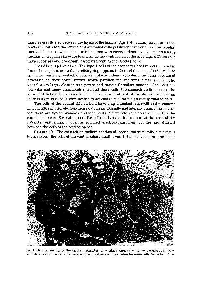
C a r d i a c s p h i n c t e r. The type 1 cells of the esophagus are far more ciliated in front of the sphincter, so that a ciliary r ing appears in front of the s tomach (Fig. 6). The sphincter consists of epithelial cells with electron-dense cytoplasm and long vacuolized processes on their apical surface which partit ion the sphincter l umen (Fig. 7). The vacuoles are large, e lectron-transparent and contain flocculent material. Each cell has few cilia and many mitochondria. Behind these cells, the stomach epi thel ium can be seen. Just beh ind the cardiac sphincter in the ventral part of the s tomach epithel ium there is a group of cells, each having m a n y ciha {Fig. 8} forming a highly cihated field.
The cells of the ventral cihated field have long branched microvilli a nd numerous mitochondria in their electron-dense cytoplasm. Dorsally and laterally beh ind the sphinc- ter, there are typical stomach epithelial cells. No muscle cells were detected in the cardiac sphincter. Several neuron- l ike cells and axonal tracts occur at the base of the sphincter epithelium. Numerous rounded electron-t ransparent cavities are situated be tween the cells of the cardiac region.
S t o m a c h. The stomach epi thel ium consists of three ultrastructurally distinct cell types {except the cells of the ventral ciliary field). Type 1 stomach cells form the major
Fig. 6. Sagittal section of the cardiac sphincter, cr - cihary ring, se - stomach epithehum, vc - vacuolated cells, vf - ventral ciliary field, arrow shows empty cavities between cells. Scale bar: 2 ~tm

113 Digestive tract of tornaria larva
4
q
) r ~
Fig. 7. Vacuolated growings of the cardiac sphincter cells, my - microvilli, vcp - vacuolated cell process. Scale bar: 2 ~m
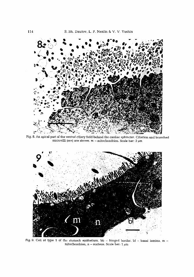
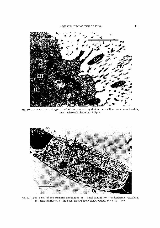
component of the epithelium. They are cuboid near the region of the cardiac and pyloric sphincters, and flat tened in the middle part of the stomach. The cytoplasm is highly electron-dense, near ly opaque and contains many mitochondria (Pig. 9). Numerous microvilli are straight. Some of them are slightly branched. Together they form a fringed border on the apical surface (Fig. 10).
Two b/pes of vesicles concentrate in the apical part of the cytoplasm: small electron- dense vesicles, which occup~ most of the cytoplasm, and large ones with electron-dense or t ransparent contents. Nucleus and extensive, rough, endoplasmic ret iculum occur in the basal region of the cell.
We found few type 2 cells in the stomach. Type 2 cells have t ransparent cytoplasm with basal ly situated nucleus and a few short mace-l ike microvilli (Fig. 11). Their cytoplasm contains mitochondria, cisternae of rough endoplasmic ret iculum and small t ransparent vesicles.
Type 3 cells have rare short microvilh of irregular shape and few cilia (Fig. 12). Cytoplasm of these cells is electron-light and filled with small and coarse electron-light vesicles.
P y 1 o r i c s p h i n c t e r a n d i n t e s t i n e. In the posterior part of the stomach the cells bear many long ciha directed towards the pyloric sphincter and intest ine lumen. Near the pyloric sphincter constriction, where the cells form the fringed border, a b a n d of cells of similar ultrastructure but almost without microvilli can be seen (Fig. 13). Still
8
f ....
. I
D ~ -
4 ~
S. S h . D a u t o v , L. P. N e z h n & V. V. Y u s h i n 114
J ~
Fig. 8. A n apical part of the ventral ci l iary field b e h i n d the cardiac sphincter. Ci l iat ion and b r a n c h e d microvi l l i (mv) are s h o w n , m - mi tochondrion . Scale bar: 2 ~m
Fig. 9. Cei l of t y p e 1 of the s t o m a c h ep i the l ium, bb - fr inged border; b! - b a s a l lamina , m - mi tochondr ion , n - nuc leus . Scale bar: 1 ~m
D i g e s t i v e t r a c t o f t o r n a r i a l a r v a 1 1 5
F ig . 10. A n a p i c a l p a r t of t y p e 1 ce l l of t h e s t o m a c h e p i t h e l i u m , c - c i l i u m , m - m i t o c h o n d r i a , m v - m ic rov i l l i . S c a l e b a r : 0 .5 ~lm
F ig . 11. T y p e 2 ce l l of t h e s t o m a c h e p i t h e l i u m , b l - b a s a l l a m i n a , e r - e n d o p l a s m i c r e t i c u l u m , m - m i t o c h o n d r i o n , n - n u c l e u s , a r r o w s s h o w c i l i a r r o o t l e t s . S c a l e b a r : 1 ~lm

116 S. Sh . D a u t o v , L. P. N e z l i n & V. V. Y u s h i n
Fig. 12. Type 3 cell of the s t o m a c h ep i the l ium, n - nuc leus , v - vacuole , a r row s h o w s b a s a l l amina . Scale bar: 1 ~m
Fig. 13. Sagi t ta l sec t ion of the pylor ic sphinc ter , a - axona l tracts, bl - b a s a l l amina , m - m i t o c h o n d - don , n - nuc leus . Scale bar: 1 ~m

Digestive tract of tornaria larva 117
further, another band of cells with electron-light cytoplasm is located. These cells have a smooth apical surface without microvilli, and bear few cilia. Then comes a b a n d of cells with relatively dense cytoplasm, few cilia and microvilli. These ceils have a variety of shapes. They form folds which resemble tight junctions. The intest ine is divided into three parts, depending on the type of epithelial cells. The anterior part is l ined with cuboid ceils having a relatively light cytoplasm and well-developed fringed border composed of elongated slightly branched microvilli (Pig. 14). These cells bear many cilia
Fig. 14. Section of the anterior part of the intestine, a - axonal tract, bb - f r i n g e d border, m - mitochondrion, n - nucleus, arrows - ciliar rootlets. Scale bar: 1
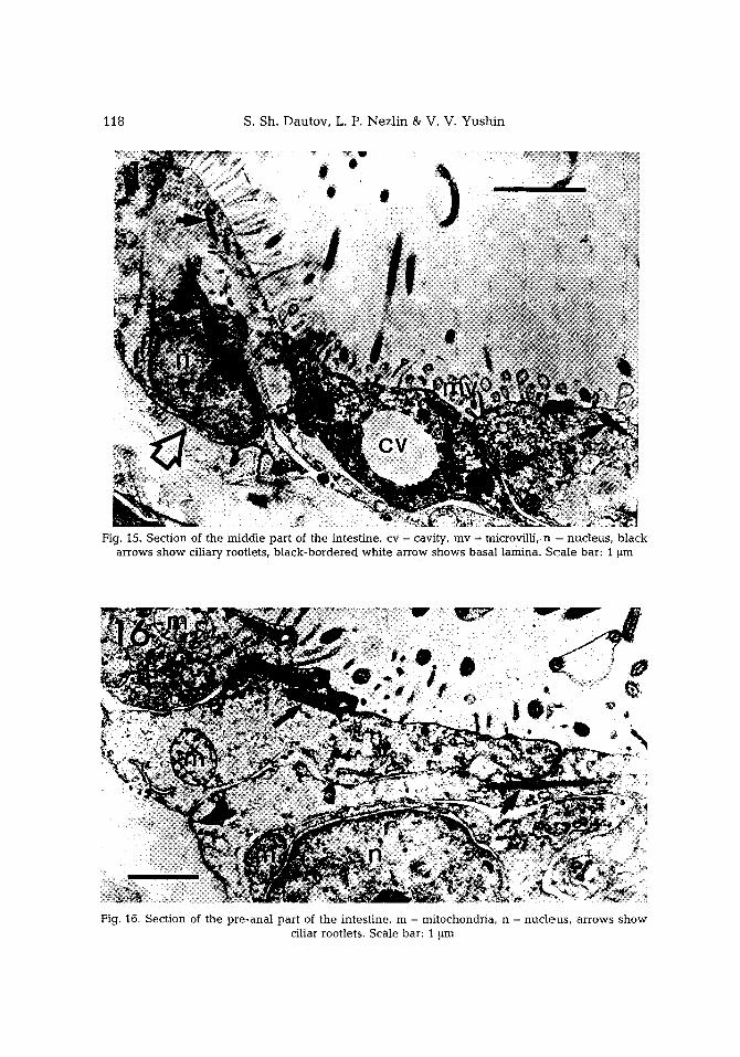
and contain numerous mitochondria in their cytoplasm. The middle part of the intest ine consists of flattened cells with short mace-l ike microvilli (Fig. 15). SuCh cells bear few cilia. There are rounded electron-light cavities be tween the cell bodies. In the pre-anal part of the intestine, the ceils have many cilia and have no fringed border (Fig. 16). On their surface, only rare, unb ranched microvilli are visible. Muscle cells are not found.
In the cardiac and pyloric parts of the stomach, and in the preanal part of the intestine, peculiarly structured cilia can be seen (Fig. 17, 18). Their distal end is greatly widened and the axoneme is spirally curved, forming one or two loops. Hence, the cross section gives an impression of ciha with two-four axonemes.
S. Sh . D a u t o v , L. P. N e z l i n & V. V. Y u s h i n 118
Fig. I5. Sec t ion of the m i d d l e pa r t of the in tes t ine , cv - cavity, m y - microv i l l i , n -- nuc leus , b l a c k a r rows s h o w ci l iary rootlets, b l a c k - b o r d e r e d w h i t e a r row shows b a s a l l amina . S c a i e bar : 1 ~m
Fig. 16. Sec t ion of the p r e - a n a l pa r t of the in tes t ine , m - mi tochondr ia , n - n u c l e u s , a r rows s h o w ci l iar rootlets . Sca le bar: 1 ~m

6
Digestive tract of tornaria larva 119
Fig. 17. Tangental section of the cilium with curved axoneme. Scale bar: 1.5 ~tm Fig. 18. Cross-section of the cilium with curved axoneme. Scale bar: 0.5 ~m
DISCUSSION
The feeding process of tornaria includes the collecting of par t ic les in the oral cavity, their passage down the esophagus , formation of a food-bolus by mucus, its t ransi t ion to the s tomach where digest ion t akes place, reject ion of the und iges t ed remains into the poster ior intest ine and excret ion through the anus (Stra thmann & Bonar, 1976). These processes are m e d i a t e d p redominan t ly by the ciliar activity of epi thel ia l cells of the digest ive tract - unl ike the ech inoderm larvae where the main role in feeding is done by contract ions of the r ing-muscles of the e sophagus and sphincters (Strathmann, 1971). Food part icles are re jec ted from the esophagus mainly by ciliary pulsat ions ra ther than by reversal peristal t ic of the e sophagea l muscles, as in the ech inoderm larvae.
This fully agrees with our data, i.e. the discovery of regions with powerful cil iation in the digest ive tract of tornaria - more powerfu l than in the ech inoderm larvae. An especia l ly high number of cilia is found near the card iac and pyloric sphincters and in the p re - ana l part of intest ine. At the same time, muscles of the digest ive tract a re not well- deve loped in tornaria. Single muscle-cel ls are found only in e sophagea l epi thel ium.
The cilia with spiral ly curved axonemes found in tornaria are similar to discocil ia first desc r ibed in po lychae te la rvae Lanice conchilega (Heimler, 1978). In discocilia, the axoneme is spiral ly curved distally and the d i sc - shaped widen ing forms terminal ly, It is supposed that discocilia are modif ied kinocil ia involved in causing strong wa te r displace- ment during swimming of the larvae (Heimler, 1978). However , Ehlers & Ehlers (1978) dec ided that discocil ia of ep ide rma l cells of mar ine Turbel lar ia may not be a genuine structure, and are an artifact of fixation.
T h e cells bea r ing many cilia are mainly (if not exclusively) found in the digest ive tract of tornaria. Perhaps more deve loped muscles in the digest ive tract of the ech inoderm larvae are re la ted to the p re sence of only monoci l ia ted cells, i.e. the impossibi l i ty of ci l iary fields with sufficient effect iveness deve lop ing in it; so that the t ranslocat ion of food part icles a long the d ige s t i ve tract is p r o v i d e d for by cil iary pulsa- tions.
Single cells of the e sophagea l epi the l ium in tornaria are ul t ras t ructural ly similar to those in the echinoid larvae. So the cells with vacuol ized apical processes in the cardiac sphincter in the tornaria r e semble the epi thel ia l cells of the lower par t of the e sophagus in the echinoplu teus of Dendraster excentricus and Psammechinus miliaris (Ryberg &

120 S. Sh. Dautov, L. P. Nezl in & V. V. Yushin
Lundgren, 1975; Burke, 1981). In both cases, the long processes are f i l led with coarse vacuoles conta in ing loose fibriilar mater ia l d i rec ted towards the e s o p h a g u s lumen. It was sugges t ed that these cells have secre tory functions, cons ider ing that th is par t of the e sophagus ensures the collection of food par t ic les and format ion of a bo lus by mucus. Apparen t ly , similar cells in tornar ia also perform secre tory functions, b u t are local ized only nea r the bo rde r b e t w e e n the e sophagus and the stomach, ra ther than a l o n g the main par t of the e sophagus (as in echinopluteus) .
The f inding of neurona l bodies in the ventra l s tomach ep i the l ium conforms with the results of h is tochemical invest igat ions of monoamine -con ta in ing n e u r o n s in t0rnaria (Nezlin & Dautov, 1988). A group of such cells in the region of the ven t r a l par t of the e sophagus has b e e n found. Evident ly the neurona l bod ies are s i tua ted in t he epi the l ium and their processes run a long the body of tornaria b e t w e e n the e p i t he l i um and basa l lamina.
In echinopluteus , food par t ic les r emain for a i o n g t ime in the l o w e r par t of the e sophagus where the bolus is forming. Then the card iac sphinc te r opens and the bolus passes to the s tomach (Strathmann, 1971). In tornaria, muscle cells have no t been found in the card ium region. Moreover , it is k n o w n that food par t ic les do not r e m a i n for long in the e sophagus of tornar ia (St ra thmann & Bonar, 1976). It can b e s u p p o s e d that the cell processes p rov ided in the sphinc ter lumen, and a lmost par t i t ioning it, conta in many vesicles, and that these ensure the adhes ion of food par t ic les by m u c u s and their t ransi t ion in doses to the stomach, thus compensa t ing for the a b s e n c e of muscle sphincter .
Three we l l -de f ined types of cells were found in the s tomach ep i the l ium, The type 1 ceils with e lec t ron-dense cytoplasm and fr inged border on their surface p r o b a b l y con- sume and store nutr ients c leaved by enzymes. The type 2 and 3 cei ls with l ight cytoplasm, w e l l - d e v e l o p e d rough endop lasmic re t iculum and numerous vesicles , appa - rent ly fulfil secre tory functions. Similar cells hav ing secretory functions a r e found in the echinoplu teus stomach, but they are not d is t r ibuted all over the s tomach ep i the l ium and are concen t ra ted in its anter ior part. Besides, these ceils in ech inop lu teus (in contrast to tornaria) a p p e a r no t to have cilia (Burke, 1981).
In the reg ion of the pyloric sphinc ter in tornaria, muscle-cel ls w e r e not found. However , there is a b a n d of cells bea r i ng many e longa ted cilia w h i c h are d i rec ted towards the in tes t ine lumen. Appa ren t ly these ci l ia ted cells p lay the role of the muscle sphincter .
At the bo rde r b e t w e e n the s tomach and the in tes t ine epithel ial , ce l ls are a lmost l ack ing in microvil i l bu t the cells of the poster ior in tes t ine wal l bea r w e l l - d e v e l o p e d microvilli. In the anter ior par t of the intest ine, microvill i form a w e l l - d e v e l o p e d f r i nged border . In the midd le par t of the intest ine, ceils bea r less microvilh and a re shorter, but these ceils are h igh ly special ized. They have we l l -deve loped rough endop lasmic ret iculum and Golgi complex. In these ceils, coarse e lec t ron- l ight vacuo le s and regions which lack any organe l les a re found. Similar regions were desc r ibed in the ech inop lu teus s tomach and they are cons ide red to b e the remains of consumed algal cei ls (Burke, 1981). Thus, the tornar ia in tes t ine appa ren t ly takes par t in food consumpt ion in contrast to ech inoplu teus of Dendraster excentricus in which the intest inal ep i the l ium is composed of unspec ia l i zed cells which lack microvilli, we l l -deve loped endop lasmic re t icu lum and Golgi complex (Burke, 1981).

D i g e s t i v e t r ac t of t o r n a r i a l a r v a 121
T h e d a t a o b t a i n e d d e m o n s t r a t e t h a t t h e u l t r a s t r u c t u r e of t h e d i g e s t i v e t r ac t s of
t o r n a r i a a n d e c h i n o d e r m l a r v a e a re e s s e n t i a l l y d i f fe rent . Firs t of all, i t is t h e p r e s e n c e of
m u l t i c i l i a t e d cells, l a c k of m u s c l e s in t h e m a i n p a r t of t h e d i g e s t i v e t ract , a n d t h e s t r u c t u r e
of t h e i n t e s t i n a l e p i t h e l i u m in to rna r i a .
Ear l ier , w e i n v e s t i g a t e d t h e u l t r a s t r u c t u r e of t he n e r v o u s s y s t e m of t o r n a r i a a n d t he
t o p o g r a p h y of c a t e c h o l a m i n e - c o n t a i n i n g cel ls in t o r n a r i a (Nez l in & Dau tov , 1988; D a u t o v
& Nez l in , 1992). C o m p a r i s o n of t h e s e d a t a w i t h t h e i n f o r m a t i o n o b t a i n e d f r o m l i t e r a t u r e
a lso d e m o n s t r a t e s e s s e n t i a l d i f f e rences . For e x a m p l e , in t he e c h i n o d e r m l a r v a e , m o n o -
a m i n e - c o n t a i n i n g cel ls a re l o c a t e d all o v e r t he c i l iary b a n d e p i t h e l i u m , w h e r e a s in
t o r n a r i a t h e y a re c o n c e n t r a t e d in t he a b o r a i a n d e s o p h a g e a l g a n g l i a (Nez l in e t al., 1984;
B i s g r o v e & B u r k e , 1987).
O u r d a t a c o n f i r m t h e s u g g e s t i o n t h a t t h e s imi la r i ty of e m b r y o n i c a n d l a r v a l d e v e l o p -
m e n t of t h e e c h i n o d e r m s a n d e n t e r o p n e u s t s b e a r s c o n v e r g e n t f ea tu r e s .
LITERATURE CITED
Bisgrove, B. W. & Burke, D. B., 1987. Development of the nervous system of the pluteus larva of Stronffylocentrotus droebachiensis. - Cell Tiss. Res. 248, 335-343.
Burke, D. R., 1981. Structure of the digestive tract of the pluteus larva of Dendraster excentricus (Echinodermata: Echinoidea). - Zoomorphology 98, 209-225.
Dautov, S. Sh. & Nezlin, L. P., 1992. Nervous system of the tornaria larva (Hemichordata: Enterop- neusta). Histochemical and ultrastructural study. - Biol. Bull. mar. biol. Lab., Woods Hole 183, 463-475.
Ehlers, U. & Ehlers, B., 1978. Paddle cilia and discocilia - Genuine structure? - Cell Tiss. Res. 192, 489-501.
Heimler, W., 1978, Discocilia - a new type of kinocilia in the larvae of Lanice conchilega (Polychaeta, Terebellimorpha). - Cell Tiss. Res. 187, 271-280.
Horst, K. van-der, 1933. Enteropneusta of the seas of the USSR. - Issled. Morel SSSR 19, 73-78 (in Russian).
Ivanova-Kazas, O. M., 1978. Comparative embryology of invertebrates. Nauka, Moscow, 163 pp (in Russian).
Nezlin, L. P. & Dautov, S. Sh., 1988. Histochemical investigation of nervous system of Tornaria - planktonic larvae of Hemichordates. - Biol. Morja Vladiv. 1988 (2), 70-71 (in Russian).
Nezlin, L. P., Dautov, S. Sh. & Malakhov, V. V., 1984. Topography of catecholamine-containing neurons in starfish larvae. - Dokl. Acad. Nauk SSSR. 278, 983-985 (in Russian).
Ryberg, E. & Lundgren, B., 1975. Secretory cells in the foregut of the echinopluteus. - Wilhelm Roux Arch. Entw. Mech. Org. 177, 255-262.
Strathmann, R. R., 1971. The feeding behavior of planktotrophic echinoderm larvae: mechanisms, regulation, and rates of suspension-feeding. - J. exp. mar. Biol. Ecol. 6, 109-160.
Strathmann, R. R., 1975. Larval feeding in echinoderms. - Am. Zool. 15, 717-731. Strathmann, R. R. & Bonar, D., 1976. Suspension feeding of tornaria larvae of Ptichodera flava
(Hemichordata: Enteropneusta). - Mar. Biol. 34, 317-324.