Structure of hepatitis C virus IRES subdomain IIa -...
9
electronic reprint Acta Crystallographica Section D Biological Crystallography ISSN 0907-4449 Editors: E. N. Baker and Z. Dauter Structure of hepatitis C virus IRES subdomain IIa Qiang Zhao, Qing Han, Charles R. Kissinger, Thomas Hermann and Peggy A. Thompson Acta Cryst. (2008). D64, 436–443 Copyright c International Union of Crystallography Author(s) of this paper may load this reprint on their own web site or institutional repository provided that this cover page is retained. Republication of this article or its storage in electronic databases other than as specified above is not permitted without prior permission in writing from the IUCr. For further information see http://journals.iucr.org/services/authorrights.html Acta Crystallographica Section D: Biological Crystallography welcomes the submission of papers covering any aspect of structural biology, with a particular emphasis on the struc- tures of biological macromolecules and the methods used to determine them. Reports on new protein structures are particularly encouraged, as are structure–function papers that could include crystallographic binding studies, or structural analysis of mutants or other modified forms of a known protein structure. The key criterion is that such papers should present new insights into biology, chemistry or structure. Papers on crystallo- graphic methods should be oriented towards biological crystallography, and may include new approaches to any aspect of structure determination or analysis. Crystallography Journals Online is available from journals.iucr.org Acta Cryst. (2008). D64, 436–443 Zhao et al. · Hepatitis C virus IRES subdomain IIa
Transcript of Structure of hepatitis C virus IRES subdomain IIa -...
electronic reprintActa Crystallographica Section D
BiologicalCrystallography
ISSN 0907-4449
Editors: E. N. Baker and Z. Dauter
Structure of hepatitis C virus IRES subdomain IIa
Qiang Zhao, Qing Han, Charles R. Kissinger, Thomas Hermann and PeggyA. Thompson
Acta Cryst. (2008). D64, 436–443
Copyright c© International Union of Crystallography
Author(s) of this paper may load this reprint on their own web site or institutional repository provided thatthis cover page is retained. Republication of this article or its storage in electronic databases other than asspecified above is not permitted without prior permission in writing from the IUCr.
For further information see http://journals.iucr.org/services/authorrights.html
Acta Crystallographica Section D: Biological Crystallography welcomes the submission ofpapers covering any aspect of structural biology, with a particular emphasis on the struc-tures of biological macromolecules and the methods used to determine them. Reportson new protein structures are particularly encouraged, as are structure–function papersthat could include crystallographic binding studies, or structural analysis of mutants orother modified forms of a known protein structure. The key criterion is that such papersshould present new insights into biology, chemistry or structure. Papers on crystallo-graphic methods should be oriented towards biological crystallography, and may includenew approaches to any aspect of structure determination or analysis.
Crystallography Journals Online is available from journals.iucr.org
Acta Cryst. (2008). D64, 436–443 Zhao et al. · Hepatitis C virus IRES subdomain IIa

research papers
436 doi:10.1107/S0907444908002011 Acta Cryst. (2008). D64, 436–443
Acta Crystallographica Section D
BiologicalCrystallography
ISSN 0907-4449
Structure of hepatitis C virus IRES subdomain IIa
Qiang Zhao,a* Qing Han,a
Charles R. Kissinger,a Thomas
Hermanna‡ and Peggy A.
Thompsonb*
aDepartment of Structural Chemistry, Anadys
Pharmaceuticals Inc., 3115 Merryfield Row,
San Diego, CA 92121, USA, and bDepartment
of Biology, Anadys Pharmaceuticals Inc.,
3115 Merryfield Row, San Diego, CA 92121,
USA
‡ Current address: Departments of Chemistry
and Biochemistry, University of California, San
Diego, 9500 Gilman Drive, La Jolla, CA 92093,
USA.
Correspondence e-mail:
# 2008 International Union of Crystallography
Printed in Singapore – all rights reserved
The hepatitis C (HCV) internal ribosome entry site (IRES)
element plays a central role in cap-independent translation of
the viral genomic RNA. The unique conformation of IRES
domain II is critical for 80S ribosomal assembly and initiation
of viral translation. Here, the crystal structure of subdomain
IIa of the HCV IRES has been determined at 2.3 A resolution,
revealing the positions of divalent metal ions and complex
inter-strand interactions that stabilize the L-shaped conforma-
tion of the RNA. The presence of divalent metal ions was
necessary for crystal formation. Magnesium ions occupy
specific sites that appear to be critical for the formation of
the folded conformation. Subdomain IIa also was crystallized
in the presence of strontium, which improved the diffraction
quality of the crystals and the ability to identify interactions of
the RNA with metal ions and tightly bound water molecules.
The hinge region and noncanonical G–U base-pair motifs are
stabilized by divalent metal ions and provide unique structural
features that are potential interaction sites for small-molecule
ligands. The information obtained from the crystal structure
provides a basis for structure-guided design of HCV transla-
tion inhibitors targeting disruption of ribosomal assembly.
Received 12 November 2007
Accepted 18 January 2008
PDB References:
Mg-subdomain IIa, 2pn3,
r2pn3sf; Sr-subdomain IIa,
2pn4, r2pn4sf.
1. Introduction
Hepatitis C virus (HCV) is an enveloped single-stranded
positive-sense RNA virus. The HCV genome contains an
internal ribosome entry site (IRES), located within the
50-untranslated region (50-UTR), that is responsible for
directing cap-independent translation of the viral mRNA
(Tsukiyama-Kohara et al., 1992; Wang et al., 1993; Reynolds et
al., 1996). In contrast to the HCV mRNA open reading frame,
the �340 nucleotide IRES RNA sequence is highly conserved
among all genotypes (Bukh et al., 1992; Davidson et al., 1995).
Assembly of the 48S complex and subsequently the 80S
ribosome is initiated when the host-cell 40S ribosomal subunit
and eukaryotic initiation factor 3 (eIF3) bind to the HCV
IRES RNA (Pestova et al., 1998, 2001; Kieft et al., 1999; Ji et
al., 2004; Siridechadilok et al., 2005; Yu et al., 2005). Formation
of this complex is dependent on the IRES adopting a complex
cation-dependent fold (Kieft et al., 1999, 2001; Klinck et al.,
2000). The high sequence conservation among viral isolates
and the unique structural features of the IRES, along with its
critical role in translation, make it an attractive target for
RNA-directed HCV therapeutics.
The IRES has been divided into four structural domains
designated I, II, III and IV (Fig. 1a), which have been shown to
electronic reprint

exist as independently folded regions (Brown et al., 1992;
Wang et al., 1995; Honda et al., 1996). The helical and stem-
loop regions within domains II and III, along with stem loop
IV, which contains the start AUG codon, all contribute to
IRES-dependent translation (Brown et al., 1992; Kieft et al.,
2001; Kikuchi et al., 2003; Ji et al., 2004; Otto & Puglisi, 2004;
Spahn et al., 2004; Boehringer et al., 2005; Laletina et al., 2006).
The precise roles of individual IRES domains in the formation
of a functional 80S complex continue to be elucidated,
furthering the understanding of IRES-mediated cap-
independent translation (Ji et al., 2004; Lu et al., 2004; Otto &
Puglisi, 2004; Locker et al., 2007).
The correctly folded tertiary structure of the HCV IRES
has been shown to be essential for binding both the 40S
ribosomal subunit and eIF3. Cryo-electron microscopy (cryo-
EM) of the IRES in complex with the 40S ribosomal subunit at
20 A resolution (Spahn et al., 2004) and the 80S ribosome at
15 A resolution (Boehringer et al., 2005) confirmed that the
IRES induces a significant conformational change in the 40S
subunit upon binding, altering the mRNA-binding region.
These studies revealed that the apical loop of domain II
reaches deep into the mRNA cleft near the tRNA-exit site of
the 40S ribosome complex (Spahn et al., 2001). Removal of
domain II does not alter the binding affinity of either the 40S
ribosomal subunit or eIF3 to the IRES; however, it has been
shown that this domain is responsible for inducing a large
conformational rearrangement at the decoding center of the
40S subunit. This conformational change may be necessary to
stabilize the viral AUG start codon in position and for
assembly of the ribosomal subunits (Brown et al., 1992;
Kolupaeva et al., 2000; Spahn et al., 2001; Lafuente et al., 2002;
Ji et al., 2004; Otto & Puglisi, 2004; Boehringer et al., 2005;
Locker et al., 2007). The NMR solution structure of domain II
revealed that a bulge within subdomain IIa induces a bend in
the helix, resulting in an overall L-shaped conformation. The
structure of the isolated domain II is consistent with its
conformation when bound to the 40S ribosomal subunit as
determined by cryo-EM (Lukavsky et al., 2003; Spahn et al.,
2004). Conserved structural motifs within domain II, including
the unique bent conformation of subdomain IIa, play a novel
functional role in 80S ribosomal assembly by promoting eIF5-
induced GTP hydrolysis and mediating eIF2 release (Locker
et al., 2007). The connection between the uniquely folded
structure of subdomain IIa and its function in 80S ribosomal
formation suggests that HCV IRES IIa is a possible target for
small-molecule antiviral drugs that bind and disrupt ribosomal
complex formation and translation of the viral mRNA.
To gain further understanding of the interactions that
stabilize the unique conformation of IRES domain II, we have
determined the X-ray crystal structure of subdomain IIa in the
presence of either Sr2+ or Mg2+ ions at resolutions of 2.3 and
2.9 A, respectively. A 3.0 A resolution crystal structure of
subdomain IIa obtained in the presence of a mixture of Mg2+
and Mn2+ ions has also been reported (Dibrov et al., 2007). We
observe differences in the overall bend angle and the positions
of key metal ion-binding sites. These results provide additional
insight into the forces that stabilize the unique L-shaped
conformation of the molecule and the possibility of molecular
intervention at this step in ribosomal complex formation.
2. Materials and methods
2.1. Constructs and crystallization
Gel-purified and desalted synthetic IRES RNA oligo-
nucleotides for subdomain IIa (50-CGG AGG AAC UAC
UGU CUU CAC GCC-30 and 50-GCG UGU CGUGCAGCC
UCC GG-30) were purchased from Dharmacon Research
(Lafayette, Colorado, USA). The brominated oligonucleotide
50-GCG (5-Br-U)GUCGUGCAGCC (5-Br-U)CCGG-30 wasused for multi-wavelength anomalous dispersion (MAD) data
collection.
RNA was annealed by heating in buffer (5 mM sodium
cacodylate pH 6.2, 2 mM MgCl2) at 353 K for 3 min and
cooling slowly to room temperature. Crystals were obtained
by the hanging-drop vapor-diffusion method, in which 1 ml0.5 mM RNA in annealing buffer was mixed with an equal
volume of a precipitant buffer containing 50 mM sodium
cacodylate pH 6.2, 10 mMMgSO4, 25 mMNaCl, 100 mMKCl,
10% 2-methyl-2,4-pentanediol (MPD) and equilibrated over a
well solution consisting of 65% MPD at 290 K or room
temperature. Crystals were obtained at 290 K after 2–3 weeks.
The crystals belonged to space group P43212, with unit-cell
parameters a = b = 48.95, c = 120.94 A and one molecule per
asymmetric unit.
For crystals grown in the presence of strontium, the RNA
was annealed as above but without MgCl2. The RNAwas then
mixed in a 1:1 ratio with 50 mM sodium cacodylate pH 6.2,
research papers
Acta Cryst. (2008). D64, 436–443 Zhao et al. � Hepatitis C virus IRES subdomain IIa 437
Figure 1(a) Secondary structure of the �340-nucleotide HCV 50-UTR containingthe four structural domains of the IRES element. Subdomain IIa isoutlined with a dashed box. (b) Sequence of the subdomain IIa constructused for crystallization and structure determination. Residues identical tothe HCV IRES domain II are shown in bold. Non-Watson–Crick base-pair interactions are indicated by dots. 5-Br-U-labeled uridines aremarked with asterisks.
electronic reprint
80 mM SrCl2, 25 mMNaCl, 30%MPD and equilibrated over a
well solution consisting of 60% MPD at 290 K. Sr-subdomain
IIa crystals had unit-cell parameters that were nearly identical
to those of the Mg-subdomain IIa crystals, but belonged to the
lower symmetry space group P212121.
2.2. Data collection and structure determination
The structure of IRES subdomain IIa with Mg2+ bound
(Mg-IIa) was determined by MAD phasing using the anom-
alous scattering from bromines incorporated into the RNA by
replacing U101 and U113 with 5-Br-U. These two nucleotides
were chosen because they form stable canonical A–U base
pairs and are distant from the hinge region, minimizing the
potential for altering the folded conformation.
Prior to data collection, crystals were flash-cooled in liquid
nitrogen and transferred to a cold nitrogen stream. Attempts
at molecular replacement using the structure determined by
NMR (Lukavsky et al., 2003; PDB code 1p5m) were not
successful. Therefore, MAD data sets (Table 1) were collected
from a single crystal at three wavelengths using bromine as the
anomalous scattering atom at 100 K on beamline 14-ID-B at
the Advanced Photon Source (APS), Argonne National
Laboratory. The X-ray data for the Sr-IIa crystal were
collected at 100 K on beamline 8-BM also at the APS. Data
were processed with DENZO/SCALEPACK (Otwinowski &
Minor, 1997). Phases were calculated using the programs
SOLVE/RESOLVE (Terwilliger & Berendzen, 1999; Terwil-
liger, 2000). The resulting MAD electron-density map at 3.2 A
resolution was used to build the initial model with the program
O (Jones et al., 1991), which gave an initial Rfree value of 0.4798
and an R factor of 0.4621 after rigid-body refinement. The
structure was refined by several rounds of simulated annealing
with restrained individual B-factor refinement in the program
CNX (Accelrys, San Diego, California, USA; Brunger et al.,
1998) and manual examination and rebuilding using the
inflection-point data.
The Sr-IIa crystal structure was solved by the molecular-
replacement method using the refined Mg-IIa model as the
search model. The Sr-IIa and Mg-IIa structures were refined at
2.3 and 2.9 A resolution to final free R factors of 32.0% and
28.3%, respectively (see Table 1).
3. Results and discussion
3.1. Construct design and crystallization approach
Both our own enzymatic digestion studies (data not shown)
and NMR solution structures (Lukavsky et al., 2003) indicate
that oligonucleotide constructs comprising only the IIa
subdomain adopt the same conformation as found in the full-
length IRES RNA. Subdomain IIa oligonucleotide constructs
of varying lengths were screened in crystallization trials (see
supplementary material1). Terminal G–C pairs were intro-
duced to stabilize the duplex, including single-base overhangs
to facilitate the formation of pseudo-continuous helices in the
crystal. The flexible unpaired U48 nucleotide was also
removed in most constructs in order to aid crystallization.
Crystallization was carried out at both room temperature and
290 K using a variety of commercial screening kits and
modified versions of an RNA-specific screen (Wahl et al.,
1996).
Construct length and crystallization temperature had the
most impact on crystal quality. Experiments at 290 K
produced higher quality crystals compared with room-
temperature crystallization. Shorter constructs comprising
only the hinge region produced few hits in initial screens and
did not lead to well diffracting crystals. Construct lengths were
progressively increased until promising crystals were obtained
at 290 K under conditions containing either MgSO4, NaCl and
MPD or magnesium acetate, MPD and spermidine. The initial
research papers
438 Zhao et al. � Hepatitis C virus IRES subdomain IIa Acta Cryst. (2008). D64, 436–443
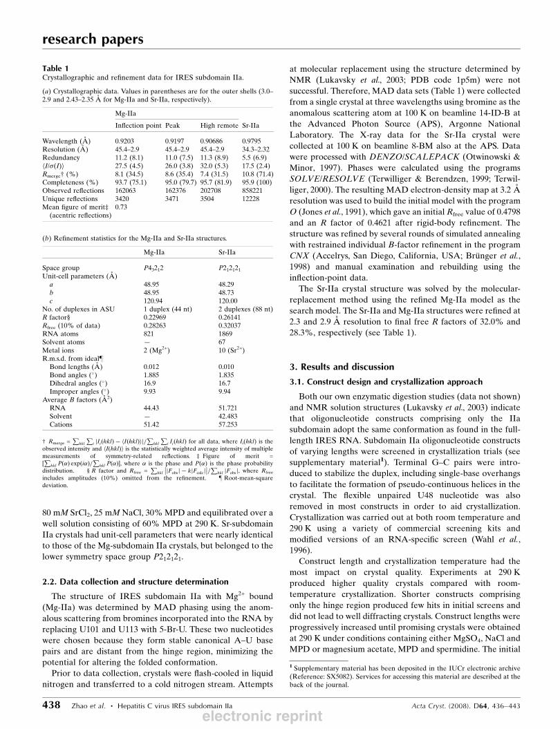
Table 1Crystallographic and refinement data for IRES subdomain IIa.
(a) Crystallographic data. Values in parentheses are for the outer shells (3.0–2.9 and 2.43–2.35 A for Mg-IIa and Sr-IIa, respectively).
Mg-IIa
Inflection point Peak High remote Sr-IIa
Wavelength (A) 0.9203 0.9197 0.90686 0.9795Resolution (A) 45.4–2.9 45.4–2.9 45.4–2.9 34.3–2.32Redundancy 11.2 (8.1) 11.0 (7.5) 11.3 (8.9) 5.5 (6.9)hI/�(I)i 27.5 (4.5) 26.0 (3.8) 32.0 (5.3) 17.5 (2.4)Rmerge† (%) 8.1 (34.5) 8.6 (35.4) 7.4 (31.5) 10.8 (71.4)Completeness (%) 93.7 (75.1) 95.0 (79.7) 95.7 (81.9) 95.9 (100)Observed reflections 162063 162376 202708 858221Unique reflections 3420 3471 3504 12228Mean figure of merit‡(acentric reflections)
0.73
(b) Refinement statistics for the Mg-IIa and Sr-IIa structures.
Mg-IIa Sr-IIa
Space group P43212 P212121Unit-cell parameters (A)a 48.95 48.29b 48.95 48.73c 120.94 120.00
No. of duplexes in ASU 1 duplex (44 nt) 2 duplexes (88 nt)R factor§† 0.22969 0.26141Rfree (10% of data) 0.28263 0.32037RNA atoms 821 1869Solvent atoms — 67Metal ions 2 (Mg2+) 10 (Sr2+)R.m.s.d. from ideal}‡Bond lengths (A) 0.012 0.010Bond angles (�) 1.885 1.835Dihedral angles (�) 16.9 16.7Improper angles (�) 9.93 9.94
Average B factors (A2)RNA 44.43 51.721Solvent — 42.483Cations 51.42 57.253
† Rmerge =P
hkl
Pi jIiðhklÞ � hIðhklÞij=Phkl
Pi IiðhklÞ for all data, where Ii(hkl) is the
observed intensity and hI(hkl)i is the statistically weighted average intensity of multiplemeasurements of symmetry-related reflections. ‡ Figure of merit =½Phkl Pð�Þ expði�Þ=
Phkl Pð�Þ�, where � is the phase and P(�) is the phase probability
distribution. † § R factor and Rfree =P
hkl
��jFobsj � kjFcalcj
��=P
hkl jFobsj, where Rfree
includes amplitudes (10%) omitted from the refinement. ‡ } Root-mean-squaredeviation.
1 Supplementary material has been deposited in the IUCr electronic archive(Reference: SX5082). Services for accessing this material are described at theback of the journal.
electronic reprint

crystals were optimized by sampling different combinations of
10–100 mM MgCl2, MgSO4, magnesium acetate, Co(NH3)6Cl3with or without NaCl, KCl and spermidine in the pH range
4.2–7.4.
Further construct optimization substantially increased the
resolution limit of the diffraction from 5 to 3 A. Single-base
changes in the overall length of the strands or the overhangs
dramatically affected the diffraction quality of the crystals.
The RNA molecule that gave the best diffracting crystals
consisted of two strands with 20 and 24 bases, respectively,
with single-base overhangs (Fig. 1b). Substituting uracil for
5-Br-U did not affect the crystal quality.
The presence of divalent metal ions was essential for crystal
formation. Initial crystals were obtained using magnesium
(Mg2+). Unambiguous identification of bound Mg2+ ions and
differentiation from water molecules in an electron-density
map is difficult except at very high resolutions. Therefore,
subdomain IIa was also crystallized in the presence of stron-
tium ions. Although the Sr2+ ion has a larger ionic radius than
that of Mg2+, it has previously been used to
substitute for Mg2+ in the crystallization of
RNA (Mueller et al., 1999; Baugh et al.,
2000; Deng et al., 2001; Wedekind & McKay,
2003). The increased diffraction from the
heavier strontium ions provided a significant
advantage in differentiating the bound
metal ions from water molecules. This
allowed us to clearly identify additional
binding sites for divalent cations in the
subdomain IIa structure. The resolution of
the data obtained from the crystals grown
with Sr2+ also was higher (2.3 A versus
2.9 A; Table 1). The Sr-subdomain IIa crys-
tals had very similar unit-cell parameters to
those of the Mg-subdomain IIa crystals, but
belonged to the lower symmetry space
group P212121 instead of P43212.
3.2. Overall structure
Both the Sr-bound and Mg-bound sub-
domain IIa crystal structures show the same
L-shaped conformation (Fig. 2), with indi-
vidual RNA molecules aligned in a pseudo-
continuous end-to-end arrangement in the
crystal lattice. The two crystal forms are
nearly identical; the presence of Sr led to a
slight breakdown in symmetry along the
fourfold axis of the tetragonal Mg form,
resulting in orthorhombic crystals. In both
structures, the asymmetric internal loop
comprising single-stranded nucleotides
A53–A57 causes the helix to bend with an
angle of about 110� between the terminal
vectors making up the curved axis, as
calculated by the program CURVES
(Lavery & Sklenar, 1989) with the bulged
nucleotides omitted. This compares with an angle of 129�
measured by the same method for the corresponding region in
the average NMR structure of isolated subdomain IIa
(Lukavsky et al., 2003) and 117� for the recently reported
subdomain IIa crystal structure solved at 3.0 A resolution
(Dibrov et al., 2007). In addition to the slightly smaller helical
bend angle, the bulge region is more compact, with the
distance between the ribose backbone C1 atoms of the two
external bases, U56 and U106, being 3–4 A less than
previously reported (Dibrov et al., 2007). In the structure
determined by Dibrov and coworkers, the external U56 base
re-orients to point towards the phosphate backbone of A54 on
an adjacent molecule, forming a hydrogen bond. This crystal-
packing interaction is not observed in the structures reported
here and could provide one explanation for the different
helical bend angles.
The unpaired nucleotide A57 is stacked below the C58–
G110 base pair as a result of a change in backbone direction at
nucleotide U56, which projects out into the solvent. The bases
research papers
Acta Cryst. (2008). D64, 436–443 Zhao et al. � Hepatitis C virus IRES subdomain IIa 439
Figure 2(a) Stereoview of the 2Fo � Fc electron-density map of the IRES subdomain IIa hinge regionwith the refined Sr-IIa atomic model superimposed, contoured at 0.26 e A�3. (b) The tertiarystructure of subdomain IIa shows an L-shaped helical conformation. Superposition of theSr-IIa (blue) and Mg-IIa (green) structures demonstrates that subdomain IIa adopts the sameconformation in the presence of strontium or magnesium metal ions. Two of the Sr2+ metal-binding sites are interchangeable with Mg2+ (Sr1/Mg1 and Sr2/Mg2). Mg2+ atoms are coloredred; Sr2+ atoms are colored orange. Figs. 2–5 were generated using PyMOL (DeLano, 2002)and CCP4MG (Potterton et al., 2002).
electronic reprint

of nucleotides A53, A54 and C55 are also unpaired and stack
continuously with the adjacent G52–C111 base pair. The
pocket that is formed by the stacked unpaired bases provides a
cluster of hydrogen-bond donors and acceptors that bind
metal ions that stabilize the L-shaped conformation and
facilitate the interaction of domain IIb with the ribosome. The
L-shaped bend in the helical axis is stabilized by a hydrogen-
bond interaction between the 20-hydroxyl group of C55 and
the N7 atom of A57. On the opposing strand, a hydrogen bond
between the N4 amino group of C111 and the G110 phosphate
group provides additional stabilization in the hinge region.
The five-nucleotide internal loop of subdomain IIa has
structural features similar to the �-turn motif (Wadley & Pyle,
2004) with nucleotides 1, 2 (A53, A54) and 5 (A57) stacking
on adjacent helices. A characteristic signature of �-turns is the
flipped-out nucleotide 4 (U56), which is commonly involved in
RNA–RNA or RNA–protein interactions. However, an
internal hydrogen bond between the 20-hydroxyl groups of
nucleotides 1 and 5 in the �-turn is absent in the subdomain
IIa structure.
Stacking of C62–G105 and the noncanonical U61–G107
base pairs is facilitated by rotation of the U106 base out of the
helix and into the solvent (Fig. 3). The noncanonical U61–
G107 base pair adjacent to the flipped-out U106 base alters
the conformation of the RNA backbone. This brings the
backbone of adjacent strands into close proximity in this
region. The weak electron density and resulting high atomic
temperature factors for the base portions of U56 and U106
indicate that they are highly flexible. In the Sr-IIa structure
these two external uridine bases are stacked parallel to each
other at a distance of 3.4–3.7 A (Fig. 3a),
while in the Mg-IIa crystal structure
they form an angle of approximately
130� to each other (Fig. 3b). A hydrogen
bond is formed between O2 of U56 and
O4 of U106, implying tautomerization
of one of the bases. These observations
are consistent with a UV cross-linking
study of domain II that indicated that
bases U56 and U106 could be covalently
linked (Lyons et al., 2001), suggesting
that these residues also are flipped out
of the helix and in close contact in
solution.
The upper and lower stem regions of
loop IIa form conventional A-form
helical structures. Watson–Crick base
pairing resumes at G52–C111 after the
bend and continues to the end of the
lower stem (C47–G116). The upper
stem region (C58–G110 through C69–
G98) contains three noncanonical base
pairs that do not significantly alter the
helical structure.
3.3. Metal-ion interactions
The presence of divalent cations is
essential for HCV IRES to adopt a
correctly folded structure and for
formation of the IRES–40S ribosomal
complex (Kieft et al., 1999). Cations
generally promote folding and stabil-
ization of RNA tertiary structure by
reducing the electrostatic repulsion
between negatively charged phosphate
groups or by coordinating directly to
electronegative atoms of the nucleotide
bases (Draper, 2004). While the
majority of cations stabilize RNA
structures through long-range diffuse
interactions, X-ray crystal structures
research papers
440 Zhao et al. � Hepatitis C virus IRES subdomain IIa Acta Cryst. (2008). D64, 436–443
Figure 3Hinge region of subdomain IIa showing the close proximity of the opposing backbone strands nearthe solvent-exposed uridines U56 and U106. The binding of Sr1/Mg1 and Sr2/Mg2 to the hingeregion is shown. (a) The two looped-out uridines of the Sr-IIa model have their bases stackedparallel to each other at a distance of 3.4–3.7 A. (b) The two bases of the looped-out uridines in theMg-IIa model form an angle of about 130� relative to each other and participate in hydrogen-bondformation.
Figure 4Bound metal ions stabilize the unpaired internal loop and non-Watson–Crick base pairs in the hingeregion. (a) Sr1 binds directly to nucleotide C55 and makes a water-mediated interaction with G110.(b) Sr2 coordinates directly to nucleotides G60, U61 and G107 and indirectly to U59 through atightly bound water molecule. The hydrogen-bond network that is formed by binding metal ions andwater is depicted.
electronic reprint

and NMR spectroscopic methods have allowed the identifi-
cation of the specific binding sites of metal ions that partici-
pate in modulating RNA structure and function (Allain &
Varani, 1995; Cate et al., 1997; Keift & Tinoco, 1997; Hermann
& Patel, 1999; Draper, 2004).
Five strontium ions were identified in association with each
subdomain IIa RNA molecule in the higher resolution (2.3 A)
Sr-IIa structure (Fig. 2b). Two of these metal-binding sites
were also clearly occupied by Mg2+ ions in the Mg-IIa struc-
ture, demonstrating the ability to interchange Mg2+ with Sr2+
without altering the RNA fold. The presence of Mg2+ or Sr2+
was essential for the generation of well diffracting crystals.
Interestingly, from our experiments, the divalent cations could
be added after annealing the RNA, suggesting that metal ions
may not be necessary in the initial RNA-annealing step but
are required for the formation of a stable folded structure.
Direct binding studies indicated that Mg2+ binds with a Kd of
�30 mM to the highest affinity sites on subdomain IIa
(manuscript in preparation).
In both crystal structures, a metal ion (Mg1, Sr1) is directly
coordinated to the O2 carbonyl O atom of nucleotide C55 and
makes a water-mediated contact to O6 of G110 (Fig. 4a). The
single-stranded stacked nucleotides in the hinge region form a
metal ion-binding pocket that provides direct coordination
with unpaired bases. The bound metal ion acts to further
stabilize the bend at the RNA internal loop.
A second metal ion (Mg2, Sr2) binds to the mismatched
G–U base pair in the major groove through direct coordina-
tion to O4 of U61, O6 of G107 and O6 of G60 (Fig. 4b). A
hydrogen-bond network in this region, which includes a water
molecule that coordinates the metal ion and forms hydrogen
bonds to the O6 atoms of U59 and G60, contributes to
stabilization of the non-Watson–Crick base pair as well as the
unique conformation of the RNA backbone that results in
U106 being rotated out into the solvent. The potential
instability caused by the close proximity of the backbone
strands at U56 and U106 may be mitigated by the binding of
Sr2 (Mg2), along with the presence of a tightly bound water
molecule that bridges the metal ion and bases. This metal-
binding site may also serve a functional role, as base-pair
mismatches are often found in ribosomal RNAs, where they
provide recognition sites for proteins and small molecules
(Gautheret et al., 1995; Chandrasekhar & Malathi, 2003).
The crystal structure shows that the hinge
region is stabilized by coordinated metal
ions, �-stacking of single-stranded nucleo-
tides and a hydrogen-bonding network that
involves tightly bound water molecules as
well as nucleotide bases. The perpendicular
orientation of bases C55 and G110 is fixed
though mutual coordination of Mg1/Sr1 and
appears to be critical for stabilizing the
conformation of subdomain IIa. The loca-
tions of the two metal-binding sites are
consistent with the NMR study of sub-
domain IIa, which suggested the presence of
one or two metal-binding sites near
nucleotides A54 and C55 (Lukavsky et al.,
2003). The existence of an Mg2+-binding site
in the hinge region near A109 and G110 has
also been suggested by FeII–EDTA foot-
printing (Kieft et al., 1999).
Three additional Sr2+-binding sites were
observed in the subdomain IIa crystal
structure. Sr3 binds directly to the
phosphate group of A54, whereas Sr4 is
research papers
Acta Cryst. (2008). D64, 436–443 Zhao et al. � Hepatitis C virus IRES subdomain IIa 441
Figure 5Metal ion-binding sites of Sr3, Sr4 and Sr5 in the Sr-IIa structure. The Sr3metal ion is directly coordinated to the phosphate group of A54. Sr4makes a water-mediated contact with the phosphate groups of G52 andA53. Sr5 forms an indirect interaction with the base of G51 in the majorgroove via a tightly bound water molecule. Strontium metal ions arecolored orange, waters are colored red and hydrogen bonds are shown asdashed lines.
Figure 6Stereo superposition of the Sr-subdomain IIa model (RNA is colored brown, strontium ionsare orange and yellow) with Mg-Mn subdomain IIa model (Dibrov et al., 2007; RNA is coloredteal, metal ions are light-blue and teal). For each model, metal ions from both copies in theasymmetric unit are superimposed and represented as spheres. For clarity, water moleculeshave been removed and only one RNAmolecule from each model is shown. The Sr1 metal ion-binding site corresponds to the Mn1 site in the Mg-Mn IIa model. The remaining metal ion-binding sites differ.
electronic reprint

indirectly coordinated to the phosphate groups of G52 and
A53 via a water-mediated interaction (Fig. 5). Sr5 is bound in
the major groove of the lower stem. This metal ion does not
form direct interactions with the RNA, but a coordinating
water molecule is hydrogen bonded to N7 of G51. These three
Sr2+ ions have relatively high temperature factors and appear
to be less tightly bound than Sr1 and Sr2 in the hinge region.
The metal-binding sites that we observe differ significantly
from those reported by Dibrov and coworkers for the same
subdomain IIa construct determined at 3.0 A resolution in the
presence of a mixture of Mg2+ and Mn2+ ions (Dibrov et al.,
2007). Only one metal ion, Mg1/Sr1, coordinated to C55 (see
Fig. 6), corresponds closely in position to a metal ion (Mn1) in
the Mg-Mn subdomain IIa structure. Mg1 in the Mg-Mnmodel
is in the same proximity as Mg2/Sr2; however, Mg2/Sr2 is
significantly closer to the base of U61 (2.5 A versus 5.3–6 A)
and stabilizes the wobble base pair through direct contacts
with U61 and G107. In the Mg-Mn subdomain IIa structure,
the Mg1 metal ion makes only one contact with N7 of G107
rather than coordinating directly to both base pairs to stabilize
their conformation. The metal ion Mn2 in the Mg-Mn model is
near Sr5 in our structure; however, it is in contact with the N7
atom of G52 rather than N7 of G51 as seen for Sr5.
We observed no convincing electron density in either of our
crystal structures for the Mg5 and Mg6 metal ions located at
both ends of the duplex or for Mg2 near G48. Also, no density
was observed at sites corresponding to the Mg3, Mg4 and Mn3
ions found between the two RNA molecules in the Mg-Mn
subdomain IIa structure (Dibrov et al., 2007).
The 2.3 A resolution data obtained from the Sr-subdomain
IIa crystals allowed us to accurately identify metal ion-binding
sites in the electron-density map. In addition, we were able to
assign the positions of bound water molecules (see Figs. 4 and
5) which could not be reliably modeled in the lower resolution
Mg-subdomain IIa structure and also are absent from
previously reported structures (Lukavsky et al., 2003; Dibrov
et al., 2007). This has provided an initial view of the role that
these water molecules play in coordinating the bound metal
ions and stabilizing the RNA conformation.
3.4. Potential for small-molecule interactions
The uniquely folded conformation of IRES subdomain IIa
and its critical role in 80S ribosomal assembly (Locker et al.,
2007) suggest that it is a possible target for small molecules
that bind to subdomain IIa, alter the conformation of the
IRES element and thereby inhibit translation of viral mRNA.
Recently, a novel class of benzimidazole compounds was
identified that bind to HCV IRES subdomain IIa with sub-
micromolar affinity (Seth et al., 2005). The binding of these
compounds was mapped to the hinge region by RNA-foot-
printing studies, which showed that nucleotides C55 and U56
had the greatest protection from enzymatic cleavage in the
presence of the compound (Seth et al., 2005).
The crystal structure of IRES subdomain IIa, coupled with
the identification of small molecules that bind to this region,
will allow a new structure-guided approach for designing
small-molecule inhibitors of HCV IRES-mediated translation.
We thank the staff at APS beamlines 14-ID and 8-BM for
providing access to their synchrotron-radiation facility and for
their help with data collection and we are grateful to Dr Ke
Shi at UMN for helpful discussions during the initial Mg-IIa
structure determination.
References
Allain, F. H. & Varani, G. (1995). Nucleic Acids Res. 23, 341–350.Baugh, C., Grate, D. & Wilson, C. (2000). J. Mol. Biol. 301, 117–128.Boehringer, D., Theremann, R., Ostareck-Lederer, A., Lewis, J. D. &Stark, H. (2005). Structure, 13, 1695–1706.
Brown, E. A., Zhang, H., Ping, L. H. & Lemon, S. M. (1992). NucleicAcids Res. 20, 5041–5045.
Brunger, A. T., Adams, P. D., Clore, G. M., DeLano, W. L., Gros, P.,Grosse-Kunstleve, R. W., Jiang, J.-S., Kuszewski, J., Nilges, M.,Pannu, N. S., Read, R. J., Rice, L. M., Simonson, T. & Warren, G. L.(1998). Acta Cryst. D54, 905–921.
Bukh, J., Purcell, R. H. & Miller, R. H. (1992). Proc. Natl Acad. Sci.USA, 89, 4942–4946.
Cate, J. H., Hanna, R. L. & Doudna, J. A. (1997). Nature Struct. Biol.4, 553–558.
Chandrasekhar, K. & Malathi, R. (2003). J. Biosci. 28, 547–555.Davidson, F., Simmonds, P., Ferguson, J. C., Jarvis, L. M., Dow, B. C.,Follett, E. A., Seed, C. R., Krusius, T., Lin, C. & Medgyesi, G. A.(1995). J. Gen. Virol. 76, 1197–1204.
DeLano, W. L. (2002). The PyMOLMolecular Graphics System. PaloAlto, California, USA: DeLano Scientific.
Deng, J., Xiong, Y. & Sundaralingam, M. (2001). Proc. Natl Acad. Sci.USA, 98, 13665–13670.
Dibrov, S. M., Johnston-Cox, H., Weng, Y. H. & Hermann, T. (2007).Angew. Chem. Int. Ed. Engl. 46, 226–229.
Draper, D. E. (2004). RNA, 10, 335–343.Gautheret, D., Konings, D. & Gutell, R. R. (1995). RNA, 1, 807–814.Hermann, T. & Patel, D. J. (1999). J. Mol. Biol. 294, 829–849.Honda, M., Brown, E. A. & Lemon, S. M. (1996). RNA, 2, 955–968.Ji, H., Fraser, C. S., Yu, Y., Leary, J. & Doudna, J. A. (2004). Proc.Natl. Acad. Sci. USA, 101, 16990–16995.
Jones, T. A., Zou, J.-Y., Cowan, S. W. & Kjeldgaard, M. (1991). ActaCryst. A47, 110–119.
Keift, J. S. & Tinoco, I. J. (1997). Structure, 5, 713–721.Kieft, J. S., Zhou, K., Jubin, R. & Doudna, J. A. (2001). RNA, 7,194–206.
Kieft, J. S., Zhou, K., Jubin, R., Murray, M. G., Lau, J. Y. & Doudna,J. A. (1999). J. Mol. Biol. 292, 513–529.
Kikuchi, K., Umehara, T., Fukuda, K., Hwang, J., Kuno, A.,Hasegawa, T. & Nishikawa, S. (2003). J. Biochem. (Tokyo), 133,263–270.
Klinck, R., Westhof, E., Walker, S., Afshar, M., Collier, A. & Aboul-Ela, F. (2000). RNA, 6, 1423–1431.
Kolupaeva, V. G., Pestova, T. V. & Hellen, C. U. (2000). J. Virol. 74,6242–6250.
Lafuente, E., Ramos, R. & Martinez-Salas, E. (2002). J. Gen. Virol.83, 1113–1121.
Laletina, E., Graifer, D., Malygin, A., Ivanov, A., Shatsky, I. &Karpova, G. (2006). Nucleic Acids Res, 34, 2027–2036.
Lavery, R. & Sklenar, H. (1989). J. Biomol. Struct. Dyn. 6, 655–667.Locker, N., Easton, L. E. & Lukavsky, P. J. (2007). EMBO J. 26,795–805.
Lu, H., Li, W., Noble, W. S., Payan, D. & Anderson, D. C. (2004). J.Proteome Res. 3, 949–957.
Lukavsky, P. J., Kim, I., Otto, G. A. & Puglisi, J. D. (2003). NatureStruct. Biol. 10, 1033–1038.
research papers
442 Zhao et al. � Hepatitis C virus IRES subdomain IIa Acta Cryst. (2008). D64, 436–443
electronic reprint

Lyons, A. J., Lytle, J. R., Gomez, J. & Robertson, H. D. (2001).NucleicAcids Res. 29, 2535–2541.
Mueller, U., Schubel, H., Sprinzl, M. & Heinemann, U. (1999). RNA,5, 670–677.
Otto, G. A. & Puglisi, J. D. (2004). Cell, 119, 369–380.Otwinowski, Z. & Minor, W. (1997).Methods Enzymol. 276, 307–326.Pestova, T. V., Kolupaeva, V. G., Lomakin, I. B., Pilipenko, E. V.,Shatsky, I. N., Agol, V. I. & Hellen, C. U. (2001). Proc. Natl Acad.Sci. USA, 98, 7029–7036.
Pestova, T. V., Shatsky, I. N., Fletcher, S. P., Jackson, R. J. & Hellen,C. U. (1998). Genes Dev. 12, 67–83.
Potterton, E., McNicholas, S., Krissinel, E., Cowtan, K. & Noble, M.(2002). Acta Cryst. D58, 1955–1957.
Reynolds, J. E., Kaminski, A., Carroll, A. R., Clarke, B. E., Rowlands,D. J. & Jackson, R. J. (1996). RNA, 2, 867–878.
Seth, P. P., Miyaji, A., Jefferson, E. A., Sannes-Lowery, K. A., Osgood,S. A., Propp, S. S., Ranken, R., Massire, C., Sampath, R., Ecker,D. J., Swayze, E. E. & Griffey, R. H. (2005). J. Med. Chem. 48, 7099–7102.
Siridechadilok, B., Fraser, C. S., Hall, R. J., Doudna, J. A. & Nogales,E. (2005). Science, 310, 1513–1515.
Spahn, C. M., Jan, E., Mulder, A., Grassucci, R. A., Sarnow, P. &Frank, J. (2004). Cell, 118, 465–475.
Spahn, C. M., Kieft, J. S., Grassucci, R. A., Penczek, P. A., Zhou, K.,Doudna, J. A. & Frank, J. (2001). Science, 291, 1959–1962.
Terwilliger, T. C. (2000). Acta Cryst. D56, 965–972.Terwilliger, T. C. & Berendzen, J. (1999). Acta Cryst. D55, 849–861.Tsukiyama-Kohara, K., Iizuka, N., Kohara, M. & Nomoto, A. (1992).J. Virol. 66, 1476–1483.
Wadley, L. M. & Pyle, A. M. (2004). Nucleic Acids Res. 32, 6650–6659.Wahl, M. C., Ramakrishnan, B., Ban, C., Chen, X. & Sundaralingam,M. (1996). Acta Cryst. D52, 668–675.
Wang, C., Le, S. Y., Ali, N. & Siddiqui, A. (1995). RNA, 1, 526–537.Wang, C., Sarnow, P. & Siddiqui, A. (1993). J. Virol. 67, 3338–3344.Wedekind, J. E. & McKay, D. B. (2003). Biochemistry, 42, 9554–9563.
Yu, Y., Ji, H., Doudna, J. A. & Leary, J. A. (2005). Protein Sci. 14,1438–1446.
research papers
Acta Cryst. (2008). D64, 436–443 Zhao et al. � Hepatitis C virus IRES subdomain IIa 443electronic reprint