Structure-FunctionElucidationofaNew -Conotoxin,Lo1a, from ... · ml/min, with 30-s washout periods...
12
Structure-Function Elucidation of a New -Conotoxin, Lo1a, from Conus longurionis Received for publication, February 6, 2014, and in revised form, February 18, 2014 Published, JBC Papers in Press, February 24, 2014, DOI 10.1074/jbc.M114.556175 Eline K. M. Lebbe ‡ , Steve Peigneur ‡ , Mohitosh Maiti §1 , Prabha Devi ¶ , Samuthirapandian Ravichandran , Eveline Lescrinier § , Chris Ulens** 2 , Etienne Waelkens ‡‡ , Lisette D’Souza ¶ , Piet Herdewijn § , and Jan Tytgat ‡3 From ‡ Toxicology and Pharmacology, University of Leuven (KU Leuven), Campus Gasthuisberg, O&N2, 3000 Leuven, Belgium, § Medicinal Chemistry, University of Leuven (KU Leuven), Rega Institute for Medical Research, 3000 Leuven, Belgium, ¶ Council of Scientific and Industrial Research CSIR-National Institute of Oceanography, Dona Paula, Goa 403 004, India, the Center of Advanced Study in Marine Biology, Annamalai University, Parangipettai, Tamil Nadu 608 502, India, and the Laboratories for **Structural Neurobiology and ‡‡ Protein Phosphorylation and Proteomics, University of Leuven (KU Leuven), O&N1, 3000 Leuven, Belgium Background: -Conotoxins are small toxins produced by cone snails and antagonists of nicotinic acetylcholine receptors. Results: Two mutants were created to investigate the unusual C terminus of a novel -conotoxin from Conus longurionis. Conclusion: We characterized an important residue for discrimination between neuronal and muscle subtype nicotinic ace- tylcholine receptors. Significance: This opens perspectives for designing new ligands to affect brain disorders. -Conotoxins are peptide toxins found in the venom of marine cone snails and potent antagonists of various subtypes of nicotinic acetylcholine receptors (nAChRs). nAChRs are cho- linergic receptors forming ligand-gated ion channels in the plasma membranes of certain neurons and the neuromuscular junction. Because nAChRs have an important role in regulating transmitter release, cell excitability, and neuronal integration, nAChR dysfunctions have been implicated in a variety of severe pathologies such as epilepsy, myasthenic syndromes, schizo- phrenia, Parkinson disease, and Alzheimer disease. To expand the knowledge concerning cone snail toxins, we examined the venom of Conus longurionis. We isolated an 18-amino acid pep- tide named -conotoxin Lo1a, which is active on nAChRs. To the best of our knowledge, this is the first characterization of a conotoxin from this species. The peptide was characterized by electrophysiological screening against several types of cloned nAChRs expressed in Xenopus laevis oocytes. The three-dimen- sional solution structure of the -conotoxin Lo1a was deter- mined by NMR spectroscopy. Lo1a, a member of the 4/7 fam- ily, blocks the response to acetylcholine in oocytes expressing 7 nAChRs with an IC 50 of 3.24 0.7 M. Furthermore, Lo1a shows a high selectivity for neuronal versus muscle subtype nAChRs. Because Lo1a has an unusual C terminus, we designed two mutants, Lo1a-D and Lo1a-RRR, to investigate the influ- ence of the C-terminal residue. Lo1a-D has a C-terminal Asp deletion, whereas in Lo1a-RRR, a triple-Arg tail replaces the Asp. They blocked the neuronal nAChR 7 with a lower IC 50 value, but remarkably, both adopted affinity for the muscle sub- type 1 1 . Nicotinic acetylcholine receptors (nAChRs) 4 are expressed in the central and peripheral nervous systems where they are involved in many neuronal functions. These neuronal functions include differentiation and synaptic plasticity, which are the basis for learning and memory (1–3). The nicotinic acetylcho- line receptor family is classified into two subtypes based on their primary sites of expression, namely neuronal and muscle subtype nAChRs. Both subtypes are pentameric integral mem- brane protein complexes classified as ligand-gated ion channels that open in response to binding of the neurotransmitter ace- tylcholine (ACh) (4, 5). The neuronal subtype nAChRs can include either exclusively -subunits such as 7 , 8 , and 9 , called homomeric ion channels or a combination of two or more different types of subunits, in a heteromeric assembly. These heteromeric channels are compiled of at least one sub- unit ( 2 – 10 ) and one subunit ( 2 – 4 ). The muscle subtype nAChRs are composed of four different types of subunits ( 1 , 1 , , and /) (6 – 8). One of the neuronal nAChRs, 7 , has received much atten- tion since its discovery thanks to the role of 7 in the central nervous system (CNS) (9). This is because 7 nAChRs are highly distributed in the brain, including regions involved in learning and memory, hippocampus, and cerebral cortex (10 – 12). Consequently, nAChR dysfunctions have been implicated in a variety of severe pathologies such as certain types of epi- lepsy, myasthenic syndromes, schizophrenia, Parkinson dis- ease, and Alzheimer disease (13–15). Therefore, the discovery of new ligands binding with high affinity and selectivity to nAChR subtypes is of prime interest to study these receptors 1 Postdoctoral research fellow of Fonds Wetenschappelijk Onderzoek (F.W.O.) Vlaanderen. 2 Supported by Grants G.0939.11 and G.0762.13 (F.W.O. Vlaanderen). 3 Supported by Grants G.0433.12, G.A071.10N, and G.0257.08 from F.W.O. Vlaanderen; EU-FP7-MAREX, IUAP 7/10, and IUAP 7/24 from the Inter-Uni- versity Attraction Poles Program (Belgian State, Belgian Science Policy); OT/12/081 and GOA 12/016 from the University of Leuven; and 2013/146 from Academische Stichting Leuven. To whom correspondence should be addressed: Toxicology and Pharmacology, KU Leuven, Herestraat 49, 3000 Leuven, Belgium. Tel.: 32-16-32-34-04; Fax: 32-16-32-34-05; E-mail: jan. [email protected]. 4 The abbreviations used are: nAChR, nicotinic acetylcholine receptor; ACh, acetylcholine; ACN, acetonitrile; TOCSY, total correlation spectroscopy; HSQC, heteronuclear single quantum correlation. THE JOURNAL OF BIOLOGICAL CHEMISTRY VOL. 289, NO. 14, pp. 9573–9583, April 4, 2014 © 2014 by The American Society for Biochemistry and Molecular Biology, Inc. Published in the U.S.A. APRIL 4, 2014 • VOLUME 289 • NUMBER 14 JOURNAL OF BIOLOGICAL CHEMISTRY 9573 at BIOMEDISCHE BIBLIOTHEEK on July 1, 2015 http://www.jbc.org/ Downloaded from
Transcript of Structure-FunctionElucidationofaNew -Conotoxin,Lo1a, from ... · ml/min, with 30-s washout periods...
Structure-Function Elucidation of a New �-Conotoxin, Lo1a,from Conus longurionisReceived for publication, February 6, 2014, and in revised form, February 18, 2014 Published, JBC Papers in Press, February 24, 2014, DOI 10.1074/jbc.M114.556175
Eline K. M. Lebbe‡, Steve Peigneur‡, Mohitosh Maiti§1, Prabha Devi¶, Samuthirapandian Ravichandran�,Eveline Lescrinier§, Chris Ulens**2, Etienne Waelkens‡‡, Lisette D’Souza¶, Piet Herdewijn§, and Jan Tytgat‡3
From ‡Toxicology and Pharmacology, University of Leuven (KU Leuven), Campus Gasthuisberg, O&N2, 3000 Leuven, Belgium,§Medicinal Chemistry, University of Leuven (KU Leuven), Rega Institute for Medical Research, 3000 Leuven, Belgium, ¶Council ofScientific and Industrial Research CSIR-National Institute of Oceanography, Dona Paula, Goa 403 004, India, the �Center ofAdvanced Study in Marine Biology, Annamalai University, Parangipettai, Tamil Nadu 608 502, India, and the Laboratories for**Structural Neurobiology and ‡‡Protein Phosphorylation and Proteomics, University of Leuven (KU Leuven), O&N1,3000 Leuven, Belgium
Background: �-Conotoxins are small toxins produced by cone snails and antagonists of nicotinic acetylcholine receptors.Results: Two mutants were created to investigate the unusual C terminus of a novel �-conotoxin from Conus longurionis.Conclusion: We characterized an important residue for discrimination between neuronal and muscle subtype nicotinic ace-tylcholine receptors.Significance: This opens perspectives for designing new ligands to affect brain disorders.
�-Conotoxins are peptide toxins found in the venom ofmarine cone snails and potent antagonists of various subtypes ofnicotinic acetylcholine receptors (nAChRs). nAChRs are cho-linergic receptors forming ligand-gated ion channels in theplasma membranes of certain neurons and the neuromuscularjunction. Because nAChRs have an important role in regulatingtransmitter release, cell excitability, and neuronal integration,nAChR dysfunctions have been implicated in a variety of severepathologies such as epilepsy, myasthenic syndromes, schizo-phrenia, Parkinson disease, and Alzheimer disease. To expandthe knowledge concerning cone snail toxins, we examined thevenom of Conus longurionis. We isolated an 18-amino acid pep-tide named �-conotoxin Lo1a, which is active on nAChRs. Tothe best of our knowledge, this is the first characterization of aconotoxin from this species. The peptide was characterized byelectrophysiological screening against several types of clonednAChRs expressed in Xenopus laevis oocytes. The three-dimen-sional solution structure of the �-conotoxin Lo1a was deter-mined by NMR spectroscopy. Lo1a, a member of the �4/7 fam-ily, blocks the response to acetylcholine in oocytes expressing �7nAChRs with an IC50 of 3.24 � 0.7 �M. Furthermore, Lo1ashows a high selectivity for neuronal versus muscle subtypenAChRs. Because Lo1a has an unusual C terminus, we designedtwo mutants, Lo1a-�D and Lo1a-RRR, to investigate the influ-ence of the C-terminal residue. Lo1a-�D has a C-terminal Aspdeletion, whereas in Lo1a-RRR, a triple-Arg tail replaces theAsp. They blocked the neuronal nAChR �7 with a lower IC50
value, but remarkably, both adopted affinity for the muscle sub-type �1�1��.
Nicotinic acetylcholine receptors (nAChRs)4 are expressedin the central and peripheral nervous systems where they areinvolved in many neuronal functions. These neuronal functionsinclude differentiation and synaptic plasticity, which are thebasis for learning and memory (1–3). The nicotinic acetylcho-line receptor family is classified into two subtypes based ontheir primary sites of expression, namely neuronal and musclesubtype nAChRs. Both subtypes are pentameric integral mem-brane protein complexes classified as ligand-gated ion channelsthat open in response to binding of the neurotransmitter ace-tylcholine (ACh) (4, 5). The neuronal subtype nAChRs caninclude either exclusively �-subunits such as �7, �8, and �9,called homomeric ion channels or a combination of two ormore different types of subunits, in a heteromeric assembly.These heteromeric channels are compiled of at least one � sub-unit (�2–�10) and one � subunit (�2–�4). The muscle subtypenAChRs are composed of four different types of subunits (�1,�1, �, and �/�) (6 – 8).
One of the neuronal nAChRs, �7, has received much atten-tion since its discovery thanks to the role of �7 in the centralnervous system (CNS) (9). This is because �7 nAChRs arehighly distributed in the brain, including regions involved inlearning and memory, hippocampus, and cerebral cortex (10 –12). Consequently, nAChR dysfunctions have been implicatedin a variety of severe pathologies such as certain types of epi-lepsy, myasthenic syndromes, schizophrenia, Parkinson dis-ease, and Alzheimer disease (13–15). Therefore, the discoveryof new ligands binding with high affinity and selectivity tonAChR subtypes is of prime interest to study these receptors
1 Postdoctoral research fellow of Fonds Wetenschappelijk Onderzoek(F.W.O.) Vlaanderen.
2 Supported by Grants G.0939.11 and G.0762.13 (F.W.O. Vlaanderen).3 Supported by Grants G.0433.12, G.A071.10N, and G.0257.08 from F.W.O.
Vlaanderen; EU-FP7-MAREX, IUAP 7/10, and IUAP 7/24 from the Inter-Uni-versity Attraction Poles Program (Belgian State, Belgian Science Policy);OT/12/081 and GOA 12/016 from the University of Leuven; and 2013/146from Academische Stichting Leuven. To whom correspondence should beaddressed: Toxicology and Pharmacology, KU Leuven, Herestraat 49, 3000Leuven, Belgium. Tel.: 32-16-32-34-04; Fax: 32-16-32-34-05; E-mail: [email protected].
4 The abbreviations used are: nAChR, nicotinic acetylcholine receptor; ACh,acetylcholine; ACN, acetonitrile; TOCSY, total correlation spectroscopy;HSQC, heteronuclear single quantum correlation.
THE JOURNAL OF BIOLOGICAL CHEMISTRY VOL. 289, NO. 14, pp. 9573–9583, April 4, 2014© 2014 by The American Society for Biochemistry and Molecular Biology, Inc. Published in the U.S.A.
APRIL 4, 2014 • VOLUME 289 • NUMBER 14 JOURNAL OF BIOLOGICAL CHEMISTRY 9573
at BIO
ME
DISC
HE
BIB
LIO
TH
EE
K on July 1, 2015
http://ww
w.jbc.org/
Dow
nloaded from
and to potentially discover new drugs for the treatment of thesepathologies (16).
New ligands may be found in cone snail species, from whichthe so-called family of �-conotoxins is a group of potentnAChRs antagonists (17, 18). These �-conotoxins are a series ofstructurally and functionally related peptides found in thevenom of cone snail species. They are classified into subfamiliesbased on the number of residues in their two “loops” betweenconserved Cys residues, with 3/5 (CCX3CX5C), 4/3 (CCX4CX3C),and 4/7 (CCX4CX7C) subfamilies the most common (19). The tox-ins of Conus sp. are usually potent, selective, and small (12–25amino acids), which is an advantage for cost-effective synthesis(20). Moreover, they are shown to function as specific probes toinvestigate the structure-function relationship of nAChRs (17).
In this study, we report the isolation of a novel 18-amino acid�-conotoxin from the venom of the marine snail Conus longu-rionis and its electrophysiological screening against six differ-ent types of nAChRs. To the best of our knowledge, this is thefirst conotoxin to be characterized from this species found inthe Indian Ocean near Tamil Nadu, India. The peptide, calledLo1a, has a W-shaped structural conformation with two loopsthat are reinforced by two disulfide bonds. The function of thepeptide revealed that Lo1a was most active against neuronalhomomeric �7 nAChRs. To further determine the structure-function relationship of Lo1a and its target �7, we engineeredtwo synthetic analogues, namely Lo1a-�D and Lo1a-RRR,based on the protein sequence of Lo1a and its homology toother conotoxins from the �4/7 family. The first peptide, Lo1a-�D, has an Asp deletion at the C terminus, whereas in the sec-ond peptide, an Arg tail replaces this Asp. Both analogues werefound to block the neuronal nAChR �7 with a lower IC50,but remarkably, they adopted affinity for the muscle subtype�1�1��. These results revealed an unexpected role for the Cterminus in determining subtype selectivity and efficacy. Con-sequently, our findings might be relevant in the context ofdesigning novel therapeutic compounds with potential utilityin diseases such as Alzheimer disease, schizophrenia, and atten-tion deficit hyperactivity disorder because, as previously men-tioned, �7 nAChRs are thought to play important roles in thebrain (21).
EXPERIMENTAL PROCEDURES
Cone Snail Specimens and Venom Extraction—Specimens ofC. longurionis (identified by Kiener in 1845 and classified byTucker and Tenorio (22)) were collected from the Indian Oceannear Tamil Nadu, India. The venomous apparatuses (venombulbs and venom ducts) were extracted from the specimens aspreviously described (23). The collected tissue was preserved inRNAlater solution (Ambion) and stored at �20 °C. The venom-ous apparatuses were used for total RNA extraction and pep-tide/protein extraction.
Peptide and Purification—Sample fractionation occurred byreversed phase HPLC (Gilson, Middleton, WI). Two steps werefollowed for the separation of the venom compounds. In thefirst step, the lyophilized crude venom powder was solubilizedinto 50% acetonitrile (ACN)/water, and aliquots were loaded ona gel filtration SuperdexTM peptide 10/300 GL column with50% ACN/water as mobile phase (flow rate, 0.5 ml/min) to sep-
arate the peptides and proteins based on their size. Two samplecollections were made that were stored overnight at �80 °C,freeze-dried and finally solubilized in 5% ACN/water. For thesecond step, an analytical Vydac C18 column (218MS54, 4.6 �250 mm, 5-�m particle size; Grace, Deerfield, IL) with a two-solvent system was used: (A) 0.1% TFA/H2O and (B) 0.085%TFA/ACN. The sample was eluted at a constant flow rate of 1ml min�1 with a 0 – 80% gradient of solvent B over 90 min (1%ACN per min after 10 min of solvent A). The HPLC columnfractions were monitored by a UV/VIS-155 detector (Gilson)scanning both 214 and 280 nm.
Peptide Sequencing—Isolated Lo1a was collected and freeze-dried for direct peptide sequencing and molecular mass analy-sis (MALDI-TOF). A Protein Sequencer PPSQ-31A/33A (Shi-madzu, Japan) was used to determine the amino acid sequenceof the separated compound. In this Edman degradationmethod, the sample was loaded onto a polybrene-pretreated,precycled glass fiber disk and Edman sequenced for 24 residuecycles.
Peptide Synthesis and Folding—Lo1a was synthesized usingFmoc (N-(9-fluorenyl)methoxycarbonyl) chemistry by Gene-Cust (Luxemburg). Lo1a-�D and Lo1a-RRR were synthesizedby GenicBio Limited (Shanghai, China). Formation of the twodisulfide bridges was carried out by adopting the selective pro-tection and deprotection strategy in vitro. The resulting bicyclicpeptides were subsequently purified by HPLC and analyzedwith ESI-MS, then freeze-dried, and stored at �20 °C until use.
Functional Characterization—Complementary DNA encodingthe nAChR channels was subcloned into the corresponding vec-tor: human �3/pcDNA3(XbaI), human �4/pGEM-HE(NheI),chick �7/pBlueScript(NotI), human �2/pSP64(PvuII), human�4/pcDNA3(XbaI), rat �1/pSP0oD(SalI), rat �1/pSP0oD(SalI), rat�/pSP0oD(SalI), rat �/pSP0oD(SalI), and rat �/pSP0oD(SalI). Thelinearized plasmids (respective restriction enzymes are indicatedin parentheses) were transcribed using the T7 (�3, �4, �7, or �4) orthe SP6 (�2, �1, �1, �, �, or �) mMESSAGE mMACHINE transcrip-tion kit (Ambion, Austin, TX).
The harvesting of stage V–VI oocytes from anesthetizedfemale Xenopus laevis frogs was previously described (24).Oocytes were injected with 50 –70 nl of cRNA at a concentra-tion of 1–3 ng/nl using a micro-injector (Drummond Scientific,Broomall, PA). The oocytes were incubated in a ND-96 solutioncontaining: 96 mM NaCl, 2 mM KCl, 1.8 mM CaCl2, 2 mM MgCl2,and 5 mM HEPES (pH 7.4), supplemented with 1.25 ml/litergentamicin and 90 mg/liter theophylline. The oocytes werestored for 1–5 days at 16 °C until sufficient expression ofnAChRs was achieved.
Whole cell currents from oocytes were recorded at roomtemperature (18 –22 °C) by the two-electrode voltage clamptechnique using a GeneClamp 500 amplifier (MolecularDevices, Sunnyvale, CA) controlled by a pClamp data acquisi-tion system (Molecular Devices). The oocytes were placed in abath containing ND-96 solution. Voltage and current elec-trodes were filled with 3 M KCl, and the resistances of bothelectrodes were between 0.5 and 1.5 M�. The elicited currentswere sampled at 100 Hz and filtered at 50 Hz using a four-pole,low pass Bessel filter. To eliminate the effect of the voltage dropacross the bath grounding electrode, the bath potential was
Structure-Function Elucidation of a New �-Conotoxin
9574 JOURNAL OF BIOLOGICAL CHEMISTRY VOLUME 289 • NUMBER 14 • APRIL 4, 2014
at BIO
ME
DISC
HE
BIB
LIO
TH
EE
K on July 1, 2015
http://ww
w.jbc.org/
Dow
nloaded from
actively controlled by a two-electrode bath clamp. Duringrecordings, oocytes were continuously perfused with ND96-A(ND96 with 1 �M atropine; except for �7) at a rate of 2 ml/min,with the conopeptides applied for 30 s before ACh was added.ACh (200 �M for �7, 70 �M for �3�4, 50 �M for �4�2 and �4�4,and 10 �M for �1�1�� and �1�1��) was applied for 2 s at 2ml/min, with 30-s washout periods between different AChapplications and 200 s after toxin application. The percentageresponse or percentage inhibition was obtained by averagingthe peak amplitude of three control responses (two directlybefore exposure to the peptide and one after 200 s of washout).
Whole cell current traces were evoked from a holding poten-tial of �90 mV. Concentration-response curves were con-structed by application of different toxin concentrations to thenAChR-expressing oocytes. The percentage of nAChR block-ade was plotted against the logarithm of the applied concentra-tions and fitted with the Hill equation: y � 100/[1 � (IC50/[toxin])h], where y is the amplitude of the toxin-induced effect,IC50 is the toxin concentration at half-maximal efficacy, [toxin]is the toxin concentration, and h is the Hill coefficient.
Comparison of two sample means was made using a pairedStudent’s t test (p � 0.05). All data are presented as the means S.E. of at least three independent experiments (n � 3). All datawere analyzed using pClamp Clampfit 10.0 (Molecular Devices)and Origin 7.5 software (Originlab, Northampton, MA).
NMR Spectroscopy—NMR spectra were recorded with a 2.6mM solution (200 �l, pH 5.9) of the folded conopeptide in D2Oand in H2O:D2O (9:1) mixture at 5 °C on a 600 MHz BrukerAvance II spectrometer equipped with a 5-mm TCI HCN Zgradient cryoprobe. Spectra were processed using Topspin 2.1(Bruker Biospin) and analyzed by using CARA program (ver-sion 1.8.4) (25).
In the one-dimensional and two-dimensional spectra thatwere recorded in H2O:D2O mixture, the water signal was sup-pressed by using excitation sculpting with gradients (26). Thetwo-dimensional NOESY in H2O (mixing time, 150 and 300ms) was recorded with a sweep width of 7210 Hz in both dimen-sions, 64 scans, 2048 data points in t2, and 1024 free inductiondecays in t1.
A two-dimensional total correlation spectroscopy (TOCSY)(27) in 90% H2O was recorded with DIPSI2 sequence for mixing(mixing time, 80 ms). A double quantum-filtered COSY (28) inH2O was acquired using excitation sculpting with gradients forwater suppression with a sweep width of 7210 Hz in bothdimensions, 64 scans, 2048 data points in t2, and 1024 freeinduction decays in t1. In the processing of two-dimensionalspectra, the data were apodized with a shifted sine bell squarefunction in both dimensions. Proton chemical shifts were cali-brated by using residual HOD (water in which one of the pro-tons has been exchanged with a deuteron) signal as reference(4.9745 ppm at 5 °C).
Natural abundance 1H,13C heteronuclear single quantumcorrelation (1H-13C HSQC) spectrum in D2O was recordedwith sensitivity enhancement and gradient coherence selectionoptimized for selection of aliphatic CH groups (JCH � 135 Hz)using 64 scans, 1024/2048 complex data points, and 12,072/7210 Hz spectral widths in t1 and t2, respectively. For the selec-tion of aromatic CH groups, 170 Hz was used for JCH along with
32 scans and 64/2048 complex data points. The two-dimen-sional HSQC-TOCSY spectrum was recorded with a pulse pro-gram consisted of an HSQC building block followed by a cleanMLEV-17 TOCSY transfer step of 80 ms of mixing time justprior to the refocusing gradients with exactly the same spectralwidths and number of points as in the aliphatic 1H-13C HSQC.
Distance restraints were derived from cross-peak volumes ofthe NOESY spectrum recorded with 150 ms of mixing time.Estimated interproton distances were derived using the isolatedspin pair approximation, rij � rref (vref/vij)1/6, where rij is theestimated interproton distance, rref is the fixed internal refer-ence distance, and vref and vij are the NOE cross-peak volumesof the reference and estimated cross-peaks, respectively. Aver-age cross-peak volume of the geminal methylene proton pairswas used as reference volume, which corresponds to the fixedreference distance of 1.8 Å. Generally an experimental errorof 20% on the calculated interproton distances was used asupper and lower bounds. The 3JHNH� coupling constants weremeasured from the one-dimensional proton spectrumrecorded in H2O and then converted to dihedral restraints asfollows: 3JHNH� 8 Hz, � �120 30°; 3JHNH� � 6 Hz; ��60 30°; � 180 30° to define the trans X-Pro7/14 confor-mation as confirmed by the observation of strong NOE inter-actions between H�(n) and HD2, HD3 (n�1) Pro.
All structure calculations were performed by using Xplor-NIH program, version 2.25 (29). A set of 100 structures wasgenerated by torsion angle molecular dynamics, starting froman extended strand and by using NMR-derived restraints. Afterthe torsion angle molecular dynamics round (30), the majorityof the structures had converged to very similar structures withsimilar total energies and having no violations of the NOE anddihedral restraints. The 15 lowest energy structures were usedfor further refinement during a “gentle molecular dynamics”round in explicit water (31). A box of water was constructed andoptimized around selected structures obtained from the previ-ous torsion angle dynamics step. The final stage of refinementcommenced with a 20-ps constant temperature moleculardynamics simulation at 300 K (20,000 steps of 0.001 ps) and wasfollowed by a 200-step conjugate gradient energy minimizationof the average structure of the last 10 ps of the 20-ps simulation.Structures were analyzed by using PROCHECK (32). Visualrepresentations of the molecule were created by using the Uni-versity of California, San Francisco Chimera program (version1.8rc).
RESULTS
Isolation of a Novel �-Conotoxin from C. longurionis Venom—N-terminal Edman degradation of the purified active peptiderevealed a novel 18-residue �-conotoxin, called Lo1a, with thesequence H-EGCCSNPACRTNHPEVCD-NH2 (bridges Cys3–Cys9 and Cys4–Cys17) and a molecular mass of 1930.12 Da,determined by MALDI-TOF (4800 Analyzer; Applied Biosys-tems). To the best of our knowledge, Lo1a is the first conotoxinisolated and pharmacologically characterized from C. longurio-nis, a species of a (vermivorous) cone snail commonly found inthe Indian Ocean in Tamil Nadu, India. It has highest sequencehomology with Qc1.5 (81%; Fig. 1), which is isolated fromConus quercinus, another vermivorous cone snail (33).
Structure-Function Elucidation of a New �-Conotoxin
APRIL 4, 2014 • VOLUME 289 • NUMBER 14 JOURNAL OF BIOLOGICAL CHEMISTRY 9575
at BIO
ME
DISC
HE
BIB
LIO
TH
EE
K on July 1, 2015
http://ww
w.jbc.org/
Dow
nloaded from
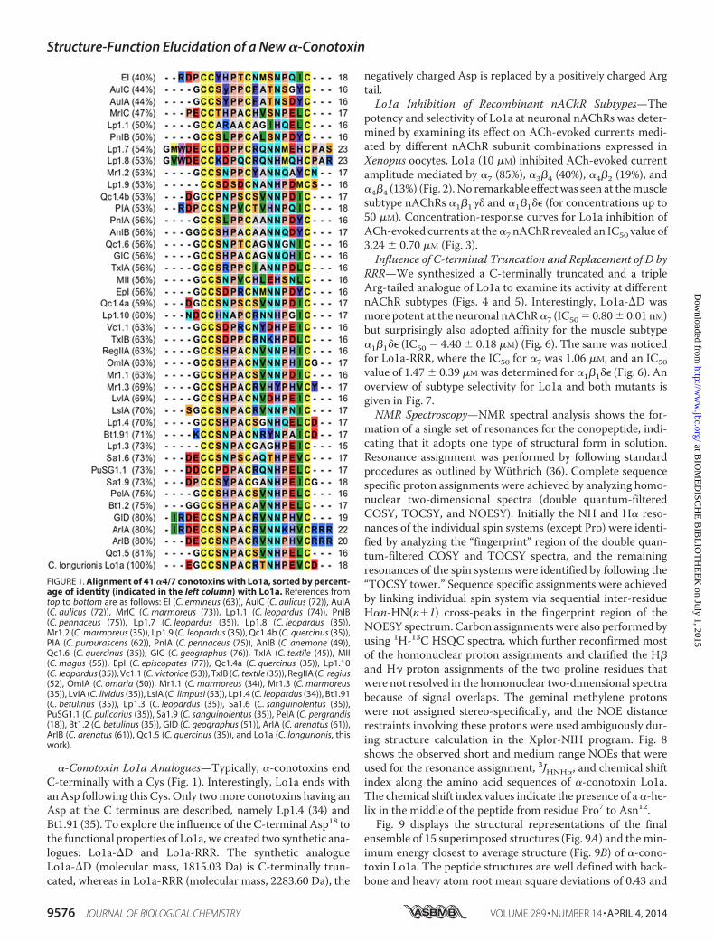
�-Conotoxin Lo1a Analogues—Typically, �-conotoxins endC-terminally with a Cys (Fig. 1). Interestingly, Lo1a ends withan Asp following this Cys. Only two more conotoxins having anAsp at the C terminus are described, namely Lp1.4 (34) andBt1.91 (35). To explore the influence of the C-terminal Asp18 tothe functional properties of Lo1a, we created two synthetic ana-logues: Lo1a-�D and Lo1a-RRR. The synthetic analogueLo1a-�D (molecular mass, 1815.03 Da) is C-terminally trun-cated, whereas in Lo1a-RRR (molecular mass, 2283.60 Da), the
negatively charged Asp is replaced by a positively charged Argtail.
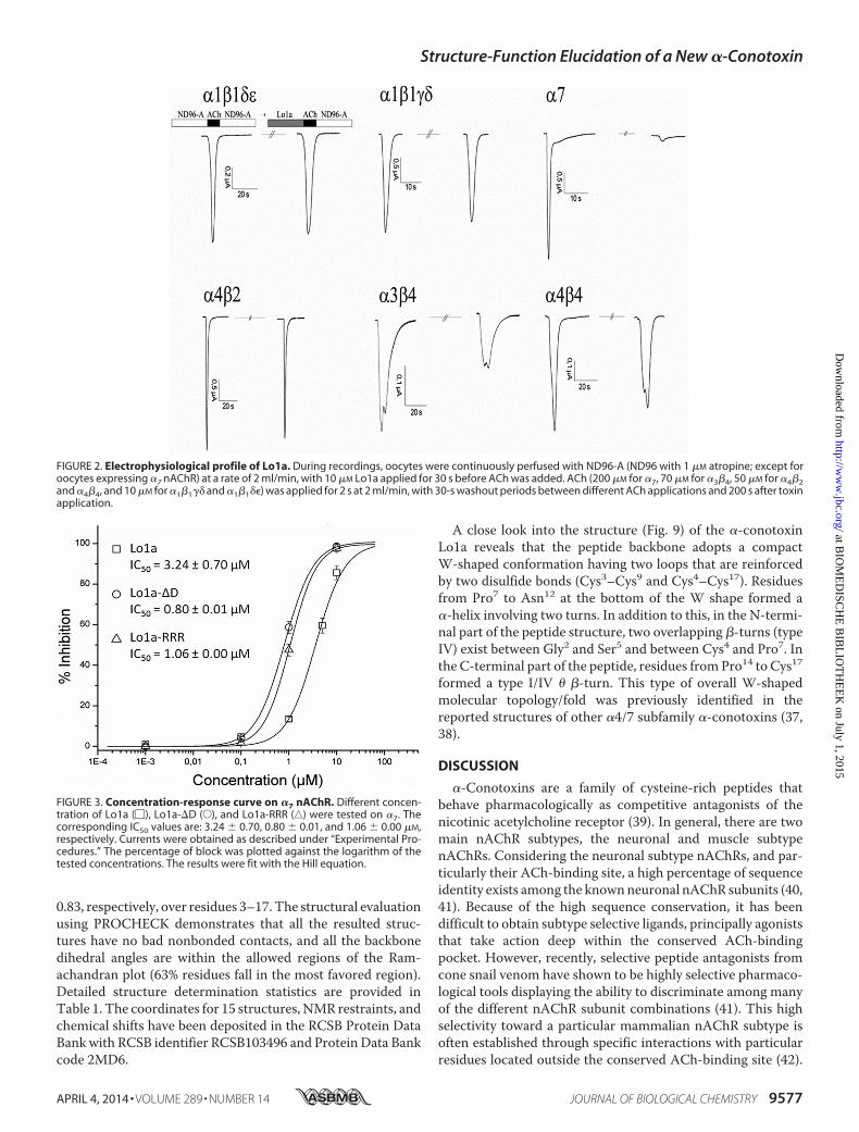
Lo1a Inhibition of Recombinant nAChR Subtypes—Thepotency and selectivity of Lo1a at neuronal nAChRs was deter-mined by examining its effect on ACh-evoked currents medi-ated by different nAChR subunit combinations expressed inXenopus oocytes. Lo1a (10 �M) inhibited ACh-evoked currentamplitude mediated by �7 (85%), �3�4 (40%), �4�2 (19%), and�4�4 (13%) (Fig. 2). No remarkable effect was seen at the musclesubtype nAChRs �1�1�� and �1�1�� (for concentrations up to50 �M). Concentration-response curves for Lo1a inhibition ofACh-evoked currents at the �7 nAChR revealed an IC50 value of3.24 0.70 �M (Fig. 3).
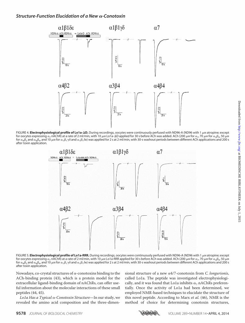
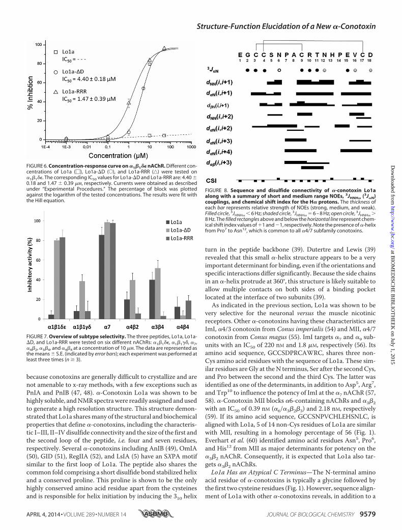
Influence of C-terminal Truncation and Replacement of D byRRR—We synthesized a C-terminally truncated and a tripleArg-tailed analogue of Lo1a to examine its activity at differentnAChR subtypes (Figs. 4 and 5). Interestingly, Lo1a-�D wasmore potent at the neuronal nAChR �7 (IC50 � 0.80 0.01 nM)but surprisingly also adopted affinity for the muscle subtype�1�1�� (IC50 � 4.40 0.18 �M) (Fig. 6). The same was noticedfor Lo1a-RRR, where the IC50 for �7 was 1.06 �M, and an IC50value of 1.47 0.39 �M was determined for �1�1�� (Fig. 6). Anoverview of subtype selectivity for Lo1a and both mutants isgiven in Fig. 7.
NMR Spectroscopy—NMR spectral analysis shows the for-mation of a single set of resonances for the conopeptide, indi-cating that it adopts one type of structural form in solution.Resonance assignment was performed by following standardprocedures as outlined by Wüthrich (36). Complete sequencespecific proton assignments were achieved by analyzing homo-nuclear two-dimensional spectra (double quantum-filteredCOSY, TOCSY, and NOESY). Initially the NH and H� reso-nances of the individual spin systems (except Pro) were identi-fied by analyzing the “fingerprint” region of the double quan-tum-filtered COSY and TOCSY spectra, and the remainingresonances of the spin systems were identified by following the“TOCSY tower.” Sequence specific assignments were achievedby linking individual spin system via sequential inter-residueH�n-HN(n�1) cross-peaks in the fingerprint region of theNOESY spectrum. Carbon assignments were also performed byusing 1H-13C HSQC spectra, which further reconfirmed mostof the homonuclear proton assignments and clarified the H�and H� proton assignments of the two proline residues thatwere not resolved in the homonuclear two-dimensional spectrabecause of signal overlaps. The geminal methylene protonswere not assigned stereo-specifically, and the NOE distancerestraints involving these protons were used ambiguously dur-ing structure calculation in the Xplor-NIH program. Fig. 8shows the observed short and medium range NOEs that wereused for the resonance assignment, 3JHNH�, and chemical shiftindex along the amino acid sequences of �-conotoxin Lo1a.The chemical shift index values indicate the presence of a �-he-lix in the middle of the peptide from residue Pro7 to Asn12.
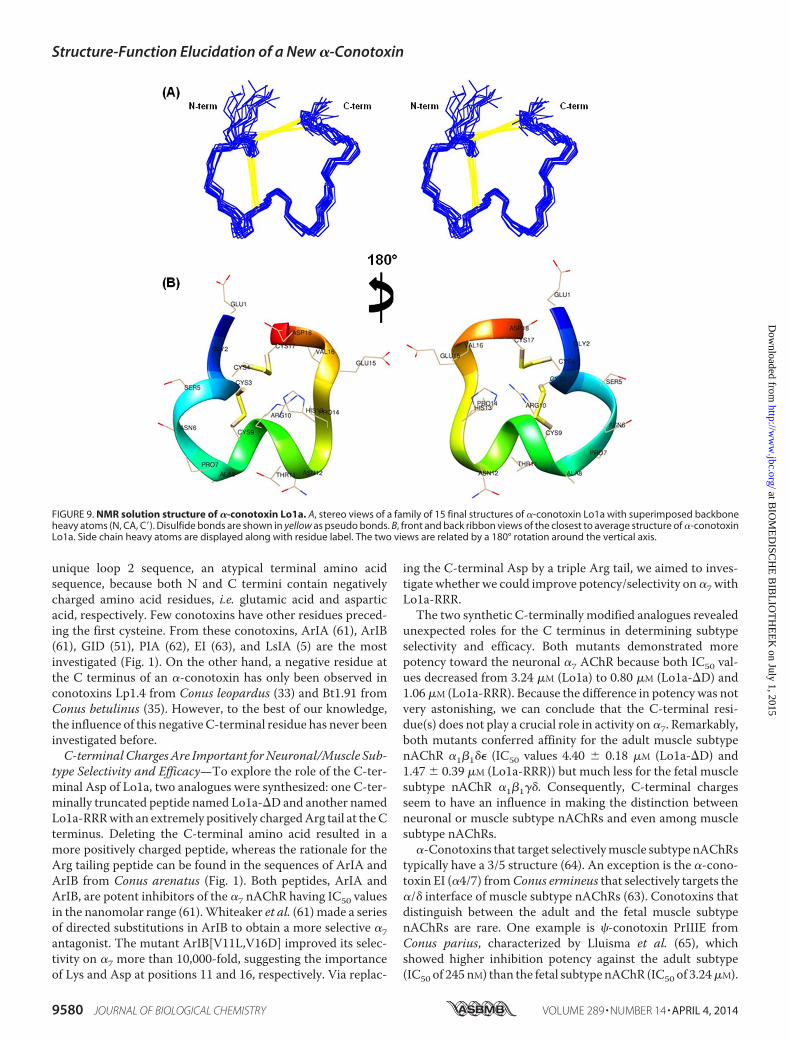
Fig. 9 displays the structural representations of the finalensemble of 15 superimposed structures (Fig. 9A) and the min-imum energy closest to average structure (Fig. 9B) of �-cono-toxin Lo1a. The peptide structures are well defined with back-bone and heavy atom root mean square deviations of 0.43 and
FIGURE 1. Alignment of 41 �4/7 conotoxins with Lo1a, sorted by percent-age of identity (indicated in the left column) with Lo1a. References fromtop to bottom are as follows: EI (C. ermineus (63)), AuIC (C. aulicus (72)), AuIA(C. aulicus (72)), MrIC (C. marmoreus (73)), Lp1.1 (C. leopardus (74)), PnIB(C. pennaceus (75)), Lp1.7 (C. leopardus (35)), Lp1.8 (C. leopardus (35)),Mr1.2 (C. marmoreus (35)), Lp1.9 (C. leopardus (35)), Qc1.4b (C. quercinus (35)),PIA (C. purpurascens (62)), PnIA (C. pennaceus (75)), AnIB (C. anemone (49)),Qc1.6 (C. quercinus (35)), GIC (C. geographus (76)), TxIA (C. textile (45)), MII(C. magus (55)), EpI (C. episcopates (77)), Qc1.4a (C. quercinus (35)), Lp1.10(C. leopardus (35)), Vc1.1 (C. victoriae (53)), TxIB (C. textile (35)), RegIIA (C. regius(52), OmIA (C. omaria (50)), Mr1.1 (C. marmoreus (34)), Mr1.3 (C. marmoreus(35)), LvIA (C. lividus (35)), LsIA (C. limpusi (53)), Lp1.4 (C. leopardus (34)), Bt1.91(C. betulinus (35)), Lp1.3 (C. leopardus (35)), Sa1.6 (C. sanguinolentus (35)),PuSG1.1 (C. pulicarius (35)), Sa1.9 (C. sanguinolentus (35)), PeIA (C. pergrandis(18)), Bt1.2 (C. betulinus (35)), GID (C. geographus (51)), ArIA (C. arenatus (61)),ArIB (C. arenatus (61)), Qc1.5 (C. quercinus (35)), and Lo1a (C. longurionis, thiswork).
Structure-Function Elucidation of a New �-Conotoxin
9576 JOURNAL OF BIOLOGICAL CHEMISTRY VOLUME 289 • NUMBER 14 • APRIL 4, 2014
at BIO
ME
DISC
HE
BIB
LIO
TH
EE
K on July 1, 2015
http://ww
w.jbc.org/
Dow
nloaded from
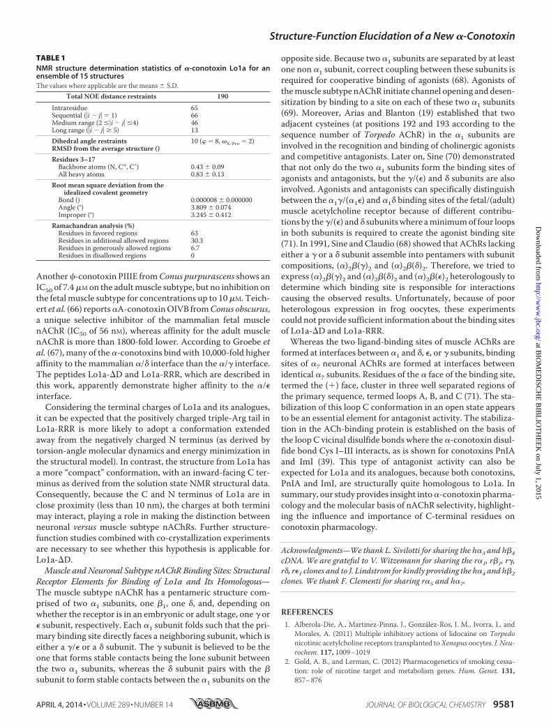
0.83, respectively, over residues 3–17. The structural evaluationusing PROCHECK demonstrates that all the resulted struc-tures have no bad nonbonded contacts, and all the backbonedihedral angles are within the allowed regions of the Ram-achandran plot (63% residues fall in the most favored region).Detailed structure determination statistics are provided inTable 1. The coordinates for 15 structures, NMR restraints, andchemical shifts have been deposited in the RCSB Protein DataBank with RCSB identifier RCSB103496 and Protein Data Bankcode 2MD6.
A close look into the structure (Fig. 9) of the �-conotoxinLo1a reveals that the peptide backbone adopts a compactW-shaped conformation having two loops that are reinforcedby two disulfide bonds (Cys3–Cys9 and Cys4–Cys17). Residuesfrom Pro7 to Asn12 at the bottom of the W shape formed a�-helix involving two turns. In addition to this, in the N-termi-nal part of the peptide structure, two overlapping �-turns (typeIV) exist between Gly2 and Ser5 and between Cys4 and Pro7. Inthe C-terminal part of the peptide, residues from Pro14 to Cys17
formed a type I/IV � �-turn. This type of overall W-shapedmolecular topology/fold was previously identified in thereported structures of other �4/7 subfamily �-conotoxins (37,38).
DISCUSSION
�-Conotoxins are a family of cysteine-rich peptides thatbehave pharmacologically as competitive antagonists of thenicotinic acetylcholine receptor (39). In general, there are twomain nAChR subtypes, the neuronal and muscle subtypenAChRs. Considering the neuronal subtype nAChRs, and par-ticularly their ACh-binding site, a high percentage of sequenceidentity exists among the known neuronal nAChR subunits (40,41). Because of the high sequence conservation, it has beendifficult to obtain subtype selective ligands, principally agoniststhat take action deep within the conserved ACh-bindingpocket. However, recently, selective peptide antagonists fromcone snail venom have shown to be highly selective pharmaco-logical tools displaying the ability to discriminate among manyof the different nAChR subunit combinations (41). This highselectivity toward a particular mammalian nAChR subtype isoften established through specific interactions with particularresidues located outside the conserved ACh-binding site (42).
FIGURE 2. Electrophysiological profile of Lo1a. During recordings, oocytes were continuously perfused with ND96-A (ND96 with 1 �M atropine; except foroocytes expressing �7 nAChR) at a rate of 2 ml/min, with 10 �M Lo1a applied for 30 s before ACh was added. ACh (200 �M for �7, 70 �M for �3�4, 50 �M for �4�2and �4�4, and 10 �M for �1�1�� and �1�1��) was applied for 2 s at 2 ml/min, with 30-s washout periods between different ACh applications and 200 s after toxinapplication.
FIGURE 3. Concentration-response curve on �7 nAChR. Different concen-tration of Lo1a (�), Lo1a-�D (E), and Lo1a-RRR (‚) were tested on �7. Thecorresponding IC50 values are: 3.24 0.70, 0.80 0.01, and 1.06 0.00 �M,respectively. Currents were obtained as described under “Experimental Pro-cedures.” The percentage of block was plotted against the logarithm of thetested concentrations. The results were fit with the Hill equation.
Structure-Function Elucidation of a New �-Conotoxin
APRIL 4, 2014 • VOLUME 289 • NUMBER 14 JOURNAL OF BIOLOGICAL CHEMISTRY 9577
at BIO
ME
DISC
HE
BIB
LIO
TH
EE
K on July 1, 2015
http://ww
w.jbc.org/
Dow
nloaded from
Nowadays, co-crystal structures of �-conotoxins binding to theACh-binding protein (43), which is a protein model for theextracellular ligand-binding domain of nAChRs, can offer use-ful information about the molecular interactions of these smallpeptides (44, 45).
Lo1a Has a Typical �-Conotoxin Structure—In our study, werevealed the amino acid composition and the three-dimen-
sional structure of a new �4/7-conotoxin from C. longurionis,called Lo1a. The peptide was investigated electrophysiologi-cally, and it was found that Lo1a inhibits �7 nAChRs preferen-tially. Once the activity of Lo1a had been determined, weemployed NMR-based techniques to elucidate the structure ofthis novel peptide. According to Marx et al. (46), NMR is themethod of choice for determining conotoxin structures,
FIGURE 4. Electrophysiological profile of Lo1a-�D. During recordings, oocytes were continuously perfused with ND96-A (ND96 with 1 �M atropine; exceptfor oocytes expressing �7 nAChR) at a rate of 2 ml/min, with 10 �M Lo1a-�D applied for 30 s before ACh was added. ACh (200 �M for �7, 70 �M for �3�4, 50 �M
for �4�2 and �4�4, and 10 �M for �1�1�� and �1�1��) was applied for 2 s at 2 ml/min, with 30-s washout periods between different ACh applications and 200 safter toxin application.
FIGURE 5. Electrophysiological profile of Lo1a-RRR. During recordings, oocytes were continuously perfused with ND96-A (ND96 with 1 �M atropine; exceptfor oocytes expressing �7 nAChR) at a rate of 2 ml/min, with 10 �M Lo1a-RRR applied for 30 s before ACh was added. ACh (200 �M for �7, 70 �M for �3�4, 50 �M
for �4�2 and �4�4, and 10 �M for �1�1�� and �1�1��) was applied for 2 s at 2 ml/min, with 30-s washout periods between different ACh applications and 200 safter toxin application.
Structure-Function Elucidation of a New �-Conotoxin
9578 JOURNAL OF BIOLOGICAL CHEMISTRY VOLUME 289 • NUMBER 14 • APRIL 4, 2014
at BIO
ME
DISC
HE
BIB
LIO
TH
EE
K on July 1, 2015
http://ww
w.jbc.org/
Dow
nloaded from
because conotoxins are generally difficult to crystallize and arenot amenable to x-ray methods, with a few exceptions such asPnIA and PnIB (47, 48). �-Conotoxin Lo1a was shown to behighly soluble, and NMR spectra were readily assigned and usedto generate a high resolution structure. This structure demon-strated that Lo1a shares many of the structural and biochemicalproperties that define �-conotoxins, including the characteris-tic I–III, II–IV disulfide connectivity and the size of the first andthe second loop of the peptide, i.e. four and seven residues,respectively. Several �-conotoxins including AnIB (49), OmIA(50), GID (51), RegIIA (52), and LsIA (5) have an SXPA motifsimilar to the first loop of Lo1a. The peptide also shares thecommon fold comprising a short disulfide bond stabilized helixand a conserved proline. This proline is shown to be the onlyhighly conserved amino acid residue apart from the cysteinesand is responsible for helix initiation by inducing the 310 helix
turn in the peptide backbone (39). Dutertre and Lewis (39)revealed that this small �-helix structure appears to be a veryimportant determinant for binding, even if the orientations andspecific interactions differ significantly. Because the side chainsin an �-helix protrude at 360°, this structure is likely suitable toallow multiple contacts on both sides of a binding pocketlocated at the interface of two subunits (39).
As indicated in the previous section, Lo1a was shown to bevery selective for the neuronal versus the muscle nicotinicreceptors. Other �-conotoxins having these characteristics areImI, �4/3 conotoxin from Conus imperialis (54) and MII, �4/7conotoxin from Conus magus (55). ImI targets �7 and �9 sub-units with an IC50 of 220 nM and 1.8 �M, respectively (56). Itsamino acid sequence, GCCSDPRCAWRC, shares three non-Cys amino acid residues with the sequence of Lo1a. These sim-ilar residues are Gly at the N terminus, Ser after the second Cys,and Pro between the second and the third Cys. The latter wasidentified as one of the determinants, in addition to Asp5, Arg7,and Trp10 to influence the potency of ImI at the �7 nAChR (57,58). �-Conotoxin MII blocks �6-containing nAChRs and �3�2with an IC50 of 0.39 nM (�6/�3�2�3) and 2.18 nM, respectively(59). If its amino acid sequence, GCCSNPVCHLEHSNLC, isaligned with Lo1a, 5 of 14 non-Cys residues of Lo1a are similarwith MII, resulting in a homology percentage of 56 (Fig. 1).Everhart et al. (60) identified amino acid residues Asn5, Pro6,and His12 from MII as major determinants for potency on the�3�2 nAChR. Consequently, it is expected that Lo1a also tar-gets �3�2 nAChRs.
Lo1a Has an Atypical C Terminus—The N-terminal aminoacid residue of �-conotoxins is typically a glycine followed bythe first two cysteine residues (Fig. 1). However, sequence align-ment of Lo1a with other �-conotoxins reveals, in addition to a
FIGURE 6. Concentration-response curve on �1�1�� nAChR. Different con-centrations of Lo1a (�), Lo1a-�D (E), and Lo1a-RRR (‚) were tested on�1�1��. The corresponding IC50 values for Lo1a-�D and Lo1a-RRR are: 4.40 0.18 and 1.47 0.39 �M, respectively. Currents were obtained as describedunder “Experimental Procedures.” The percentage of block was plottedagainst the logarithm of the tested concentrations. The results were fit withthe Hill equation.
FIGURE 7. Overview of subtype selectivity. The three peptides, Lo1a, Lo1a-�D, and Lo1a-RRR were tested on six different nAChRs: �1�1��, �1�1��, �7,�4�2, �3�4, and �4�4 at a concentration of 10 �M. The data are represented asthe means S.E. (indicated by error bars); each experiment was performed atleast three times (n � 3).
FIGURE 8. Sequence and disulfide connectivity of �-conotoxin Lo1aalong with a summary of short and medium range NOEs, 3JHNH� (3J�N)couplings, and chemical shift index for the H� protons. The thickness ofeach bar represents relative strength of NOEs (strong, medium, and weak).Filled circle, 3JHNH� � 6 Hz; shaded circle, 3JHNH� � 6 – 8 Hz; open circle, 3JHNH� 8 Hz. The filled rectangles above and below the horizontal line represent chem-ical shift index values of �1 and �1, respectively. Note the presence of �-helixfrom Pro7 to Asn12, which is common to all �4/7 subfamily conotoxins.
Structure-Function Elucidation of a New �-Conotoxin
APRIL 4, 2014 • VOLUME 289 • NUMBER 14 JOURNAL OF BIOLOGICAL CHEMISTRY 9579
at BIO
ME
DISC
HE
BIB
LIO
TH
EE
K on July 1, 2015
http://ww
w.jbc.org/
Dow
nloaded from
unique loop 2 sequence, an atypical terminal amino acidsequence, because both N and C termini contain negativelycharged amino acid residues, i.e. glutamic acid and asparticacid, respectively. Few conotoxins have other residues preced-ing the first cysteine. From these conotoxins, ArIA (61), ArIB(61), GID (51), PIA (62), EI (63), and LsIA (5) are the mostinvestigated (Fig. 1). On the other hand, a negative residue atthe C terminus of an �-conotoxin has only been observed inconotoxins Lp1.4 from Conus leopardus (33) and Bt1.91 fromConus betulinus (35). However, to the best of our knowledge,the influence of this negative C-terminal residue has never beeninvestigated before.
C-terminal Charges Are Important for Neuronal/Muscle Sub-type Selectivity and Efficacy—To explore the role of the C-ter-minal Asp of Lo1a, two analogues were synthesized: one C-ter-minally truncated peptide named Lo1a-�D and another namedLo1a-RRR with an extremely positively charged Arg tail at the Cterminus. Deleting the C-terminal amino acid resulted in amore positively charged peptide, whereas the rationale for theArg tailing peptide can be found in the sequences of ArIA andArIB from Conus arenatus (Fig. 1). Both peptides, ArIA andArIB, are potent inhibitors of the �7 nAChR having IC50 valuesin the nanomolar range (61). Whiteaker et al. (61) made a seriesof directed substitutions in ArIB to obtain a more selective �7antagonist. The mutant ArIB[V11L,V16D] improved its selec-tivity on �7 more than 10,000-fold, suggesting the importanceof Lys and Asp at positions 11 and 16, respectively. Via replac-
ing the C-terminal Asp by a triple Arg tail, we aimed to inves-tigate whether we could improve potency/selectivity on �7 withLo1a-RRR.
The two synthetic C-terminally modified analogues revealedunexpected roles for the C terminus in determining subtypeselectivity and efficacy. Both mutants demonstrated morepotency toward the neuronal �7 AChR because both IC50 val-ues decreased from 3.24 �M (Lo1a) to 0.80 �M (Lo1a-�D) and1.06 �M (Lo1a-RRR). Because the difference in potency was notvery astonishing, we can conclude that the C-terminal resi-due(s) does not play a crucial role in activity on �7. Remarkably,both mutants conferred affinity for the adult muscle subtypenAChR �1�1�� (IC50 values 4.40 0.18 �M (Lo1a-�D) and1.47 0.39 �M (Lo1a-RRR)) but much less for the fetal musclesubtype nAChR �1�1��. Consequently, C-terminal chargesseem to have an influence in making the distinction betweenneuronal or muscle subtype nAChRs and even among musclesubtype nAChRs.
�-Conotoxins that target selectively muscle subtype nAChRstypically have a 3/5 structure (64). An exception is the �-cono-toxin EI (�4/7) from Conus ermineus that selectively targets the�/� interface of muscle subtype nAChRs (63). Conotoxins thatdistinguish between the adult and the fetal muscle subtypenAChRs are rare. One example is �-conotoxin PrIIIE fromConus parius, characterized by Lluisma et al. (65), whichshowed higher inhibition potency against the adult subtype(IC50 of 245 nM) than the fetal subtype nAChR (IC50 of 3.24 �M).
FIGURE 9. NMR solution structure of �-conotoxin Lo1a. A, stereo views of a family of 15 final structures of �-conotoxin Lo1a with superimposed backboneheavy atoms (N, CA, C�). Disulfide bonds are shown in yellow as pseudo bonds. B, front and back ribbon views of the closest to average structure of �-conotoxinLo1a. Side chain heavy atoms are displayed along with residue label. The two views are related by a 180° rotation around the vertical axis.
Structure-Function Elucidation of a New �-Conotoxin
9580 JOURNAL OF BIOLOGICAL CHEMISTRY VOLUME 289 • NUMBER 14 • APRIL 4, 2014
at BIO
ME
DISC
HE
BIB
LIO
TH
EE
K on July 1, 2015
http://ww
w.jbc.org/
Dow
nloaded from
Another �-conotoxin PIIIE from Conus purpurascens shows anIC50 of 7.4 �M on the adult muscle subtype, but no inhibition onthe fetal muscle subtype for concentrations up to 10 �M. Teich-ert et al. (66) reports �A-conotoxin OIVB from Conus obscurus,a unique selective inhibitor of the mammalian fetal musclenAChR (IC50 of 56 nM), whereas affinity for the adult musclenAChR is more than 1800-fold lower. According to Groebe etal. (67), many of the �-conotoxins bind with 10,000-fold higheraffinity to the mammalian �/� interface than the �/� interface.The peptides Lo1a-�D and Lo1a-RRR, which are described inthis work, apparently demonstrate higher affinity to the �/�interface.
Considering the terminal charges of Lo1a and its analogues,it can be expected that the positively charged triple-Arg tail inLo1a-RRR is more likely to adopt a conformation extendedaway from the negatively charged N terminus (as derived bytorsion-angle molecular dynamics and energy minimization inthe structural model). In contrast, the structure from Lo1a hasa more “compact” conformation, with an inward-facing C ter-minus as derived from the solution state NMR structural data.Consequently, because the C and N terminus of Lo1a are inclose proximity (less than 10 nm), the charges at both terminimay interact, playing a role in making the distinction betweenneuronal versus muscle subtype nAChRs. Further structure-function studies combined with co-crystallization experimentsare necessary to see whether this hypothesis is applicable forLo1a-�D.
Muscle and Neuronal Subtype nAChR Binding Sites: StructuralReceptor Elements for Binding of Lo1a and Its Homologous—The muscle subtype nAChR has a pentameric structure com-prised of two �1 subunits, one �1, one �, and, depending onwhether the receptor is in an embryonic or adult stage, one � or� subunit, respectively. Each �1 subunit folds such that the pri-mary binding site directly faces a neighboring subunit, which iseither a �/� or a � subunit. The � subunit is believed to be theone that forms stable contacts being the lone subunit betweenthe two �1 subunits, whereas the � subunit pairs with the �subunit to form stable contacts between the �1 subunits on the
opposite side. Because two �1 subunits are separated by at leastone non �1 subunit, correct coupling between these subunits isrequired for cooperative binding of agonists (68). Agonists ofthe muscle subtype nAChR initiate channel opening and desen-sitization by binding to a site on each of these two �1 subunits(69). Moreover, Arias and Blanton (19) established that twoadjacent cysteines (at positions 192 and 193 according to thesequence number of Torpedo AChR) in the �1 subunits areinvolved in the recognition and binding of cholinergic agonistsand competitive antagonists. Later on, Sine (70) demonstratedthat not only do the two �1 subunits form the binding sites ofagonists and antagonists, but the �/(�) and � subunits are alsoinvolved. Agonists and antagonists can specifically distinguishbetween the �1�/(�1�) and �1� binding sites of the fetal/(adult)muscle acetylcholine receptor because of different contribu-tions by the �/(�) and � subunits where a minimum of four loopsin both subunits is required to create the agonist binding site(71). In 1991, Sine and Claudio (68) showed that AChRs lackingeither a � or a � subunit assemble into pentamers with subunitcompositions, (�)2�(�)2 and (�)2�(�)2. Therefore, we tried toexpress (�)2�(�)2 and (�)2�(�)2 and (�)2�(�)2 heterologously todetermine which binding site is responsible for interactionscausing the observed results. Unfortunately, because of poorheterologous expression in frog oocytes, these experimentscould not provide sufficient information about the binding sitesof Lo1a-�D and Lo1a-RRR.
Whereas the two ligand-binding sites of muscle AChRs areformed at interfaces between �1 and �, �, or � subunits, bindingsites of �7 neuronal AChRs are formed at interfaces betweenidentical �7 subunits. Residues of the � face of the binding site,termed the (�) face, cluster in three well separated regions ofthe primary sequence, termed loops A, B, and C (71). The sta-bilization of this loop C conformation in an open state appearsto be an essential element for antagonist activity. The stabiliza-tion in the ACh-binding protein is established on the basis ofthe loop C vicinal disulfide bonds where the �-conotoxin disul-fide bond Cys I–III interacts, as is shown for conotoxins PnIAand ImI (39). This type of antagonist activity can also beexpected for Lo1a and its analogues, because both conotoxins,PnIA and ImI, are structurally quite homologous to Lo1a. Insummary, our study provides insight into �-conotoxin pharma-cology and the molecular basis of nAChR selectivity, highlight-ing the influence and importance of C-terminal residues onconotoxin pharmacology.
Acknowledgments—We thank L. Sivilotti for sharing the h�3 and h�4
cDNA. We are grateful to V. Witzemann for sharing the r�1, r�1, r�,r�, r�1 clones and to J. Lindstrom for kindly providing the h�4 and h�2
clones. We thank F. Clementi for sharing r�5 and h�7.
REFERENCES1. Alberola-Die, A., Martinez-Pinna, J., González-Ros, J. M., Ivorra, I., and
Morales, A. (2011) Multiple inhibitory actions of lidocaine on Torpedonicotinic acetylcholine receptors transplanted to Xenopus oocytes. J. Neu-rochem. 117, 1009 –1019
2. Gold, A. B., and Lerman, C. (2012) Pharmacogenetics of smoking cessa-tion: role of nicotine target and metabolism genes. Hum. Genet. 131,857– 876
TABLE 1NMR structure determination statistics of �-conotoxin Lo1a for anensemble of 15 structuresThe values where applicable are the means S.D.
Total NOE distance restraints 190
Intraresidue 65Sequential (�i � j� � 1) 66Medium range (2 �i � j� 4) 46Long range (�i � j� � 5) 13Dihedral angle restraints 10 ( � 8, X-Pro � 2)RMSD from the average structure ()Residues 3–17
Backbone atoms (N, C�, C�) 0.43 0.09All heavy atoms 0.83 0.13
Root mean square deviation from theidealized covalent geometry
Bond () 0.000008 0.000000Angle (°) 3.809 0.074Improper (°) 3.245 0.412
Ramachandran analysis (%)Residues in favored regions 63Residues in additional allowed regions 30.3Residues in generously allowed regions 6.7Residues in disallowed regions 0
Structure-Function Elucidation of a New �-Conotoxin
APRIL 4, 2014 • VOLUME 289 • NUMBER 14 JOURNAL OF BIOLOGICAL CHEMISTRY 9581
at BIO
ME
DISC
HE
BIB
LIO
TH
EE
K on July 1, 2015
http://ww
w.jbc.org/
Dow
nloaded from
3. Rezvani, A. H., and Levin, E. D. (2001) Cognitive effects of nicotine. Biol.Psychiatry 49, 258 –267
4. Cooper, E., Couturier, S., and Ballivet, M. (1991) Pentameric structure andsubunit stoichiometry of a neuronal nicotinic acetylcholine receptor. Na-ture 350, 235–238
5. Inserra, M. C., Kompella, S. N., Vetter, I., Brust, A., Daly, N. L., Cuny, H.,Craik, D. J., Alewood, P. F., Adams, D. J., and Lewis, R. J. (2013) Isolationand characterization of �-conotoxin LsIA with potent activity at nicotinicacetylcholine receptors. Biochem. Pharmacol. 86, 791–799
6. Colquhoun, L. M., and Patrick, J. W. (1997) Pharmacology of neuronalnicotinic acetylcholine receptor subtypes. Adv. Pharmacol. 39, 191–220
7. Le Novère, N., and Changeux, J. P. (1995) Molecular evolution of thenicotinic acetylcholine receptor: an example of multigene family in excit-able cells. J. Mol. Evol. 40, 155–172
8. Gotti, C., Fornasari, D., and Clementi, F. (1997) Human neuronal nicotinicreceptors. Prog. Neurobiol. 53, 199 –237
9. Couturier, S., Bertrand, D., Matter, J. M., Hernandez, M. C., Bertrand, S.,Millar, N., Valera, S., Barkas, T., and Ballivet, M. (1990) A neuronal nico-tinic acetylcholine receptor subunit (�7) is developmentally regulated andforms a homo-oligomeric channel blocked by �-BTX. Neuron 5, 847– 856
10. Rubboli, F., Court, J. A., Sala, C., Morris, C., Chini, B., Perry, E., and Cle-menti, F. (1994) Distribution of nicotinic receptors in the human hip-pocampus and thalamus. Eur. J. Neurosci. 6, 1596 –1604
11. Wevers, A., Jeske, A., Lobron, C., Birtsch, C., Heinemann, S., Maelicke, A.,Schröder, R., and Schröder, H. (1994) Cellular distribution of nicotinicacetylcholine receptor subunit mRNAs in the human cerebral cortex asrevealed by non-isotopic in situ hybridization. Brain Res. Mol. Brain Res.25, 122–128
12. Breese, C. R., Adams, C., Logel, J., Drebing, C., Rollins, Y., Barnhart, M.,Sullivan, B., Demasters, B. K., Freedman, R., and Leonard, S. (1997) Com-parison of the regional expression of nicotinic acetylcholine receptor �7mRNA and [125I]�-bungarotoxin binding in human postmortem brain.J. Comp. Neurol. 387, 385–398
13. Gotti, C., and Clementi, F. (2004) Neuronal nicotinic receptors: fromstructure to pathology. Prog. Neurobiol. 74, 363–396
14. Sacco, K. A., Bannon, K. L., and George, T. P. (2004) Nicotinic receptormechanisms and cognition in normal states and neuropsychiatric disor-ders. J. Psychopharmacol. 18, 457– 474
15. Steinlein, O. K., and Bertrand, D. (2010) Nicotinic receptor channelopa-thies and epilepsy. Pflugers Arch. 460, 495–503
16. Quinton, L., Servent, D., Girard, E., Molgó, J., Le Caer, J. P., Malosse, C.,Haidar el, A., Lecoq, A., Gilles, N., and Chamot-Rooke, J. (2013) Identifi-cation and functional characterization of a novel �-conotoxin (EIIA) fromConus ermineus. Anal. Bioanal. Chem. 405, 5341–5351
17. Sine, S. M., Kreienkamp, H. J., Bren, N., Maeda, R., and Taylor, P. (1995)Molecular dissection of subunit interfaces in the acetylcholine receptor:identification of determinants of �-conotoxin M1 selectivity. Neuron 15,205–211
18. McIntosh, J. M., Plazas, P. V., Watkins, M., Gomez-Casati, M. E., Olivera,B. M., and Elgoyhen, A. B. (2005) A novel �-conotoxin, PeIA, cloned fromConus pergrandis, discriminates between rat �9�10 and �7 nicotinic cho-linergic receptors. J. Biol. Chem. 280, 30107–30112
19. Arias, H. R., and Blanton, M. P. (2000) �-Conotoxins. Int. J. Biochem. CellBiol. 32, 1017–1028
20. Olivera, B. M., Rivier, J., Clark, C., Ramilo, C. A., Corpuz, G. P., Abogadie,F. C., Mena, E. E., Woodward, S. R., Hillyard, D. R., and Cruz, L. J. (1990)Diversity of Conus neuropeptides. Science 249, 257–263
21. Grønlien, J. H., Håkerud, M., Ween, H., Thorin-Hagene, K., Briggs, C. A.,Gopalakrishnan, M., and Malysz, J. (2007) Distinct profiles of �7 nAChRpositive allosteric modulation revealed by structurally diverse chemo-types. Mol. Pharmacol. 72, 715–724
22. Tucker, J. K., and Tenorio, M. J. (2009) Systematic Classification of Recentand Fossil Conoidean Gastropods: With Keys to the Genera of Cone Shells,ConchBooks, Hackenheim, Germany
23. Olivera, B. M., Gray, W. R., Zeikus, R., McIntosh, J. M., Varga, J., Rivier, J.,de Santos, V., and Cruz, L. J. (1985) Peptide neurotoxins from fish-huntingcone snails. Science 230, 1338 –1343
24. Van Der Haegen, A., Peigneur, S., and Tytgat, J. (2011) Importance of
position 8 in mu-conotoxin KIIIA for voltage-gated sodium channel se-lectivity. FEBS J. 278, 3408 –3418
25. Keller, M., and Steiger, R. (2004) The pi plate: an implant for unstableextension fractures of the distal radius in patients with osteoporotic bone.Tech. Hand Up Extrem. Surg. 8, 212–218
26. Hwang, T. L., and Shaka, A. J. (1998) Multiple-pulse mixing sequences thatselectively enhance chemical exchange or cross-relaxation peaks in high-resolution NMR spectra. J. Magn. Reson. 135, 280 –287
27. Shaka, A. J., Lee, C. J., and Pines, A. (1988) Iterative schemes for bilinearoperators: application to spin decoupling. J. Magn. Reson. 77, 274 –293
28. Derome, A. E., and Williamson, M. P. (1990) Rapid-pulsing artifacts indouble-quantum-filtered COSY. J. Magn. Reson. 88, 177–185
29. Schwieters, C. D., Kuszewski, J. J., Tjandra, N., and Clore, G. M. (2003) TheXplor-NIH NMR molecular structure determination package. J. Magn.Reson. 160, 65–73
30. Stein, E. G., Rice, L. M., and Brünger, A. T. (1997) Torsion-angle moleculardynamics as a new efficient tool for NMR structure calculation. J. Magn.Reson. 124, 154 –164
31. Spronk, C. A., Linge, J. P., Hilbers, C. W., and Vuister, G. W. (2002) Im-proving the quality of protein structures derived by NMR spectroscopy.J. Biomol. NMR 22, 281–289
32. Laskowski, R. A., Macarthur, M. W., Moss, D. S., and Thornton, J. M.(1993) Procheck: a program to check the stereochemical quality of proteinstructures. J. Appl. Crystallogr. 26, 283–291
33. Yuan, D. D., Han, Y. H., Wang, C. G., and Chi, C. W. (2007) From theidentification of gene organization of � conotoxins to the cloning of noveltoxins. Toxicon 49, 1135–1149
34. Peng, C., Chen, W., Sanders, T., Chew, G., Liu, J., Hawrot, E., and Chi, C.(2010) Chemical synthesis and characterization of two �4/7-conotoxins.Acta Biochim. Biophys. Sin. 42, 745–753
35. Kaas, Q., Yu, R., Jin, A. H., Dutertre, S., and Craik, D. J. (2012) ConoServer:updated content, knowledge, and discovery tools in the conopeptide da-tabase. Nucleic Acids Res. 40, D325–D330
36. Wüthrich, K. (1986) NMR of Proteins and Nucleic Acids, John Wiley &Sons Inc., New York
37. Park, K. H., Suk, J. E., Jacobsen, R., Gray, W. R., McIntosh, J. M., and Han,K. H. (2001) Solution conformation of �-conotoxin EI, a neuromusculartoxin specific for the �1/� subunit interface of Torpedo nicotinic acetyl-choline receptor. J. Biol. Chem. 276, 49028 – 49033
38. Chi, S. W., Kim, D. H., Olivera, B. M., McIntosh, J. M., and Han, K. H.(2004) Solution conformation of �-conotoxin GIC, a novel potent antag-onist of �3�2 nicotinic acetylcholine receptors. Biochem. J. 380, 347–352
39. Dutertre, S., and Lewis, R. J. (2006) Toxin insights into nicotinic acetyl-choline receptors. Biochem. Pharmacol. 72, 661– 670
40. Le Novère, N., Corringer, P. J., and Changeux, J. P. (2002) The diversity ofsubunit composition in nAChRs: evolutionary origins, physiologic andpharmacologic consequences. J. Neurobiol. 53, 447– 456
41. Tsetlin, V. I., and Hucho, F. (2004) Snake and snail toxins acting on nico-tinic acetylcholine receptors: fundamental aspects and medical applica-tions. FEBS Lett. 557, 9 –13
42. Dutertre, S., and Lewis, R. J. (2004) Computational approaches to under-stand �-conotoxin interactions at neuronal nicotinic receptors. Eur.J. Biochem. 271, 2327–2334
43. Brejc, K., van Dijk, W. J., Klaassen, R. V., Schuurmans, M., van Der Oost, J.,Smit, A. B., and Sixma, T. K. (2001) Crystal structure of an ACh-bindingprotein reveals the ligand-binding domain of nicotinic receptors. Nature411, 269 –276
44. Armishaw, C., Jensen, A. A., Balle, T., Clark, R. J., Harpsøe, K., Skonberg,C., Liljefors, T., and Strømgaard, K. (2009) Rational design of �-conotoxinanalogues targeting �7 nicotinic acetylcholine receptors: improved antag-onistic activity by incorporation of proline derivatives. J. Biol. Chem. 284,9498 –9512
45. Dutertre, S., Ulens, C., Büttner, R., Fish, A., van Elk, R., Kendel, Y., Hop-ping, G., Alewood, P. F., Schroeder, C., Nicke, A., Smit, A. B., Sixma, T. K.,and Lewis, R. J. (2007) AChBP-targeted �-conotoxin correlates distinctbinding orientations with nAChR subtype selectivity. EMBO J. 26,3858 –3867
46. Marx, U. C., Daly, N. L., and Craik, D. J. (2006) NMR of conotoxins:
Structure-Function Elucidation of a New �-Conotoxin
9582 JOURNAL OF BIOLOGICAL CHEMISTRY VOLUME 289 • NUMBER 14 • APRIL 4, 2014
at BIO
ME
DISC
HE
BIB
LIO
TH
EE
K on July 1, 2015
http://ww
w.jbc.org/
Dow
nloaded from
structural features and an analysis of chemical shifts of post-translation-ally modified amino acids. Magn. Reson. Chem. 44, S41–S50
47. Hu, S. H., Gehrmann, J., Guddat, L. W., Alewood, P. F., Craik, D. J., andMartin, J. L. (1996) The 1.1 A crystal structure of the neuronal acetylcho-line receptor antagonist, �-conotoxin PnIA from Conus pennaceus. Struc-ture 4, 417– 423
48. Hu, S. H., Gehrmann, J., Alewood, P. F., Craik, D. J., and Martin, J. L. (1997)Crystal structure at 1.1 A resolution of �-conotoxin PnIB: comparisonwith �-conotoxins PnIA and GI. Biochemistry 36, 11323–11330
49. Loughnan, M. L., Nicke, A., Jones, A., Adams, D. J., Alewood, P. F., andLewis, R. J. (2004) Chemical and functional identification and character-ization of novel sulfated �-conotoxins from the cone snail Conus anem-one. J. Med. Chem. 47, 1234 –1241
50. Talley, T. T., Olivera, B. M., Han, K. H., Christensen, S. B., Dowell, C.,Tsigelny, I., Ho, K. Y., Taylor, P., and McIntosh, J. M. (2006) �-ConotoxinOmIA is a potent ligand for the acetylcholine-binding protein as well as�3�2 and �7 nicotinic acetylcholine receptors. J. Biol. Chem. 281,24678 –24686
51. Nicke, A., Loughnan, M. L., Millard, E. L., Alewood, P. F., Adams, D. J.,Daly, N. L., Craik, D. J., and Lewis, R. J. (2003) Isolation, structure, andactivity of GID, a novel � 4/7-conotoxin with an extended N-terminalsequence. J. Biol. Chem. 278, 3137–3144
52. Franco, A., Kompella, S. N., Akondi, K. B., Melaun, C., Daly, N. L., Luetje,C. W., Alewood, P. F., Craik, D. J., Adams, D. J., and Marí, F. (2012) RegIIA:an �4/7-conotoxin from the venom of Conus regius that potently blocks�3�4 nAChRs. Biochem. Pharmacol. 83, 419 – 426
53. Sandall, D. W., Satkunanathan, N., Keays, D. A., Polidano, M. A., Liping,X., Pham, V., Down, J. G., Khalil, Z., Livett, B. G., and Gayler, K. R. (2003)A novel �-conotoxin identified by gene sequencing is active in suppressingthe vascular response to selective stimulation of sensory nerves in vivo.Biochemistry 42, 6904 – 6911
54. McIntosh, J. M., Yoshikami, D., Mahe, E., Nielsen, D. B., Rivier, J. E., Gray,W. R., and Olivera, B. M. (1994) A nicotinic acetylcholine receptor ligandof unique specificity, �-conotoxin ImI. J. Biol. Chem. 269, 16733–16739
55. Cartier, G. E., Yoshikami, D., Gray, W. R., Luo, S., Olivera, B. M., andMcIntosh, J. M. (1996) A new �-conotoxin which targets �3�2 nicotinicacetylcholine receptors. J. Biol. Chem. 271, 7522–7528
56. Johnson, D. S., Martinez, J., Elgoyhen, A. B., Heinemann, S. F., and McIn-tosh, J. M. (1995) �-Conotoxin ImI exhibits subtype-specific nicotinicacetylcholine receptor blockade: preferential inhibition of homomeric �7and �9 receptors. Mol. Pharmacol. 48, 194 –199
57. Quiram, P. A., and Sine, S. M. (1998) Structural elements in �-conotoxinImI essential for binding to neuronal �7 receptors. J. Biol. Chem. 273,11007–11011
58. Quiram, P. A., and Sine, S. M. (1998) Identification of residues in theneuronal �7 acetylcholine receptor that confer selectivity for conotoxinImI. J. Biol. Chem. 273, 11001–11006
59. McIntosh, J. M., Azam, L., Staheli, S., Dowell, C., Lindstrom, J. M., Kurya-tov, A., Garrett, J. E., Marks, M. J., and Whiteaker, P. (2004) Analogs of�-conotoxin MII are selective for �6-containing nicotinic acetylcholinereceptors. Mol. Pharmacol. 65, 944 –952
60. Everhart, D., Cartier, G. E., Malhotra, A., Gomes, A. V., McIntosh, J. M.,and Luetje, C. W. (2004) Determinants of potency on �-conotoxin MII, apeptide antagonist of neuronal nicotinic receptors. Biochemistry 43,2732–2737
61. Whiteaker, P., Christensen, S., Yoshikami, D., Dowell, C., Watkins, M.,Gulyas, J., Rivier, J., Olivera, B. M., and McIntosh, J. M. (2007) Discovery,
synthesis, and structure activity of a highly selective �7 nicotinic acetyl-choline receptor antagonist. Biochemistry 46, 6628 – 6638
62. Dowell, C., Olivera, B. M., Garrett, J. E., Staheli, S. T., Watkins, M., Kurya-tov, A., Yoshikami, D., Lindstrom, J. M., and McIntosh, J. M. (2003)�-Conotoxin PIA is selective for �6 subunit-containing nicotinic acetyl-choline receptors. J. Neurosci. 23, 8445– 8452
63. Martinez, J. S., Olivera, B. M., Gray, W. R., Craig, A. G., Groebe, D. R.,Abramson, S. N., and McIntosh, J. M. (1995) �-Conotoxin EI, a new nic-otinic acetylcholine receptor antagonist with novel selectivity. Biochemis-try 34, 14519 –14526
64. Lewis, R. J., Dutertre, S., Vetter, I., and Christie, M. J. (2012) Conus venompeptide pharmacology. Pharmacol. Rev. 64, 259 –298
65. Lluisma, A. O., López-Vera, E., Bulaj, G., Watkins, M., and Olivera, B. M.(2008) Characterization of a novel psi-conotoxin from Conus pariusReeve. Toxicon 51, 174 –180
66. Teichert, R. W., Rivier, J., Torres, J., Dykert, J., Miller, C., and Olivera, B. M.(2005) A uniquely selective inhibitor of the mammalian fetal neuromus-cular nicotinic acetylcholine receptor. J. Neurosci. 25, 732–736
67. Groebe, D. R., Dumm, J. M., Levitan, E. S., and Abramson, S. N. (1995)�-Conotoxins selectively inhibit one of the two acetylcholine binding sitesof nicotinic receptors. Mol. Pharmacol. 48, 105–111
68. Sine, S. M., and Claudio, T. (1991) �- and �-subunits regulate the affinityand the cooperativity of ligand binding to the acetylcholine receptor.J. Biol. Chem. 266, 19369 –19377
69. Changeux, J. P. (1990) The TiPS lecture: the nicotinic acetylcholine recep-tor: an allosteric protein prototype of ligand-gated ion channels. TrendsPharmacol. Sci. 11, 485– 492
70. Sine, S. M. (1993) Molecular dissection of subunit interfaces in the acetyl-choline receptor: identification of residues that determine curare selectiv-ity. Proc. Natl. Acad. Sci. U.S.A. 90, 9436 –9440
71. Prince, R. J., and Sine, S. M. (1996) Molecular dissection of subunit inter-faces in the acetylcholine receptor. Identification of residues that deter-mine agonist selectivity. J. Biol. Chem. 271, 25770 –25777
72. Luo, S., Kulak, J. M., Cartier, G. E., Jacobsen, R. B., Yoshikami, D., Olivera,B. M., and McIntosh, J. M. (1998) �-Conotoxin AuIB selectively blocks�3�4 nicotinic acetylcholine receptors and nicotine-evoked norepineph-rine release. J. Neurosci. 18, 8571– 8579
73. Jin, A. H., Vetter, I., Dutertre, S., Abraham, N., Emidio, N. B., Inserra, M.,Murali, S. S., Christie, M. J., Alewood, P. F., and Lewis, R. J. (2014) MrIC, anovel �-conotoxin agonist in the presence of PNU at endogenous �7nicotinic acetylcholine receptors. Biochemistry 53, 1–3
74. Peng, C., Han, Y., Sanders, T., Chew, G., Liu, J., Hawrot, E., Chi, C., andWang, C. (2008) �4/7-Conotoxin Lp1.1 is a novel antagonist of neuronalnicotinic acetylcholine receptors. Peptides 29, 1700 –1707
75. Luo, S., Nguyen, T. A., Cartier, G. E., Olivera, B. M., Yoshikami, D., andMcIntosh, J. M. (1999) Single-residue alteration in �-conotoxin PnIAswitches its nAChR subtype selectivity. Biochemistry 38, 14542–14548
76. McIntosh, J. M., Dowell, C., Watkins, M., Garrett, J. E., Yoshikami, D., andOlivera, B. M. (2002) �-Conotoxin GIC from Conus geographus, a novelpeptide antagonist of nicotinic acetylcholine receptors. J. Biol. Chem. 277,33610 –33615
77. Loughnan, M., Bond, T., Atkins, A., Cuevas, J., Adams, D. J., Broxton,N. M., Livett, B. G., Down, J. G., Jones, A., Alewood, P. F., and Lewis, R. J.(1998) �-Conotoxin EpI, a novel sulfated peptide from Conus episcopatusthat selectively targets neuronal nicotinic acetylcholine receptors. J. Biol.Chem. 273, 15667–15674
Structure-Function Elucidation of a New �-Conotoxin
APRIL 4, 2014 • VOLUME 289 • NUMBER 14 JOURNAL OF BIOLOGICAL CHEMISTRY 9583
at BIO
ME
DISC
HE
BIB
LIO
TH
EE
K on July 1, 2015
http://ww
w.jbc.org/
Dow
nloaded from
Piet Herdewijn and Jan TytgatUlens, Etienne Waelkens, Lisette D'Souza, Ravichandran, Eveline Lescrinier, ChrisMaiti, Prabha Devi, Samuthirapandian Eline K. M. Lebbe, Steve Peigneur, Mohitosh
Conus longurionis-Conotoxin, Lo1a, from αStructure-Function Elucidation of a New
Neurobiology:
doi: 10.1074/jbc.M114.556175 originally published online February 24, 20142014, 289:9573-9583.J. Biol. Chem.
10.1074/jbc.M114.556175Access the most updated version of this article at doi:
.JBC Affinity SitesFind articles, minireviews, Reflections and Classics on similar topics on the
Alerts:
When a correction for this article is posted•
When this article is cited•
to choose from all of JBC's e-mail alertsClick here
http://www.jbc.org/content/289/14/9573.full.html#ref-list-1
This article cites 75 references, 27 of which can be accessed free at
at BIO
ME
DISC
HE
BIB
LIO
TH
EE
K on July 1, 2015
http://ww
w.jbc.org/
Dow
nloaded from