Structure-function elucidation of a new α-conotoxin 1 Structure ...
26
Structure-function elucidation of a new α-conotoxin 1 Structure-Function Elucidation of a New α-conotoxin, Lo1a, from Conus longurionis* Eline K. M. Lebbe 1 ; Steve Peigneur 1 ; Mohitosh Maiti 2 ; Prabha Devi 3 ; Samuthirapandian Ravichandran 4 ; Eveline Lescrinier 2 ; Chris Ulens 5 ; Etienne Waelkens 6 ; Lisette D'Souza 3 ; Piet Herdewijn 2 ; Jan Tytgat 1 1 Toxicology and Pharmacology, University of Leuven (KU Leuven), Campus Gasthuisberg, O&N2, Herestraat 49, PO Box 922, 3000 Leuven, Belgium 2 Medicinal Chemistry, University of Leuven (KU Leuven), Rega Institute for Medical Research, Minderbroedersstraat 10, 3000 Leuven, Belgium 3 CSIR-National Institute of Oceanography, Dona Paula, Goa, India 4 Center of Advanced Study in Marine Biology, Annamalai University, Parangipettai, Tamil Nadu, India 5 Laboratory for Structural Neurobiology, University of Leuven (KU Leuven), O&N1 Herestraat 49, Box 601, 3000 Leuven, Belgium 6 Laboratory for Protein Phosphorylation and Proteomics, University of Leuven (KU Leuven), O&N1 Herestraat 49, Box 901, 3000 Leuven, Belgium *Running title: Structure-function elucidation of a new α-conotoxin To whom correspondence should be addressed: Jan Tytgat, Toxicology and Pharmacology, University of Leuven (KU Leuven), Herestraat 49, 3000 Leuven, Belgium, Tel: +32 16 32 34 04; Fax: +32 16 32 34 05; Email: [email protected] . Keywords: conotoxin; nicotinic acetylcholine receptor; electrophysiology; NMR; protein engineering; Alzheimer’s disease; schizophrenia; conotoxin structure-function relationship Background: α-Conotoxins are small toxins produced by cone snails and antagonists of nicotinic acetylcholine receptors. Results: Two mutants were created to investigate the unusual C-terminus of a novel α- conotoxin from Conus longurionis. Conclusions: We characterized an important residue for discrimination between neuronal and muscle subtype nicotinic acetylcholine receptors. Significance: This opens perspectives for designing new ligands to affect brain disorders. ABSTRACT α-Conotoxins are peptide toxins found in the venom of marine Cone snails and potent antagonists of various subtypes of nicotinic acetylcholine receptors (nAChRs). nAChRs are cholinergic receptors forming ligand-gated ion channels in the plasma membranes of certain neurons and the neuromuscular junction. As nAChRs have an important role in regulating transmitter release, cell excitability, and neuronal integration, nAChR dysfunctions have been implicated in a variety of severe pathologies such as epilepsy, myasthenic syndromes, schizophrenia, Parkinson’s and Alzheimer’s diseases. In order to expand the knowledge concerning Cone snail toxins, we examined the venom of Conus longurionis. We isolated an 18-amino acid peptide named α-conotoxin Lo1a, which is active on nAChRs. To the best of our knowledge, this is the first characterization of a conotoxin from this species. The peptide was characterized by electrophysiological screening against several types of cloned nAChRs expressed in Xenopus laevis oocytes. The three-dimensional solution structure of the α-conotoxin Lo1a was determined by NMR spectroscopy. Lo1a, member of the α4/7 family, blocks the response to acetylcholine in oocytes expressing α 7 nAChRs with an IC 50 of 3.24 ± 0.7 μM. Furthermore, Lo1a shows a high selectivity for neuronal versus muscle subtype nAChRs. As Lo1a has an unusual C-terminus, we designed two mutants, Lo1a-∆D and Lo1a- RRR, in order to investigate the influence of the C-terminal residue. Lo1a-∆D has a C- terminal Asp-deletion whereas in Lo1a-RRR, a triple Arg-tail replaces the Asp. They block the neuronal nAChR α 7 with a lower IC 50 value, but remarkably, both adopted affinity for the muscle subtype α 1 β 1 δε. http://www.jbc.org/cgi/doi/10.1074/jbc.M114.556175 The latest version is at JBC Papers in Press. Published on February 24, 2014 as Manuscript M114.556175 Copyright 2014 by The American Society for Biochemistry and Molecular Biology, Inc. by guest on February 15, 2018 http://www.jbc.org/ Downloaded from
Transcript of Structure-function elucidation of a new α-conotoxin 1 Structure ...

Structure-function elucidation of a new α-conotoxin
1
Structure-Function Elucidation of a New α-conotoxin, Lo1a, from Conus longurionis*
Eline K. M. Lebbe1; Steve Peigneur1; Mohitosh Maiti 2; Prabha Devi3; Samuthirapandian Ravichandran4; Eveline Lescrinier2; Chris Ulens5; Etienne Waelkens6; Lisette D'Souza3; Piet
Herdewijn2; Jan Tytgat1
1Toxicology and Pharmacology, University of Leuven (KU Leuven), Campus Gasthuisberg, O&N2, Herestraat 49, PO Box 922, 3000 Leuven, Belgium
2Medicinal Chemistry, University of Leuven (KU Leuven), Rega Institute for Medical Research, Minderbroedersstraat 10, 3000 Leuven, Belgium
3CSIR-National Institute of Oceanography, Dona Paula, Goa, India 4Center of Advanced Study in Marine Biology, Annamalai University, Parangipettai, Tamil Nadu,
India 5Laboratory for Structural Neurobiology, University of Leuven (KU Leuven), O&N1 Herestraat 49,
Box 601, 3000 Leuven, Belgium 6Laboratory for Protein Phosphorylation and Proteomics, University of Leuven (KU Leuven), O&N1
Herestraat 49, Box 901, 3000 Leuven, Belgium
*Running title : Structure-function elucidation of a new α-conotoxin To whom correspondence should be addressed: Jan Tytgat, Toxicology and Pharmacology, University of Leuven (KU Leuven), Herestraat 49, 3000 Leuven, Belgium, Tel: +32 16 32 34 04; Fax: +32 16 32 34 05; Email: [email protected]. Keywords: conotoxin; nicotinic acetylcholine receptor; electrophysiology; NMR; protein engineering; Alzheimer’s disease; schizophrenia; conotoxin structure-function relationship Background: α-Conotoxins are small toxins produced by cone snails and antagonists of nicotinic acetylcholine receptors. Results: Two mutants were created to investigate the unusual C-terminus of a novel α-conotoxin from Conus longurionis. Conclusions: We characterized an important residue for discrimination between neuronal and muscle subtype nicotinic acetylcholine receptors. Significance: This opens perspectives for designing new ligands to affect brain disorders. ABSTRACT
α-Conotoxins are peptide toxins found in the venom of marine Cone snails and potent antagonists of various subtypes of nicotinic acetylcholine receptors (nAChRs). nAChRs are cholinergic receptors forming ligand-gated ion channels in the plasma membranes of certain neurons and the neuromuscular junction. As nAChRs have an important role in regulating transmitter release, cell excitability, and neuronal integration, nAChR dysfunctions have been implicated in a variety of severe pathologies such as epilepsy, myasthenic syndromes, schizophrenia, Parkinson’s and Alzheimer’s diseases.
In order to expand the knowledge concerning Cone snail toxins, we examined the venom of Conus longurionis. We isolated an 18-amino acid peptide named α-conotoxin Lo1a, which is active on nAChRs. To the best of our knowledge, this is the first characterization of a conotoxin from this species. The peptide was characterized by electrophysiological screening against several types of cloned nAChRs expressed in Xenopus laevis oocytes. The three-dimensional solution structure of the α-conotoxin Lo1a was determined by NMR spectroscopy. Lo1a, member of the α4/7 family, blocks the response to acetylcholine in oocytes expressing α7 nAChRs with an IC50 of 3.24 ± 0.7 µM. Furthermore, Lo1a shows a high selectivity for neuronal versus muscle subtype nAChRs. As Lo1a has an unusual C-terminus, we designed two mutants, Lo1a-∆D and Lo1a-RRR, in order to investigate the influence of the C-terminal residue. Lo1a-∆D has a C-terminal Asp-deletion whereas in Lo1a-RRR, a triple Arg-tail replaces the Asp. They block the neuronal nAChR α7 with a lower IC50 value, but remarkably, both adopted affinity for the muscle subtype α1β1δε.
http://www.jbc.org/cgi/doi/10.1074/jbc.M114.556175The latest version is at JBC Papers in Press. Published on February 24, 2014 as Manuscript M114.556175
Copyright 2014 by The American Society for Biochemistry and Molecular Biology, Inc.
by guest on February 15, 2018http://w
ww
.jbc.org/D
ownloaded from

Structure-function elucidation of a new α-conotoxin
2
Nicotinic acetylcholine receptors (nAChRs) are expressed in the central and peripheral nervous systems where they are involved in many neuronal functions. These neuronal functions include differentiation and synaptic plasticity, which are the basis for learning and memory (1-3). The nicotinic acetylcholine receptor family is classified into two subtypes based on their primary sites of expression, namely neuronal and muscle subtype nAChRs. Both subtypes are pentameric integral membrane protein complexes classified as ligand-gated ion channels that open in response to binding of the neurotransmitter acetylcholine (ACh) (4,5). The neuronal subtype nAChRs can include either only α-subunits such as α7, α8 and α9, called homomeric ion channels or a combination of two or more different types of subunits, in a heteromeric assembly. These heteromeric channels are compiled of at least one α (α2-α10) and one β (β2-β4) subunit. The muscle subtype nAChRs are composed of four different types of subunits (α1, β1, δ, ε/γ) (6-8).
One of the neuronal nAChRs, α7, has received much attention since its discovery thanks to the role of α7 in the central nervous system (CNS) (9). This is because α7 nAChRs are highly distributed in the brain, including regions involved in learning and memory, hippocampus and cerebral cortex (10-12). Consequently, nAChR dysfunctions have been implicated in a variety of severe pathologies such as certain types of epilepsy, myasthenic syndromes, schizophrenia, Parkinson’s and Alzheimer’s diseases (13-15). Therefore, the discovery of new ligands binding with high affinity and selectivity to nAChR subtypes is of prime interest in order to study these receptors and to potentially discover new drugs for the treatment of these pathologies (16).
New ligands may be found in Cone snail species, from which the so-called family of α-conotoxins is a group of potent nAChRs antagonists (17,18). These α-conotoxins are a series of structurally and functionally related peptides found in the venom of cone snail species. They are classified into subfamilies based on the number of residues in their two ‘loops’ between conserved Cys residues, with 3/5 (CCX3CX5C), 4/3 (CCX4CX3C) and 4/7 (CCX4CX7C) subfamilies the most common (19). The toxins of Conus sp. are usually potent, selective and small (12-25 amino acids) which is an advantage for cost-effective synthesis (20). Moreover, they are shown to function as specific
probes to investigate structure-function relationship of nAChRs (17).
In this study, we report the isolation of a novel 18-amino acid α-conotoxin from the venom of the marine snail Conus longurionus and its electrophysiological screening against six different types of nAChRs. To the best of our knowledge, this is the first conotoxin to be characterized from this species found in the Indian Ocean near Tamil Nadu, India. The peptide, called Lo1a, has a “W-shape” structural conformation with two loops that are reinforced by two disulfide bonds. The function of the peptide revealed that Lo1a was most active against neuronal homomeric α7 nAChRs. To further determine the structure-function relationship of Lo1a and its target α7, we engineered two synthetic analogues, namely Lo1a-∆D and Lo1a-RRR, based on the protein sequence of Lo1a and its homology to other conotoxins from the α4/7 family. The first peptide, Lo1a-∆D, has an Asp deletion at the C-terminus, while in the second peptide an Arg-tail replaces this Asp. Both analogues are found to block the neuronal nAChR α7 with a lower IC50, but remarkably, they adopted affinity for the muscle subtype α1β1δε. These results reveal an unexpected role for the C-terminus in determining subtype selectivity and efficacy. As a consequence, our findings might be relevant in the context of designing novel therapeutic compounds with potential utility in diseases such as Alzheimer’s disease, schizophrenia, and attention deficit hyperactivity disorder (ADHD), because as previously mentioned, α7 nAChRs are thought to play important roles in the brain (21).
EXPERIMENTAL PROCEDURES Cone snail specimens and venom
extraction – Specimens of C. longurionis (identified by Kiener in 1845 and classified by Tucker and Tenorio (22)) were collected from the Indian Ocean near Tamil Nadu, India. The venomous apparatuses (venom bulbs and venom ducts) were extracted from the specimens as previously described (23). The collected tissue was preserved in RNAlater solution (Ambion) and stored at -20 °C. The venomous apparatuses were used for total RNA extraction and peptide/protein extraction
Peptide Fractionation and Purification –Sample fractionation occurred by reversed-phase high performance liquid chromatography (RP-HPLC, Gilson, Middleton, WI, USA). Two steps were followed for the separation of the venom
by guest on February 15, 2018http://w
ww
.jbc.org/D
ownloaded from

Structure-function elucidation of a new α-conotoxin
3
compounds. In the first step, the lyophilized crude venom powder was solubilized into 50% acetonitrile (ACN)/water and aliquots were loaded on a Gel filtration SuperdexTM Peptide 10/300 GL column with 50% ACN/water as mobile phase (flow rate 0.5 ml/min) to separate the peptides and proteins based on their size. Two sample collections were made that were stored overnight at -80°C, freeze-dried and finally solubilized in 5% ACN/water. For the second step, an analytical Vydac C18 column (218MS54, 4.6 x 250 mm, 5-µm particle size; Grace, Deerfield, IL) with a two solvent system was used: (A) 0.1% trifluoroacetic acid (TFA)/H2O and (B) 0.085% TFA/ACN. The sample was eluted at a constant flow rate of 1ml*min-1 with a 0-80% gradient of solvent B over 90 min (1% ACN per minute after 10 minutes of solvent A). The HPLC column elutes were monitored by a UV/VIS-155 detector (Gilson) scanning both 214 nm and 280 nm.
Peptide Sequencing – Isolated Lo1a was collected and freeze-dried for direct peptide sequencing and molecular mass analysis (MALDI-TOF). A Protein Sequencer PPSQ-31A/33A (Shimadzu, Japan) was used to determine the amino acid sequence of the separated compound. In this Edman degradation method, the sample was loaded onto a polybrene-pretreated, precycled glass fibre disk, and Edman sequenced for 24 residue cycles.
Peptide synthesis and folding –Lo1a was synthesized using Fmoc chemistry by GeneCust (Luxemburg). Lo1a-∆D and Lo1a-RRR were synthesized by GenicBio Limited (Shanghai, China). Formation of the two disulfide bridges was carried out by adopting the selective protection and deprotection strategy in vitro. The resulting bicyclic peptides were subsequently purified by HPLC and analyzed with ESI-MS, then freeze-dried and stored at -20°C until use.
Functional characterization – Complementary DNA encoding the nAChR-channels was subcloned into the corresponding vector: human α3/pcDNA3(XbaI), human α4/pGEM-HE(NheI), chick α7/pBlueScript(NotI), human β2/pSP64(PvuII), human β4/pcDNA3(XbaI), rat α1/pSP0oD(SalI), rat β1/pSP0oD(SalI), rat γ/pSP0oD(SalI), rat δ/pSP0oD(SalI), rat ε/pSP0oD(SalI). The linearized plasmids - respective restriction enzymes are indicated in parentheses - were transcribed using the T7 (α3, α4, α7, β4) or the SP6 (β2, α1, β1, γ, δ, ε) mMESSAGE-
mMACHINE transcription kit (Ambion, Austin, TX).
The harvesting of stage V-VI oocytes from anaesthetized female Xenopus laevis frogs was previously described (24). Oocytes were injected with 50-70 nl of cRNA at a concentration of 1-3 ng/nl using a micro-injector (Drummond Scientific, Broomall, PA). The oocytes were incubated in a ND-96 solution containing: 96 mM NaCl, 2 mM KCl, 1.8 mM CaCl2, 2 mM MgCl2 and 5 mM HEPES (pH 7.4), supplemented with 1.25 ml/l gentamicin and 90 mg/l theophylline. Oocytes were stored for 1-5 days at 16°C until sufficient expression of nAChRs was achieved.
Whole-cell currents from oocytes were recorded at room temperature (18-22°C) by the two-electrode voltage clamp technique using a GeneClamp 500 amplifier (Molecular Devices, Sunnyvale, CA, USA) controlled by a pClamp data acquisition system (Molecular Devices, Sunnyvale, CA, USA). Oocytes were placed in a bath containing ND-96 solution. Voltage and current electrodes were filled with 3M KCl, and the resistances of both electrodes were between 0.5 and 1.5 MΩ. The elicited currents were sampled at 100 Hz and filtered at 50 Hz using a four-pole, low pass Bessel filter. To eliminate the effect of the voltage drop across the bath grounding electrode, the bath potential was actively controlled by a two-electrode bath clamp. During recordings, oocytes were continuously perfused with ND96-A (ND96 with 1 µM atropine; except for α7) at a rate of 2 mL/min, with the conopeptides applied during 30s before ACh was added. ACh (200 µM for α7, 70 µM for α3β4, 50 µM for α4β2 and α4β4, 10 µM for α1β1γδ and α1β1δε) was applied for 2s at 2 mL/min, with 30s washout periods between different ACh applications and 200s after toxin application. The percentage response or percentage inhibition was obtained by averaging the peak amplitude of three control responses (two directly before exposure to the peptide and one after 200s washout).
Whole-cell current traces were evoked from a holding potential of -90 mV. Concentration-response curves were constructed by application of different toxin concentrations to the nAChR-expressing oocytes. The percentage of nAChR-blockade was plotted against the logarithm of the applied concentrations and fitted with the Hill equation: y = 100/[1 + (IC50/[toxin])h], where y is the amplitude of the toxin-induced effect, IC50 is the
by guest on February 15, 2018http://w
ww
.jbc.org/D
ownloaded from

Structure-function elucidation of a new α-conotoxin
4
toxin concentration at half maximal efficacy, [toxin] is the toxin concentration and h is the Hill coefficient.
Comparison of two sample means was made using a paired Student’s t test (p < 0.05). All data are presented as mean ± standard error (S.E.M) of at least 3 independent experiments (n ≥ 3). All data was analyzed using pClamp Clampfit 10.0 (Molecular Devices®, Downingtown, PA) and Origin 7.5 software (Originlab®, Northampton, MA).
NMR Spectroscopy – NMR spectra were recorded with a 2.6 mM solution (200 µL, pH 5.9) of the folded conopeptide in D2O and in H2O:D2O (9:1) mixture at 5 ºC on a 600 MHz Bruker Avance II spectrometer equipped with a 5mm TCI HCN Z-gradient cryoprobe. Spectra were processed using Topspin 2.1 (Bruker Biospin) and analyzed by using CARA program (version 1.8.4) (25).
In the one-dimensional and two-dimensional spectra that were recorded in H2O:D2O mixture, the water signal was suppressed by using excitation sculpting with gradients (26). The two-dimensional NOESY in H2O (mixing time 150 and 300 ms) was recorded with a sweep width of 7210 Hz in both dimensions, 64 scans, 2048 data points in t2, and 1024 free induction decays (FIDs) in t1.
A two-dimensional total correlation spectroscopy (2D-TOCSY) (27) in 90% H2O was recorded with DIPSI2 sequence for mixing (mixing time 80 ms). A double quantum-filtered correlation spectrum (DQF-COSY) (28) in H2O was acquired using excitation sculpting with gradients for water suppression with a sweep width of 7210 Hz in both dimensions, 64 scans, 2048 data points in t2, and 1024 FIDs in t1. In the processing of two-dimensional spectra the data were apodized with a shifted sine-bell square function in both dimensions. Proton chemical shifts were calibrated by using residual HOD signal as reference (4.9745 ppm at 5 ºC).
Natural abundance 1H, 13C heteronuclear single quantum correlation (1H-13C HSQC) spectrum in D2O was recorded with sensitivity enhancement and gradient coherence selection optimized for selection of aliphatic CH groups (JCH = 135 Hz) using 64 scans, 1024/2048 complex data points, and 12072/7210 Hz spectral widths in t1 and t2 respectively. For the selection of aromatic CH groups 170 Hz was used for JCH along with 32 scans, and 64/2048 complex data points. The two-dimensional heteronuclear single quantum correlation-total
correlation spectroscopy (HSQC-TOCSY) spectrum was recorded with a pulse program consisted of an HSQC building block followed by a clean MLEV-17 TOCSY transfer step of 80 ms mixing time just prior to the refocusing gradients with exactly the same spectral widths and number of points as in the aliphatic 1H-13C HSQC.
Distance restraints were derived from cross-peak volumes of the NOESY spectrum recorded with 150 ms mixing time. Estimated interproton distances were derived using the isolated spin pair approximation, r ij = rref (vref/vij)
1/6 where r ij is the estimated interproton distance, rref is the fixed internal reference distance, and vref and vij
are the NOE cross-peak volumes of the reference and estimated cross-peaks respectively. Average cross-peak volume of the geminal methylene proton pairs was used as reference volume which corresponds to the fixed reference distance of 1.8 Ǻ. Generally an experimental error of ± 20% on the calculated interproton distances was used as upper and lower bounds. The 3JHNHα coupling constants were measured from the one-dimensional proton spectrum recorded in H2O and then converted to dihedral restraints as follows: 3JHNHα > 8 Hz, ϕ = -120 ± 30º; 3JHNHα < 6 Hz; ϕ = -60 ± 30º; ω = 180 ± 30º to define the trans X-Pro7/14 conformation as confirmed by the observation of strong NOE interactions between Hα(n) and HD2, HD3 (n+1) Pro.
All structure calculations were performed by using Xplor-NIH program, version 2.25 (29). A set of 100 structures was generated by torsion angle molecular dynamics, starting from an extended strand and by using NMR-derived restraints. After the torsion angle molecular dynamics round (30), the majority of the structures had converged to very similar structures with similar total energies and having no violations of the NOE and dihedral restraints. Fifteen lowest energy structures were used for further refinement during a “gentle molecular dynamics” round in explicit water (31). A box of water was constructed and optimized around selected structures obtained from the previous torsion angle dynamics step. The final stage of refinement commenced with a 20 ps constant temperature molecular dynamics simulation at 300K (20,000 steps of 0.001 ps) and was followed by a 200-step conjugate gradient energy minimization of the average structure of the last 10 ps of the 20 ps simulation. Structures were analyzed by using PROCHECK (32). Visual representations of the molecule were
by guest on February 15, 2018http://w
ww
.jbc.org/D
ownloaded from

Structure-function elucidation of a new α-conotoxin
5
created by using UCSF Chimera program (version 1.8rc).
RESULTS
Isolation of a novel α-conotoxin from C. longurionis venom – N-terminal Edman degradation of the purified active peptide revealed a novel 18-residue α-conotoxin, called Lo1a, with the sequence H-EGCCSNPACRTNHPEVCD-NH2 (bridges Cys3-Cys9 and Cys4-Cys17) and a molecular mass of 1930.12 Da, determined by MALDI-TOF (4800 Analyzer, Applied Biosystems, USA). To the best of our knowledge, Lo1a is the first conotoxin isolated and pharmacologically characterized from C. longurionis, a species of a (vermivorous) cone snail commonly found in the Indian Ocean in Tamil Nadu, India. It has highest sequence homology with Qc1.5 (81%, Figure 1), which is isolated from C. quercinus, another vermivorous cone snail (33) . α-Conotoxin Lo1a analogues – Typically, α-
conotoxins end C-terminally with a Cys (Figure 1). Interestingly, Lo1a ends with an Asp following this Cys. Only two more conotoxins having an Asp at the C-terminus are described, namely Lp1.4 (34) and Bt1.91 (35). To explore the influence of the C-terminal Asp-18 to the functional properties of Lo1a, we created two synthetic analogues: Lo1a-∆D and Lo1a-RRR. The synthetic analogue Lo1a-∆D (molecular mass 1815.03 Da) is C-terminally truncated whereas in Lo1a-RRR (molecular mass 2283.60 Da) the negatively charged Asp is replaced by a positively charged Arg-tail.
Lo1a inhibition of recombinant nAChR subtypes – The potency and selectivity of Lo1a at neuronal nAChRs was determined by examining its effect on ACh-evoked currents mediated by different nAChR subunit combinations expressed in Xenopus oocytes. Lo1a (10µM) inhibited ACh-evoked current amplitude mediated by α7 (85%), α3β4 (40%), α4β2 (19%), and α4β4 (13%) (Figure 2). No remarkable effect was seen at the muscle subtype nAChRs α1β1γδ and α1β1δε (for concentrations up to 50 µM). Concentration-response curves for Lo1a inhibition of ACh-evoked currents at the α7 nAChR revealed an IC50 value of 3.24 ± 0.70 µM (Figure 3).
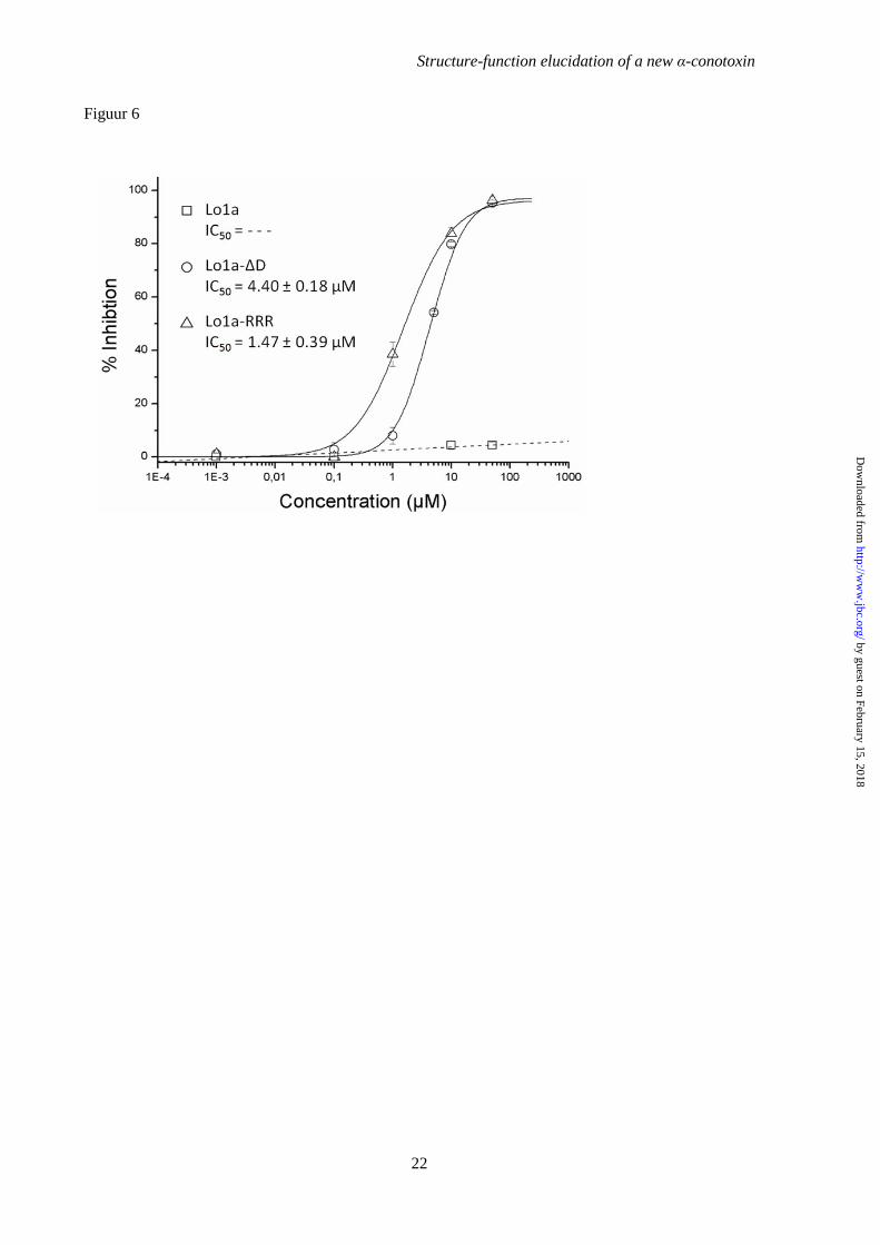
Influence of C-terminal truncation and replacement of -D by –RRR – We synthesized a C-terminally truncated analogue of Lo1a to examine its activity at different nAChR subtypes (Figures 4 and 5). Interestingly, Lo1a-∆D was
more potent at the neuronal nAChR α7 (IC50 = 0.80 ± 0.01 nM) but surprisingly also adopted affinity for the muscle subtype α1β1δε (IC50 = 4.40 ± 0.18 µM) (Figure 6). The same was noticed for Lo1a-RRR where the IC50 for α7 was 1.06 µM and an IC50 value of 1.47 ± 0.39 µM was determined for α1β1δε (Figure 6). An overview of subtype selectivity for Lo1a and both mutants is given in Figure 7.
NMR spectroscopy – NMR spectral analysis shows the formation of a single set of resonances for the conopeptide, indicating that it adopts one type of structural form in solution. Resonance assignment was performed by following standard procedures as outlined by Wüthrich and co-workers (36). Complete sequence specific proton assignments were achieved by analyzing homo-nuclear two-dimensional (2D) spectra (DQF-COSY, TOCSY, and NOESY). Initially the NH and Hα resonances of the individual spin systems (except Pro) were identified by analyzing the so-called “fingerprint” region of the DQF-COSY and TOCSY spectra and the remaining resonances of the spin systems were identified by following the so-called “TOCSY-tower”. Sequence specific assignments were achieved by linking individual spin system via sequential inter-residue Hαn-HN(n+1) cross-peaks in the “fingerprint” region of the NOESY spectrum. Carbon assignments were also performed by using 1H-13C HSQC spectra, which further reconfirmed most of the homo-nuclear proton assignments and clarified the Hβ and Hγ proton assignments of the two proline residues that were not resolved in the homo-nuclear 2D spectra due to signal overlaps. The geminal methylene protons were not assigned stereo-specifically and the NOE distance restraints involving these protons were used ambiguously during structure calculation in the Xplor-NIH program. Figure 8 shows the observed short and medium range NOEs that were used for the resonance assignment, 3JHNHα, and chemical shift index (CSI) along the amino acid sequences of α-conotoxin Lo1a. The CSI values indicate the presence of a α-helix in the middle of the peptide from residue Pro7 to Asn12.
Figure 9 displays the structural representations of the final ensemble of 15 superimposed structures (Figure 9A) and the minimum-energy closest-to-average structure (Figure 9B) of α-conotoxin Lo1a. The peptide structures are well defined with backbone and heavy atom RMSD of 0.43 and 0.83 respectively over residues 3 to 17. The structural evaluation
by guest on February 15, 2018http://w
ww
.jbc.org/D
ownloaded from
Structure-function elucidation of a new α-conotoxin
6
using PROCHECK demonstrates that all the resulted structures are having no bad non-bonded contacts and all the backbone dihedral angles are within the allowed regions of the Ramachandran plot (63% residues fall in the most favored region). Detailed structure determination statistics are provided in Table 1. The coordinates for 15 structures, NMR restraints and chemical shifts have been deposited in the RCSB Protein Data Bank with RCSB ID: RCSB103496 and PDB ID: 2MD6.
A close look into the structure (Figure 9 ) of the α-conotoxin Lo1a reveals that the peptide backbone adopts a compact “W-shaped” conformation having two loops that are reinforced by two disulfide bonds (Cys3-Cys9 and Cys4-Cys17). Residues from Pro7 to Asn12 at the bottom of the “W-shape” formed a α-helix involving two-turns. Besides this, in the N-terminal part of the peptide structure two overlapping beta-turns (type IV) exist between Gly2 to Ser5 and Cys4 to Pro7. In the C-terminal part of the peptide, residues from Pro14 to Cys17 formed a type I/IV beta-turn. This type of overall “W-shaped” molecular topology/fold was previously identified in the reported structures of other α4/7 subfamily α-conotoxins (37,38). DISCUSSION
Alpha-conotoxins are a family of cysteine-rich peptides that behave pharmacologically as competitive antagonists of the nicotinic acetylcholine receptor (39). In general, there are two main nAChR subtypes, the neuronal and muscle subtype nAChRs. Considering the neuronal subtype nAChRs and particularly their ACh binding site, a high percentage of sequence identity exists among the known neuronal nAChR subunits (40,41). Due to the high sequence conservation, it has been difficult to obtain subtype selective ligands, principally agonists that take action deep within the conserved ACh binding pocket. However recently, selective peptide antagonists from cone snail venom have shown to be highly selective pharmacological tools displaying the ability to discriminate amongst many of the different nAChR subunit combinations (41). This high selectivity toward a particular mammalian nAChR subtype is often established through specific interactions with particular residues located outside the conserved ACh binding site (42). Nowadays, co-crystal structures of α-conotoxins binding to the acetylcholine binding protein (AChBP) (43), which is a protein model
for the extracellular ligand-binding domain of nAChRs, can offer useful information about the molecular interactions of these small peptides (44,45).
Lo1a has a typical α-conotoxin structure – In our study we revealed the amino acid composition and the 3-dimensional structure of a new α4/7-conotoxin from Conus longurionis, called Lo1a. The peptide was investigated electrophysiologically and it was found that Lo1a inhibits α7 nAChRs preferentially. Once the activity of Lo1a had been determined we employed NMR based techniques to elucidate the structure of this novel peptide. According to Marx (46), NMR is the method of choice for determining conotoxin structures, because conotoxins are generally difficult to crystallize and are not amenable to X-ray methods, with a few exceptions such as PnIA and PnIB (47,48). α-Conotoxin Lo1a showed to be highly soluble and NMR spectra were readily assigned and used to generate a high-resolution structure. This structure demonstrated that Lo1a shares many of the structural and biochemical properties that define α-conotoxins, including the characteristic I-III, II-IV disulfide connectivity and the size of the first and the second loop of the peptide, i.e. four and seven residues respectively. Several α-conotoxins including AnIB (49), OmIA (50), GID (51), RegIIA (52) and LsIA (53) have an SXPA motif similar to the first loop of Lo1a. The peptide also shares the common fold comprising a short disulfide bond stabilized helix and a conserved proline. This proline is shown to be the only highly conserved amino acid residue apart from the cysteines and is responsible for helix initiation by inducing the 310 helix turn in the peptide backbone (39). Dutertre and Lewis (2006) revealed that this small α-helix structure appears to be a very important determinant for binding, even if the orientations and specific interactions differ significantly. As the side chains in an α-helix protrude at 360°, this structure is likely suitable to allow multiple contacts on both sides of a binding pocket located at the interface of two subunits (39).
As indicated in the previous section, Lo1a showed to be very selective for the neuronal versus the muscle nicotinic receptors. Other α-conotoxins having these characteristics are ImI, a α4/3 conotoxin from C. imperialis (54) and MII, a α4/7 conotoxin from C. magus (55). ImI targets α7 and α9 subunits with an IC50 of 220 nM and 1.8 µM respectively (56). Its amino acid sequence, GCCSDPRCAWRC, shares three non-
by guest on February 15, 2018http://w
ww
.jbc.org/D
ownloaded from

Structure-function elucidation of a new α-conotoxin
7
Cys amino acid residues with the sequence of Lo1a. These similar residues are Gly at the N-terminus, Ser after the second Cys and Pro between the second and the third Cys. The latter was identified as one of the determinants, besides Asp5, Arg7 and Trp10 to influence the potency of ImI at the α7 nAChR (57,58). α-Conotoxin MII blocks α6-containing nAChRs and α3β2 with an IC50 of 0.39 nM (α6/α3β2β3) and 2.18 nM respectively (59). If its amino acid sequence, GCCSNPVCHLEHSNLC, is aligned with Lo1a, 5 out of 14 non-Cys residues of Lo1a are similar with MII, resulting in a homology percentage of 56 (Figure 1). Everhart et al. (2004) (60) identified amino acid residues Asn5, Pro6 and His12 from MII as major determinants for potency on the α3β2 nAChR. Consequently, it is expected that Lo1a also targets α3β2 nAChRs.
Lo1a has an atypical C-terminus – The N-terminal amino acid residue of α-conotoxins is typically a glycine followed by the first two cysteine residues (Figure 1). However, sequence alignment of Lo1a with other α-conotoxins reveals besides a unique loop-2 sequence an atypical terminal amino acid sequence as both N- and C-termini contain negatively charged amino acid residues, i.e. glutamic acid and aspartic acid respectively. Few conotoxins have other residues preceding the first cysteine. From these conotoxins, ArIA (61), ArIB (61), GID (51), PIA (62), EI (63) and LsIA (53) are the most investigated (Figure 1). On the other hand, a negative residue at the C-terminus of a α-conotoxin has only been observed in conotoxins Lp1.4 from Conus leopardus (33) and Bt1.91 from Conus betulinus (35). But to the best of our knowledge, the influence of this negative C-terminal residue has never been investigated before.
C-terminal charges are important for neuronal/muscle subtype selectivity and efficacy – To explore the role of the C-terminal Asp of Lo1a, two analogues were synthesized: one C-terminally truncated peptide, named Lo1a-∆D and another named Lo1a-RRR with an extremely positively charged Arg-tail at the C-terminus. Deleting the C-terminal amino acid resulted in a more positively charged peptide, while the rationale for the Arg-tailing peptide can be found in the sequences of ArIA and ArIB from C. arenatus (Figure 1). Both peptides, ArIA and ArIB, are potent inhibitors of the α7 nAChR having IC50 values in the nanomolar range (61). Whiteaker et al. (2007) made a series of directed substitutions in ArIB to obtain a more selective
α7 antagonist. The mutant ArIB[V11L,V16D] improved its selectivity on α7 more than 10.000-fold suggesting the importance of Lys and Asp on position 11 and 16 respectively. Via replacing the C-terminal Asp by a triple Arg-tail, we aimed to investigate whether we could improve potency/selectivity on α7 with Lo1a-RRR.
The two synthetic C-terminally modified analogues revealed unexpected roles for the C-terminus in determining subtype selectivity and efficacy. Both mutants demonstrated more potency towards the neuronal α7 AChR as both IC50 values decreased from 3.24 µM (Lo1a) to 0.80 µM (Lo1a-∆D) and 1.06 µM (Lo1a-RRR). As the difference in potency was not very astonishing, we can conclude that the C-terminal residue(s) doesn’t play a crucial role in activity on α7. Remarkably, both mutants conferred affinity for the adult muscle subtype nAChR α1β1δε (IC50 values 4.40 ± 0.18 µM (Lo1a-∆D) and 1.47 ± 0.39 µM (Lo1a-RRR)) but much less for the fetal muscle subtype nAChR α1β1γδ. Consequently, C-terminal charges seem to have an influence in making the distinction between neuronal or muscle subtype nAChRs and even among muscle subtype nAChRs. α-Conotoxins which target selectively muscle
subtype nAChRs typically have a 3/5 structure (64). An exception is the α-conotoxin EI (α4/7) from C. ermineus that selectively targets the α/δ interface of muscle subtype nAChRs (63). Conotoxins that distinguish between the adult and the fetal muscle subtype nAChRs are rare. One example is ψ-conotoxin PrIIIE from C. parius, characterized by Luisma et al. (2008), which showed higher inhibition potency against the adult subtype (IC50 of 245 nM) than the fetal-subtype nAChR (IC50 of 3.24 µM) (65). Another ψ-conotoxin PIIIE from C. purpurascens shows an IC50 of 7.4 µM on the adult muscle subtype, but no inhibition on the fetal muscle subtype for concentrations up to 10 µM. Teichert et al. (2005) reports αA-conotoxin OIVB from C. obscurus, a unique selective inhibitor of the mammalian fetal muscle nAChR (IC50 of 56 nM), whereas affinity for the adult muscle nAChR is more than 1800-fold lower (66). According to Groebe et al. (1995), many of the α-conotoxins bind with 10.000-fold higher affinity to the mammalian α/δ interface than the α/γ interface (67). The peptides Lo1a-∆D and Lo1a-RRR which are described in this work, apparently demonstrate higher affinity to the α/ε interface.
by guest on February 15, 2018http://w
ww
.jbc.org/D
ownloaded from

Structure-function elucidation of a new α-conotoxin
8
Considering the terminal charges of Lo1a and its analogous, it can be expected that the positively charged triple Arg-tail in Lo1a-RRR is more likely to adopt a conformation extended away from the negatively charged N-terminus (as derived by torsion-angle molecular dynamics and energy minimization in the structural model). In contrast, the structure from Lo1a has a more ‘compact’ conformation, with an inward-facing C-terminus as derived from the solution state NMR structural data. Consequently, as the C- and N-terminus of Lo1a are in close proximity (less than 10 nm), the charges at both termini may interact, playing a role in making the distinction between neuronal versus muscle subtype nAChRs. Further structure-function studies combined with co-crystallization experiments are necessary to see whether this hypothesis is applicable for Lo1a-∆D.
Muscle and neuronal subtype nAChRs binding sites: structural receptor elements for binding of Lo1a and its homologous – The muscle subtype nAChR has a pentameric structure comprised of two α1 subunits, one β1, one δ, and depending on whether the receptor is in an embryonic or adult stage, one γ or ε subunit respectively. Each α1 subunit folds such that the primary binding site directly faces a neighboring subunit, which is either a γ/ε or a δ subunit. The γ subunit is believed to be the one that forms stable contacts being the lone subunit between the two α1 subunits, while the δ subunit pairs with the β subunit to form stable contacts between the α1 subunits on the opposite side. As two α1 subunits are separated by at least one non α1 subunit, correct coupling between these subunits is required for cooperative binding of agonists (68). Agonists of the muscle subtype nAChR initiate channel opening and desensitization by binding to a site on each of these two α1 subunits (69). Moreover, Arias and Blanton (2000) established that two adjacent cysteines (at position 192 and 193 according to the sequence number of Torpedo AChR) in the α1 subunits are involved in the recognition and binding of cholinergic agonists and competitive antagonists (19). Later on, Sine (1993)
demonstrated that not only the two α1 subunits form the binding sites of agonists and antagonists, but that also the γ/(ε) and δ subunits are involved (70). Agonists and antagonists can specifically distinguish between the α1γ/(α1ε) and α1δ binding sites of the fetal/(adult) muscle acetylcholine receptor because of different contributions by the γ/(ε) and δ subunits where a minimum of four loops in both subunits is required to create the agonist binding site (71). In 1991, Sine and Claudio showed that AChRs lacking either a γ or a δ subunit assemble into pentamers with subunit compositions, (α)2β(γ)2 and (α)2β(δ)2 (68). Therefore, we tried to express (α)2β(γ)2 and (α)2β(δ)2 and (α)2β(ε)2 heterologously to determine which binding site is responsible for interactions causing the observed results. Unfortunately, due to poor heterologous expression in frog oocytes, these experiments could not provide sufficient information about the binding sites of Lo1a-∆D and Lo1a-RRR.
Whereas the two ligand binding sites of muscle AChRs are formed at interfaces between α1 and δ, ε or γ subunits, binding sites of α7 neuronal AChRs are formed at interfaces between identical α7 subunits. Residues of the α face of the binding site, termed the (+) face, cluster in three well separated regions of the primary sequence, termed loops A, B, and C (71). The stabilization of this loop C conformation in an open state appears to be an essential element for antagonist activity. The stabilization in the AChBP is established on the basis of the loop C vicinal disulfide bonds where the α-conotoxin disulfide bond Cys I-III interacts, as is shown for conotoxins PnIA and ImI (39). This type of antagonist activity can also be expected for Lo1a and its analogues, as both conotoxins, PnIA and ImI, are structurally quite homologous to Lo1a.
In summary, our study provides insight into α-conotoxin pharmacology and the molecular basis of nAChR selectivity, highlighting the influence and importance of C-terminal residues on conotoxin pharmacology.
by guest on February 15, 2018http://w
ww
.jbc.org/D
ownloaded from
Structure-function elucidation of a new α-conotoxin
9
REFERENCES 1. Alberola-Die, A., Martinez-Pinna, J., Gonzalez-Ros, J. M., Ivorra, I., and Morales, A. (2011)
Multiple inhibitory actions of lidocaine on Torpedo nicotinic acetylcholine receptors transplanted to Xenopus oocytes. J Neurochem 117, 1009-1019
2. Gold, A. B., and Lerman, C. (2012) Pharmacogenetics of smoking cessation: role of nicotine target and metabolism genes. Hum Genet
3. Rezvani, A. H., and Levin, E. D. (2001) Cognitive effects of nicotine. Biol Psychiatry 49, 258-267
4. Cooper, E., Couturier, S., and Ballivet, M. (1991) Pentameric structure and subunit stoichiometry of a neuronal nicotinic acetylcholine receptor. Nature 350, 235-238
5. Inserra, M. C., Kompella, S. N., Vetter, I., Brust, A., Daly, N. L., Cuny, H., Craik, D. J., Alewood, P. F., Adams, D. J., and Lewis, R. J. (2013) Isolation and characterization of alpha-conotoxin LsIA with potent activity at nicotinic acetylcholine receptors. Biochem Pharmacol
6. Colquhoun, L. M., and Patrick, J. W. (1997) Pharmacology of neuronal nicotinic acetylcholine receptor subtypes. Adv Pharmacol 39, 191-220
7. Le Novere, N., and Changeux, J. P. (1995) Molecular evolution of the nicotinic acetylcholine receptor: an example of multigene family in excitable cells. J Mol Evol 40, 155-172
8. Gotti, C., Fornasari, D., and Clementi, F. (1997) Human neuronal nicotinic receptors. Prog Neurobiol 53, 199-237
9. Couturier, S., Bertrand, D., Matter, J. M., Hernandez, M. C., Bertrand, S., Millar, N., Valera, S., Barkas, T., and Ballivet, M. (1990) A neuronal nicotinic acetylcholine receptor subunit (alpha 7) is developmentally regulated and forms a homo-oligomeric channel blocked by alpha-BTX. Neuron 5, 847-856
10. Rubboli, F., Court, J. A., Sala, C., Morris, C., Chini, B., Perry, E., and Clementi, F. (1994) Distribution of nicotinic receptors in the human hippocampus and thalamus. Eur J Neurosci 6, 1596-1604
11. Wevers, A., Jeske, A., Lobron, C., Birtsch, C., Heinemann, S., Maelicke, A., Schroder, R., and Schroder, H. (1994) Cellular distribution of nicotinic acetylcholine receptor subunit mRNAs in the human cerebral cortex as revealed by non-isotopic in situ hybridization. Brain Res Mol Brain Res 25, 122-128
12. Breese, C. R., Adams, C., Logel, J., Drebing, C., Rollins, Y., Barnhart, M., Sullivan, B., Demasters, B. K., Freedman, R., and Leonard, S. (1997) Comparison of the regional expression of nicotinic acetylcholine receptor alpha7 mRNA and [125I]-alpha-bungarotoxin binding in human postmortem brain. J Comp Neurol 387, 385-398
13. Gotti, C., and Clementi, F. (2004) Neuronal nicotinic receptors: from structure to pathology. Prog Neurobiol 74, 363-396
14. Sacco, K. A., Bannon, K. L., and George, T. P. (2004) Nicotinic receptor mechanisms and cognition in normal states and neuropsychiatric disorders. J Psychopharmacol 18, 457-474
15. Steinlein, O. K., and Bertrand, D. (2010) Nicotinic receptor channelopathies and epilepsy. Pflugers Arch 460, 495-503
16. Quinton, L., Servent, D., Girard, E., Molgo, J., Le Caer, J. P., Malosse, C., Haidar el, A., Lecoq, A., Gilles, N., and Chamot-Rooke, J. (2013) Identification and functional characterization of a novel alpha-conotoxin (EIIA) from Conus ermineus. Anal Bioanal Chem 405, 5341-5351
17. Sine, S. M., Kreienkamp, H. J., Bren, N., Maeda, R., and Taylor, P. (1995) Molecular dissection of subunit interfaces in the acetylcholine receptor: identification of determinants of alpha-conotoxin M1 selectivity. Neuron 15, 205-211
18. McIntosh, J. M., Plazas, P. V., Watkins, M., Gomez-Casati, M. E., Olivera, B. M., and Elgoyhen, A. B. (2005) A novel alpha-conotoxin, PeIA, cloned from Conus pergrandis, discriminates between rat alpha9alpha10 and alpha7 nicotinic cholinergic receptors. J Biol Chem 280, 30107-30112
19. Arias, H. R., and Blanton, M. P. (2000) Alpha-conotoxins. Int J Biochem Cell Biol 32, 1017-1028
by guest on February 15, 2018http://w
ww
.jbc.org/D
ownloaded from

Structure-function elucidation of a new α-conotoxin
10
20. Olivera, B. M., Rivier, J., Clark, C., Ramilo, C. A., Corpuz, G. P., Abogadie, F. C., Mena, E. E., Woodward, S. R., Hillyard, D. R., and Cruz, L. J. (1990) Diversity of Conus Neuropeptides. Science 249, 257-263
21. Gronlien, J. H., Hakerud, M., Ween, H., Thorin-Hagene, K., Briggs, C. A., Gopalakrishnan, M., and Malysz, J. (2007) Distinct profiles of alpha7 nAChR positive allosteric modulation revealed by structurally diverse chemotypes. Mol Pharmacol 72, 715-724
22. Tucker, J. K., and Tenorio, M. J. (2009) Systematic Classification of Recent and Fossil Conoidean Gastropods: With Keys to the Genera of Cone Shells, ConchBooks
23. Olivera, B. M., Gray, W. R., Zeikus, R., McIntosh, J. M., Varga, J., Rivier, J., de Santos, V., and Cruz, L. J. (1985) Peptide neurotoxins from fish-hunting cone snails. Science 230, 1338-1343
24. Van Der Haegen, A., Peigneur, S., and Tytgat, J. (2011) Importance of position 8 in mu-conotoxin KIIIA for voltage-gated sodium channel selectivity. FEBS J 278, 3408-3418
25. Keller, M., and Steiger, R. (2004) The pi plate: an implant for unstable extension fractures of the distal radius in patients with osteoporotic bone. Tech Hand Up Extrem Surg 8, 212-218
26. Hwang, T. L., and Shaka, A. J. (1998) Multiple-pulse mixing sequences that selectively enhance chemical exchange or cross-relaxation peaks in high-resolution NMR spectra. J Magn Reson 135, 280-287
27. Shaka, A. J., Lee, C. J., and Pines, A. (1988) Iterative Schemes for Bilinear Operators - Application to Spin Decoupling. Journal of Magnetic Resonance 77, 274-293
28. Derome, A. E., and Williamson, M. P. (1990) Rapid-Pulsing Artifacts in Double-Quantum-Filtered Cosy. Journal of Magnetic Resonance 88, 177-185
29. Schwieters, C. D., Kuszewski, J. J., Tjandra, N., and Clore, G. M. (2003) The Xplor-NIH NMR molecular structure determination package. J Magn Reson 160, 65-73
30. Stein, E. G., Rice, L. M., and Brunger, A. T. (1997) Torsion-angle molecular dynamics as a new efficient tool for NMR structure calculation. J Magn Reson 124, 154-164
31. Spronk, C. A., Linge, J. P., Hilbers, C. W., and Vuister, G. W. (2002) Improving the quality of protein structures derived by NMR spectroscopy. J Biomol NMR 22, 281-289
32. Laskowski, R. A., Macarthur, M. W., Moss, D. S., and Thornton, J. M. (1993) Procheck - a Program to Check the Stereochemical Quality of Protein Structures. J Appl Crystallogr 26, 283-291
33. Yuan, D. D., Han, Y. H., Wang, C. G., and Chi, C. W. (2007) From the identification of gene organization of alpha conotoxins to the cloning of novel toxins. Toxicon 49, 1135-1149
34. Peng, C., Chen, W., Sanders, T., Chew, G., Liu, J., Hawrot, E., and Chi, C. (2010) Chemical synthesis and characterization of two alpha4/7-conotoxins. Acta Biochim Biophys Sin (Shanghai) 42, 745-753
35. Kaas, Q., Yu, R., Jin, A. H., Dutertre, S., and Craik, D. J. (2012) ConoServer: updated content, knowledge, and discovery tools in the conopeptide database. Nucleic Acids Res 40, D325-330
36. Wüthrich, K. (1986) NMR of Proteins and Nucleic Acids. John Wiley & Sons Inc., New York 37. Park, K. H., Suk, J. E., Jacobsen, R., Gray, W. R., McIntosh, J. M., and Han, K. H. (2001)
Solution conformation of alpha-conotoxin EI, a neuromuscular toxin specific for the alpha 1/delta subunit interface of torpedo nicotinic acetylcholine receptor. J Biol Chem 276, 49028-49033
38. Chi, S. W., Kim, D. H., Olivera, B. M., McIntosh, J. M., and Han, K. H. (2004) Solution conformation of alpha-conotoxin GIC, a novel potent antagonist of alpha3beta2 nicotinic acetylcholine receptors. Biochem J 380, 347-352
39. Dutertre, S., and Lewis, R. J. (2006) Toxin insights into nicotinic acetylcholine receptors. Biochem Pharmacol 72, 661-670
40. Le Novere, N., Corringer, P. J., and Changeux, J. P. (2002) The diversity of subunit composition in nAChRs: evolutionary origins, physiologic and pharmacologic consequences. J Neurobiol 53, 447-456
41. Tsetlin, V. I., and Hucho, F. (2004) Snake and snail toxins acting on nicotinic acetylcholine receptors: fundamental aspects and medical applications. FEBS Lett 557, 9-13
by guest on February 15, 2018http://w
ww
.jbc.org/D
ownloaded from
Structure-function elucidation of a new α-conotoxin
11
42. Dutertre, S., and Lewis, R. J. (2004) Computational approaches to understand alpha-conotoxin interactions at neuronal nicotinic receptors. Eur J Biochem 271, 2327-2334
43. Brejc, K., van Dijk, W. J., Klaassen, R. V., Schuurmans, M., van Der Oost, J., Smit, A. B., and Sixma, T. K. (2001) Crystal structure of an ACh-binding protein reveals the ligand-binding domain of nicotinic receptors. Nature 411, 269-276
44. Armishaw, C., Jensen, A. A., Balle, T., Clark, R. J., Harpsoe, K., Skonberg, C., Liljefors, T., and Stromgaard, K. (2009) Rational design of alpha-conotoxin analogues targeting alpha7 nicotinic acetylcholine receptors: improved antagonistic activity by incorporation of proline derivatives. J Biol Chem 284, 9498-9512
45. Dutertre, S., Ulens, C., Buttner, R., Fish, A., van Elk, R., Kendel, Y., Hopping, G., Alewood, P. F., Schroeder, C., Nicke, A., Smit, A. B., Sixma, T. K., and Lewis, R. J. (2007) AChBP-targeted alpha-conotoxin correlates distinct binding orientations with nAChR subtype selectivity. EMBO J 26, 3858-3867
46. Marx, U. C., Daly, N. L., and Craik, D. J. (2006) NMR of conotoxins: structural features and an analysis of chemical shifts of post-translationally modified amino acids. Magn Reson Chem 44 Spec No, S41-50
47. Hu, S. H., Gehrmann, J., Guddat, L. W., Alewood, P. F., Craik, D. J., and Martin, J. L. (1996) The 1.1 A crystal structure of the neuronal acetylcholine receptor antagonist, alpha-conotoxin PnIA from Conus pennaceus. Structure 4, 417-423
48. Hu, S. H., Gehrmann, J., Alewood, P. F., Craik, D. J., and Martin, J. L. (1997) Crystal structure at 1.1 A resolution of alpha-conotoxin PnIB: comparison with alpha-conotoxins PnIA and GI. Biochemistry 36, 11323-11330
49. Loughnan, M. L., Nicke, A., Jones, A., Adams, D. J., Alewood, P. F., and Lewis, R. J. (2004) Chemical and functional identification and characterization of novel sulfated alpha-conotoxins from the cone snail Conus anemone. J Med Chem 47, 1234-1241
50. Talley, T. T., Olivera, B. M., Han, K. H., Christensen, S. B., Dowell, C., Tsigelny, I., Ho, K. Y., Taylor, P., and McIntosh, J. M. (2006) Alpha-conotoxin OmIA is a potent ligand for the acetylcholine-binding protein as well as alpha3beta2 and alpha7 nicotinic acetylcholine receptors. J Biol Chem 281, 24678-24686
51. Nicke, A., Loughnan, M. L., Millard, E. L., Alewood, P. F., Adams, D. J., Daly, N. L., Craik, D. J., and Lewis, R. J. (2003) Isolation, structure, and activity of GID, a novel alpha 4/7-conotoxin with an extended N-terminal sequence. J Biol Chem 278, 3137-3144
52. Franco, A., Kompella, S. N., Akondi, K. B., Melaun, C., Daly, N. L., Luetje, C. W., Alewood, P. F., Craik, D. J., Adams, D. J., and Mari, F. (2012) RegIIA: an alpha4/7-conotoxin from the venom of Conus regius that potently blocks alpha3beta4 nAChRs. Biochem Pharmacol 83, 419-426
53. Inserra, M. C., Kompella, S. N., Vetter, I., Brust, A., Daly, N. L., Cuny, H., Craik, D. J., Alewood, P. F., Adams, D. J., and Lewis, R. J. (2013) Isolation and characterization of alpha-conotoxin LsIA with potent activity at nicotinic acetylcholine receptors. Biochem Pharmacol 86, 791-799
54. McIntosh, J. M., Yoshikami, D., Mahe, E., Nielsen, D. B., Rivier, J. E., Gray, W. R., and Olivera, B. M. (1994) A nicotinic acetylcholine receptor ligand of unique specificity, alpha-conotoxin ImI. J Biol Chem 269, 16733-16739
55. Cartier, G. E., Yoshikami, D., Gray, W. R., Luo, S., Olivera, B. M., and McIntosh, J. M. (1996) A new alpha-conotoxin which targets alpha3beta2 nicotinic acetylcholine receptors. J Biol Chem 271, 7522-7528
56. Johnson, D. S., Martinez, J., Elgoyhen, A. B., Heinemann, S. F., and McIntosh, J. M. (1995) alpha-Conotoxin ImI exhibits subtype-specific nicotinic acetylcholine receptor blockade: preferential inhibition of homomeric alpha 7 and alpha 9 receptors. Mol Pharmacol 48, 194-199
57. Quiram, P. A., and Sine, S. M. (1998) Structural elements in alpha-conotoxin ImI essential for binding to neuronal alpha7 receptors. J Biol Chem 273, 11007-11011
58. Quiram, P. A., and Sine, S. M. (1998) Identification of residues in the neuronal alpha7 acetylcholine receptor that confer selectivity for conotoxin ImI. J Biol Chem 273, 11001-11006
by guest on February 15, 2018http://w
ww
.jbc.org/D
ownloaded from
Structure-function elucidation of a new α-conotoxin
12
59. McIntosh, J. M., Azam, L., Staheli, S., Dowell, C., Lindstrom, J. M., Kuryatov, A., Garrett, J. E., Marks, M. J., and Whiteaker, P. (2004) Analogs of alpha-conotoxin MII are selective for alpha6-containing nicotinic acetylcholine receptors. Mol Pharmacol 65, 944-952
60. Everhart, D., Cartier, G. E., Malhotra, A., Gomes, A. V., McIntosh, J. M., and Luetje, C. W. (2004) Determinants of potency on alpha-conotoxin MII, a peptide antagonist of neuronal nicotinic receptors. Biochemistry 43, 2732-2737
61. Whiteaker, P., Christensen, S., Yoshikami, D., Dowell, C., Watkins, M., Gulyas, J., Rivier, J., Olivera, B. M., and McIntosh, J. M. (2007) Discovery, synthesis, and structure activity of a highly selective alpha7 nicotinic acetylcholine receptor antagonist. Biochemistry 46, 6628-6638
62. Dowell, C., Olivera, B. M., Garrett, J. E., Staheli, S. T., Watkins, M., Kuryatov, A., Yoshikami, D., Lindstrom, J. M., and McIntosh, J. M. (2003) Alpha-conotoxin PIA is selective for alpha6 subunit-containing nicotinic acetylcholine receptors. J Neurosci 23, 8445-8452
63. Martinez, J. S., Olivera, B. M., Gray, W. R., Craig, A. G., Groebe, D. R., Abramson, S. N., and McIntosh, J. M. (1995) alpha-Conotoxin EI, a new nicotinic acetylcholine receptor antagonist with novel selectivity. Biochemistry 34, 14519-14526
64. Lewis, R. J., Dutertre, S., Vetter, I., and Christie, M. J. (2012) Conus venom peptide pharmacology. Pharmacol Rev 64, 259-298
65. Lluisma, A. O., Lopez-Vera, E., Bulaj, G., Watkins, M., and Olivera, B. M. (2008) Characterization of a novel psi-conotoxin from Conus parius Reeve. Toxicon 51, 174-180
66. Teichert, R. W., Rivier, J., Torres, J., Dykert, J., Miller, C., and Olivera, B. M. (2005) A uniquely selective inhibitor of the mammalian fetal neuromuscular nicotinic acetylcholine receptor. J Neurosci 25, 732-736
67. Groebe, D. R., Dumm, J. M., Levitan, E. S., and Abramson, S. N. (1995) alpha-Conotoxins selectively inhibit one of the two acetylcholine binding sites of nicotinic receptors. Mol Pharmacol 48, 105-111
68. Sine, S. M., and Claudio, T. (1991) Gamma- and delta-subunits regulate the affinity and the cooperativity of ligand binding to the acetylcholine receptor. J Biol Chem 266, 19369-19377
69. Changeux, J. P. (1990) The TiPS lecture. The nicotinic acetylcholine receptor: an allosteric protein prototype of ligand-gated ion channels. Trends Pharmacol Sci 11, 485-492
70. Sine, S. M. (1993) Molecular dissection of subunit interfaces in the acetylcholine receptor: identification of residues that determine curare selectivity. Proc Natl Acad Sci U S A 90, 9436-9440
71. Prince, R. J., and Sine, S. M. (1996) Molecular dissection of subunit interfaces in the acetylcholine receptor. Identification of residues that determine agonist selectivity. J Biol Chem 271, 25770-25777
72. Luo, S., Kulak, J. M., Cartier, G. E., Jacobsen, R. B., Yoshikami, D., Olivera, B. M., and McIntosh, J. M. (1998) alpha-conotoxin AuIB selectively blocks alpha3 beta4 nicotinic acetylcholine receptors and nicotine-evoked norepinephrine release. J Neurosci 18, 8571-8579
73. Jin, A. H., Vetter, I., Dutertre, S., Abraham, N., Emidio, N. B., Inserra, M., Murali, S. S., Christie, M. J., Alewood, P. F., and Lewis, R. J. (2013) MrIC, a Novel alpha-Conotoxin Agonist in the Presence of PNU at Endogenous alpha7 Nicotinic Acetylcholine Receptors. Biochemistry
74. Peng, C., Han, Y., Sanders, T., Chew, G., Liu, J., Hawrot, E., Chi, C., and Wang, C. (2008) alpha4/7-conotoxin Lp1.1 is a novel antagonist of neuronal nicotinic acetylcholine receptors. Peptides 29, 1700-1707
75. Luo, S., Nguyen, T. A., Cartier, G. E., Olivera, B. M., Yoshikami, D., and McIntosh, J. M. (1999) Single-residue alteration in alpha-conotoxin PnIA switches its nAChR subtype selectivity. Biochemistry 38, 14542-14548
76. McIntosh, J. M., Dowell, C., Watkins, M., Garrett, J. E., Yoshikami, D., and Olivera, B. M. (2002) Alpha-conotoxin GIC from Conus geographus, a novel peptide antagonist of nicotinic acetylcholine receptors. J Biol Chem 277, 33610-33615
by guest on February 15, 2018http://w
ww
.jbc.org/D
ownloaded from
Structure-function elucidation of a new α-conotoxin
13
77. Loughnan, M., Bond, T., Atkins, A., Cuevas, J., Adams, D. J., Broxton, N. M., Livett, B. G., Down, J. G., Jones, A., Alewood, P. F., and Lewis, R. J. (1998) alpha-conotoxin EpI, a novel sulfated peptide from Conus episcopatus that selectively targets neuronal nicotinic acetylcholine receptors. J Biol Chem 273, 15667-15674
78. Sandall, D. W., Satkunanathan, N., Keays, D. A., Polidano, M. A., Liping, X., Pham, V., Down, J. G., Khalil, Z., Livett, B. G., and Gayler, K. R. (2003) A novel alpha-conotoxin identified by gene sequencing is active in suppressing the vascular response to selective stimulation of sensory nerves in vivo. Biochemistry 42, 6904-6911
by guest on February 15, 2018http://w
ww
.jbc.org/D
ownloaded from

Structure-function elucidation of a new α-conotoxin
14
Acknowledgements–We would like to thank to L. Sivilotti for sharing the hα3 and hβ4 cDNA. We are grateful to V. Witzemann for sharing the rα1, rβ1, rγ, rδ, rε1 clones and to J. Lindstrom for kindly providing the hα4 and hβ2 clones. We thank F. Clementi for sharing rα5 and for hα7. FOOTNOTES JT was supported by the following grants: G.0433.12, G.A071.10N and G.0257.08 (F.W.O. Vlaanderen), EU – FP7 – MAREX, IUAP 7/10 & IUAP 7/24 (Inter-University Attraction Poles Program, Belgian State, Belgian Science Policy) and OT/12/081 & GOA 12/016 (KU Leuven), 2013/146 (Academische Stichting Leuven). Dr. M. Maiti is a postdoctoral research fellow of F.W.O. Vlaanderen, Belgium. CU was supported by G.0939.11 and G.0762.13. To whom correspondence should be addressed: Jan Tytgat, Department of Pharmacy, Toxicology and Pharmacology, KU Leuven, Herestraat 49, 3000 Leuven, Belgium, Tel: +32 16 32 34 04; Fax: +32 16 32 34 05; Email: [email protected]. 1Toxicology and Pharmacology, University of Leuven (KU Leuven), Campus Gasthuisberg, O&N2, Herestraat 49, PO Box 922, 3000 Leuven, Belgium 2Medicinal Chemistry, University of Leuven (KU Leuven), Rega Institute for Medical Research, Minderbroedersstraat 10, 3000 Leuven, Belgium 3CSIR-National Institute of Oceanography, Dona Paula, Goa, India 4Center of Advanced Study in Marine Biology, Annamalai University, Parangipettai, Tamil Nadu, India 5Laboratory for Structural Neurobiology, University of Leuven (KU Leuven), O&N1 Herestraat 49, Box 601, 3000 Leuven, Belgium 6Laboratory for Protein Phosphorylation and Proteomics, University of Leuven (KU Leuven), O&N1 Herestraat 49, Box 901, 3000 Leuven, Belgium 7The abbreviations used were: ACh, Acetylcholine; AChBP, Acetylcholine binding protein; ADHD, Attention deficit hyperactivity disorder; IC50, Half-maximal inhibitory concentration; MALDI-TOF, Matrix-assisted laser desorption/ionization - time of flight; nAChR, Nicotinic acetylcholine receptor; NMR, Nuclear magnetic resonance; NOE, Nuclear overhauser effect; NOESY, Nuclear overhauser effect spectroscopy; RMSD, Root mean square deviation; RP-HPLC, Reversed phase-high performance liquid chromatography; S.D., Standard deviation; TFA, trifluoroacetic acid TABLE LEGENDS TABLE 1. NMR structure determination statistics of α-conotoxin Lo1a for an ensemble of 15 structures. Values where applicable are the means ± S.D. FIGURE LEGENDS FIGURE 1. Alignment of 41 α4/7 conotoxins with Lo1a, sorted by percentage of identity (indicated in the left column) with Lo1a. References from top to bottom: EI (C. ermineus (63)), AuIC (C. aulicus, (72)), AuIA (C. aulicus (72)), MrIC (C. marmoreus, (73)), Lp1.1 (C. leopardus (74)), PnIB (C. pennaceus (75)), Lp1.7 (C. leopardus, (35)), Lp1.8 (C. leopardus, (35)), Mr1.2 (C. marmoreus, (35)), Lp1.9 (C. leopardus, (35)), Qc1.4b (C.quercinus, (35)), PIA (C. purpurascens (62)), PnIA (C. pennaceus, (75)), AnIB (C. anemone, (49)), Qc1.6 (C. quercinus, (35)), GIC (C. geographus, (76)), TxIA (C. textile, (45)), MII (C. magus, (55)), EpI (C. episcopates,(77)), Qc1.4a (C. quercinus, (35)), Lp1.10 (C. leopardus, (35)), Vc1.1 (C. victoriae, (78)), TxIB (C. textile, (35)), RegIIA (C. regius, (52), OmIA (C. omaria, (50)), Mr1.1 (C. marmoreus, (34)), Mr1.3 (C. marmoreus, (35)), LvIA (C. lividus, (35)), LsIA (C. limpusi, (53)), Lp1.4 (C. leopardus, (34)), Bt1.91 (C. betulinus, (35)), Lp1.3 (C. leopardus, (35)), Sa1.6 (C. sanguinolentus, (35)), PuSG1.1 (C. pulicarius, (35)), Sa1.9 (C. sanguinolentus, (35)), PeIA (C. pergrandis, (18)), Bt1.2 (C. betulinus, (35)), GID (C. geographus, (51)), ArIA (C. arenatus (61)), ArIB (C. arenatus, (61)), Qc1.5 (C. quercinus, (35)), Lo1a (C. longurionis, this work).
by guest on February 15, 2018http://w
ww
.jbc.org/D
ownloaded from

FIGURE 2. Electrophysiological profile of Lo1a. perfused with ND96-A (ND96 with 1 µM atropine; except for oocytes expressing of 2 mL/min, with 10 µM Lo1a applied during 30s before ACh was added. ACh (200 µM for µM for α3β4, 50 µM for α4β2 and αwith 30s washout periods between different ACh applications and 200s af FIGURE 3. Concentration-response curve on ∆D () and Lo1a-RRR (∆) were tested on 0.80 ± 0.01 µM and 1.06 ± 0.00 µM respectively. “Materials and Methods”. The percentage of block was plotted against the logarithm of the tested concentrations. The results were fit wit FIGURE 4. Electrophysiological profile of Lo1aperfused with ND96-A (ND96 with 1 µM atropine; except for oocytes expressing of 2 mL/min, with 10 µM Lo1a-70 µM for α3β4, 50 µM for α4βmL/min, with 30s washout periods between different ACh applications and 200s after toxin application. FIGURE 5. Electrophysiological profile of Lo1aperfused with ND96-A (ND96 with 1 µM atropine; except for oocytes expressing of 2 mL/min, with 10 µM Lo1a-RRR applied during 30s before ACh70 µM for α3β4, 50 µM for α4βmL/min, with 30s washout periods between different ACh applications and 200s after toxin application. FIGURE 6. Concentration-response curve on Lo1a-∆D () and Lo1a-RRR (∆) were tested on and Lo1a-RRR are: 4.40 ± 0.18 µM and 1.47 ± 0.39 µM respectively. described under “Materials and Methods”. The percentage of block was plotted against the logarithm of the tested concentrations. The results were fit with the Hill equation. FIGURE 7. Overview of subtype selectivity. The three peptides, Lo1a, were tested on 6 different nAChRs: µM. Data are represented as the means ± S.E. (indicated by error bars); each experiment was performed at least three times (n FIGURE 8. Sequence and disulfide connectivity of and medium range NOEs, 3JHNHα
The thickness of the bar represents relative strength of NOEs (strong, medium and weak). 6 Hz,
3JHNHα = 6-8 Hz, 3JHNH
represent CSI values of +1 and -common to all α4/7 subfamily conotoxins. FIGURE 9. NMR solution structure of structures of α-conotoxin Lo1a with superimposed backbone heavy atoms (N, CA, C’). Disulfide bonds are shown in yellow as pseudo bonds. average structure of α-conotoxin Lo1a.label. The two views are related by a 180° rotation around the vertical axis.
Structure-function elucidation of a new
15
. Electrophysiological profile of Lo1a. During recordings, oocytes were continuously A (ND96 with 1 µM atropine; except for oocytes expressing α
/min, with 10 µM Lo1a applied during 30s before ACh was added. ACh (200 µM for and α4β4, 10 µM for α1β1γδ and α1β1δε) was applied for 2s at 2 mL/min,
with 30s washout periods between different ACh applications and 200s after toxin application.
response curve on α7 nAChR. Different concentration of Lo1a (∆) were tested on α7. The corresponding IC50 values are: 3.24 ± 0.70 µM,
0.80 ± 0.01 µM and 1.06 ± 0.00 µM respectively. Currents were obtained as described under “Materials and Methods”. The percentage of block was plotted against the logarithm of the tested concentrations. The results were fit with the Hill equation.
Electrophysiological profile of Lo1a-∆D. During recordings, oocytes were continuously A (ND96 with 1 µM atropine; except for oocytes expressing α
-∆D applied during 30s before ACh was added. ACh (200 µM for β2 and α4β4, 10 µM for α1β1γδ and α1β1δε) was applied for 2s at 2
mL/min, with 30s washout periods between different ACh applications and 200s after toxin
Electrophysiological profile of Lo1a-RRR. During recordings, oocytes were continuously A (ND96 with 1 µM atropine; except for oocytes expressing α
RRR applied during 30s before ACh was added. ACh (200 µM for β2 and α4β4, 10 µM for α1β1γδ and α1β1δε) was applied for 2s at 2
mL/min, with 30s washout periods between different ACh applications and 200s after toxin
ponse curve on α1β1δε nAChR. Different concentration of Lo1a (∆) were tested on α1β1δε. The corresponding IC50
RRR are: 4.40 ± 0.18 µM and 1.47 ± 0.39 µM respectively. Currents were obtained as described under “Materials and Methods”. The percentage of block was plotted against the logarithm of the tested concentrations. The results were fit with the Hill equation.
Overview of subtype selectivity. The three peptides, Lo1a, Lo1aon 6 different nAChRs: α1β1δε, α1β1γδ, α7, α4β2, α3β4 and α4β4 at a concentration of 10
µM. Data are represented as the means ± S.E. (indicated by error bars); each experiment was performed at least three times (n ≥ 3).
Sequence and disulfide connectivity of α-conotoxin Lo1a along withα (
3JαN) couplings, and chemical shift index (CSI) for the HThe thickness of the bar represents relative strength of NOEs (strong, medium and weak).
HNHα > 8 Hz. The filled squares above and below the horizontal line -1 respectively. Note the presence of α-helix from Pro
4/7 subfamily conotoxins.
NMR solution structure of α-conotoxin Lo1a. (A) Stereo views of a family of 15 final conotoxin Lo1a with superimposed backbone heavy atoms (N, CA, C’). Disulfide
bonds are shown in yellow as pseudo bonds. (B) Front and back ribbon views of the closestconotoxin Lo1a. Side chains heavy atoms are displayed along wit
label. The two views are related by a 180° rotation around the vertical axis.
function elucidation of a new α-conotoxin
During recordings, oocytes were continuously A (ND96 with 1 µM atropine; except for oocytes expressing α7 nAChR) at a rate
/min, with 10 µM Lo1a applied during 30s before ACh was added. ACh (200 µM for α7, 70 ) was applied for 2s at 2 mL/min,
ter toxin application.
nAChR. Different concentration of Lo1a (), Lo1a-values are: 3.24 ± 0.70 µM,
Currents were obtained as described under “Materials and Methods”. The percentage of block was plotted against the logarithm of the tested
During recordings, oocytes were continuously A (ND96 with 1 µM atropine; except for oocytes expressing α7 nAChR) at a rate
uring 30s before ACh was added. ACh (200 µM for α7, ε) was applied for 2s at 2
mL/min, with 30s washout periods between different ACh applications and 200s after toxin
During recordings, oocytes were continuously A (ND96 with 1 µM atropine; except for oocytes expressing α7 nAChR) at a rate
was added. ACh (200 µM for α7, ε) was applied for 2s at 2
mL/min, with 30s washout periods between different ACh applications and 200s after toxin
nAChR. Different concentration of Lo1a (), 50 values for Lo1a-∆D
Currents were obtained as described under “Materials and Methods”. The percentage of block was plotted against the logarithm
Lo1a-∆D and Lo1a-RRR at a concentration of 10
µM. Data are represented as the means ± S.E. (indicated by error bars); each experiment was
conotoxin Lo1a along with a summary of short ) couplings, and chemical shift index (CSI) for the Hα protons.
The thickness of the bar represents relative strength of NOEs (strong, medium and weak). 3JHNHα < > 8 Hz. The filled squares above and below the horizontal line
helix from Pro7-Asn12 which is
views of a family of 15 final conotoxin Lo1a with superimposed backbone heavy atoms (N, CA, C’). Disulfide
Front and back ribbon views of the closest-to-Side chains heavy atoms are displayed along with residue
by guest on February 15, 2018http://w
ww
.jbc.org/D
ownloaded from

Structure-function elucidation of a new α-conotoxin
16
Table 1 Total NOE distance restraints 190 Intra residue 65 Sequential (׀i-j1=׀) 66 Medium range (2≤׀i-j4≥׀) 46 Long range (׀i-j5≤׀) 13 Dihedral angle restraints 10 (ϕ=8, ωX-Pro=2) RMSD from the average structure (Ǻ) Residue 3-17 Backbone atoms (N, Cα, C’) 0.43 ± 0.09 All heavy atoms 0.83 ± 0.13 RMS deviation from the idealized covalent geometry Bond (Ǻ) 0.000008 ± 0.000000 Angle (º) 3.809 ± 0.074 Improper (º) 3.245 ± 0.412 Ramachandran analysis (%) Residues in favored regions 63 Residues in additional allowed regions 30.3 Residues in generously allowed regions 6.7 Residues in disallowed regions 0
by guest on February 15, 2018http://w
ww
.jbc.org/D
ownloaded from

Figure 1
Structure-function elucidation of a new
17
function elucidation of a new α-conotoxin
by guest on February 15, 2018http://w
ww
.jbc.org/D
ownloaded from

Structure-function elucidation of a new α-conotoxin
18
Figure 2
by guest on February 15, 2018http://w
ww
.jbc.org/D
ownloaded from
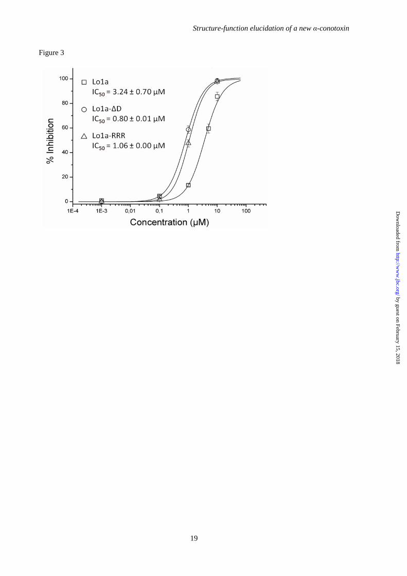
Figure 3
Structure-function elucidation of a new
19
function elucidation of a new α-conotoxin
by guest on February 15, 2018http://w
ww
.jbc.org/D
ownloaded from

Structure-function elucidation of a new α-conotoxin
20
Figure 4
by guest on February 15, 2018http://w
ww
.jbc.org/D
ownloaded from

Structure-function elucidation of a new α-conotoxin
21
Figure 5
by guest on February 15, 2018http://w
ww
.jbc.org/D
ownloaded from
Figuur 6
Structure-function elucidation of a new
22
function elucidation of a new α-conotoxin
by guest on February 15, 2018http://w
ww
.jbc.org/D
ownloaded from

Figure 7
Structure-function elucidation of a new
23
function elucidation of a new α-conotoxin
by guest on February 15, 2018http://w
ww
.jbc.org/D
ownloaded from

Figure 8
Structure-function elucidation of a new
24
function elucidation of a new α-conotoxin
by guest on February 15, 2018http://w
ww
.jbc.org/D
ownloaded from

Structure-function elucidation of a new α-conotoxin
25
N-term N-termC-term C-term(A)
(B)
180°
Figure 9
by guest on February 15, 2018http://w
ww
.jbc.org/D
ownloaded from
Herdewijn and Jan TytgatRavichandran, Eveline Lescrinier, Chris Ulens, Etienne Waelkens, Lisette D'Souza, Piet
Eline K. M. Lebbe, Steve Peigneur, Mohitosh Maiti, Prabha Devi, Samuthirapandianlongurionis
Conus-conotoxin, Lo1a, from αStructure-Function Elucidation of a New
published online February 24, 2014J. Biol. Chem.
10.1074/jbc.M114.556175Access the most updated version of this article at doi:
Alerts:
When a correction for this article is posted•
When this article is cited•
to choose from all of JBC's e-mail alertsClick here
by guest on February 15, 2018http://w
ww
.jbc.org/D
ownloaded from