
Structure-Function Analysis of Rotavirus NSP2 Octamer by Using a Novel Complementation System
11
JOURNAL OF VIROLOGY, Aug. 2006, p. 7984–7994 Vol. 80, No. 16 0022-538X/06/$08.000 doi:10.1128/JVI.00172-06 Copyright © 2006, American Society for Microbiology. All Rights Reserved. Structure-Function Analysis of Rotavirus NSP2 Octamer by Using a Novel Complementation System† Zenobia F. Taraporewala, 1 ‡ Xiaofang Jiang, 2 ‡ Rodrigo Vasquez-Del Carpio, 1 Hariharan Jayaram, 2 B. V. Venkataram Prasad, 2 * and John T. Patton 1 * Laboratory of Infectious Diseases, National Institutes of Allergy and Infectious Diseases, National Institutes of Health, Bethesda, Maryland 20892, 1 and Verna and Marrs McLean Department of Biochemistry and Molecular Biology, Baylor College of Medicine, Houston, Texas 77030 2 Received 25 January 2006/Accepted 31 May 2006 Viral inclusion bodies, or viroplasms, that form in rotavirus-infected cells direct replication and packaging of the segmented double-stranded RNA (dsRNA) genome. NSP2, one of two rotavirus proteins needed for viroplasm assembly, possesses NTPase, RNA-binding, and helix-unwinding activities. NSP2 of the rotavirus group causing endemic infantile diarrhea (group A) was shown to self-assemble into large doughnut-shaped octamers with circumferential grooves and deep clefts containing nucleotide-binding histidine triad (HIT)-like motifs. Here, we demonstrate that NSP2 of group C rotavirus, a group that fails to reassort with group A viruses, retains the unique architecture of the group A octamer but differs in surface charge distribution. By using an NSP2-dependent complementation system, we show that the HIT-dependent NTPase activity of NSP2 is necessary for dsRNA synthesis, but not for viroplasm formation. The complementation system also showed that despite the retention of the octamer structure and the HIT-like fold, group C NSP2 failed to rescue replication and viroplasm formation in NSP2-deficient cells infected with group A rotavirus. The distinct differences in the surface charges on the Bristol and SA11 NSP2 octamers suggest that charge complementarity of the viroplasm-forming proteins guides the specificity of viroplasm formation and, possibly, reassortment restriction between rotavirus groups. A characteristic of many viral infections is the formation of large inclusion bodies, or viroplasms, within the cell that serve as sites of genome replication and capsid morphogenesis. Al- though the complex protein-protein and protein-RNA interac- tions that make up these viral factories are poorly defined, they must provide a microenvironment with the necessary fluidity required for replication and particle-assembly processes to proceed efficiently while supporting the structural integrity of the inclusion. In the case of rotaviruses, members of the Reo- viridae, two nonstructural proteins, NSP2 and NSP5, interact to form a membrane-free viroplasm scaffold (12) in which the segmented double-stranded RNA (dsRNA) of the virus is rep- licated and packaged into previrion core particles (32). X-ray crystallographic studies have shown that NSP2 self-assembles into large doughnut-shaped octamers, through tail-to-tail stacking of tetramers (18). Besides having affinity for the di- meric phosphoprotein NSP5 (10), the NSP2 octamer also pos- sesses nonspecific RNA-binding activity (34), possibly medi- ated by the highly electropositive grooves that extend diagonally across the tetramer-tetramer interface of the octa- mer. NSP2 possesses an Mg 2 -dependent hydrolysis activity, capable of removing the -phosphate from nucleoside triphos- phates (NTPs) and thus serving as an NTPase (34). This hy- drolytic activity is mediated by residues in the cleft region of each NSP2 monomer that form a histidine triad (HIT)-like motif (4), an element common to a large and ubiquitous family of nucleotidyl hydrolases (2) that heretofore has not been seen in a viral protein. Although the unique structural and enzy- matic properties of the octamer can be anticipated to be im- portant for virus replication, how these features contribute to viroplasm formation and to processes occurring within the viroplasm has not been resolved. The rotavirion is an icosahedron consisting of three concen- tric protein layers that encapsidates 11 segments of dsRNA. The antigenicity of the protein that forms the intermediate layer (VP6) is used to classify rotaviruses into groups (e.g., A, B, and C) (11). Virus strains belonging to the same group can undergo genetic recombination (reassortment) upon coinfec- tion, while strains belonging to different groups cannot. The basis of reassortment restriction between groups is undefined. During cell entry, rotavirions are converted to transcriptionally active double-layered particles that synthesize the template positive-strand RNAs for translation and genome replication. The positive-strand RNAs are packaged into and replicated by core replication intermediates in viroplasms (28). These cores represent icosahedral T A 1 particles surrounded by the core lattice protein, VP2, that contain minor amounts of the viral RNA-dependent RNA polymerase (RdRP), VP1, and the 5- capping enzyme, VP3. NSP2 is associated with these interme- diates, possibly through its affinity for single-stranded RNA (ssRNA) or the RdRP. Although much has been learned about NSP2 through stud- * Corresponding author. Mailing address for J. T. Patton: Labora- tory of Infectious Diseases, National Institutes of Allergy and Infec- tious Diseases, National Institutes of Health, Bethesda, MD 20892. Phone: (301) 594-1615. Fax: (301) 496-8312. E-mail: jpatton@niaid .nih.gov. Mailing address for B. V. V. Prasad: Verna and Marrs McLean Department of Biochemistry and Molecular Biology, Baylor College of Medicine, Houston, TX 77030. Phone: (713) 798-0124. Fax: (713) 796-9438. E-mail: [email protected]. † Supplemental material for this article may be found at http://jvi .asm.org/. ‡ Z.F.T. and X.J. made equal contributions to this study. 7984 Downloaded from https://journals.asm.org/journal/jvi on 26 October 2021 by 211.206.33.7.
Transcript of Structure-Function Analysis of Rotavirus NSP2 Octamer by Using a Novel Complementation System

JOURNAL OF VIROLOGY, Aug. 2006, p. 7984–7994 Vol. 80, No. 160022-538X/06/$08.00�0 doi:10.1128/JVI.00172-06Copyright © 2006, American Society for Microbiology. All Rights Reserved.
Structure-Function Analysis of Rotavirus NSP2 Octamerby Using a Novel Complementation System†
Zenobia F. Taraporewala,1‡ Xiaofang Jiang,2‡ Rodrigo Vasquez-Del Carpio,1Hariharan Jayaram,2 B. V. Venkataram Prasad,2* and John T. Patton1*
Laboratory of Infectious Diseases, National Institutes of Allergy and Infectious Diseases, National Institutes of Health,Bethesda, Maryland 20892,1 and Verna and Marrs McLean Department of Biochemistry and
Molecular Biology, Baylor College of Medicine, Houston, Texas 770302
Received 25 January 2006/Accepted 31 May 2006
Viral inclusion bodies, or viroplasms, that form in rotavirus-infected cells direct replication and packagingof the segmented double-stranded RNA (dsRNA) genome. NSP2, one of two rotavirus proteins needed forviroplasm assembly, possesses NTPase, RNA-binding, and helix-unwinding activities. NSP2 of the rotavirusgroup causing endemic infantile diarrhea (group A) was shown to self-assemble into large doughnut-shapedoctamers with circumferential grooves and deep clefts containing nucleotide-binding histidine triad (HIT)-likemotifs. Here, we demonstrate that NSP2 of group C rotavirus, a group that fails to reassort with group Aviruses, retains the unique architecture of the group A octamer but differs in surface charge distribution. Byusing an NSP2-dependent complementation system, we show that the HIT-dependent NTPase activity of NSP2is necessary for dsRNA synthesis, but not for viroplasm formation. The complementation system also showedthat despite the retention of the octamer structure and the HIT-like fold, group C NSP2 failed to rescuereplication and viroplasm formation in NSP2-deficient cells infected with group A rotavirus. The distinctdifferences in the surface charges on the Bristol and SA11 NSP2 octamers suggest that charge complementarityof the viroplasm-forming proteins guides the specificity of viroplasm formation and, possibly, reassortmentrestriction between rotavirus groups.
A characteristic of many viral infections is the formation oflarge inclusion bodies, or viroplasms, within the cell that serveas sites of genome replication and capsid morphogenesis. Al-though the complex protein-protein and protein-RNA interac-tions that make up these viral factories are poorly defined, theymust provide a microenvironment with the necessary fluidityrequired for replication and particle-assembly processes toproceed efficiently while supporting the structural integrity ofthe inclusion. In the case of rotaviruses, members of the Reo-viridae, two nonstructural proteins, NSP2 and NSP5, interact toform a membrane-free viroplasm scaffold (12) in which thesegmented double-stranded RNA (dsRNA) of the virus is rep-licated and packaged into previrion core particles (32). X-raycrystallographic studies have shown that NSP2 self-assemblesinto large doughnut-shaped octamers, through tail-to-tailstacking of tetramers (18). Besides having affinity for the di-meric phosphoprotein NSP5 (10), the NSP2 octamer also pos-sesses nonspecific RNA-binding activity (34), possibly medi-ated by the highly electropositive grooves that extenddiagonally across the tetramer-tetramer interface of the octa-mer. NSP2 possesses an Mg2�-dependent hydrolysis activity,
capable of removing the �-phosphate from nucleoside triphos-phates (NTPs) and thus serving as an NTPase (34). This hy-drolytic activity is mediated by residues in the cleft region ofeach NSP2 monomer that form a histidine triad (HIT)-likemotif (4), an element common to a large and ubiquitous familyof nucleotidyl hydrolases (2) that heretofore has not been seenin a viral protein. Although the unique structural and enzy-matic properties of the octamer can be anticipated to be im-portant for virus replication, how these features contribute toviroplasm formation and to processes occurring within theviroplasm has not been resolved.
The rotavirion is an icosahedron consisting of three concen-tric protein layers that encapsidates 11 segments of dsRNA.The antigenicity of the protein that forms the intermediatelayer (VP6) is used to classify rotaviruses into groups (e.g., A,B, and C) (11). Virus strains belonging to the same group canundergo genetic recombination (reassortment) upon coinfec-tion, while strains belonging to different groups cannot. Thebasis of reassortment restriction between groups is undefined.During cell entry, rotavirions are converted to transcriptionallyactive double-layered particles that synthesize the templatepositive-strand RNAs for translation and genome replication.The positive-strand RNAs are packaged into and replicated bycore replication intermediates in viroplasms (28). These coresrepresent icosahedral T A 1 particles surrounded by the corelattice protein, VP2, that contain minor amounts of the viralRNA-dependent RNA polymerase (RdRP), VP1, and the 5�-capping enzyme, VP3. NSP2 is associated with these interme-diates, possibly through its affinity for single-stranded RNA(ssRNA) or the RdRP.
Although much has been learned about NSP2 through stud-
* Corresponding author. Mailing address for J. T. Patton: Labora-tory of Infectious Diseases, National Institutes of Allergy and Infec-tious Diseases, National Institutes of Health, Bethesda, MD 20892.Phone: (301) 594-1615. Fax: (301) 496-8312. E-mail: [email protected]. Mailing address for B. V. V. Prasad: Verna and MarrsMcLean Department of Biochemistry and Molecular Biology, BaylorCollege of Medicine, Houston, TX 77030. Phone: (713) 798-0124. Fax:(713) 796-9438. E-mail: [email protected].
† Supplemental material for this article may be found at http://jvi.asm.org/.
‡ Z.F.T. and X.J. made equal contributions to this study.
7984
Dow
nloa
ded
from
http
s://j
ourn
als.
asm
.org
/jour
nal/j
vi o
n 26
Oct
ober
202
1 by
211
.206
.33.
7.

ies on the purified recombinant form of the protein, our un-derstanding of the role of NSP2 in the biological system islimited. Some insight has been gained by analysis of cells in-fected with the tsE(1400) strain, a mutant SA11 rotavirus witha temperature-sensitive (ts) lesion mapping to the genomesegment (gene 8) encoding NSP2 (29). Notably, at the non-permissive temperature, tsE-infected cells are defective in vi-roplasm formation, synthesize low levels of viral dsRNA, andoveraccumulate empty particles (6). RNA interference exper-iments have shown similar changes in infected cells in whichNSP2 expression has been suppressed (32). These findingssuggest a role for NSP2 not only in viroplasm formation, butalso in coordinating genome packaging and replication withparticle assembly. The critical role of NSP2 in viroplasm for-mation has been confirmed by transient expression studies,which have shown that the expression of NSP2 in combinationwith NSP5 promotes assembly of viroplasm-like structures(VLS) (12).
To further understand the role of NSP2 in rotavirus repli-cation, we have initiated a series of structural and biochemicalstudies designed (i) to address whether the architecture ofNSP2 monomer and octamer is conserved across the variousgroups of rotavirus; (ii) to identify properties of heterologousNSP2 and NSP5 required in support of genome replication andviroplasm formation; and (iii) to define how the various func-tional properties of NSP2, such as NTPase and RNA-bindingactivities and viroplasm formation, are partitioned structurally.In this study, we have determined the atomic structure of NSP2encoded by the group C Bristol strain of rotavirus (NSP2c),which exhibits only 35% sequence identity with group A NSP2,and compared it to that encoded by the group A SA11 strain(NSP2a). Although the results revealed extensive conservationof octamer architecture, significant differences were noted inthe overall surface potential of the octamers. To explore thecontribution of the conserved structural elements to the func-tion of NSP2 in the context of infection, we created a novelNSP2-dependent complementation system that supports ge-nome replication and particle assembly in tsE-infected cellscontaining a gene 8-specific siRNA, but only when NSP2a isexpressed in trans from siRNA-resistant transcripts. Using thisassay system, we demonstrate that the HIT-dependent NTPaseactivity of NSP2a is necessary for dsRNA synthesis but not forviroplasm formation and that despite overall structural homol-ogy, NSP2c fails to functionally substitute for NSP2a in sup-porting viral replication and this failure is likely to due tosignificant differences in the surface charge distribution.
MATERIALS AND METHODS
Expression and purification of NSP2. The NSP2 cDNA in the plasmid RotaCseg9/M13 of the Bristol strain (7) was amplified by PCR and cloned into thebacterial expression vector pQE60 (pQE60g8C), such that six His residues werefused to the 3� end of the NSP2c open reading frame (ORF). Bacterially ex-pressed NSP2c was initially purified by Ni-nitrilotriacetic acid (NTA) affinitychromatography as described for SA11 NSP2 (NSP2a) (34). Polyclonal antiserato NSP2c were produced in guinea pigs by immunization with 50 �g of proteinin Freund’s complete adjuvant and again in incomplete adjuvant at weeks 2, 4,and 6.
Preparation of NSP2 for crystallization. NSP2a was expressed in Escherichiacoli strain SG13009 (QIAGEN) and purified as described before (18). NSP2c wasexpressed and purified in the same manner with slight modification. E. coliSG13009 cells containing pQE60g8C were grown to an optical density of 0.7 at600 nm in Terrific Broth (Invitrogen), when the expression of NSP2c was induced
by adding isopropyl-�-D-thiogalactopyranoside (IPTG) to a final concentrationof 1 mM. After incubation for 8 to 10 h at 37°C, the bacteria were recovered bycentrifugation at 4,200 � g for 30 min. The cell pellet from a 4-liter culture wasresuspended in 200 ml of lysis buffer (50 mM NaH2PO4, 300 mM NaCl, 10 mMimidazole, pH 8.0, 40 �g/ml RNase A) and processed twice using a Microfluidizerhigh-shear processor (model 110Y; Microfluidics, Newton, Mass.). To recoverHis-tagged NSP2c, the clarified lysate was incubated with 8 ml of Ni-NTAagarose beads (QIAGEN). Subsequently, the beads were washed four times withbuffer containing 50 mM NaH2PO4, 300 mM NaCl, 20 mM imidazole, pH 8.0,and once with the same buffer, except containing 50 mM imidazole. Protein waseluted with 70 ml of the same buffer containing 250 mM imidazole, and thenusing a Centricon centrifugation cartridge, the buffer was changed to 10 mMTris–100 mM NaCl, pH 8.0, with a protein concentration of 4 to 6 mg/ml.
Crystallization. NSP2a was crystallized as described before (18). NSP2c wascrystallized by hanging drop vapor diffusion. Microseeding technique was used togrow crystals of better quality. Drops were assembled with 4 �l of protein mixedwith 4 �l of well solution [1.5 to 1.7 M (NH4)2SO4, 100 mM morpholineethane-sulfonic acid (MES), pH 6.4], and then preequilibrated overnight. A very smallamount of solution from a drop in which a crystal had been crushed was placedat one point of the overnight-preequilibrated protein drop mixture withoutmixing. Crystals typically grew after several hours.
Data collection and processing. NSP2c crystals were suspended in motherliquor with 0.25 M and then 0.5 M Li2SO4 for 1 min each and transferred into 1M Li2SO4. Crystals were then plunge-frozen in liquid nitrogen. Diffraction datawere collected on SBC-CAT beam line 19ID at the Advanced Photon Source(Argonne National Laboratory). Data were processed using the HKL2000 (26)software package. Diffraction data for NSP2a crystals under cryo-conditions werecollected on the F2 beam line at CHESS (Cornell University).
Structure determination and refinement. The structures of NSP2c and NSP2a(to 2.2-Å resolution) were solved by molecular replacement methods using thepreviously published 2.6-Å structure of NSP2a as a search model. The molecularreplacement solution was obtained using PHASER (24). Model building wascarried out manually using the program O (19). Any model bias was examined bycomputing composite-OMIT maps using CNS (3). Iterative rounds of refinementwith simulated annealing in CNS and model building with O were carried toobtain the final model. Details of the diffraction data analysis and structurerefinement are provided in Table S1 of the supplemental material. Optimizedmultiple superposition was obtained with LSQMAN in the CCP4 suite of pro-grams (9). An electrostatic potential map of the octamer was calculated usingDelphi (17). Figures were generated with PYMOL (http://www.pymol.org). Theaccession code for NSP2c in the Protein Data Base is 2GUO.
Expression vectors. Wild-type (wt) and mutated NSP2a ORFs were preparedby PCR and subcloned in the T7 transcription vector, SP72. The g8* nomencla-ture identifies pSP72 with NSP2a-encoding gene 8 cDNAs that have been mu-tated in the target site for the g8D siRNA, such that the siRNA no longer inducesdegradation of transcripts made from the vector. The mutations were introducedinto the codon wobble positions of the target site and did not alter the NSP2aamino acid sequence. The g8D siRNA target site in gene 8 cDNAs was mutatedwith a QuikChange kit (Stratagene) and the overlapping primer pair 5�-GATGTTCATGTTAAGGAGCTAGTGGCAGAGCTGCGATGGCAATATAAC-3�and 5�-GTTATATTGCCATCGCAGCTCTGCCACTAG CTCCTTAACATGAACATC-3�.
To prepare the T7 transcription vectors pSP72g8*/D36A and pSP72g9C, theNSP2 coding region in pQE60g8/D36A and pQE60g9C was amplified using anExpand high-fidelity PCR kit. The amplified products were digested with EcoRIand XhoI and inserted into the corresponding restriction sites of pSP72. Thevectors pSP72g8*-HA and pSP72g8*/K188A-HA were generated by outwardPCR using an Expand high-fidelity PCR kit. The amplified DNAs were digestedwith HindIII and then self-ligated to produce plasmids. The vectorspSP72g8*�C4 and pSP72g8*�C7 were similarly generated using PCR. The am-plified DNAs were digested with EcoRI and XhoI and ligated into correspondingsites of pSP72.
siRNAs and transfection of cells. The g8D and irrelevant (IR) siRNAs (Dhar-macon Research) were described previously (32). The control IR siRNA targetsthe Wa gene 5 RNA, but not any of the SA11 RNAs.
Fetal rhesus monkey kidney (MA104) cells grown in 12-well plates were rinsedwith minimal essential medium (MEM) and then overlaid with 1 ml of (QualityBiological Inc., Gaithersburg, MD) MEM per well. Transfections were carriedout by adding to each well a 200-�l volume of Opti-MEM I containing 2%Lipofectamine 2000 (Invitrogen), 0.5 �g of the appropriate T7 transcriptionalvector, and/or 0.3 �M siRNA. After incubation for 4 h, 1 ml of MEM containing20% fetal bovine serum was added to each well. Incubation was then continuedfor an additional 2 h, at which point cells were infected.
VOL. 80, 2006 STRUCTURE-FUNCTION ANALYSIS OF ROTAVIRUS NSP2 OCTAMER 7985
Dow
nloa
ded
from
http
s://j
ourn
als.
asm
.org
/jour
nal/j
vi o
n 26
Oct
ober
202
1 by
211
.206
.33.
7.

Virus infection. Transfected cells were infected with trypsin-activated tsEstrain and/or vaccinia virus vTF7.3 (13), each at a multiplicity of infection (MOI)of 10. At 1 h postinfection (p.i.), the inoculum was replaced with MEM contain-ing 40 �g of cytosine arabinoside and 100 �g of rifampin (Sigma) per ml. Toradiolabel proteins, the inoculum was replaced instead with 80% methionine-and cysteine-free MEM and 20% MEM, containing 25 �Ci of [35S]-Expressprotein labeling mix (Perkin-Elmer Life Sciences) per ml. To radiolabel RNAs,the inoculum was replaced with 80% phosphate-free Dulbecco’s MEM and 20%MEM supplemented with 25 �Ci of 32Pi (8,000 to 9,000 Ci/mmol) per ml. Cellswere typically harvested at 12 h p.i.
Protein analysis. Infected cell lysates were analyzed by electrophoresis on 10%NuPAGE Bis-Tris gels (Invitrogen). Proteins were detected by autoradiography.In Western blot assays, proteins were detected on nitrocellulose membranes byincubation with a 1:500 dilution of guinea pig antisera prepared against NSP2aor NSP2c. Hemagglutinin (HA)-tagged protein was detected with rat anti-HAmonoclonal antibody (1:2,000). Horseradish peroxidase-conjugated goat anti-guinea pig, anti-mouse, and anti-rat antisera were used as secondary antibodies(1:10,000). Blots were developed with SuperSignal West Pico chemiluminescentsubstrate (Pierce).
RNA analysis. Infected cells were lysed by freeze-thawing in water and cen-trifuged to remove large cellular debris. After treatment with 160 U of DNase I(Invitrogen) per ml for 30 min at 37°C, RNA was purified by phenol-chloroformextraction, resolved by electrophoresis on 12% polyacrylamide gels, detected byautoradiography, and quantified using a phosphorimager. The levels of dsRNAsynthesis were compared among samples by measuring the intensity of gene 1after correcting for background in the corresponding lanes.
Isolation of virus particles. Infected MA104 cells labeled with 35S-labeledamino acids were scraped into the media at 24 h p.i. and lysed by freeze-thawing.After removal of cell debris by low-speed centrifugation, virus was pelleted bycentrifugation at 160,000 � g for 2 h. The pellet was suspended in Tris-bufferedsaline (TBS) buffer (25 mM Tris-HCl, pH 7.4, 137 mM NaCl, 5 mM KCl, 1 mMMgCl2, 0.7 mM CaCl2, 0.7 mM Na2HPO4, 5.5 mM dextrose), and virus wasbanded by CsCl centrifugation. Fractions (0.5 ml) from the gradient were ana-lyzed by electrophoresis on 10% NuPAGE Bis-Tris gels and autoradiography.
IFF assay. The infectious focus-forming (IFF) assay was performed as follows.CsCl gradient fractions containing triple-layered particles were pooled andtreated with 5 �g of trypsin per ml. The samples were diluted with medium 199(Invitrogen), and virus in the dilutions was adsorbed onto monolayers of MA104cells. The inoculum was replaced with medium 199 containing 40 �g of cytosinearabinoside, 100 �g of rifampin (Sigma), and 10 �g of actinomycin D (Invitro-gen) per ml to inhibit the growth of vaccinia virus. The cells were maintained at31°C for 30 h, washed three times with phosphate-buffered saline (PBS), andfixed by incubation for 2 min in methanol. The cells were immunostained bysequential incubation with guinea pig anti-VP6 polyclonal sera (1:2,000) andhorseradish peroxidase-conjugated goat anti-guinea pig antibody (1:3,000), eachfor 30 min at room temperature. The cells were washed four times with PBS priorto developing for 20 min in a solution containing 0.266 mg per ml of 3-amino-9-ethylcarbazole in 0.1 M sodium acetate, pH 5.2, and 0.045% hydrogen perox-ide. Stained cells were counted with an inverted light microscope.
IF analysis. MA104 cells were infected at an MOI of 10 with vTF7.3 and/or thetsE strain. At 1 h p.i., the inoculum was removed and replaced with a mixture of4% Lipofectamine (Invitrogen) and 0.5 �g each of one or more T7 transcriptionvectors in medium 199. At 6 h p.i., the transfection mixture was replaced withmedium 199 containing 10% FBS. The cells were processed for immunofluores-cence (IF) at 16 h p.i. by fixing with formaldehyde and permeabilizing with TritonX-100 (4). Afterwards, the cells were incubated with a 1:500 to 1:1,000 dilutionof one or more of the following: guinea pig polyclonal antisera prepared againstrecombinant NSP2a or NSP2c, rat anti-HA monoclonal antibody, and mouseanti-NSP5 monoclonal antibody 158G37. After washing, the cells were incubatedwith a 1:1,000 dilution of one or more of the following: Alexa Fluor 488 goatanti-guinea pig immunoglobulin G (IgG), Alexa Fluor 594 goat anti-mouse IgG,and Alexa Fluor 568 goat anti-rat IgG (Molecular Probes). Nuclei were detectedby staining with Hoechst 33258 (4�,6�-diamino-2-phenylindole [DAPI]).
Protein structure accession number. The atomic coordinates for NSP2c havebeen deposited in the Protein Data Bank (PDB) under accession code 2GUO.
RESULTS
Structure of NSP2c. The group A rotaviruses, which includethe prototypic strain SA11, are a widespread cause of life-threatening dehydrating diarrhea in young children (21). Incontrast, group C rotaviruses are generally associated with
limited outbreaks of diarrheal disease. The group C Bristolstrain originated from a family outbreak of gastroenteritis thatresulted in the death of a young child (5). Sequencing showsthat NSP2c shares limited similarity (57%) and identity (35%)with NSP2a (Fig. 1). As an approach for identifying structuralfeatures of NSP2 that are vital to rotavirus replication, wedetermined the atomic structure of NSP2c and contrasted itsarchitectural features with those of NSP2a.
Recombinant NSP2c with a C-terminal His tag was ex-pressed in bacteria and purified under conditions similar toNSP2a (18). NSP2c crystallized in space group I4 with twomolecules in the asymmetric unit of the crystal, in contrast toNSP2a (18), which crystallized in space group I422 with onemolecule in the asymmetric unit. Using the X-ray structure ofNSP2a (PDB identification no. 1L9V) as an initial phasingmodel for molecular replacement, we determined the X-raystructure of NSP2c to a 2.8-Å resolution (Fig. 2). After severaliterative cycles of model building and refinement, the structurewas refined with a final R factor of 0.23 (Rfree � 0.281). Res-idues Ala 2 to Lys 246 and Tyr 254 to Met 309 from eachmolecule could be built into the electron density. Recently, weobtained crystals of NSP2a that diffracted to a 2.2-Å resolution(Fig. 2). The structure of NSP2a was determined to 2.2 Å usingthe previously published 2.6-Å structure (PDB identificationno. 1L9V) with an overall R factor of 0.233 (Rfree � 0.268). Therefined 2.2-Å structure consists of residues Ala 2 to Pro 295
FIG. 1. Sequence alignment of NSP2a with NSP2c. Identical, sim-ilar, and catalytic residues are highlighted in green, cyan, and red,respectively. The secondary structural elements as observed in theX-ray structures of NSP2a and NSP2c are indicated above and belowtheir respective sequences. The HIT homology region is underlined inblue. Sequence alignment was carried out using the program ClustalW.Sequences shown are from the SwissProt Database: NSP2a, accessionno. Q03243; and NSP2c, accession no. Q9PY93.
7986 TARAPOREWALA ET AL. J. VIROL.
Dow
nloa
ded
from
http
s://j
ourn
als.
asm
.org
/jour
nal/j
vi o
n 26
Oct
ober
202
1 by
211
.206
.33.
7.

and Pro 299 to Val 312. The 2.2-Å NSP2a structure is essen-tially the same as the 2.6-Å structure, except that some loopsare better defined and several water molecules are easily iden-tified.
Despite only 35% identity, the overall tertiary structure ofthe NSP2c monomer is very similar to that of the NSP2amonomer with distinct N-terminal and C-terminal domainsseparated by a deep cleft (Fig. 1 and 2). The root mean squaredeviation between the two structures is 1.8 Å for the matchingC atoms. Located in the cleft are the residues forming aHIT-like motif implicated in nucleotide binding and hydrolysis(4). When the structures of the NSP2a and NSP2c monomersare superimposed, the proposed catalytic residues of the cleft(His 225 and His 221 of NSP2a and His 222 and His 218 ofNSP2c) are closely matched (Fig. 2C), indicating a conserva-tion of the active site.
The major difference between the NSP2a and NSP2c mono-mers concerns the loop formed by residues 170 to 185 (Fig.2B). In particular, NSP2c lacks residues corresponding to res-idues 183 to 185 of NSP2a, thus leaving NSP2c with a shorterloop structure (arrow in Fig. 2A and B). This difference ap-pears to influence the overall positioning of the C-terminaldomain, with noticeable displacements in the positions of four-helices in the C-terminal domain of NSP2c. In the NSP2ccrystal, the two molecules (A and B) in the asymmetric unithave nearly identical structures, superimposing with a rootmean square deviation of 0.58 Å for matching C atoms. Themain difference, however, between the two monomeric formsin the NSP2c asymmetric unit is that, in molecule A, the C-terminal domain is moved slightly further away from the N-
terminal domain. This domain movement appears to be facil-itated by the small changes within the same loop region(residues 170 to 182) that distinguish the NSP2a and NSP2cmonomers.
Despite different space groups, both NSP2a and NSP2c formoctameric structures with tail-to-tail stacking of two four-foldrelated tetramers using crystallographic symmetry (Fig. 3A andB). For NSP2a, the octamer represents a 422 (d4) crystallo-graphic symmetry, whereas for NSP2c the octamer representsonly four-fold (c4) symmetry. The two octamers are essentiallyof the same dimensions with NSP2c octamer being slightlyshorter by about 5 Å along the four-fold axis, and wider bythe same amount along the axis perpendicular to the four-foldaxis. Comparison of the NSP2 octamers by superposition of theC backbone of each subunit shows that their minor differ-ences reflect the small variations described above in their mo-nomeric subunit structures. The most striking difference be-tween NSP2a and NSP2c octamers is in the distribution ofsurface electrostatic charges (Fig. 3C and D). The surface ofthe NSP2a octamer is significantly more basic than that of theNSP2c octamer, especially along the four grooves that rundiagonally across the tetramer-tetramer interface. Based ontheir basic nature, the grooves were proposed to serve as theRNA-binding sites of the octamer (18). More recently, cryo-electron microscopy studies have shown that grooves not onlyrepresent RNA-binding sites but also NSP5-binding sites aswell (Jiang et al., unpublished data). Notably, while each sub-unit of the NSP2 octamer contains an NTP-binding site, theRNA- and NSP5-binding grooves are formed by intersubunitinteractions in the octamer. Thus, the ability of NSP2 to self-
FIG. 2. Comparison of the monomeric subunits of NSP2a and NSP2c. (A) Ribbon diagram of NSP2a (cyan) and the two molecules (A and B)in the crystallographic asymmetric unit of NSP2c (green). The locations of the N and C termini are denoted. The catalytic residues of NSP2a (His225, His 221, and Lys 188) are shown in red, and those of NSP2c (His 222, His 218, and Lys 185) are shown in blue. The loop (residues 170 to185) is shown by an arrow. (B) A close-up view of the superposition of the loop regions showing noticeable differences between NSP2a and NSP2cstructures. In both panels B and C, the same color scheme as in panel A is used. (C) A close-up view of the superposition of catalytic residues inNSP2a and NSP2c structures.
VOL. 80, 2006 STRUCTURE-FUNCTION ANALYSIS OF ROTAVIRUS NSP2 OCTAMER 7987
Dow
nloa
ded
from
http
s://j
ourn
als.
asm
.org
/jour
nal/j
vi o
n 26
Oct
ober
202
1 by
211
.206
.33.
7.

assemble into an octamer is critical for its interaction withRNA and NSP5.
Novel NSP2-dependent complementation system. Biochem-ical analyses have indicated that NSP2c, like NSP2a, hasssRNA-binding and NTPase activity, albeit with somewhatvarying specific activities (data not shown). The conserved na-ture of the architecture of the NSP2a and NSP2c octamers andtheir NTPase and RNA-binding activities indicate that thesestructural and biochemical features are critical to rotavirus
replication. Moreover, the extensive similarity in the featuresof NSP2a and NSP2c raises the possibility that one can substi-tute for the other during virus replication.
To establish a biologically relevant system to study the fea-tures of NSP2, particularly in the context of genome replica-tion and viroplasm formation, we developed a novel comple-mentation system wherein rotavirus replication was madedependent on the availability of NSP2 provided in trans. Twocritical aspects of the system are as follows. (i) Instead ofinfecting cells with wt SA11, cells were infected with thetsE(1400) strain, a mutant virus encoding NSP2 with a ts mu-tation (A152V). At permissive temperature (31°C), the tsEstrain grows to a titer that is 4 logs higher than at nonper-missive temperature (39°C). As shown by IF staining (Fig. 4A),tsE-infected cells maintained at 39°C are defective in viroplasmformation. Although well-developed viroplasms can be de-tected in tsE-infected cells maintained at 31°C, such viroplasmscan be detected in SA11-infected cells maintained at either 31or 39°C (Fig. 4A). Based on 32Pi labeling, the amount ofdsRNA produced in tsE-infected cells at 39°C is 100-fold lessthan that produced at 31°C (Fig. 4B). (ii) An SA11 gene 8-spe-cific (g8D) siRNA was transfected into tsE-infected cells tosuppress the expression of virus-encoded NSP2a. In previousstudies, g8D siRNA was shown to reduce NSP2a expression inSA11-infected cells by 10-fold, thereby inhibiting viroplasmformation (32). Thus, in this NSP2-dependent complementa-tion system, the supply of functional NSP2a required to sup-port dsRNA synthesis and viroplasm formation was suppressedby not only using a ts form of NSP2a, but also by using ansiRNA to reduce the expression of ts NSP2a. Because thetransfection efficiency of MA104 cells was limited to 80 to 90%(32), 10 to 20% of the cells in the complementation systemwere infected with the tsE strain, but failed to receive g8DsiRNA. The presence of the tsE strain in the g8D siRNA-freecells provided the advantage of preventing the high levels ofbackground genome replication and particle assembly that
FIG. 3. Comparison of NSP2a and NSP2c octamers. (A) NSP2aoctamer (in ribbon representation) as viewed along the four-fold axis(top) and one of the two two-fold axes (bottom) of the 422 symmetricstructure. (B) NSP2c octamer in the same orientations (top and bot-tom). In panel B, the molecules A and B, which represent the crystal-lographic asymmetric unit, are shown in green and brown, respectively.In panel A, although shown in different colors, the molecules are samebecause of 422 symmetry. (C) Electrostatic surface potentials ofNSP2a octamers in the same orientations as in panel A. (D) Electro-static potential surfaces of NSP2c octamers in the same orientations asin panel B. Blue and red represent positive and negatively chargedresidues, respectively. The groove across the two-fold axis is shown byblack arrows.
FIG. 4. Viroplasm formation and dsRNA synthesis in SA11- andtsE strain-infected cells. MA104 cells infected with SA11 or the tsEstrain were maintained at either 31°C or 39°C until 12 h p.i. in mediumcontaining no label or 32Pi and processed for either IF assay or dsRNAsynthesis. (A) Viroplasm formation was visualized by IF assay usingprimary antibodies for the detection of NSP2 (green) and NSP5 (red).Colocalization of signals is indicated by yellow, and cell nuclei werevisualized by DAPI. (B) 32P-labeled viral dsRNA samples (g1 to g11)harvested from infected cells maintained at 39°C were analyzed byelectrophoresis on 12% polyacrylamide gels and autoradiography.
7988 TARAPOREWALA ET AL. J. VIROL.
Dow
nloa
ded
from
http
s://j
ourn
als.
asm
.org
/jour
nal/j
vi o
n 26
Oct
ober
202
1 by
211
.206
.33.
7.
would have occurred if wt SA11 virus had been used in thecomplementation system instead of the tsE strain.
To provide a source of recombinant NSP2a in the comple-mentation system, we used vaccinia virus vTF7.3 (13) to syn-thesize NSP2a mRNAs from T7 transcription vectors trans-fected into tsE-infected cells. This system was selected overcytomegalovirus-driven plasmids to express proteins in vivobecause it provided for more rapid and higher levels of proteinexpression, mimicking more closely the NSP2 expression pat-tern during rotavirus infection. Earlier studies have also shownthat productive rotavirus replication occurs despite coinfectionof cells with vaccinia virus (8, 23). Vector-derived NSP2 tran-scripts, unlike the parallel NSP2 transcripts generated by thevirus, were not degraded by g8D siRNA-induced RISC com-plexes due to the introduction of silent mutations into thesiRNA target sequence of the NSP2a cDNA. In this article,NSP2a originating from vector-derived transcripts resistant todegradation by g8D siRNA-induced RISC complexes is iden-tified with an asterisk (e.g., NSP2a*).
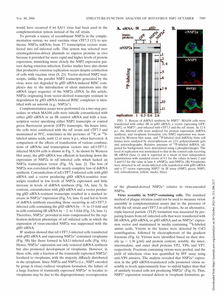
Complementation assays were performed via a two-step pro-cedure in which MA104 cells were initially cotransfected witheither g8D siRNA or an IR control siRNA and with a tran-scription vector specifying either NSP2 transcripts or controlgreen fluorescent protein (GFP) transcripts. Six hours later,the cells were coinfected with the tsE strain and vTF7.3 andmaintained at 39°C, sometimes in the presence of 32Pi or 35S-labeled amino acids, until 12 or more h p.i., when harvested. Acomparison of the effects of transfection of various combina-tions of siRNAs and transcription vectors into tsE/vTF7.3-infected MA104 cells is shown in Fig. 5. As shown by Westernblot assay, the g8D siRNA was efficient in knocking down theexpression of NSP2a in tsE-infected cells which lacked anNSP2a transcription vector (Fig. 5A, lane 2). The loss ofNSP2a was correlated with the nearly complete loss of dsRNAsynthesis. Cotransfection of tsE/vTF7.3-infected cells with g8DsiRNA and a vector producing g8D siRNA-sensitive tran-scripts resulted in low levels of NSP2a expression and littleincrease in levels of dsRNA synthesis (Fig. 5A, lane 3). Incontrast, cotransfection with g8D siRNA and a vector produc-ing g8D siRNA-resistant transcripts resulted in a marked in-crease in NSP2a* expression (Fig. 5A, lane 4) and led to levelsof dsRNA synthesis exceeding those occurring in tsE/vTF7.3-infected cells containing the g8D siRNA by 5- to 15-fold andin cells containing IR siRNA by 2- to 3-fold (Fig. 5A, lane 1).Therefore, NSP2a* provided in trans compensated for the rep-lication-deficient phenotype of tsE-infected cells in which theexpression of virus-encoded NSP2a was suppressed with theg8D siRNA.
IF analysis showed that tsE/vTF7.3-infected cells transfectedwith g8D siRNA and expressing NSP2a* contained viroplasms(Fig. 5B) like those formed in SA11-infected cells (Fig. 4A).Hence, NSP2a* expression not only restored dsRNA synthesisbut also promoted development of viroplasms. However, inthese cells, only a fraction of the transiently expressed NSP2a*localized to viroplasms, with the majority diffusely distributedin the cytoplasm. Since NSP2a and NSP5a (i.e., NSP5 encodedby group A virus) codirect viroplasm formation, the failure ofa large fraction of transiently expressed NSP2a* to localize toviroplasms may be due to the disproportionate overexpression
of the plasmid-derived NSP2a* relative to virus-encodedNSP5a.
Virus assembly in NSP2*-containing cells. The standardmethod of plaque titration could not be used to measure virionassembly in complementation assays due to the presence ofboth the tsE strain and vTF7.3 in cell lysates. As an alternative,triple-layered particle (TLP) formation was measured by pre-paring lysates from tsE-infected cells that were transfected withIR siRNA, g8D siRNA, or g8D siRNA and an NSP2a* expres-sion vector and maintained in media containing 35S-labeledamino acids. Virions in the lysates were detected by CsClcentrifugation, followed by electrophoresis of the gradientfractions (Fig. 6). Virions were identified based on their den-sity (� � 1.36 g/ml) and protein content, notably, the inner,intermediate, and outer shell proteins VP2, VP6, and VP7,respectively. Fractions containing virions were pooled, and theyield of infectious virus was quantified by IFF assay usinganti-VP6 antsiera. The analysis revealed that NSP2a* expres-sion in the g8D siRNA-transfected cells promoted virion as-sembly to levels approximately four- to sixfold higher than thatof similarly treated cells not producing NSP2a* (Fig. 6). Thus,NSP2* expression rescued defects in viroplasm formation, ge-
FIG. 5. Rescue of dsRNA synthesis by NSP2*. MA104 cells weretransfected with either IR or g8D siRNA; a vector expressing GFP,NSP2, or NSP2*; and infected with vTF7.3 and the tsE strain. At 12 hp.i., the infected cells were analyzed for protein expression, dsRNAsynthesis, and viroplasm formation. (A) NSP2 expression was moni-tored by Western blot assay, and 32P-labeled viral dsRNAs from celllysates were analyzed by electrophoresis on 12% polyacrylamide gelsand autoradiography. Relative amounts of 32P-labeled dsRNA, ad-justed for background, were determined using a phosphorimager. Thelevel of replication was normalized to that in the control cells receivingIR siRNA (lane 1) and is reported as a mean of four independentquantitations with standard errors of 0.1 for the values in lanes 2 and3 and 0.5 for the value in lane 4. NSP2a, anti-NSP2a. (B) Viroplasmswere detected in tsE strain-infected cells transfected with g8D siRNAand a T7 vector expressing NSP2* by IF assay (NSP2, green; NSP5,red; colocalization, yellow; nuclei, blue).
VOL. 80, 2006 STRUCTURE-FUNCTION ANALYSIS OF ROTAVIRUS NSP2 OCTAMER 7989
Dow
nloa
ded
from
http
s://j
ourn
als.
asm
.org
/jour
nal/j
vi o
n 26
Oct
ober
202
1 by
211
.206
.33.
7.

nome replication, and virion assembly in tsE-infected cells con-taining g8D siRNA, findings validating the potential of thecomplementation system as a tool for dissecting the structureand function of NSP2.
NTPase activity of NSP2 is required for dsRNA synthesis.NSP2 has been implicated in rotavirus replication and pack-aging (6, 27, 29), but the contribution of the NTPase activity ofNSP2 to these processes is uncertain. The enzymatic activitycould be anticipated to have an essential function, given theconservation of the HIT-like motif in the NSP2a and NSP2coctamer and the retention of the activity. To examine thispossibility, the abilities of two mutant forms of NSP2a (K188Aand H225A) possessing little or no NTPase activity were as-sayed in the complementation system for the ability to supportgenome replication and viroplasm formation. The NTPase-defective phenotype of K188A* and H225A* results from mu-tation of conserved residues that make up the HIT-like motif(4). The sedimentation properties of the bacterially expressedK188A and H225A were indistinguishable from that of NSP2a(4), indicating that the mutant forms of NSP2 retained theoctamer conformation. The complementation assay showedthat K188A* failed to promote dsRNA synthesis above thelevel detected in control cells lacking transiently expressedNSP2 (Fig. 7A, lanes 2 and 4). The level of dsRNA synthesis
with K188A* was less than that in cells containing NSP2a*(Fig. 7A, lane 3). Thus, the NTPase-defective phenotype wascorrelated with an inability of NSP2a* to support genomereplication, indicating that the NTPase activity of NSP2 iscritical for virus replication. Similar results were obtained us-ing H225A* in the complementation system (data not shown).
To determine whether K188A* and H225A* were defectivein supporting viroplasm formation, cells expressing these pro-teins in complementation assays were analyzed by IF. Asshown in Fig. 7B, K188A* supported the formation of numer-ous large viroplasms, like those formed in cells expressingNSP2a*. Similar viroplasms were detected in cells expressing
FIG. 6. Virus assembly in NSP2*-complemented cells. MA104 cellswere transfected with either IR (A) or g8D siRNA (B and C) and witha vector expressing NSP2* (C) and then infected with vTF7.3 and thetsE strain. Cells were maintained in the presence of 35S-labeled aminoacids and harvested 18 h p.i. Virus particles pelleted (p) from celllysates were analyzed by CsCl centrifugation. Gradient fractions wereanalyzed for density and for protein content by electrophoresis. Thetiter of the virus in the fractions containing TLPs (*) was determinedby IFF assay. FIG. 7. Effect of NSP2 mutation on genome replication. MA104
cells were transfected with an siRNA (IR or g8D) and a transcriptionvector expressing GFP or wt or mutant NSP2 and then infected withvTF7.3 and the tsE strain and maintained in medium containing nolabel or 32Pi. At 12 h p.i., the infected cells were analyzed for proteinexpression, dsRNA synthesis, and viroplasm formation. (A) NSP2aand HA fused to NSP2a were detected by Western blot assay, and32P-labeled dsRNA samples were analyzed by electrophoresis on 12%polyacrylamide gels and autoradiography. (B) Viroplasm formation incells producing the indicated vector-derived NSP2a was monitored byIF assay using primary antibodies (anti-NSP2a [NSP2a] and anti-HA[HA]) for the detection of NSP2a* (green) and NSP5 (red) in theupper panel and for the detection of the HA-tagged NSP2a* (red) andNSP5 (green) in the lower panel. Colocalization of signals is indicatedby yellow, and cell nuclei were visualized by DAPI.
7990 TARAPOREWALA ET AL. J. VIROL.
Dow
nloa
ded
from
http
s://j
ourn
als.
asm
.org
/jour
nal/j
vi o
n 26
Oct
ober
202
1 by
211
.206
.33.
7.

H225A* (data not shown). The fact that K188A* failed tosupport dsRNA synthesis, despite supporting viroplasm forma-tion, suggests that the NTPase activity of NSP2 contributes tovirus replication at a step independent of viroplasm formation.These results also show that the K188A mutation does notprevent NSP2a from interacting with NSP5a, since viroplasmformation is dependent on this event. Our findings are consis-tent with the results of previous coexpression assays, whichshowed that both the K188A and H225A mutant forms caninteract with NSP5 to form VLS in uninfected cells (4).
C-terminal HA tag interferes with NSP2 function in repli-cation. To distinguish between virus-encoded NSP2 and tran-siently expressed NSP2a* and K188A* in IF assays, andthereby confirm that NSP2a* and K188A* promoted viroplasmformation in the complementation system, a C-terminal HAtag (YPYDVPDYA) was added to these proteins. The result-ing products, NSP2a/HA* and K188A-HA*, when transientlyexpressed in the complementation system, supported viro-plasm formation (Fig. 7B). Hence, addition of short C-terminalsequences such as the HA tag does not interfere with thecapacity of NSP2a to support viroplasm formation. This findingis consistent with earlier results showing that NSP2a linked atits C terminus to GFP supported VLS formation when tran-siently coexpressed with NSP5a (10). Thus, the addition ofextra residues to the C terminus of NSP2 does not prevent thenecessary interaction between the protein and NSP5a requiredfor viroplasm formation.
The functionality of NSP2a/HA* and K188A-HA* was fur-ther evaluated by analyzing their capacity to support dsRNAsynthesis in the complementation system. In agreement withthe results described above with the K188A* mutant (Fig. 7A),the tagged version of the protein, K188A-HA*, failed to stim-ulate dsRNA synthesis above levels detected in cells lackingNSP2a* (lane 6). Surprisingly, the HA-tagged form of NSP2a*(lane 5), unlike the untagged form (lane 3), also failed tosupport dsRNA synthesis above background levels (lane 2).These results indicate that although addition of HA tag toNSP2a* did not affect its function in viroplasm formation, themodification did render the protein biologically inactive basedon its failure to support genome replication.
The C-terminal tail of NSP2 is dispensable for replication.The rationale for adding the HA tag to the C terminus ofNSP2a was, in part, based on the observation that the C-terminal amino acids 314 to 317 of NSP2 and the fused six-Histag were not detectable in the NSP2a (and NSP2c) crystalstructure and therefore were assumed to be disordered (18).However, despite its disordered status, the complementationassay suggested that the addition of the HA tag to the Cterminus interfered with an undefined, but important biologi-cal function of the protein. To further investigate the impor-tance of the C terminus, transcription vectors were constructedthat encoded NSP2a* with C-terminal deletions of 4 (�C4*) or7 (�C7*) amino acids. These modified proteins were comparedwith NSP2a* for the ability to support dsRNA synthesis incomplementation assays. As shown in Fig. 7A (lanes 7 to 9),dsRNA levels in cells expressing �C4* or �C7* were like thosein cells expressing NSP2a*. Thus, the C terminus of NSP2a(311 to 317) has no structure or function that is essential forrotavirus genome replication. Our results showing that theterminal amino acids are not required for replication suggest
that the HA tag interfered with NSP2 function not by perturb-ing a direct role of the C terminus in dsRNA synthesis, butmore likely by obstructing other undefined functional domainsof the octamer.
Group C NSP2 fails to substitute for group A NSP2. Thesimilarity of the NSP2a and NSP2c octamers both in theirstructure and activity (NTPase and RNA binding) suggests thatthese proteins are capable of substituting for one another inthe viral life cycle. To address this possibility, we used thecomplementation system to test whether transiently expressedNSP2c*, in place of NSP2a*, could support genome replicationand viroplasm formation in tsE-infected cells. As shown inFig. 8A, little or no dsRNA was produced in cells containingNSP2c (lane 6), similar to results with K188A* (lane 4). On theother hand, cells containing NSP2a* (lane 3) synthesizeddsRNA to levels similar to that of cells containing virion-encoded NSP2a (lane 1). NSP2a/D36A*, a form of NSP2a witha nondisruptive mutation mapping to the tetramer-tetramerinterface of the octamer and positioned away from any puta-tive ligand-binding site, also supported dsRNA to levels com-parable to that of NSP2a*. Taken together, these results showthat despite structural similarity, the NSP2c octamer cannotfunctionally replace NSP2a in supporting replication of the tsEstrain, a group A rotavirus.
The complementation system showed that NSP2c* not onlyfailed to support dsRNA synthesis in tsE-infected cells, butbased on IF analysis, also failed to nucleate viroplasm forma-
FIG. 8. Failure of NSP2c to support group A rotavirus replication.MA104 cells were transfected with an siRNA (IR or g8D) and atranscription vector expressing GFP, wt or mutant NSP2a*, or NSP2c,and then infected with vTF7.3 and the tsE strain. (A) At 12 h p.i., theinfected cells were analyzed for protein expression by Western blotassay and 32P-labeled viral dsRNA was analyzed by 12% polyacryl-amide gel electrophoresis and autoradiography. NSP2a, anti-NSP2a;NSP2c, anti-NSP2c; NSP5a, anti-NSP5a. (B) Formation of viro-plasms in g8D-treated tsE strain-infected MA104 cells expressingNSP2c and VLS in MA104 cells transiently expressing either NSP2cand NSP5a or NSP2a and NSP5a was monitored at 12 h p.i. by IF assay(NSP2, green; NSP5, red; colocalization, yellow; nuclei, blue).
VOL. 80, 2006 STRUCTURE-FUNCTION ANALYSIS OF ROTAVIRUS NSP2 OCTAMER 7991
Dow
nloa
ded
from
http
s://j
ourn
als.
asm
.org
/jour
nal/j
vi o
n 26
Oct
ober
202
1 by
211
.206
.33.
7.

tion with NSP5a (Fig. 8B). Instead, NSP2c* and tsE-encodedNSP5a were diffusely distributed throughout the cytoplasm.Similarly, transient coexpression of NSP2c and NSP5a in un-infected cells did not generate VLS. These data indicate thatNSP2c cannot interact with NSP5a for VLS formation. Be-cause both the NSP2a and NSP2c octamers share similar non-specific RNA-binding and NTPase activities, it seems doubtfulthat either of these has something to do with the failure ofNSP2c to support viroplasm formation. Instead, given the ob-vious difference in the electrostatic surface potential of theNSP2a and NSP2c octamers, it seems more reasonable to pre-dict that the unique surface charge on the Bristol species pre-vented it from interacting with NSP5a in such a way as to formthe NSP2-NSP5 complexes required for viroplasm assembly.
DISCUSSION
In this study, we report that NSP2a and NSP2c display aconsiderable degree of structural conservation, both in theirfold as monomers and in the arrangement of NSP2a andNSP2c monomers within octamers. NSP2a and NSP2c alsoretain a common HIT-like fold and use a nearly identical set ofresidues to create the HIT-like signature motif that is associ-ated with the NTPase activity of the protein. Although sharinga similar architecture, assays performed with an NSP2-depen-dent complementation system show that NSP2c cannot func-tionally replace NSP2a, as it fails to rescue genome replicationin NSP2a-depleted cells infected with a group A virus. Thisfailure appears connected to the inability of NSP2c to interactappropriately with NSP5a to form viroplasms in rotavirus-in-fected cells. This is also consistent with the observation thatcoexpression of NSP2c and NSP5a fails to stimulate VLS for-mation. Likewise, the coexpression of NSP2a and NSP5c doesnot result in VLS formation (unpublished data). The molecu-lar interactions that are required to generate NSP2-NSP5 com-plexes have not been defined; however, considering thatNSP2a (pI 9.02) is more basic than NSP2c (pI 8.6) and thatNSP5c (pI 4.9) is significantly more acidic than NSP5a (pI6.7), it is likely that charge complementarity plays an impor-tant role. GRASP (graphical representation of surface poten-tial) analysis shows that the distribution of charged residues onthe NSP2c octamer is quite distinct from that on the NSP2aoctamer (Fig. 3C and D). The different arrangement of chargeson the NSP2c octamer may make the NSP2c incompatible forthe necessary interactions with NSP5a, required to generatethe complexes that serve as building blocks in viroplasm for-mation. However, we cannot exclude the possibility that one ofthe minor structural differences observed between the NSP2aand NSP2c octamers (e.g., 170-to-185 loop) accounts for theirfunctional incompatibility.
Previous studies analyzing the formation of TLPs by coex-pression of different combinations of group A and C capsidproteins have shown that these proteins can functionally sub-stitute for each other in assembly, thereby allowing the pro-duction of chimeric particles (22). The fact that such chimericparticles can be generated indicates that surface residues onthe group A and C capsid proteins that drive the necessaryprotein-protein interactions required for particle assembly areconserved. Thus, while some interactions between group A andC proteins are possible, such as those involving capsid proteins,
other interactions are not, such as those for the nonstructuralproteins NSP2 and NSP5. The inability of NSP2 and NSP5 ofdifferent rotavirus groups to collaborate in forming chimericviroplasms may preclude the establishment of viral factories incoinfected cells with the capacity to replicate and package,simultaneously, genomes belonging to two different groups.Without such chimeric viroplasms, formation of reassortantsby the mixed packaging of newly replicated dsRNA into prog-eny cores is, presumably, not possible. Indeed, given the es-sential contribution of viroplasms to packaging and replicationof the viral genome, an inability of the NSP2 and NSP5 pro-teins of different rotavirus groups to function together in viro-plasm assembly would impose a dominating insurmountablerestriction on reassortment, one that could be not be overcomeeven if all other viral proteins were fully compatible in theirfunctions.
To establish a biologically relevant assay system for the studyof NSP2 function, we infected cells with a mutant virus con-taining a ts lesion mapping to the NSP2 gene (tsE) and used ansiRNA to induce the degradation of the NSP2 gene in thetsE-infected cells. This combination at the nonpermissive tem-perature resulted in little or no NSP2 expression, dsRNA rep-lication, and viroplasm formation in the cell, establishing thebasis for the creation of an NSP2-dependent complementationsystem. The success in applying the NSP2-dependent comple-mentation system to the analysis of NSP2 mutants in the virallife cycle suggests that combinations of other SA11 ts mutantsand siRNAs may be similarly used for defining the functions ofother rotavirus proteins: VP1 (tsC), VP2 (tsF), VP3 (tsB), VP4(tsA), and VP6 (tsG) (14). Interestingly, although providingNSP2 in trans in the NSP2-dependent complementation systemstimulated dsRNA synthesis, viroplasm formation, and TLPassembly, the levels of these events were not as high as thosethat occur in wt SA11-infected cells. The explanation for this isnot known, but may, to some extent, reflect the technical im-possibility of successfully introducing two viruses, one siRNA,and one plasmid into all the cells used in these experiments.Others have also encountered difficulties in creating protein-dependent complementation systems that can fully restore lev-els of virus replication. An investigation of this limitation for acomplementation system developed for flock house virus indi-cates that the failure of the protein provided in trans to supportreplication events may be a reflection of not just how muchprotein is made, but also where the protein is made in the cell(36). That is, fully restoring levels of virus replication mayrequire the trans protein to be produced in a manner that isspatially coordinated with the site in which the protein is uti-lized. In the case of the NSP2-dependent complementationsystem, it may be reasoned that NSP2 in trans does not supportrotavirus replication with maximum efficiency because much ofthe protein is made distantly from the viroplasms in which theprotein is needed, instead of being made in polysomal fieldsadjoining viroplasms.
By using the complementation assay, we provide the firstevidence of the dependency of rotavirus dsRNA synthesis onnucleotide hydrolysis by NSP2. The importance of this findingis underscored by the uniqueness of this activity. Unlike otherviral NTPases, NSP2 does not belong to the large superfamilyof viral NTPases/helicases that carry one or more of the char-acteristic Walker motifs (15, 16, 20). Instead, based on struc-
7992 TARAPOREWALA ET AL. J. VIROL.
Dow
nloa
ded
from
http
s://j
ourn
als.
asm
.org
/jour
nal/j
vi o
n 26
Oct
ober
202
1 by
211
.206
.33.
7.

tural homology, a HIT-like motif was identified as the putativesite for nucleotide binding and hydrolysis in NSP2 (18) andlater confirmed by site-directed mutagenesis (4). This was thefirst report of a viral NTPase that shared features with a pro-totype member in the large and ubiquitous nucleotide-bindingHIT family of proteins. Another uncommon feature of theNTPase activity of NSP2 is that it is not stimulated by ssRNAand does not drive a helicase-like function. However, NSP2does possess an NTP- and Mg2�-independent helix-destabiliz-ing activity (35), a passive function that is purely stoichiometricand not kinetic and that is often found associated with viralssDNA binding proteins (1, 25, 30, 33).
Most viral NTPases are believed to be involved in duplexunwinding during transcription, packaging, replication,translation, recombination, or repair (15). In this study, weshow that genome replication does not proceed withoutnucleotide hydrolysis by NSP2 in rotavirus-infected cellsdespite the formation of viroplasm. A simple interpretationof this finding is that the NTPase activity of NSP2 comesinto play after the establishment of subcellular sites forreplication and assembly, possibly during the formation ofreplication intermediates. Because nucleotide binding oc-curs in the interdomain cleft region in each NSP2 monomer(4) and induces conformation shifts in the NSP2 octamerfrom a more relaxed to a compact conformation (31), onewould predict that nucleotide-induced conformational shiftstransduce structural changes in the neighboring electropos-itive grooves of the NSP2 octamer. These deep grooves aresites where the ssRNA is believed to wrap around the pe-riphery of the octamer (18). Hence, nucleotide binding andhydrolysis could regulate the interaction of NSP2 withssRNA. The recent finding that an ssRNA molecule with a5� �-phosphate is a better substrate for the hydrolytic activ-ity of NSP2 than an NTP and involves the same HIT-likeactive site (R. V. Carpio et al., unpublished observations)further supports the notion that the hydrolytic activity ofNSP2 may regulate the ssRNA binding activity, or viceversa, possibly, during genome packaging or the initiation ofdsRNA synthesis.
ACKNOWLEDGMENTS
We appreciate the contribution of Bristol gene 9 cDNA by Ian Clark(Southampton Hospital, United Kingdom), the tsE(1400) strain byFrank Ramig (Baylor College of Medicine, Houston, Tex.), and mouseanti-NSP5 monoclonal antibody 158G37 by Didier Poncet (CNRS-INRA, France).
This work was supported by the Intramural Research Program ofNIAID, NIH, to J.T.P. and grants from the NIH (AI36040) and R.Welch Foundation to B.V.V.P.
REFERENCES
1. Boehmer, P. E., and I. R. Lehman. 1993. Herpes simplex virus type 1 ICP8:helix-destabilizing properties. J. Virol. 67:711–715.
2. Brenner, C., P. Bieganowski, H. C. Pace, and K. Huebner. 1999. The histi-dine triad superfamily of nucleotide-binding proteins. J. Cell Physiol. 181:179–187.
3. Brunger, A. T., P. D. Adams, G. M. Clore, W. L. DeLano, P. Gros, R. W.Grosse-Kunstleve, J. S. Jiang, J. Kuszewski, M. Nilges, N. S. Pannu, R. J.Read, L. M. Rice, T. Simonson, and G. L. Warren. 1998. Crystallography andNMR system: a new software suite for macromolecular structure determi-nation. Acta Crystallogr. Sect. D Biol. Crystallogr. 54:905–921.
4. Carpio, R. V., F. D. Gonzalez-Nilo, H. Jayaram, E. Spencer, B. V. Prasad,J. T. Patton, and Z. F. Taraporewala. 2004. Role of the histidine triad-likemotif in nucleotide hydrolysis by the rotavirus RNA-packaging proteinNSP2. J. Biol. Chem. 279:10624–10633.
5. Caul, E. O., C. R. Ashley, J. M. Darville, and J. C. Bridger. 1990. Group Crotavirus associated with fatal enteritis in a family outbreak. J. Med. Virol.30:201–205.
6. Chen, D., J. L. Gombold, and R. F. Ramig. 1990. Intracellular RNA synthesisdirected by temperature-sensitive mutants of simian rotavirus SA11. Virol-ogy 178:143–151.
7. Chen, Z., P. R. Lambden, J. Lau, E. O. Caul, and I. N. Clark. 2002. Humangroup C rotavirus: completion of the genome sequences and gene codingassignments of a non-cultivatable rotavirus. Virus Res. 83:179–187.
8. Chizhikov, V., and J. T. Patton. 2000. A four-nucleotide translation enhancerin the 3�-terminal consensus sequence of the nonpolyadenylated mRNAs ofrotavirus. RNA 6:814–825.
9. Collaborative Computational Project. 1994. The CCP4 suite: programs forprotein crystallography. Acta Crystallogr. Sect. D Biol. Crystallogr. 50:760–763.
10. Eichwald, C., J. F. Rodriguez, and O. R. Burrone. 2004. Characterization ofrotavirus NSP2/NSP5 interactions and the dynamics of viroplasm formation.J. Gen. Virol. 85:625–634.
11. Estes, M. K. 2001. Rotaviruses and their replication, 4th ed. LippincottWilliams & Wilkins, Philadelphia, Pa.
12. Fabbretti, E., I. Afrikanova, F. Vascotto, and O. R. Burrone. 1999. Twonon-structural rotavirus proteins, NSP2 and NSP5, form viroplasm-likestructures in vivo. J. Gen. Virol. 80:333–339.
13. Fuerst, T. R., E. G. Niles, F. W. Studier, and B. Moss. 1986. Eucaryotictransient-expression system based on recombinant vaccinia virus that syn-thesizes bacteriophage T7 RNA polymerase. Proc. Natl. Acad. Sci. USA83:8122–8126.
14. Gombold, J. L., and R. F. Ramig. 1987. Assignment of simian rotavirus SA11temperature-sensitive mutant groups A, C, F, and G to genome segments.Virology 161:463–473.
15. Gorbalenya, A. E., and E. V. Koonin. 1989. Viral proteins containing thepurine NTP-binding sequence pattern. Nucleic Acids Res. 17:8413–8440.
16. Gorbalenya, A. E., E. V. Koonin, A. P. Donschenko, and V. M. Blinov. 1988.A novel superfamily of nucleoside triphosphate-binding motif containingproteins which are probably involved in duplex unwinding in DNA and RNAreplication and recombination. FEBS Lett. 235:16–24.
17. Honig, B., and A. Nicholls. 1995. Classical electrostatics in biology andchemistry. Science. 268:1144–1149.
18. Jayaram, H., Z. Taraporewala, J. T. Patton, and B. V. Prasad. 2002. Rota-virus protein involved in genome replication and packaging exhibits a HIT-like fold. Nature 417:311–315.
19. Jones, T. A., J. Y. Zou, S. W. Cowan, and Kjeldgaard. 1991. Improvedmethods for building protein models in electron density maps and the loca-tion of errors in these models. Acta Crystallogr. A 47:110–119.
20. Kadare, G., and A.-L. Haenni. 1997. Virus-encoded RNA helicases. J. Virol.71:2583–2590.
21. Kapikian, A. 2001. A rotavirus vaccine for prevention of severe diarrhoea ofinfants and young children: development, utilization and withdrawal. Novar-tis Found. Symp. 238:153–171.
22. Kim, Y., K. O. Chang, W. Y. Kim, and L. J. Saif. 2002. Production of hybriddouble- or triple-layered virus-like particles of group A and C rotavirusesusing a baculovirus expression system. Virology 302:1–8.
23. Komoto, S., J. Sasaki, and K. Taniguchi. 2006. Reverse genetics system forintroduction of site-specific mutations into the double-stranded RNA ge-nome of infectious rotavirus. Proc. Natl. Acad. Sci. USA 103:4646–4651.
24. McCoy, A. J., R. W. Grosse-Kunstleve, L. C. Storoni, and R. J. Read. 2005.Likelihood-enhanced fast translation functions. Acta Crystallogr. Sect. DBiol. Crystallogr. 61:458–464.
25. Monaghan, A., A. Webster, and R. T. Hay. 1994. Adenovirus DNA bindingprotein: helix destabilising properties. Nucleic Acids Res. 22:742–748.
26. Otwinowski, Z., and W. Minor. 1997. Processing of X-ray diffraction datacollected in oscillation mode. Methods Enzymol. 276:307–326.
27. Patton, J. T., and C. O. Gallegos. 1988. Structure and protein composition ofthe rotavirus replicase particle. Virology 166:358–365.
28. Patton, J. T., and E. Spencer. 2000. Genome replication and packaging ofsegmented double-stranded RNA viruses. Virology 277:217–225.
29. Ramig, R. F., and B. L. Petrie. 1984. Characterization of temperature-sensitive mutants of simian rotavirus SA11: protein synthesis and morpho-genesis. J. Virol. 49:665–673.
30. Rochester, S. C., and P. Traktman. 1998. Characterization of the single-stranded DNA binding protein encoded by the vaccinia virus I3 gene. J. Vi-rol. 72:2917–2926.
31. Schuck, P., Z. Taraporewala, P. McPhie, and J. T. Patton. 2001. Rotavirusnonstructural protein NSP2 self-assembles into octamers that undergo li-gand-induced conformational changes. J. Biol. Chem. 276:9679–9687.
32. Silvestri, L. S., Z. F. Taraporewala, and J. T. Patton. 2004. Rotavirus rep-lication: plus-sense templates for double-stranded RNA synthesis are madein viroplasms. J. Virol. 78:7763–7774.
VOL. 80, 2006 STRUCTURE-FUNCTION ANALYSIS OF ROTAVIRUS NSP2 OCTAMER 7993
Dow
nloa
ded
from
http
s://j
ourn
als.
asm
.org
/jour
nal/j
vi o
n 26
Oct
ober
202
1 by
211
.206
.33.
7.

33. Soengas, M. S., C. Gutierrez, and M. Salas. 1995. Helix-destabilizing activityof phi 29 single-stranded DNA binding protein: effect on the elongation rateduring strand displacement DNA replication. J. Mol. Biol. 253:517–529.
34. Taraporewala, Z., D. Chen, and J. T. Patton. 1999. Multimers formed by therotavirus nonstructural protein NSP2 bind to RNA and have nucleosidetriphosphatase activity. J. Virol. 73:9934–9943.
35. Taraporewala, Z. F., and J. T. Patton. 2001. Identification and characteriza-tion of the helix-destabilizing activity of rotavirus nonstructural proteinNSP2. J. Virol. 75:4519–4527.
36. Venter, P. A., N. K. Krishna, and A. Schneemann. 2005. Capsid proteinsynthesis from replicating RNA directs specific packaging of the genome ofa multipartite, positive-strand RNA virus. J. Virol. 79:6239–6248.
7994 TARAPOREWALA ET AL. J. VIROL.
Dow
nloa
ded
from
http
s://j
ourn
als.
asm
.org
/jour
nal/j
vi o
n 26
Oct
ober
202
1 by
211
.206
.33.
7.


