Structure and Function of Prokaryotes Structures External to the Cell Wall Cell Walls Biochemistry...
13
Structure and Function of Prokaryotes • Structures External to the Cell Wall • Cell Walls Biochemistry (Gram +/-)
-
date post
22-Dec-2015 -
Category
Documents
-
view
221 -
download
3
Transcript of Structure and Function of Prokaryotes Structures External to the Cell Wall Cell Walls Biochemistry...

Structure and Function of Prokaryotes
• Structures External to the Cell Wall
• Cell Walls Biochemistry (Gram +/-)

Prokaryote “Anatomy” Overview
Cell envelope: Collectively all the structures outside from the plasma membrane.

Cell Wall & Osmotic Pressure• Solutes diffuse from high to low concentration.
• Net movement of water is toward higher solute concentrations.
• Cells have semi-permeable membranes (water passes; solutes don’t).
• Intracellular and external environment strive for isosmotic equilibrium.
• Osmotic pressure, refers to that required to hold back the net movement of water.
• Hypotonic environments promote “osmolysis”; unless there’s a rigid cell wall.
• Hypertonic environments promote “plasmolysis”

Two Cell Wall Designs:

Gram StainDifferential staining to distinguish cell wall types.
(Christian Gram 1884)

Cell Wall BiochemistryWhat is peptidoglycan?
Gram -
Gram +

Cell Wall BiochemistryWhat is peptidoglycan?
(Inter-)(gly)
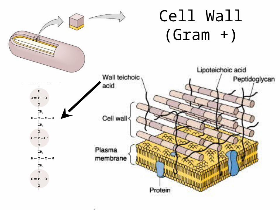
Cell Wall (Gram +)

Cell Wall (Gram -)(w/ Outer Membrane)
Periplasmic space
Braun’s lipoprotein
(LPS = endotoxin)core

Glycocalyx: means sugar coating; often polysaccharide or polypeptide layer external to the cell wall.
• Capsules: organized, consolidated, well attached.
• Slime Layer: unorganized; loose; removed easily.
• Function in attachment; protection; virulence.
Structures External to the Cell Wall
S-layer: extremely well organized layer of protein subunits that forms a rigid mesh, or mail, next to cell wall. Functions in protection and in resisting hypoosmotic stress

Structures External to the Cell Wall
peritrichous
monotrichous
amphitrichous
lophotrichous
Flagella:• Mostly made of flagellin.
• Filament thick (20 nm) & long (10-20 µm).
• Filament possibly sheathed.
• Varied locations on cell:
Fimbriae:• 1000’s of thin (~5 nm) & short appendages of helical proteins.
• Attachment to (specific) surfaces.
Sex Pili:• 1-10 slightly larger than fimbriae.
• Only in cells with a fertility plasmid (F factor), called donors.
• Attaches to like cells without F factor, called recipients.
• Facilitates genetic transfer between cells; with recipient gaining the F factor and possibly other genes.

Flagellar Motility & Chemotaxis
Other kinds of motility:
Spirochete; axial flagella
Gliding motility; no flagella

Random (searching)
Positive Chemotaxis
(“follow that smell”)
Low [nutrient solute] High [nutrient solute]