Structural insights into the interaction of human IgG1 ... · the same buffer, Fc was eluted in a...
8
research papers 2354 http://dx.doi.org/10.1107/S1399004715018015 Acta Cryst. (2015). D71, 2354–2361 Received 20 May 2015 Accepted 25 September 2015 Edited by S. Wakatsuki, Stanford University, USA Keywords: CD64; FcRI; IgG; protein complex; Fc receptor. PDB reference: IgG1 Fc–FcRI ECD complex, 4zne Supporting information: this article has supporting information at journals.iucr.org/d Structural insights into the interaction of human IgG1 with FccRI: no direct role of glycans in binding Vaheh Oganesyan, a * Yariv Mazor, a Chunning Yang, a Kimberly E. Cook, a Robert M. Woods, a Andrew Ferguson, b Michael A. Bowen, a Tom Martin, a Jie Zhu, c Herren Wu a and William F. Dall’Acqua a * a Department of Antibody Discovery and Protein Engineering, MedImmune LLC, 1 MedImmune Way, Gaithersburg, MD 20878, USA, b Discovery Sciences, Structure and Biophysics, AstraZeneca Pharmaceuticals, 35 Gatehouse Drive, Mailstop E3, Waltham, MA 02451, USA, and c Biopharmaceutical Development, MedImmune LLC, 1 MedImmune Way, Gaithersburg, MD 20878, USA. *Correspondence e-mail: [email protected], [email protected] The three-dimensional structure of a human IgG1 Fc fragment bound to wild- type human Fc RI is reported. The structure of the corresponding complex was solved at a resolution of 2.4 A ˚ using molecular replacement; this is the highest resolution achieved for an unmutated Fc RI molecule. This study highlights the critical structural and functional role played by the second extracellular subdomain of Fc RI. It also explains the long-known major energetic contribution of the Fc ‘LLGG’ motif at positions 234–237, and particularly of Leu235, via a ‘lock-and-key’ mechanism. Finally, a previously held belief is corrected and a differing view is offered on the recently proposed direct role of Fc carbohydrates in the corresponding interaction. Structural evidence is provided that such glycan-related effects are strictly indirect. 1. Introduction The family of IgG Fc gamma receptors (Fc Rs) play a crucial role in controlling the immune response in mammals (Nimmerjahn & Ravetch, 2006, 2008; Guilliams et al. , 2014). In humans, this family comprises a complex array of various members (Fc RI, Fc RII, Fc RIIIA and Fc RIIIB) and their allelic variants. These differ in their function (activating versus inhibitory), structural features and affinities for different IgG isotypes. Fc Rs contain two to three Ig-like C-type domains, a single transmembrane-spanning region (with the exception of Fc RIIIB) and a cytoplasmic tail of varying length. Fc RI, also known as CD64, is the only high-affinity (nanomolar range) receptor and the only one whose extracellular domain (ECD) comprises three individual subdomains (D1, D2 and D3). Fc RI binds IgG1 best, approximately tenfold better than IgG3 and IgG4, and does not bind significantly to IgG2. Mutational studies have previously attributed the high binding affinity of IgG for Fc RI to the second and third subdomains of the receptor (Harrison & Allen, 1998; Hulett & Hogarth, 1998). Recently published X-ray crystal structures of human Fc RI bound to IgG1 Fc (Lu et al., 2015; Kiyoshi et al., 2015) suggested that D3 does not directly participate in the corre- sponding interaction. While Lu et al. (2015) reported that Fc RI recognizes Fc glycans and attributed the high affinity between the two partners to this structural feature, Kiyoshi et al. (2015) found that such glycans make only little contribution to the interaction. We sought to better understand the molecular basis of IgG recognition by Fc RI. For this purpose, we solved the X-ray ISSN 1399-0047
Transcript of Structural insights into the interaction of human IgG1 ... · the same buffer, Fc was eluted in a...
research papers
2354 http://dx.doi.org/10.1107/S1399004715018015 Acta Cryst. (2015). D71, 2354–2361
Received 20 May 2015
Accepted 25 September 2015
Edited by S. Wakatsuki, Stanford
University, USA
Keywords: CD64; Fc�RI; IgG; protein complex;
Fc receptor.
PDB reference: IgG1 Fc–Fc�RI ECD complex,
4zne
Supporting information: this article has
supporting information at journals.iucr.org/d
Structural insights into the interaction of humanIgG1 with FccRI: no direct role of glycans in binding
Vaheh Oganesyan,a* Yariv Mazor,a Chunning Yang,a Kimberly E. Cook,a Robert M.
Woods,a Andrew Ferguson,b Michael A. Bowen,a Tom Martin,a Jie Zhu,c Herren
Wua and William F. Dall’Acquaa*
aDepartment of Antibody Discovery and Protein Engineering, MedImmune LLC, 1 MedImmune Way, Gaithersburg,
MD 20878, USA, bDiscovery Sciences, Structure and Biophysics, AstraZeneca Pharmaceuticals, 35 Gatehouse Drive,
Mailstop E3, Waltham, MA 02451, USA, and cBiopharmaceutical Development, MedImmune LLC, 1 MedImmune Way,
Gaithersburg, MD 20878, USA. *Correspondence e-mail: [email protected],
The three-dimensional structure of a human IgG1 Fc fragment bound to wild-
type human Fc�RI is reported. The structure of the corresponding complex was
solved at a resolution of 2.4 A using molecular replacement; this is the highest
resolution achieved for an unmutated Fc�RI molecule. This study highlights
the critical structural and functional role played by the second extracellular
subdomain of Fc�RI. It also explains the long-known major energetic
contribution of the Fc ‘LLGG’ motif at positions 234–237, and particularly of
Leu235, via a ‘lock-and-key’ mechanism. Finally, a previously held belief is
corrected and a differing view is offered on the recently proposed direct role
of Fc carbohydrates in the corresponding interaction. Structural evidence is
provided that such glycan-related effects are strictly indirect.
1. Introduction
The family of IgG Fc gamma receptors (Fc�Rs) play a crucial
role in controlling the immune response in mammals
(Nimmerjahn & Ravetch, 2006, 2008; Guilliams et al., 2014).
In humans, this family comprises a complex array of various
members (Fc�RI, Fc�RII, Fc�RIIIA and Fc�RIIIB) and their
allelic variants. These differ in their function (activating versus
inhibitory), structural features and affinities for different IgG
isotypes. Fc�Rs contain two to three Ig-like C-type domains,
a single transmembrane-spanning region (with the exception
of Fc�RIIIB) and a cytoplasmic tail of varying length. Fc�RI,
also known as CD64, is the only high-affinity (nanomolar
range) receptor and the only one whose extracellular domain
(ECD) comprises three individual subdomains (D1, D2 and
D3). Fc�RI binds IgG1 best, approximately tenfold better
than IgG3 and IgG4, and does not bind significantly to IgG2.
Mutational studies have previously attributed the high binding
affinity of IgG for Fc�RI to the second and third subdomains
of the receptor (Harrison & Allen, 1998; Hulett & Hogarth,
1998). Recently published X-ray crystal structures of human
Fc�RI bound to IgG1 Fc (Lu et al., 2015; Kiyoshi et al., 2015)
suggested that D3 does not directly participate in the corre-
sponding interaction. While Lu et al. (2015) reported that
Fc�RI recognizes Fc glycans and attributed the high affinity
between the two partners to this structural feature, Kiyoshi et
al. (2015) found that such glycans make only little contribution
to the interaction.
We sought to better understand the molecular basis of IgG
recognition by Fc�RI. For this purpose, we solved the X-ray
ISSN 1399-0047

crystal structure of the complex between the Fc portion of a
human IgG1 and unmutated Fc�RI at 2.4 A resolution. Our
data allowed a detailed description of the corresponding
interface. In particular, we confirm structurally and function-
ally the critical role played by Fc�RI D2. We also explain at a
structural level the major energetic contribution of Fc residues
spanning positions 234–237 (LLGG). Our study also confirms
that the use by Kiyoshi et al. (2015) of an Fc�RI molecule
mutated at 19 positions did not affect the overall structure and
agrees with their findings that glycans do not directly contri-
bute to the interaction.
2. Methods
2.1. Host cell-line generation
An MGAT1 knockout (KO) cell line was generated from
Chinese hamster ovary (CHO) K1 cells by knocking out the
MGAT1 gene which encodes mannosyl (�-1,3-)-glycoprotein
�-1,2-N-acetylglucosaminyltransferase. A zinc-finger nuclease
pair targeting the coding region of MGAT1 was designed to
recognize the gene at the following region: GCCTGCGACC-
CCCTCACCagccgtGATCCCCATCCTGGTC. ZFN plasmids
were transfected into host cells by nucleofection using stan-
dard protocols. MGAT1 KO cells were enriched by treatment
with phytohemagglutinin (PHA) for two passages. PHA-
resistant cells were stained with fluorescent Galanthus nivalis
lectin (GNA)-FITC to detect high-mannose glycosylation
of cell-surface proteins. Strongly staining cells were then
subcloned by FACS into 96-well plates. Genomic DNA was
isolated from individual wells, amplified using primers flanking
the ZFN cut site, denatured, re-annealed and subjected to a
CEL1 nuclease assay. CEL1 selectively cleaves re-annealed
products that have a mismatch between the two strands.
Digested products were run on an agarose gel and amplifica-
tion products generating a mismatch were further analyzed by
DNA sequencing. Clone CATSMGATKO-D4 was identified
and contains a frameshift mutation near the ZFN cutting site
on both alleles. Recombinant proteins expressed in this cell
line exhibit a homogenous Man5 glycosylation profile (Shi et
al., 2004).
2.2. FccRI production and purification
DNA encoding residues 1–277 of human Fc�RI ECD was
synthesized (Life Technologies, Grand Island, New York,
USA) and cloned into a mammalian expression vector under
the control of a Human cytomegalovirus (CMV) promoter.
Briefly, CATSMGATKO-D4 cells were transfected by
nucleofection using standard protocols and pools were
selected with methionine sulfoximide (MSX; Sigma–Aldrich,
St Louis, Missouri, USA). Cell pools were then assessed by
flow cytometry for intracellular staining with antihuman
Fc�RI APC (Life Technologies). The best-expressing pool
was expanded and used for the production of secreted Fc�RI.
Cells were grown for 13 d, after which the Fc�RI-containing
medium was collected and passed over a human IgG
Sepharose column (GE Healthcare, Piscataway, New Jersey,
USA) previously equilibrated with phosphate-buffered saline
(PBS) pH 7.2. Following washes to baseline with the same
buffer, Fc�RI was eluted using Pierce Elution Buffer (Thermo
Fisher Scientific, Waltham, Massachusetts, USA). Fractions
containing Fc�RI were pooled and loaded onto a 5 ml HiTrap
SP HP column (GE Healthcare) previously equilibrated with
50 mM sodium acetate pH 5.2. Following washes to baseline
with the same buffer, Fc�RI was eluted in a 0–0.5 M NaCl
gradient. Fc�RI was then dialyzed against 25 mM Tris–HCl
pH 7.5, 100 mM NaCl overnight at 4�C and concentrated to
�4 mg ml�1 using a Vivaspin ultrafiltration device (10 kDa
cutoff, Sartorius AG, Bohemia, New York, USA).
2.3. Fc production and purification
DNA encoding a human IgG1 Fc fragment spanning resi-
dues 221–446 (EU numbering convention; Kabat et al., 1991)
was cloned into a mammalian expression vector under the
control of a Human cytomegalovirus (CMV) promoter
(Oganesyan et al., 2008) and transiently transfected into
human embryonic kidney (HEK) 293 cells using Lipofecta-
mine (Life Technologies) and standard protocols. Purification
was carried out using a HiTrap Protein A column according
to the manufacturer’s instructions (GE Healthcare). After
overnight dialysis in 25 mM Tris–HCl pH 7.5 at 4�C, the
protein solution was further applied onto a HiTrap Q HP 5 ml
column (GE Healthcare). Following washes to baseline using
the same buffer, Fc was eluted in a 0–0.5 M NaCl gradient.
The protein was then concentrated to �10 mg ml�1 using a
Vivaspin ultrafiltration device (10 kDa cutoff, Sartorius AG).
The corresponding SDS–PAGE profile only revealed the
presence of one band around 25 or 50 kDa under reducing or
nonreducing conditions, respectively (data not shown).
2.4. Complex formation and crystallization
Previously purified Fc�RI and Fc were mixed in a 1:1 molar
ratio. Further purification of the complex was carried out using
a Superdex S200 10/300 GL column (GE Healthcare). The
purified complex was then concentrated to �5.5 mg ml�1
using a Vivaspin concentrator (30 kDa cutoff, Sartorius AG)
and subjected to crystallization trials. Sitting-drop crystal-
lization experiments were initially set up in 96-well Intelli-
Plates (Art Robbins Instruments, Sunnyvale, California,
USA) using a Phoenix crystallization robot (Art Robbins
Instruments) and commercially available screens from
Hampton Research and Molecular Dimensions. Crystal-
lization optimization was carried out in hanging-drop format
using 24-well Linbro plates using varying drop volumes and
ratios of protein to reservoir solution in the drop, typically
ranging from 1–6 ml and 1:1–5:1(v:v), respectively. Diffraction-
quality crystals were grown from a reservoir solution
consisting of 50 mM zinc acetate dehydrate, 20% PEG 3350.
For cryoprotection, crystals were transferred into the same
solution supplemented with 25% glycerol and flash-cooled in
liquid nitrogen.
research papers
Acta Cryst. (2015). D71, 2354–2361 Oganesyan et al. � Interaction of human IgG1 with Fc�RI 2355

2.5. X-ray data collection and structure determination
A diffraction data set was collected from a single crystal
on the IMCA-CAT 17-ID beamline of the Advanced Photon
Source (APS) at Argonne National Laboratory (University of
Chicago, Chicago, Illinois, USA) equipped with a PILATUS
6M detector (Dectris). 360 diffraction images were recorded at
APS using an oscillation range of 0.5�, a crystal-to-detector
distance of 401 mm, an exposure time of 1 s and a wavelength
of 1.0000 A. Diffraction data were processed using the XDS
package (Kabsch, 2010).
2.6. Generation of FccRI variants
Fc�RI:Fc�RIIIA chimeras were designed using the
following domain boundaries: Fc�RIIIA (D1), 19–106;
Fc�RIIIA (D2), 107–208; Fc�RI (D1), 21–102; Fc�RI (D2),
103–187; Fc�RI (D3), 188–282. Fc�RI and its variants were
cloned into an Orip/EBNA-1-based episomal mammalian
expression plasmid, pOE (Dimasi et al., 2009). Proteins were
produced by transient transfection of CHO cells in serum-free
medium using standard protocols. Cell-culture supernatants
were harvested 10 d after transfection and passed through a
0.22 mm sterile filter (PALL Life Sciences, Port Washington,
New York, USA). Variants were purified by affinity chroma-
tography using IgG Sepharose 6
Fast Flow (GE Healthcare) and
buffer-exchanged into PBS pH
7.2. Fc�RIIIA was produced as
described by Dall’Acqua et al.
(2006). The concentration of the
purified proteins was determined
from their absorbance at 280 nm.
2.7. Binding of FccRI variants toIgG1
ELISA plates were coated with
Fc�RI, Fc�RIIIA, individual
Fc�RI variants or control gp130
at 2 mg ml�1 in PBS pH 7.2 at 4�C
for 20 h and then blocked with
3%(v/v) nonfat milk containing
0.1%(v/v) Tween 20 in PBS pH
7.2 for 1 h at room temperature
(RT). A human IgG1 (R347) at
concentrations of 10, 5 or
2.5 mg ml�1 in PBS pH 7.2 was
then added to the wells and
incubated for 1 h at RT. HRP-
conjugated donkey F(ab0)2 frag-
ment antihuman IgG (H+L)
(Jackson ImmunoResearch, West
Grove, Pennsylvania, USA) was
used as a secondary antibody for
45 min at RT and the plates were
developed using tetramethyl-
benzidine (TMB; Dako, Carpin-
teria, California, USA). The
signal was quenched with 1 M H2SO4 and read at 450 nm using
an EnVision plate reader (PerkinElmer, Waltham, Massa-
chusetts, USA).
3. Results and discussion
3.1. Structure of the FccRI–Fc complex
We carried out a crystallographic study of the complex
formed between Fc�RI ECD and the Fc portion of a human
IgG1 in an effort to account for the high affinity of the
corresponding interaction. Both proteins were expressed in
mammalian cells, purified, complexed and crystallized. The
crystals had C2 symmetry, with unit-cell parameters a = 134.7,
b = 126.8, c = 71.8 A, � = 118.4�, and diffracted to 2.4 A
resolution. The structure was determined by molecular
replacement using MOLREP (Vagin & Teplyakov, 2010). The
search model for Fc�RI consisted of PDB entry 3rjd (Lu et al.,
2011). The search model for Fc consisted of another Fc
portion exhibiting the same amino-acid sequence as in this
study, expressed and purified in the same conditions as
described above (see x2) and the structure of which had been
determined at high resolution (1.5 A; unpublished data). The
carbohydrate moiety was not included in the search model.
research papers
2356 Oganesyan et al. � Interaction of human IgG1 with Fc�RI Acta Cryst. (2015). D71, 2354–2361
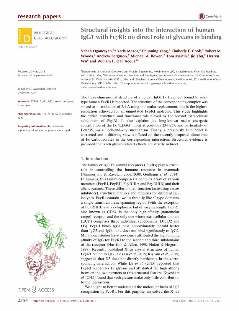
Figure 1General view of the complex formed between human IgG1 Fc and the ECD of human Fc�RI. (a) The Fc–Fc�RI (salmon/green) complex is shown as ribbons and carbohydrates as sticks. Fc�RI D2 appears to bethe lone structural contributor to the interface with Fc. (b) Surface representation of the Fc–Fc�RIcomplex, in which Fc�RI (green) has been moved 20 A away from Fc (orange) to show the shapecomplementarity. This and other figures were prepared using PyMOL (Schrodinger).

One Fc (two identical polypeptides) and one Fc�RI molecule
were found in the asymmetric part of the unit cell (Fig. 1a).
Iterative refinement/rebuilding of
the model was performed with
REFMAC5 (Murshudov et al.,
2011) and O (Jones et al., 1991).
The overall folds of both
members of the complex were
similar to those of the corre-
sponding templates. Electron
density accounted for residues
232–446 of one of the Fc poly-
peptides (chain E) and for amino
acids 236–444 of the other (chain
J). Therefore, 11 amino acids at
the N-terminus of chain E and 15
amino acids at the N-terminus of
chain J, as well as amino acids 445
and 446 at the C-terminus of
chain J, were not included in the
model. Eight N-terminal and ten
C-terminal Fc�RI residues, as
well as the amino acids corre-
sponding to positions 44–54, 87–
90 and 219–222, had no traceable
electron density. Both proteins
had a number of N-linked
carbohydrate chains (Supple-
mentary Fig. S1). Those attached
to Fc Asn297 were well visible in
the electron density up to the last
N-acetylglucosamine (GlcNAc). The electron density around
Fc�RI Asn59, Asn78, Asn152, Asn159 and Asn163 accounted
for GlcNAc2/Man1, GlcNAc1, GlcNAc1, GlcNAc1/Man1 and
GlcNAc2, respectively. Some of those carbohydrates, namely
GlcNAc near Asn78 and Man near Asn159, were placed in
electron density of much lower quality than others, probably
owing to the conformational heterogeneity of sugar moieties
(Supplementary Fig. S1). The final refined model contains
5311 protein atoms, 321 carbohydrate atoms, three zinc ions
(present in the crystallization solution) and 156 solvent
molecules. Data and refinement statistics are shown in Table 1.
The interface between Fc and Fc�RI covers �1160 A2 and
involves both chains of Fc. No carbohydrate–carbohydrate or
protein–carbohydrate interactions between Fc and Fc�RI
were observed. The shape complementarity between Fc and
Fc�RI is remarkably high (Fig. 1b). It was estimated at 0.82
using Sc (0.78 for Fc chain J and 0.84 for Fc chain E; Lawrence
& Colman, 1993), which is higher than the complementarity
between the heavy and light chains of antibodies (namely
0.69). The charge complementarity is also remarkable (Figs. 2a
and 2b). Positively charged Fc�RI patches were found facing
negatively charged Fc pockets. Fc�RI D2 was found to be
solely responsible for the interaction with Fc, since no contacts
within a distance cutoff up to 5 A involving D1 and D3 were
seen.
The interfaces between Fc�RI and the two Fc chains (E and
J) are not equally important. In particular, the interface
between Fc�RI and Fc chain E constitutes a major interaction
research papers
Acta Cryst. (2015). D71, 2354–2361 Oganesyan et al. � Interaction of human IgG1 with Fc�RI 2357
Table 1X-ray data and model-refinement statistics.
Values in parentheses are for the highest resolution shell.
Data statisticsWavelength (A) 1.0000Resolution (A) 86.6–2.4 (2.43–2.42)Space group C2Unit-cell parameters (A, �) a = 134.7, b = 126.8,
c = 71.8, � = 118.4Total reflections 134782 (1507)Unique reflections 39992 (426)Completeness (%) 98.7 (100.0)Rmerge 0.062 (0.476)Mean I/�(I) 14.4 (2.4)Multiplicity 3.4 (3.5)CC1/2 0.995 (0.856)
Refinement statisticsResolution (A) 86.6–2.4Rwork 0.201Rfree 0.254Rwork+free 0.203R.m.s.d., bonds (A) 0.011R.m.s.d., angles (�) 1.566Ramachandran plot†
Residues in most favored region (%) 93.6Residues in additionally allowed region (%) 6.4Residues in generously allowed region (%) 0.0
No. of protein atoms 5311No. of nonprotein atoms 480Mean B factor (model/Wilson) (A2) 34.2/43.5
† The Ramachandran plot was produced using PROCHECK (Laskowski et al., 1993).
Figure 2Charge complementarity between Fc and Fc�RI. The positive and negative electrostatic potentials areindicated in blue and red, respectively, and were calculated using the APBS (Adaptive Poisson–BoltzmannSolver; Baker et al., 2001) plugin in PyMOL. Positively charged surfaces on Fc�RI (a) line up againstnegatively charged patches of Fc (b).

area. The complex-formation significance score from PISA
(Krissinel & Henrick, 2007) is a remarkable 0.963, and is only
slightly lower than that between two Fc polypeptides (1.000).
Six hydrogen bonds are formed between Fc�RI D2 and the
Fc region spanning residues 233-ELLGGPS-239 (Fig. 3a,
Supplementary Table S1). In addition, a very prominent ‘lock-
and-key’ feature exists. The ‘key’, corresponding to Leu235
(ELLG), is positioned inside an Fc�RI pocket exhibiting a
positively charged ‘rim’ (Fig. 3b). The pocket is made of the
hydrophobic amino acids Leu105, Trp106, Ala126, Trp127 and
Val132 in Fc�RI. In addition, these hydrophobic interactions
are strengthened by hydrogen bonds formed between the
main-chain N and O atoms of Lys173 and Leu131, respec-
tively, in Fc�RI with the main-chain O and N atoms of Fc L235
(Fig. 3a). The other Fc chain (J) established only three
hydrogen bonds with Fc�RI D2 (Fig. 3c, Supplementary Table
S1).
3.2. Comparison of FccRI–Fc X-ray structures
While our manuscript was in preparation, two X-ray crystal
structures of human Fc�RI complexed with IgG1 Fc at reso-
lutions of 3.5 and 1.8 A were published (Lu et al., 2015; Kiyoshi
et al., 2015). All three structures are generally similar in terms
of domain organization and the relative positions of all
polypeptides. Therefore, the Fc�RI mutations described by
Kiyoshi et al. (2015) do not affect its structure or mode of
interaction with Fc. These structures superimpose with r.m.s
deviations of 2.2 A (Lu et al., 2015) and 0.34 A (Kiyoshi et al.,
2015) over C� atoms. We also note that the crystal form
described here is almost identical to that presented by Kiyoshi
et al. (2015) as judged by space group and unit-cell parameters.
Lu et al. (2015) proposed that the interaction between Fc�RI
Arg175 and the Fc carbohydrates can account for the corre-
sponding high affinity. Indeed, the importance and composi-
tion of the carbohydrates at Fc Asn297 in Fc�R binding is well
established. It is believed that glycans allow the Fc CH2
domains to maintain a favorable distance and conformation
for receptor binding (Radaev et al., 2001; Jefferis & Lund,
2002; Arnold et al., 2007; Feige et al., 2009). Incremental
shortening of IgG1 carbohydrates also results in incremental
weakening of the corresponding affinity for Fc�R (Mimura et
al., 2000). Structural investigations of such Fcs with truncated
carbohydrate chains revealed shortening of the overall
research papers
2358 Oganesyan et al. � Interaction of human IgG1 with Fc�RI Acta Cryst. (2015). D71, 2354–2361
Figure 3(a) Representation of the intermolecular contacts between Fc (E chain, salmon; significance of 0.963) and Fc�RI D2 (green). (b) Fc�RI creates a pocket(‘lock’) to fit Leu235 in the Fc E chain (‘key’; salmon). (c) Representation of the intermolecular contacts between Fc (J chain, salmon; significance of0.348) and Fc�RI D2 (green). Fc residues were numbered according to the EU numbering convention (Kabat et al., 1991). Dotted lines representhydrogen bonds (distances are given in A). None of the interfaces involved carbohydrates.
Figure 4Superimposition of free (PDB entry 3rjd; magenta) and bound (thisstudy; green) Fc�RI using D2 C� atoms. No major conformational changecan be seen upon Fc binding. However, the position of Fc�RI D3, whichis not involved in any interactions with Fc, Fc�RI D1 or Fc�RI D2,appears to depend on both crystal packing and/or the flexibility of thepeptide connecting domains 2 and 3. All superimpositions wereperformed using LSQKAB (Kabsch, 1976).

CH2–CH2 distance (Krapp et al., 2003). However, owing to the
limited resolution of the structure published by Lu et al.
(2015), we find that the glycans were not accurately modeled
and exhibited forbidden sugar conformations. Our present
structure does not show any glycan-related interaction, which
strengthens the previous findings that sugars are not strictly
required for complex formation (Sazinsky et al., 2008) or
engaged in significant intermolecular interactions (Kiyoshi et
al., 2015). In particular, the closest distance between the
partner molecules in the complex involved the Man1 residue
attached to Fc�RI Asn58 and Fc chain E Ala330, and was
estimated at approximately 12 A.
Furthermore, our structure suggests that Fc Leu235 plays a
major role (Fig. 3b), in agreement with Kiyoshi et al. (2015)
and Lu et al. (2015). Our results are also in very good agree-
ment with those of Chappel et al. (1991), who demonstrated
the major involvement of this region by introducing the 234-
LLGG-237 motif into a human IgG2 and restoring high-
affinity binding to Fc�RI.
3.3. Structural comparison of FccRI in free and bound states
The X-ray structure of the entire ECD of mammalian cell-
derived Fc�RI in an unliganded state was previously deter-
mined at 2.65 A resolution by Lu et al. (2011). The three
Fc�RI subdomains of our structure and this unliganded Fc�RI
exhibit similar, but not identical, conformations and inter-
domain interactions. When full-length ECDs were used, the
r.m.s deviation over C� atoms was 1.8 A. When super-
imposition was carried out at the domain level, the deviations
were much smaller and were estimated at 0.38, 0.37 and 0.56 A
for D1, D2 and D3, respectively. We then carried out structural
superimposition of both unliganded and bound Fc�RI
through Fc�RI D2, the major contributor of the interaction
research papers
Acta Cryst. (2015). D71, 2354–2361 Oganesyan et al. � Interaction of human IgG1 with Fc�RI 2359
Figure 5(a) Schematic representation of the domain arrangement of humanFc�RI, Fc�RIIIA and variants thereof. Green and blue ovals correspondto Fc�RI and Fc�RIIIA domains, respectively. The low-affinityFc�RIIIA/F158 allotype was used to obtain a larger affinity range whencharacterizing variants. (b) Binding of human IgG1 at varyingconcentrations to Fc�R variants as measured by ELISA. Standarddeviations are indicated by error bars and represent triplicate measure-ments within the same experiment.
Figure 6Superimposition of the complex between Fc�RI (green) and Fc (orange)with Fc�RIIIA–Fc (blue; PDB entry 3sgj) and Fc�RIIA–Fc (magenta;PDB entry 3ry6) complexes. The Fc part of the Fc�RIIIA–Fc andFc�RIIA–Fc complexes is not shown. All three Fc�Rs bind in the crevicebetween Fc CH2 domains and superimpose quite well with each other.However, the details of these interactions are quite different as theFc�RIIIA–Fc interface involves carbohydrates from both the receptorand the IgG1. There is no evidence for direct protein–carbohydrateinteraction in the Fc�RIIA–Fc complex (perhaps owing to the lowresolution of the structure).

with Fc, in an effort to detect any change in relative domain
orientation upon Fc binding (Fig. 4). In particular, the acute
angle between D1 and D2, previously described in detail by
Lu et al. (2011), remained unchanged. We also find that Fc�RI
D3 is the most misaligned. Since it is not engaged in any
contact with either D1 or D2 or with Fc, its position is most
likely to be determined by crystal contacts and the flexibility
of the connecting peptide between Fc�RI D2 and D3. Minor
differences in interdomain interactions are shown in Supple-
mentary Table S2.
3.4. Relative functional importance of FccRI domains
Our structural analysis suggested that Fc�RI D2 is the sole
contributor to Fc binding. To investigate whether Fc�RI
domains I and III play any functional role, we generated
several variants in which various domains of Fc�RIIIA (F158)
and Fc�RI were swapped (Fig. 5a). Binding of a human IgG1
to these variants was then analyzed by ELISA (Fig. 5b). In the
absence of D3 (V4), binding of Fc�RI to IgG1 is only slightly
weaker when compared with the entire Fc�RI ECD,
suggesting some minor, probably indirect, role for D3. This is
supported by the slight increase in IgG1 binding of Fc�RIIIA
containing Fc�RI D3 (V3) when compared with Fc�RIIIA.
Variants missing (V1) or including (V4) Fc�RI D1 exhibit
nearly identical binding to IgG1. This suggests that D1 is
interchangeable between Fc�RI and Fc�RIIIA and does not
specifically contribute to IgG1 binding. Importantly, our
results highlight the importance of Fc�RI D2, as all constructs
that lack this domain (V2 and V3) exhibit severely impaired
binding to IgG1. Likewise, the V1 construct, in which D2
constitutes the only Fc�RI component, binds to IgG1 nearly
as well as the entire Fc�RI ECD. Taken together, our data
show that Fc�RI D2 is the most important domain to confer
high-affinity binding to Fc. This is in good agreement with
previous data using murine molecules (Hulett & Hogarth,
1998).
4. Conclusion
In an effort to identify the critical structural features respon-
sible for the high-affinity interaction of IgG1 Fc with Fc�RI,
we solved the structure of the corresponding complex. Fc�RI,
Fc�RII and Fc�RIIIA bind to nearly the same place on Fc
(Fig. 6), although the details of these interactions are very
different. Unlike the interface of the Fc�RIIIA–Fc complex,
that between Fc�RI and Fc does not contain any carbo-
hydrate. It is, however, well known that aglycosylated IgG
research papers
2360 Oganesyan et al. � Interaction of human IgG1 with Fc�RI Acta Cryst. (2015). D71, 2354–2361
Figure 7Three-dimensional views of the Fc�RI–Fc (green/orange) complex in relation to the presence of Fab arms. (a) shows a view in which the Fab arms wouldbe positioned towards the viewer, whereas (b) shows a view in which the Fab arms would point towards the right side. The N-terminus of both Fcpolypeptides points away from Fc�RI, which allows the presence of the corresponding Fab arms.

molecules exhibit very weak to no binding to Fc�RI. There-
fore, Fc glycans play an important indirect role in this inter-
action, likely by maintaining a favorable Fc conformation or
CH2 distance for engaging Fc�RI. As a result of the high
resolution of our structure, we confirm here that such glycan-
related effects are indirect only. The higher resolution also
allowed us to provide structural evidence for the important
functional role of Fc amino acid Leu235, which is in very good
agreement with the studies of Chappel et al. (1991) and
Kiyoshi et al. (2015). We have also elucidated the individual
role of Fc�RI subdomains and find a good agreement between
the structural and functional data. In particular, Fc�RI D2
constitutes an integral structural and energetic component of
the interaction with IgG1 Fc. Finally, our described mode of
interaction between Fc�RI and Fc is compatible in the context
of an interaction with a full-length IgG1 with its Fab arms.
Indeed, both N-terminal ends of Fc polypeptides are posi-
tioned so as to allow the formation of interchain disulfide
bonds and point away from Fc�RI (Figs. 7a and 7b). There-
fore, both Fab arms are expected to have little to no confor-
mational restriction owing to the flexibility of the entire hinge.
References
Arnold, J. N., Wormald, M. R., Sim, R. B., Rudd, P. M. & Dwek, R. A.(2007). Annu. Rev. Immunol. 25, 21–50.
Baker, N. A., Sept, D., Joseph, S., Holst, M. J. & McCammon, J. A.(2001). Proc. Natl Acad. Sci. USA, 98, 10037–10041.
Chappel, M. S., Isenman, D. E., Everett, M., Xu, Y.-Y., Dorrington,K. J. & Klein, M. H. (1991). Proc. Natl Acad. Sci. USA, 88, 9036–9040.
Dall’Acqua, W. F., Cook, K. E., Damschroder, M. M., Woods, R. M. &Wu, H. (2006). J. Immunol. 177, 1129–1138.
Dimasi, N., Gao, C., Fleming, R., Woods, R. M., Yao, X.-T., Shirinian,L., Kiener, P. A. & Wu, H. (2009). J. Mol. Biol. 393, 672–692.
Feige, M. J., Nath, S., Catharino, S. R., Weinfurtner, D., Steinbacher, S.& Buchner, J. (2009). J. Mol. Biol. 391, 599–608.
Guilliams, M., Bruhns, P., Saeys, Y., Hammad, H. & Lambrecht, B. N.(2014). Nature Rev. Immunol. 14, 349.
Harrison, P. T. & Allen, J. M. (1998). Protein Eng. Des. Sel. 11,225–232.
Hulett, M. D. & Hogarth, P. M. (1998). Mol. Immunol. 35, 989–996.Jefferis, R. & Lund, J. (2002). Immunol. Lett. 82, 57–65.Jones, T. A., Zou, J.-Y., Cowan, S. W. & Kjeldgaard, M. (1991). Acta
Cryst. A47, 110–119.Kabat, E. A., Wu, T. T., Perry, H. M., Gottesman, K. S. & Foeller, C.
(1991). Sequences of Proteins of Immunological Interest.Washington DC: National Institutes of Health.
Kabsch, W. (1976). Acta Cryst. A32, 922–923.Kabsch, W. (2010). Acta Cryst. D66, 125–132.Kiyoshi, M., Caaveiro, J. M., Kawai, T., Tashiro, S., Ide, T., Asaoka, Y.,
Hatayama, K. & Tsumoto, K. (2015). Nature Commun. 6, 6866.Krapp, S., Mimura, Y., Jefferis, R., Huber, R. & Sondermann, P.
(2003). J. Mol. Biol. 325, 979–989.Krissinel, E. & Henrick, K. (2007). J. Mol. Biol. 372, 774–797.Laskowski, R. A., MacArthur, M. W., Moss, D. S. & Thornton, J. M.
(1993). J. Appl. Cryst. 26, 283–291.Lawrence, M. C. & Colman, P. M. (1993). J. Mol. Biol. 234, 946–
950.Lu, J., Chu, C., Zou, Z., Hamacher, N. B., Rixon, M. W. & Sun, P. D.
(2015). Proc. Natl Acad. Sci. USA, 112, 833–838.Lu, J., Ellsworth, J. L., Hamacher, N., Oak, S. W. & Sun, P. D. (2011). J.
Biol. Chem. 286, 40608–40613.Mimura, Y., Church, S., Ghirlando, R., Ashton, P. R., Dong, S.,
Goodall, M., Lund, J. & Jefferis, R. (2000). Mol. Immunol. 37,697–706.
Murshudov, G. N., Skubak, P., Lebedev, A. A., Pannu, N. S., Steiner,R. A., Nicholls, R. A., Winn, M. D., Long, F. & Vagin, A. A. (2011).Acta Cryst. D67, 355–367.
Nimmerjahn, F. & Ravetch, J. V. (2006). Immunity, 24, 19–28.Nimmerjahn, F. & Ravetch, J. V. (2008). Nature Rev. Immunol. 8,
34–47.Oganesyan, V., Damschroder, M. M., Leach, W., Wu, H. &
Dall’Acqua, W. F. (2008). Mol. Immunol. 45, 1872–1882.Radaev, S., Motyka, S., Fridman, W. H., Sautes-Fridman, C. & Sun,
P. D. (2001). J. Biol. Chem. 276, 16469–16477.Sazinsky, S. L., Ott, R. G., Silver, N. W., Tidor, B., Ravetch, J. V. &
Wittrup, K. D. (2008). Proc. Natl Acad. Sci. USA, 105, 20167–20172.
Shi, S., Williams, S. A., Seppo, A., Kurniawan, H., Chen, W., Ye, Z.,Marth, J. D. & Stanley, P. (2004). Mol. Cell. Biol. 24, 9920–9929.
Vagin, A. & Teplyakov, A. (2010). Acta Cryst. D66, 22–25.
research papers
Acta Cryst. (2015). D71, 2354–2361 Oganesyan et al. � Interaction of human IgG1 with Fc�RI 2361