Structural Elucidation of the Disulfated Oligosaccharide … · · 2001-07-09THE JOURNAL OF...
8
THE JOURNAL OF BIOLOGICAL CHEMISTRY 0 1985 by The American Society of Biological Chemistg, Inc. Vol. 260, No. 29, Issue of December 15, pp. 15623-15630,1985 Printed in U.S.A. Structural Elucidation ofthe Disulfated Oligosaccharide from Bovine Lutropin” (Received for publication, May 30,1985) Eric D. Green,a*b*c Herman van Halbeek? Irving Boime,b and Jacques U. Baenzigera*f From the Departments of “Pathology and ’Pharmacology, Washington University Medical School, St. Louis, Missouri 63110 and the dDepartment of Bw-Organic Chemistry, Unkersity of Utrecht, Croesestrant 79, NL-3522 AD Utrecht, The Netherlands The Asn-linked oligosaccharides of the pituitaryhormone lutropin (LH) contain both sulfate and GalNAc. Bovine pituitary explants incorporate [3H]glucosamine, r3H]mannose, [3H]fucose, and [35S]sulfate into the Asn-linked oligosaccharides of LH. Endoglycosidase F or N-glycanase releases the [3H]glucosamine- and [3H]mannose-labeled oligosaccharides from the protein, which resolve on anion-exchange high pressure liquid chromatography as neutrd (S-0), mono- (S-1), anddisulfated (5-2) species.Based on sequentialenzymedigestion,methylation,periodate oxidation, and nuclear magnetic resonance studies, the proposed structure for 5-2 is as follows: so44(3 or 4)GalNAc@1+4GlcNAc@l+2Mancu1 I 6 Man@1+4GlcNAc@1+4GlcNAc+Asn 3 t F f(Fuc) so44(3 or 4)GalNAc@l+4GlcNAc@1+2Man~l Sulfate is confined to position 3 or 4 of GalNAc based on periodate and methylation data and can be removed by methanolysis. The presence of @-linked GalNAc at a position typically occupied by Gal has not previously been observed. The Asn-linked oligosaccharides of bovine, human, and ovine lutropin (LH’) contain GalNAc and sulfate (1-6). Al- though several different structures for these unique carbohy- drate moieties have been proposed (1,4-8), definitive evidence has not been obtained largely due to difficulties encountered in removal of the sulfate and GalNAc. [35S]Sulfate can be incorporated into the oligosaccharides of LH in both meta- bolic (2, 3, 6) and cell-free systems (3). For bovine LH, both labeling systems yield qualitatively the same mono- and di- sulfated oligosaccharides (3). Bovine and human LH, bovine and human thyrotropin (TSH), and bovine follitropin (FSH) are also sulfated in the cell-freesystem (9). Although the types and proportions of oligosaccharides on these 35S-su1- fated hormones differ,they all contain a similar, if not iden- tical, disulfated oligosaccharide. structure. Metabolic labeling of LH with 3H-sugars reveals the pres- ence of neutral as well as mono- and disulfated oligosaccha- rides. Understanding the relationship of sulfation and GalNAc addition in vivo and in the cell-free system requires a detailed characterization of the sulfated and neutral oligo- saccharides. Here we present the structural elucidation of the metabolically labeled, disulfated oligosaccharide using exogly- cosidase digestions, periodate oxidations, and methylation analyses. Parallel studies using oligosaccharides from purified bovine LH also support our proposedstructure. * This investigation was supported in part by National Institutes of Child Health and Human Development Grant PO-HD-13481 (to I. B.), National Cancer Institute Grallt R01-CA21923 (to J. U. B.), Metabolic Labeling of LH Using Pituitary Explants-Slices of fresh and National Institutes of Child Health and Human Development bovine pituitaries were incubated in Krebs-Ringer bicarbonate buffer Grant WD20197 (to I. B. and J. U. B.). The costs of publication of as described (2,3). For labeling of oligosaccharides with 3H-sugars, 1 this article were defrayed in part by the payment of page charges. mM cold sodium sulfate was added and the incubation times were This article must therefore be hereby marked “advertisement” in extended to 21 h. [6-3H]Glucosamine(New England Nuclear), [2-3H] accordance with 18 U.S.C. Section 1734 solely to indicate this fact. mannose (New England Nuclear), [5,6-3H]fucose(American Radio- ‘ Participant in the Medical Scientist Training Program supported labeled Chemicals), and [l-3H]galactose (American Radiolabeled through Grant GM07200 from the National Institute of General Chemicals) were used at 20 pCi/ml. For [3H]mannose labeling, 10 Medical Sciences. mM glutamine and 1 mg/ml fructose were added to inhibit cellular e Present address: Complex Carbohydrate Research Center, Rich- metabolism of the isotope. Products were immunoprecipitated with ard B. Russell Agricultural Center, Athens, GA 30613. Recipient of Research Development Award K04-CA00671 from Pansorbin (Calbiochem) to immobilize the antibody-LH complexes. antiserum raised against purified 8-subunit from bovine LH, using the National Institutes of Health. This antiserum is specific for native bovine LH and precipitates little The abbreviations used are: LH, lutropin (luteinizing hormone); if any free a-subunit or bovine FSH (25). Labeled products were FSH, follitropin (follicle-stimulating hormone); TSH, thyrotropin liberated from the Pansorbin by heating to 100 “C in the presence of (thyroid-stimulating hormone); Endo F, endo-8-N-acetylglucosamin- 6 M guanidine hydrochloride, 1 mM EDTA, 1 M Tris (pH 8.0), and idase F; Endo H, endo-8-N-acetylglucosaminidase H, S-0, neutral desalted by gelfiltration onBio-Gel P-2 (Bio-Rad). The purity of the oligosaccharides; S-1, monosulfated oligosaccharides; S-2, disulfated immunoprecipitated, radiolabeled LH was found to be >90% by oligosaccharides; S-N, monosulfated/monosialylated oligosaccha- sodium dodecyl sulfate-polyacrylamide gel electrophoresis and tryptic rides; HPLC, high pressure liquid chromatography. peptide mapping. 15623 MATERIALS AND METHODS
-
Upload
nguyencong -
Category
Documents
-
view
216 -
download
2
Transcript of Structural Elucidation of the Disulfated Oligosaccharide … · · 2001-07-09THE JOURNAL OF...

THE JOURNAL OF BIOLOGICAL CHEMISTRY 0 1985 by The American Society of Biological Chemistg, Inc.
Vol. 260, No. 29, Issue of December 15, pp. 15623-15630,1985 Printed in U.S.A.
Structural Elucidation of the Disulfated Oligosaccharide from Bovine Lutropin”
(Received for publication, May 30,1985)
Eric D. Green,a*b*c Herman van Halbeek? Irving Boime,b and Jacques U. Baenzigera*f From the Departments of “Pathology and ’Pharmacology, Washington University Medical School, St. Louis, Missouri 63110 and the dDepartment of Bw-Organic Chemistry, Unkersity of Utrecht, Croesestrant 79, NL-3522 AD Utrecht, The Netherlands
The Asn-linked oligosaccharides of the pituitary hormone lutropin (LH) contain both sulfate and GalNAc. Bovine pituitary explants incorporate [3H]glucosamine, r3H]mannose, [3H]fucose, and [35S]sulfate into the Asn-linked oligosaccharides of LH. Endoglycosidase F or N-glycanase releases the [3H]glucosamine- and [3H]mannose-labeled oligosaccharides from the protein, which resolve on anion-exchange high pressure liquid chromatography as neutrd (S-0), mono- (S-1), and disulfated (5-2) species. Based on sequential enzyme digestion, methylation, periodate oxidation, and nuclear magnetic resonance studies, the proposed structure for 5-2 is as follows:
so44(3 or 4)GalNAc@1+4GlcNAc@l+2Mancu1 I 6 Man@1+4GlcNAc@1+4GlcNAc+Asn 3 t
F f(Fuc) so44(3 or 4)GalNAc@l+4GlcNAc@1+2Man~l
Sulfate is confined to position 3 or 4 of GalNAc based on periodate and methylation data and can be removed by methanolysis. The presence of @-linked GalNAc at a position typically occupied by Gal has not previously been observed.
The Asn-linked oligosaccharides of bovine, human, and ovine lutropin (LH’) contain GalNAc and sulfate (1-6). Al- though several different structures for these unique carbohy- drate moieties have been proposed (1,4-8), definitive evidence has not been obtained largely due to difficulties encountered in removal of the sulfate and GalNAc. [35S]Sulfate can be incorporated into the oligosaccharides of LH in both meta- bolic (2, 3, 6) and cell-free systems (3). For bovine LH, both labeling systems yield qualitatively the same mono- and di- sulfated oligosaccharides (3). Bovine and human LH, bovine and human thyrotropin (TSH), and bovine follitropin (FSH) are also sulfated in the cell-free system (9). Although the
types and proportions of oligosaccharides on these 35S-su1- fated hormones differ, they all contain a similar, if not iden- tical, disulfated oligosaccharide. structure.
Metabolic labeling of LH with 3H-sugars reveals the pres- ence of neutral as well as mono- and disulfated oligosaccha- rides. Understanding the relationship of sulfation and GalNAc addition in vivo and in the cell-free system requires a detailed characterization of the sulfated and neutral oligo- saccharides. Here we present the structural elucidation of the metabolically labeled, disulfated oligosaccharide using exogly- cosidase digestions, periodate oxidations, and methylation analyses. Parallel studies using oligosaccharides from purified bovine LH also support our proposed structure.
* This investigation was supported in part by National Institutes of Child Health and Human Development Grant PO-HD-13481 (to I. B.), National Cancer Institute Grallt R01-CA21923 (to J. U. B.), Metabolic Labeling of LH Using Pituitary Explants-Slices of fresh and National Institutes of Child Health and Human Development bovine pituitaries were incubated in Krebs-Ringer bicarbonate buffer Grant WD20197 ( t o I. B. and J. U. B.). The costs of publication of as described (2,3). For labeling of oligosaccharides with 3H-sugars, 1 this article were defrayed in part by the payment of page charges. mM cold sodium sulfate was added and the incubation times were This article must therefore be hereby marked “advertisement” in extended to 21 h. [6-3H]Glucosamine (New England Nuclear), [2-3H] accordance with 18 U.S.C. Section 1734 solely to indicate this fact. mannose (New England Nuclear), [5,6-3H]fucose (American Radio- ‘ Participant in the Medical Scientist Training Program supported labeled Chemicals), and [l-3H]galactose (American Radiolabeled
through Grant GM07200 from the National Institute of General Chemicals) were used at 20 pCi/ml. For [3H]mannose labeling, 10 Medical Sciences. mM glutamine and 1 mg/ml fructose were added to inhibit cellular
e Present address: Complex Carbohydrate Research Center, Rich- metabolism of the isotope. Products were immunoprecipitated with ard B. Russell Agricultural Center, Athens, GA 30613.
Recipient of Research Development Award K04-CA00671 from Pansorbin (Calbiochem) to immobilize the antibody-LH complexes. antiserum raised against purified 8-subunit from bovine LH, using
the National Institutes of Health. This antiserum is specific for native bovine LH and precipitates little The abbreviations used are: LH, lutropin (luteinizing hormone); if any free a-subunit or bovine FSH (25). Labeled products were
FSH, follitropin (follicle-stimulating hormone); TSH, thyrotropin liberated from the Pansorbin by heating to 100 “C in the presence of (thyroid-stimulating hormone); Endo F, endo-8-N-acetylglucosamin- 6 M guanidine hydrochloride, 1 mM EDTA, 1 M Tris (pH 8.0), and idase F; Endo H, endo-8-N-acetylglucosaminidase H, S-0, neutral desalted by gel filtration on Bio-Gel P-2 (Bio-Rad). The purity of the oligosaccharides; S-1, monosulfated oligosaccharides; S-2, disulfated immunoprecipitated, radiolabeled LH was found to be >90% by oligosaccharides; S-N, monosulfated/monosialylated oligosaccha- sodium dodecyl sulfate-polyacrylamide gel electrophoresis and tryptic rides; HPLC, high pressure liquid chromatography. peptide mapping.
15623
MATERIALS AND METHODS

15624 Sulfated Oligosac
Generation of Metabolically Labeled Oligosaccharide Standards- MOPC-315 cells were grown in L-15 (Leibovitz) media supplemented with 10% fetal calf serum (Kansas City Biological) to 60,000 cells/ ml. [3H]Glucosamine or [3H]mannose (50 pCi/ml) was added and incubation was continued for 40 h. Cells were removed by centrifuga- tion, and the media was concentrated, desalted by gel filtration over Bio-Gel P-2, and exhaustively digested with Pronase in 2 mM CaC12, 0.1 M Tris (pH 7.8). Fractionation using concanavalin A-Sepharose (Pharmacia) was used (10, 11) to purify dibranched complex oligo- saccharides. Cleavage with endo-p-N-acetylglucosaminidase F (Endo F), removal of sialic acid (2 N acetic acid, 100 "C, 15 min), and successive enzymatic hydrolysis yielded the structures shown in Fig. 2. Exoglycosidase digestions and methylation analysis were used to confirm the proposed structures.
Sulfate Removal by Methanolysis-Dried samples were resuspended in 0.5 N anhydrous methanol-HC1, incubated at room temperature for 5-10 h, and dried under vacuum. Residual HCl was removed by repeated evaporation from methanol. For the oligosaccharides stud- ied, no alterations in the underlying carbohydrate structure following methanolysis have been detected.
tein by overnight digestion with either Endo F as described (3) or N- Enzyme Digestions-Oligosaccharides were released from the pro-
glycanase (Genzyme Corporation) in 0.2% Nonidet P-40, 1 mM EDTA, and 75 mM sodium phosphate (pH 8.6). For the oligosaccha- rides of bovine LH, Endo F cleaves between the 2 GlcNAc residues of the core, liberating oligosaccharides with only one core GlcNAc. In contrast, N-glycanase hydrolyzes the Asn-GlcNAc linkage, yielding an oligosaccharide with 2 core GlcNAc residues. Exoglycosidase diges- tions were performed as described (12).
NaBPHl4 Labeling of Oligosaccharides from Bovine LH-Oligosac- charides liberated by Endo F do not have a reducing terminus because of the addition of glycerol to the liberated core GlcNAc by a contam- inating enzyme.' In contrast, N-glycanase preparations are free of this contaminant and thus yield an unblocked reducing end. Purified bovine LH (13) was exhaustively digested with N-glycanase and labeled with 5 mCi of NaB[3H]4 (400 mCi/mmol, ICN) in 0.3 ml of sodium borate (pH 9.8) a t 30 "C for 3 h. Excess cold NaBH4 was added, and the reaction was terminated 1 h later by addition of several drops of acetic acid. The product was passed over Dowex AG 50-X12 (H+), dried, and repeatedly evaporated from methanol to remove borate. The remaining material was desalted by gel filtration over Bio-Gel P-2, and the oligosaccharides were purified by prepara- tive anion-exchange HPLC.
HPLC Techniques-Anion-exchange HPLC of oligosaccharides was performed on a MicroPak AX-10 (Varian) column (14); elution was achieved with a linear gradient of 2.5 to 125 mM KHzP04 (pH 4.0) over 25 min. Oligosaccharide size was determined by ion-sup- pression amine absorption HPLC with a MicroPak AX-5 (Varian) column (15). The oligosaccharides were eluted with a mobile phase consisting of 3% (v/v) acetic acid, titrated to pH 5.5 with triethyla- mine, which increased at a rate of 0.75%/min from an initial 15%. For both of these HPLC systems, a flow rate of 1.0 ml/min was used and fractions of 0.3 ml were collected.
Separation of unreduced GalNAc and GlcNAc was performed using two Aminex HPX-87H (Bio-Rad) columns connected in series. Sam- ples were eluted isocratically in 0.01 N sulfuric acid at a flow rate of 0.6 ml/min, and 0.3-min fractions were collected. Recoveries of 80- 90% were obtained. To determine the amounts of amino sugars in intact oligosaccharides, samples were dissolved in 4 N HCl and heated in vacuo at 100 "C for 3 h. Following lyophilization to remove HC1, samples were suspended in saturated sodium bicarbonate, reacety- lated with 10 p1 of acetic anhydride, and passed through Amberlite MB-3 (Mallinckrodt) to remove salts. Dried samples were dissolved in 0.01 N sulfuric acid prior to injection.
Periodate Oxidation of Oligosaccharides-Samples were dried, re- suspended in 0.05 M sodium metaperiodate, 0.05 M sodium acetate (pH 4.6), and incubated in the dark at 4 "C for 24 h. Reactions were stopped by addition of 100 pl of 0.16 M ethylene glycol. The amount of remaining GlcNAc and GalNAc was quantitated by hydrolysis and HPLC analysis as above.
Methylation Analysis of Oligosaccharides-HPLC-purified oligosac- charides were permethylated by the method of Hakomori (16, 17). For analysis of permethylated [3H]mannose residues, samples were extracted in chloroform, hydrolyzed in 2 M sulfuric acid at 100 "c overnight, and reduced with sodium borohydride. Products were
R. Trimble, personal communication.
charide Structure
separated by reverse-phase HPLC (18) using an Ultrasphere ODS (PTH) column (Beckman). Samples were injected in water and eluted by a step gradient of increasing acetonitrile as follows: 0% for 5 min, 1% for 10 min, 5% for 5 min, 10% for 5 min, and 100% for 5 min. A flow rate of 1.0 ml/min was used, and fractions of 0.3 min each were collected.
For analysis of radioactive and unlabeled GlcNAc and GalNAc, as well as unlabeled mannose, permethylated samples were extracted in chloroform, hydrolyzed, reduced, and peracetylated as described by Stellner et al. (17). Separation of permethylated alditol acetates of [3H]GlcNAc and [3H]GalNAc was performed by gas chromatography using a column (2 mm X 2 m) packed with 3% OV-17 on Gas Chrom Q (100/120 mesh). The temperature was increased 1 "C/min from an initial 160 "C. Radioactivity was collected by inverting a glass scin- tillation vial filled with glass wool for 1 min/fraction over the detector output. Recoveries of 80-90% for hexoses and 30-50% for hexosa- mines of injected radioactivity were achieved.
Unlabeled amino and neutral sugars were analyzed by gas chro- matography-mass spectrometry using 3% OV-17 (2 mm X 0.5 m). Neutral sugars were separated by an increase of 4"/min from an initial 110 "C. Amino sugars were separated isothermally at 170 "C. Electron impact ionization mass spectrometry was performed as described (12, 19, 20). Using these same gas chromatography condi- tions, amino sugar analyses were confirmed using methane chemical ionization mass spectrometry described by Laine'(21).
' H NMR Spectroscopy-Oligosaccharides were repeatedly dissolved in DzO (99.96 atom % D, Aldrich) at room temperature and pD 7, with intermediate lyophilization. The deuterium-exchanged com- pounds were subjected to 'H NMR spectroscopy performed at 500 MHz on a Bruken AM-500 machine (Bruker Spectrospin GmbH, Rheinstetten, Federal Republic of Germany). Further experimental details have been described (22). Chemical shifts are expressed in parts/million downfield from internal 4,4-dimethyl-4-silapentane-l- sulfonate and were measured at 27 "C relative to internal acetone ( 6 2.225) with an accuracy of 0.002 ppm.
RESULTS
Metabolic and Cell-free Labeling of Bovine LH Oligosaccha- rides-Metabolic and cell-free sulfation of bovine LH yields qualitatively the same mixture of mono- (S-1) and disulfated (S-2) oligosaccharides (3). In agreement with the observations of Parsons and Pierce (l), [35S]sulfate-labeled S-2 is com- pletely resistant to enzymatic deglycosylation. Basic features of the oligosaccharide structure are defined by two observa- tions. First, S-2 binds to the lectin concanavalin A, implying that it is either a high mannose or a dibranched complex oligosaccharide (11). Second, S-2 is not released from the protein by Endo H (3), which is specific for high mannose and hybrid types of oligosaccharides. Therefore, S-2 has the characteristics of a dibranched complex-type Asn-linked oli- gosaccharide. Since analysis of the underlying carbohydrate structure requires the removal of [35S]sulfate, neutral and amino sugars were labeled metabolically using pituitary ex- plants in culture.
In addition to [35S]sulfate (2, 3), bovine pituitary explants incorporate [3H]glucosamine, [3H]mannose, [3H]fucose, but not [3H]galactose, into the Asn-linked oligosaccharides of LH. [3H]Glucosamine, which is incorporated as both [3H]Gl~NA~ and [3H]GalNAc, most efficiently labels LH. Thus, bovine LH oligosaccharides contain sulfate, mannose, fucose, and GlcNAc and/or GalNAc, but not galactose.
Anion-exchange HPLC Separation of Bovine LH Oligosac- ~harides-[~H]Glucosamine- or [3H]mannose-labeled LH 01- igosaccharides are released from the protein with Endo F. [3H]Mannose-labeled oligosaccharides are quantitatively lib- erated. However, for [3H]glucosamine-labeled hormone, 15- 20% of the label remains protein-bound, because the Asn- linked [3H]GlcNAc is not cleaved by Endo F. Anion-exchange HPLC separation of Endo F-released, [3H]glucosamine- (Fig. 1,panel a) or [3H]mannose-(data not shown) labeled oligosac- charides reveals an approximately equal distribution of three

Sulfated Oligosaccharide Structure 15625
30r0 F'bLH
FRACTION
FIG. 1. Anion-exchange HPLC of [3H]glucosamine-labeled oligosaccharides. Immunoprecipiated [3H]glucosamine-labeled bo- vine LH (5000 cpm) was digested with Endo F, and oligosaccharides separated by anion-exchange HPLC (panel a). Peaks corresponding to oligosaccharides with 0 (S-0), 1 (S-1), or 2 (S-2) sulfates and those with 1 sialic acid and 1 sulfate (S-N) are indicated. This technique was used preparatively to purify each type of oligosaccharide, and S- 2 (5000 cpm) was rechromatographed before (panel b ) and after (panel c ) methanolytic removal of sulfate (De-S04). bLH, bovine LH.
classes of molecules. The first, designated S-0, is not bound by either anion or cation exchange resins and does not label with [35S]sulfate (3). The second, S-1, corresponds to oligo- saccharides bearing a single sulfate. The third, S-2, contains 2 sulfates and is characterized here. A minor (40%) peak eluting slightly before S-2 corresponds to the oligosaccharides containing one sulfate and one sialic acid (S-N). The S-N oligosaccharides are probably not derived from LH, as will be discussed below.
Preparative anion-exchange HPLC purification of Endo F- released, [3H]glucosamine-labeled S-2 yields a homogeneous population of oligosaccharides, free of S-N (panel b ) . Removal of sulfate from S-2 by methanolysis yields a neutral product (panel e), without any evidence of damage to the underlying carbohydrate chain.
Exoglycosidase Digestions of S-2"The relative sizes of sul- fated and neutral oligosaccharides can be assessed using ion- suppression amine absorption HPLC (3, 15). Suppression of the ionic interaction of oligosaccharides with the column's amine function allows oligosaccharides with the same (or no) charge to be separated on the basis of their size, with smaller ones eluting earlier than larger ones.
S-2 resolves as a single, homogeneous peak (Fig. 2, panel a) by ion-suppression amine absorption HPLC. Desulfated S-2 (panel b) also elutes as a single peak, but at an earlier position than S-2. This shift is disproportionately large due to the loss of both anionic character and mass. Desulfated S-2 does not coelute with standards corresponding to typical Endo F-re- leased, dibranched, Asn-linked oligosaccharides. Rather, the apparent size of desulfated S-2 falls between the asialo- and agalacto- dibranched complex oligosaccharide standards.
Ion-suppression' amine absorption HPLC was used to de- termine the effect of exoglycosidase treatment on these oli- gosaccharides. S-2 and desulfated S-2 are completely resistant
20 40 60 80 100 120 140 FRACTION
FIG. 2. Ion-suppression amine absorption HPLC of ['Hlglu- cosamine-labeled oligosaccharides. Endo F-released [3H]gluco- samine (5000 cpm)-labeled S-2 (panel a), desulfated (De-SOJ S-2 (panel b) , or jack bean /3-N-acetylhexosaminidase (6-Hexosaminid- me)-digested desulfated S-2 (panel c ) were resolved by ion-suppres- sion amine absorption HPLC. Arrows correspond to elution positions of either free amino sugar ( G ) or of Endo F-released, metabolically labeled oligosaccharides from MOPC cells. G, GlcNAc; M, mannose; g, galactose.
to the following enzymes: @-N-acetylhexosaminidase (Diplo- coccus), a-N-acetylhexosaminidase (pig testes), a-mannosi- dase (jack bean), @-mannosidase (Aspergillus), neuraminidase (Clostridium perfringens), a-fucosidase (bovine epididymis), @-galactosidase (Diplococcus), and @-galactosidase (jack bean). In addition, S-2 is resistant to the action of 8-N-acetylhexo- saminidase (jack bean). In contrast, following p-N-acetylhex- osaminidase (jack bean) digestion of Endo F-released desul- fated S-2, 83% of recovered radioactivity migrates as free sugar and 17% comigrates with authentic Man3GlcNAc core (panel e). Although this represents a ratio of 4.9:l.O for released hexosamine to core hexosamine, the recovery of Man,GlcNAc core is lower than that of free hexosamine when authentic standards are analyzed separately. Correction for this discrepancy in recovery indicates that @-N-acetylglucos- aminidase (jack bean) releases 4 peripheral hexosamine resi- dues from desulfated S-2 , leaving 1 in the MansGlcNAc core. Under these HPLC conditions, an amino sugar appears to contribute roughly half the mass of a neutral sugar (15). The elution of desulfated S-2 slightly earlier than the asialo-, dibranched complex oligosaccharide standard is also consist- ent with the presence of 4 terminal amino sugars as compared with 2 GlcNAc and 2 Gal residues in the standard. Therefore, S-2 is an Endo H-resistant/Endo F-sensitive, dibranched (concanavalin A binding) oligosaccharide with 2 terminal sulfates preventing the enzymatic release of 4 hexosamine residues.
Analysis of 3H-Labeled Amino Sugars in S-2"To determine the relative distribution and location of GlcNAc and GalNAc in [3H]glucosamine-labeled S-2, samples were analyzed on an Aminex HPX-87H HPLC column that separates nonreduced amino sugars. When [3H]glucosamine-labeled S-2 (Fig. 3, panel a) or desulfated S-2 (data not shown) is hydrolyzed in 4 N HC1, reacetylated, and analyzed by this HPLC technique,

15626 Sulfated Oligosaccharide Structure
0 x
E
FRACTION
FIG. 3. Amino sugar analysis of 5-2 and 8-N-acetylhexosa- minidase-digested, desulfated 5-2. Endo F-released, [3H]gluco- samine-labeled S-2 (5000 cpm) was hydrolyzed with 4 N HCl, reace- tylated, and analyzed on an Aminex HPX-87H HPLC column (panel a). Endo F-released, desulfated (De-SOJ S-2 (5000 cpm) was analyzed on the same column before (panel b ) and after (panel c) digestion with @-N-acetylhexosaminidase (jack bean). Elution positions of GlcNAc, GalNAc, and the Man3GlcNAc core standard are indicated. M , mannose; G, GlcNAc.
60% of the radioactivity is recovered as GlcNAc and 40% as GalNAc. Assuming a total of 5 labeled amino sugars/oligosac- charide, Endo F-released S-2 and desulfated S-2 contain 3 GlcNAc and 2 Endo F-released GalNAc residues/oligosaccha- ride.
[3H]Glucosamine-labeled S-2 (data not shown) and desul- fated S-2 (panel b ) void from the Aminex HPX-87H column. Following @-N-acetylhexosaminidase digestion, desulfated S- 2 yields three peaks. The first, representing 20% of the radioactivity, is slightly retarded by the column and elutes at the same position as the authentic Man3GlcNAc core. This peak contains only GlcNAc as assessed by hydrolysis with 4 N HC1 and HPLC analysis. The remaining two peaks each contain 40% of the radioactivity and elute as GlcNAc and GalNAc, respectively. The relative recoveries of Man3GlcNAc, GalNAc, and GlcNAc are virtually quantitative under these conditions, and thus the ratio of GlcNAc:GalNAc:Man3- GlcNAc is 2:2:1. Endo F-cleaved S-2, therefore, contains a single GlcNAc in the core with 2 GlcNAc and 2 GalNAc residues located peripherally.
The specificities of the two types of 0-N-acetylhexosamin- idase, jack bean and Diplococcus, suggest that GalNAc is P- linked and terminal 'to GlcNAc. Neither enzyme has any detectable activity for nitrophenyl-a-GalNAc. The jack bean enzyme has 2.00 units/ml (release of 1 pmol of nitrophenyl substrate/min at 37" is 1 unit) of activity for nitrophenyl-P- GlcNAc and 1.93 units/ml for nitrophenyl-/3-GalNAc, whereas the Diplococcal enzyme has 1.10 units/ml of activity for nitrophenyl-P-GlcNAc and 0.03 unit/ml for nitrophenyl- @-GalNAc. In practice, with low concentrations of substrate, Diplococcal ,8-N-ncetylhexosaminidase releases terminal /3- linked GlcNAc from dibranched oligosaccharides more effi- ciently than the jack bean enzyme. Therefore, if desulfated S-2 contained terminal GlcNAc, the Diplococcal enzyme would be expected to mediate its rapid hydrolysis, which it
does not. In contrast, the jack bean enzyme, containing both P-GlcNAc and P-GalNAc glycosidase activities, cleaves all 4 peripheral amino sugars from desulfated S-2.
[3H]Mannose-labeled S-2 and desulfated S-2 comigrate with their [3H]glucosamine-labeled counterparts. Digestion of Endo F-released desulfated S-2 with P-N-acetylhexosamini- dase (jack bean) converts all of the oligosaccharide to Man3GlcNAc. Only following removal of the terminal amino sugars is [3H]mannose released by a-mannosidase, yielding [3H]mannose and the [3H]mannose-GlcNAc disaccharide in a ratio of 2:1, respectively (data not shown).
Periodate Oxidation of ~H]Glucosamine-labeled S-2-Per- iodate oxidation of sugar chains results in the destruction of monosaccharides containing vicinal hydroxyl groups and thus gives information about the substitution of constituent sugars in oligosaccharides. [3H]Glucosamine-labeled S-2 samples were analyzed for amino sugar content (as shown in Fig. 3, panel a) with or without prior treatment with periodate. The recovery of GlcNAc and GalNAc is shown in Table I. Endo F-released S-2 and desulfated S-2 contain 3 GlcNAc and 2 GalNAc residues. Periodate oxidation of S-2 has no effect on the recovery of either sugar. Since GlcNAc and GalNAc have amino groups at C-2, the only vicinal hydroxyls are at C-3 and C-4. The resistance of S-2 to periodate suggests that all GlcNAc and GalNAc residues are substituted at C-3 and/or C-4. In contrast, periodate oxidation of desulfated S-2 de- stroys 90% of the GalNAc residues, without any effect on GlcNAc. Thus, sulfate removal converts all of the GalNAc from a periodate-resistant to a periodate-sensitive form. The periodate oxidations also indicate that all 3 GlcNAc residues are substituted at C-3 and/or C-4, regardless of the presence or absence of sulfate. The 2 sulfates of 5-2 are therefore attached to the 2 GalNAc residues at either C-3 and/or C-4, and these GalNAc residues become terminal with sulfate removal.
Methylation Analysis of Radiolabeled S-2-To determine the sequence and branching pattern of the constituent sugars of S-2, methylation of radiolabeled oligosaccharides was per- formed. Permethylated alditols of [3H]mannose-labeled S-2 elute from reverse-phase HPLC (Fig. 4, panel a) as two species, comigrating with authentic standards of 2,4-dimethyl- [3H]Man and 3,4,6-trimeth~l-[~H]Man. The relative recover- ies are 33 and 66%, respectively, corresponding to 1 residue of 3,6-disubstituted mannose and 2 residues of 2-monosubsti- tuted mannose (Table 11, A) per S-2 molecule. These data are consistent with the core structure found in dibranched com- plex, Asn-linked oligosaccharides. Identical data are obtained with desulfated [3H]mannose-labeled S-2 (Table 11, A), indi- cating that none of the sulfate is linked to mannose.
Reverse-phase HPLC analysis of the permethylated alditol acetates of [3H]glucosamine-labeled oligosaccharides was also carried out. Endo F-released S-2 yields only material which
TABLE I Periodate oxidation of Endo F-released 13Hlglucosamine-labeled
oligosaccharides
Sample Periodate Number of residues"
GlcNAc GalNAc
s-2 - 3 . d 1.9 Desulfated S-2 - 3.0 1.9 s-2 + 3.0 1.8 Desulfated S-2 + 3.0 0.2
a All data are based on average of at least two separate experiments. * Residue numbers were calculated by setting the value for GlcNAc
to 3.0. The recovery of GlcNAc was similar for periodate-treated and untreated samples.

Sulfated Oligosaccharide Structure 15627
,,ra.[3H]Man 2.4 S-2 (HPLC)
Ma" 1
3.4.6 2.3.4.6 Mon M""
4 t
30 40
FRACTION FIG. 4. Methylation analysis of radiolabeled 5-2 oligosac-
charides. Permethylated alditols of Endo F-released, [3H]mannose- labeled S-2 (2000 cpm) were resolved by reverse-phase HPLC (panel a). Elution positions of 2,4-dimethyl-, 3,4,6-trimethyl-, 2,3,4,6-tetra- methyl-[3H]mannitol standards are shown. Permethylated alditol acetates (6000 cpm) of N-glycanase-released, [3H]glucosamine (Gln)- labeled S-2 (panel b ) or desulfated (De-SO4) S-2 (panel c) were analyzed by gas chromatography (GC). The column was calibrated with unlabeled standards using flame ionization. The elution posi- tions of 3,6-dimethyl GlcNAc, 3,4,6-trimethyl-GlcNAc, and 3,4,6- trimethyl-GalNAc standards are indicated.
TABLE I1 Methylation analysis of radiolabeled oligosaccharides
s-20 Desulfated 5-2"
A. Endo F-releasedb 2,4-Dimeth~l-[~H]Man + (1.0) + (1.0) 3,4$-Trimeth~l-[~H]Man + (2.0) + (2.0)
3,4,6-Trimethyl-[3H]GalNAc - (0) + (2.1) 3,6-Dimethyl-[3H]GlcNAc + (4.0) + (4.0) 3,4,6-Trimethyl-[3H]GlcNAc + (1.2) + (0.7)
Integrated peak areas from Fig. 4 expressed as residue numbers in parentheses. Analysis of [3H]mannose-labeled, desulfated S-2 is not shown in Fig. 4. Mannose residue numbers were calculated by setting peak area of 2,4-dimethyl-[3H]mann~se to 1.0. GlcNAc and GalNAc residue numbers were calculated by setting peak area of 3,6- dimethyl-[3H]GlcNAc to 4.0.
B. N-Glycanase-release8
[3H]Mannose residues were analyzed as methyl alditols. "3H-labeled amino sugars were analyzed as methylated alditol
acetates.
comigrates with 3,6-dimethyl-GlcNAc, while peaks which comigrate with 3,6-dimethyl-GlcNAc and 3,4,6-trimethyl- GalNAc in the expected 3:2 ratio are obtained with desulfated S-2 (data not shown). However, due to poor resolution com- pared to [3H]mannose-labeled derivatives and a lack of labeled standards, other means of analysis were required.
The permethylated alditol acetates of [3H]glucosamine- labeled oligosaccharides are well-resolved by gas chromatog- raphy using an OV-17 column (12, 23). Following calibration with authentic methylated hexosamine standards, partially methylated [3H]hexosamines obtained from S-2 and desul- fated S-2 were collected directly from the gas chromatograph for analysis (Fig. 4). For these analyses, N-glycanase was used to release the oligosaccharides from LH, (Table 11, B), al- though similar results were obtained with Endo F. No 3,4,6- trimethyl-GalNAc was detected in S-2, whereas 2 residues of 3,4,6-trimethyl-GalNAc were found following desulfation, in agreement with periodate and enzyme degradation studies. No monosubstituted (dimethyl) GalNAc derivatives were de- tected in s-2, possibly reflecting poor release of sulfate from the methylated derivative during hydrolysis. Thus we were unable to more precisely define. the position of sulfate. 3,6- dimethyl-GlcNAc was detected in desulfated S-2. The ratio of 3,4,6-trimethyl-GalNAc to 3,6-dimethyl-GlcNAc was 2:4, consistent with the presence of 2 terminal GalNAc and 4 monosubstituted GlcNAc residues (2 core and 2 peripheral) in desulfated S-2. No disubstituted GlcNAc corresponding to the fucosylated, Asn-linked GlcNAc was detected, due to the release of fucose during methanolysis. Since the proportion of S-2 bearing fucose was not established, the amount of 3,6- dimethyl-GlcNAc in the sulfated oligosaccharides could rep- resent between 3 and 4 residues.
The presence of sulfate on GalNAc may account for the failure to recover methylated GalNAc residues from 5-2 (see above). S-2 is not extracted into chloroform following per- methylation because of the strong anionic contribution of sulfate. The permethylated oligosaccharide remaining in the aqueous phase yields the 3,6-dimethyl-GlcNAc, but no meth- ylated GalNAc species. The most likely reason for this is poor release of sulfate from the permethylated, sulfated GalNAc.
Terminal GlcNAc (3,4,6-trimethyl) was detected in both S- 2 and desulfated 5-2. This result is inconsistent with enzyme degradation, periodate oxidation, and NMR data (see below). The presence of 3,4,6-trimethyl-GlcNAc does not correlate with sulfate removal, since it is detectable before and after methanolysis. The amount of 3,4,6-trimethyl-GlcNAc ac- counts for less than a residue in methylated desulfated S-2. Thus, the presence of 3,4,6-trimethyl-GlcNAc in both S-2 and desulfated S-2 may reflect a specific degradation artifact, potentially related to the presence of the unusual GalNAc+ GlcNAc+Man sequence. Since there is no evidence of a thud branch point in the disulfated oligosaccharide, and since both sulfates can be localized to terminal GalNAc, the 3,4,6-tri- methyl-GlcNAc presumably does not reflect a structural fea- ture of S-2. This was confirmed by NMR spectroscopy, as discussed below.
Based on the data, we have presented, we would propose that the structure of metabolically labeled S-2 from bovine LH is that shown in Fig. 5.
Confirmation of S-2 Structure Using Purified Bovine LH- To confirm that the pituitary synthesizes the same carbohy- drate structures in vivo, we analyzed oligosaccharides from bovine LH purified from tissue. Introduction of radiolabel by NaB['H], reduction is not feasible following oligosaccharide release by Endo F. This is due to addition of glycerol to the reducing ends of the cleaved sugar chain by a contaminating
15628 Sulfated Oligosaccharide Structure
so,- I s o r 4 svo.4
GalNAc GalNAc
GlcNAc
~ ( F u c ) - GlcNAc
I Asn
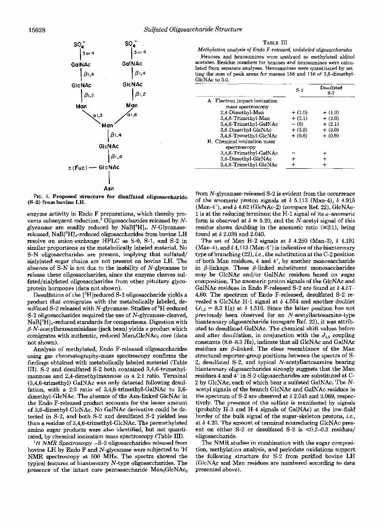
FIG. 5. Proposed structure for disulfated oligosaccharide (5-2) from bovine LH.
enzyme activity in Endo F preparations, which thereby pre- vents subsequent reduction.' Oligosaccharides released by N- glycanase are readily reduced by NaB[3H]4. N-Glycanase- released, NaB [3H]4-reduced oligosaccharides from bovine LH resolve on anion-exchange HPLC as S-0, S-1, and S-2 in similar proportions as the metabolically labeled material. .No S-N oligosaccharides are present, implying that sulfated/ sialylated sugar chains are not present on bovine LH. The absence of S-N is not due to the inability of N-glycanase to release these oligosaccharides, since the enzyme cleaves sul- fated/sialylated oligosaccharides from other pituitary glyco- protein hormones (data not shown).
Desulfation of the [3H]reduced S-2 oligosaccharide yields a product that comigrates with the metabolically labeled, de- sulfated 5-2 released with N-glycanase. Studies of 3H-reduced S-2 oligosaccharides required the use of N-glycanase-cleaved, NaB[3H]4-reduced standards for comparisons. Digestion with P-N-acetylhexosaminidase (jack bean) yields a product which comigrates with authentic, reduced MansGlcNAcz core (data not shown).
Analysis of methylated, Endo F-released oligosaccharides using gas chromatography-mass spectroscopy confirms the findings obtained with metabolically labeled material (Table 111). S-2 and desulfated S-2 both contained 3,4,6-trimethyl- mannose and 2,4-dimethylmannose in a 2:l ratio. Terminal (3,4,6-trimethyl) GalNAc was only detected following desul- fation, with a 2:3 ratio of 3,4,6-trimethyl-GalNAc to 3,6- dimethyl-GlcNAc. The absence of the Asn-linked GlcNAc in the Endo F-released product accounts for the lesser amount of 3,6-dimethyl-GlcNAc. No GalNAc derivative could be de- tected in s-2, and both s-2 and desulfated s-2 yielded less than a residue of 3,4,6-trimethyl-GlcNAc. The permethylated amino sugar products were also identified, but not quanti- tated, by chemical ionization mass spectroscopy (Table 111).
'H NMR Spectroscopy-S-2 oligosaccharides released from bovine LH by Endo F and N-glycanase were subjected to 'H NMR spectroscopy at 500 MHz. The spectra showed the typical features of biantennary N-type oligosaccharides. The presence of the intact core pentasaccharide Man3GlcNAcz
TABLE I11 Methylation analysis of Endo F-released, unlabeled oligosaccharides
Hexoses and hexosamines were analyzed as methylated alditol acetates. Residue numbers for hexoses and hexosamines were calcu- lated from separate analyses. Hexosamines were quantitated by set- ting the sum of peak areas for masses 158 and 116 of 3,6-dimethyl- GlcNAc to 3.0.
s-2 Desulfated s-2
A. Electron impact ionization
2,4-Dimethyl-Man + (1.0) + (1.0) 3,4,6-Trimethyl-Man + (2.1) + (2.0) 3,4,6-Trimethyl-GalNAc - (0) + (2.1) 3,6-Dimethyl-GlcNAc + (3.0) + (3.0) 3,4,6-Trimethyl-GlcNAc + (0.6) + (0.9)
mass spectroscopy
B. Chemical ionization mass spectroscopy
3,4,6-Trimethyl-GalNAc - + 3,6-Dimethyl-GlcNAc + + 3,4,6-Trimethyl-GlcNAc + +
from N-glycanase-released S-2 is evident from the occurrence of the anomeric proton signals at 6 5.113 (Man-4), 6 4.915 (Man-4'), and 6 4.62 (GlcNAc-2) (compare Ref. 22). GlcNAc- 1 is at the reducing terminus; the H-1 signal of its a-anomeric form is observed at 6 E 5.20, and the N-acetyl signal of this residue shows doubling in the anomeric ratio (~2:1) , being found at 6 2.038 and 2.040.
The set of Man H-2 signals at 6 4.250 (Man-3), 6 4.191 (Man-4), and 6 4.113 (Man-4') is indicative of the biantennary type of branching (22), i.e., the substitution at the C-2 position of both Man residues, 4 and 4', by another monosaccharide in @-linkage. These P-linked substituent monosaccharides may be GlcNAc and/or GalNAc residues based on sugar composition. The anomeric proton signals of the GlcNAc and GalNAc residues in Endo F-released S-2 are found at 6 4.57- 4.60. The spectrum of Endo F-released, desulfated S-2 re- vealed a GlcNAc H-1 signal at 6 4.554 and another doublet (J1,2 = 8.3 Hz) at 6 4.516. Since the latter position has not previously been observed for an N-acetyllactosamine-type biantennary oligosaccharide (compare Ref. 22), it was attrib- uted to desulfated GalNAc. The chemical shift values before and after desulfation, in conjunction with the Jls2 coupling constants (8.0-8.3 Hz), indicate that all GlcNAc and GalNAc residues are P-linked. The close resemblance of the Man structural-reporter-group positions between the spectra of S- 2, desulfated S-2, and typical N-acetyllactosamine bearing biantennary oligosaccharides strongly suggests that the Man residues 4 and 4' in S-2 oligosaccharides are substituted at C- 2 by GlcNAc, each of which bear a sulfated GalNAc. The N- acetyl signals of the branch GlcNAc and GalNAc residues in the spectrum of S-2 are observed at 6 2.045 and 2.069, respec- tively. The presence of the sulfate is manifested by signals (probably H-3 and H-4 signals of GalNAc) at the low-field border of the bulk signal of the sugar-skeleton protons, ie., at 6 4.20. The amount of terminal nonreducing GlcNAc pres- ent on either s-2 or desulfated s-2 is <0.2-0.3 residues/ oligosaccharide.
The NMR studies in combination with the sugar composi- tion, methylation analysis, and periodate oxidations support the following structure for S-2 from purified bovine LH (GlcNAc and Man residues are numbered according to data presented above).

Sulfated Oligosaccharide Structure
5 4 SO4~GalNAc(@l,4)4lcNAc(@l,2)+Man(~l,3)
1 3 2 1
7 Man(@l,4)+GlcNAc(@l,4)+GlcNAc
S04-GalNAc(@1,4)41cNAc(@1,2)+Man(al,6)
15629
5’ 4‘
The NMR spectra confirm the @-linkage of the GalNAc residues, the attachment of the 2 peripheral GlcNAc residues to core mannoses, the substitution of GalNAc by sulfate, the typical biantennary core structure, and the lack of significant quantities of terminal GlcNAc.
Determination of Fucose Location in S-2”Two observa- tions suggest that fucose is attached to the Asn-linked GlcNAc of S-2. First, Endo F digestion of [3H]fucose-labeled LH yields a disaccharide containing [3H]fucose (data not shown). This disaccharide is presumably fucose-GlcNAc, which is formed by the combined action of Endo F and the contaminating N- glycanase activity in Endo F preparations (24). In contrast, N-glycanase releases [3H]fucose-labeled oligosaccharides from LH with the same proportions of S-0, S-1, and S-2 as found with [3H]glucosamine-labeled LH. Furthermore, none of the exoglycosidase data obtained with either radioactive or unlabeled LH oligosaccharides would support a peripheral location of fucose. Although these results indicate that fucose is present on the Asn-linked GlcNAc of all the types of oligosaccharides, the proportion which is fucosylated remains to be determined.
DISCUSSION
Parsons and Pierce (1) first observed that sulfate is linked to terminal N-acetylhexosamines on LH oligosaccharides. These Asn-linked oligosaccharides are also unique due to the presence of GalNAc. Not all pituitary hormones contain both sulfate and GalNAc, and the closely related placental hor- mone, human chorionic gonadotropin, does not contain either moiety on its Asn-linked oligosaccharides. Understanding of these unusual posttranslational modifications and their reg- ulation has been hampered by incomplete information about the structure of the oligosaccharides present on LH and other pituitary hormones. A number of advances have facilitated the detailed examination of these oligosaccharides: 1) meta- bolic (2, 6) and cell-free (3) incorporation of [35S]sulfate, 2) metabolic incorporation of radiolabeled hexoses and hexosa- mines, 3) quantitative release of Asn-linked oligosaccharides with Endo F (3) and/or N-glycanase, 4) fractionation of charged oligosaccharide species by HPLC with respect to charge and size, 5) quantitative removal of sulfate without damage to underlying oligosaccharide by methanolysis, and 6) analysis of sulfated oligosaccharides by 500-MHz NMR spectroscopy.
The structure proposed for the disulfated oligosaccharide (S-2) of bovine LH (Fig. 5) is identical to other dibranched complex oligosaccharides in its core region and in the attach- ment of GlcNAc to Man at position 2 by a @-linkage. It is unique in the attachment of @1,4-linked S04-GalNAc at both nonreducing termini. The presence of sulfate exclusively on GalNAc suggests a requirement of this amino sugar for sul- fation. However, other structural features of the oligosaccha- rides or peptides may play a regulatory role as well (9).
Several other proposed structures for bovine (4), ovine (4- 6), and human (7) LH oligosaccharides have been reported. In general, these studies have used unlabeled, partially frac- tionated oligosaccharides. Since there is considerable micro- heterogeneity in the structures of LH oligosaccharides (see
Fig. 1 and Refs. 3 and 9), initial fractionation of oligosaccha- rides prior to structural characterization is essential. Thus, the previous data present an averaging of many microheter- ogeneous structures, which may account for some of the differences between our results and those of other studies. Differences between our findings and those using ovine and human LH may be due to animal-species variation.
In two reports (7, 8) GalNAc was found to be present in the chitobiose core of human pituitary hormone oligosaccha- rides. Our exoglycosidase, periodate oxidation, methylation, and NMR data indicate that GalNAc in bovine LH is exclu- sively peripheral to the chitobiose core. Since S-2 from human LH appears to be identical to that from bovine LH (9), and the presence of GalNAc in the oligosaccharide core region would represent virtually the only exception to the well- characterized pathway of Asn-linked oligosaccharide biosyn- thesis, we believe that GalNAc is strictly a peripheral sugar in the oligosaccharides of Asn-linked glycoprotein hormones.
Compositional studies have shown that the oligosaccharides of bovine LH contain mannose, GalNAc, GlcNAc, fucose, and sulfate, but not galactose or sialic acid (1,9), Thus, the origin of a minor population (<IO% of incorporated [3H]glucosa- mine) of sulfated/sialylated (S-N) oligosaccharides in meta- bolically labeled material was not obvious. These oligosaccha- rides are probably not derived from LH a-@ dimers based on the following. First, S-N oligosaccharides are not present in either cell-free 35S-sulfated-LH or N-glycanase-released, NaB[3H]4-reduced LH oligosaccharides. Thus, purified bovine LH does not contain S-N, as previously shown by sugar compositions (1, 4). Second, analysis of [3H]glucosamine- labeled tryptic peptides from immunoprecipitated bovine LH demonstrated that the a subunit, and not the @, is the source of S-N oligosaccharides. Furthermore, bovine FSH contains a significant (22%) proportion of S-N oligosaccharides, as found by cell-free sulfation (9). Therefore, our LH-specific antisera may precipitate a small amount of FSH, which con- tains S-N, or our antisera may precipitate a small amount of free, uncombined a subunit. The same enzymes responsible for the synthesis of S-N on FSH may also act on free a subunits. Therefore, contamination by either FSH or uncom- bined a subunits could account for the small amount of S-N detected in our analyses.
The presence of the two unique components of LH oligo- saccharides, GalNAc and sulfate, is probably directly related. Since only GalNAc is sulfated on LH, the presence of GalNAc is a minimal requirement for sulfation to occur. The extent to which other features of the oligosaccharide and/or peptide are required for sulfation is under examination. In addition, if GalNAc provides the “signal” required for sulfation, it may be that the pivotal step in the synthesis of sulfated, rather than nonsulfated, oligosaccharides is the addition of GalNAc. The appropriate “sulfation” determination inherent to certain pituitary glycoproteins may regulate the addition of GalNAc, which in turn acts as both the signal and acceptor for sulfate.
Acknowledgments-We thank Stuart Kornfeld and Gabriela Adelt Green for critical review of this manuscript. We also thank Sen-itiroh Hakomori, Klaus Stellner, and Rosalind Kornfeld for providing meth- ylation standards.

15630 Sulfated Oligosaccharide Structure
REFERENCES
1. Parsons, T. F., and Pierce, J. G. (1980) Proc. Nutl. Acad. Sci. U.
2. Hortin, G., Natowicz, M., Pierce, J., Baenziger, J., Parsons, T., and Boime I. (1981) Proc. Nutl. Acud. Sci. U. S. A. 78, 7468- 7472
3. Green, E. D., Gruenebaum, J., Bielinska, M., Baenziger, J. U., and Boime, I. (1984) Proc. Natl. Acad. Sci. U. S. A. 81, 5320- 5324
4. Bahl, 0. P., Reddy, M. S., and Bedi, G. S. (1980) Biochem.
5. Bedi, G. S., French, W. C., and Bahl, 0. P. (1982) J. Bwl. Chem. Biophys. Res. Commun. 96,1192-1199
6. Anumula, K. R., and Bahl, 0. P. (1983) Arch. Biochem. Biophys.
7. Hara, K., Rathnam, P., and Saxena, B. B. (1978) J. Biol. Chem.
8. Tolvo, A., Fujiki, Y., Bhavana, V. P., Rathnam, P., and Saxena,
9. Green, E. D., Baenziger, J. U., and Boime, I. (1985) J. Biol. Chem.
10. Cummings, R. D., and Kornfeld, S. (1982) J. Biol. Chem. 257,
11. Baenziger, J. U., and Fiete, D. (1979) J. Biol. Chem. 254, 2400-
S. A. 77, 7089-7093
257,4345-4355
220,645-651
253,1582-1591
B. B. (1982) Biochim. Biophys. Acta 719, 1-10
260,15631-15638
11235-11240
2407
12. Mellis, S. J., and Baenziger, J. U. (1983) J. Biol. Chem. 258,
13. Papkoff, H., and Gan, J. (1970) Arch. Biochem. Biophys. 136,
14. Baenziger, J. U., and Natowicz, M. (1981) Anal. Biochem. 112,
15. Mellis, S. J., and Baenziger, J. U. (1983) Anal. Biochem. 134,
16. Hakomori, S. (1964) J. Bwchem. (Tokyo) 55,205-208 17. Stellner, K., Saito, H., and Hakomori, S. (1973) Arch. Bwchem.
18. Saadat, S., and Ballou, C. E. (1983) Curbohydr. Res. 119, 248-
19. Mellis, S. J., and Baenziger, J. U. (1983) J. Bwl. Chem. 258,
20. Baenziger, J. U., and Fiete, D. (1979) J. Biol. Chem. 254, 789-
21. Laine, R. A. (1981) Anal. Bwchem. 116,383-388 22. Vliegenthart, J. F. G., Dorland, L., and Van Halbeek, H. (1983)
23. Tai, T., Yamashita, K., and Kobata, A. (1975) J. Biochem. 78,
24. Plummer, T. H., Elder, J. H., Alexander, S., Phelan, A. W., and Tarentino, A. L. (1984) J. Biol. Chem. 259, 10700-10704
25. Corless, C. L., and Boime, I. (1985) Endocrinology 117,
11546-11556
522-528
357-361
442-449
Biophys. 155,464472
253
11557-11563
795
Adv. Curbohydr. Chem. Biochem. 41,209-374
679-686
1699-1706