Structural Differentiation Diagnosis of the Upper and Lower … · · 2012-04-07How many Clinical...
60
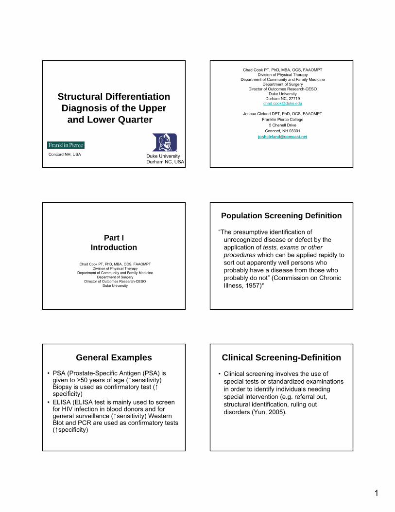
1 Structural Differentiation Diagnosis of the Upper and Lower Quarter Duke University Durham NC, USA Concord NH, USA Chad Cook PT, PhD, MBA, OCS, FAAOMPT Division of Physical Therapy Department of Community and Family Medicine Department of Surgery Director of Outcomes Research-CESO Duke University Durham NC, 27719 [email protected] Joshua Cleland DPT, PhD, OCS, FAAOMPT Franklin Pierce College 5 Chenell Drive Concord, NH 03301 [email protected] Part I Introduction Chad Cook PT, PhD, MBA, OCS, FAAOMPT Division of Physical Therapy Department of Community and Family Medicine Department of Surgery Director of Outcomes Research-CESO Duke University Population Screening Definition “The presumptive identification of unrecognized disease or defect by the application of tests, exams or other procedures which can be applied rapidly to sort out apparently well persons who probably have a disease from those who probably do not” (Commission on Chronic Illness, 1957)* General Examples • PSA (Prostate-Specific Antigen (PSA) is given to >50 years of age ( sensitivity) Biopsy is used as confirmatory test ( specificity) • ELISA (ELISA test is mainly used to screen for HIV infection in blood donors and for general surveillance ( sensitivity) Western Blot and PCR are used as confirmatory tests ( specificity) Clinical Screening-Definition • Clinical screening involves the use of special tests or standardized examinations in order to identify individuals needing special intervention (e.g. referral out, structural identification, ruling out disorders (Yun, 2005).
Transcript of Structural Differentiation Diagnosis of the Upper and Lower … · · 2012-04-07How many Clinical...
1
Structural Differentiation Diagnosis of the Upper
and Lower Quarter
Duke UniversityDurham NC, USA
Concord NH, USA
Chad Cook PT, PhD, MBA, OCS, FAAOMPTDivision of Physical Therapy
Department of Community and Family MedicineDepartment of Surgery
Director of Outcomes Research-CESODuke University
Durham NC, [email protected]
Joshua Cleland DPT, PhD, OCS, FAAOMPTFranklin Pierce College
5 Chenell DriveConcord, NH 03301
Part IIntroduction
Chad Cook PT, PhD, MBA, OCS, FAAOMPTDivision of Physical Therapy
Department of Community and Family MedicineDepartment of Surgery
Director of Outcomes Research-CESODuke University
Population Screening Definition
“The presumptive identification of unrecognized disease or defect by the application of tests, exams or other procedures which can be applied rapidly to sort out apparently well persons who probably have a disease from those who probably do not” (Commission on Chronic Illness, 1957)*
General Examples• PSA (Prostate-Specific Antigen (PSA) is
given to >50 years of age ( sensitivity) Biopsy is used as confirmatory test ( specificity)
• ELISA (ELISA test is mainly used to screen for HIV infection in blood donors and for general surveillance ( sensitivity) Western Blot and PCR are used as confirmatory tests ( specificity)
Clinical Screening-Definition
• Clinical screening involves the use of special tests or standardized examinations in order to identify individuals needing special intervention (e.g. referral out, structural identification, ruling out disorders (Yun, 2005).

2
Clinical Examples
• Canadian C-Spine Rules are used to determine who doesn’t need an x-ray ( sensitivity-99%) X-ray is used to diagnose fracture ( specificity)
• Ottawa Ankle Rules are used to determine who doesn’t need an x-ray ( sensitivity) X-ray is used to diagnose fracture ( specificity)
Criteria for Screening• Significant burden of disease in population
either in prevalence or morbidity/mortality.• Pathology screened has high prevalence
(>5%)• Pathology has a pre-clinical phase (onset of
disease to the first appearance of symptoms)• Screening tests are inexpensive to
administer.
Obuchowski et al. Ten Criteria for Effective Screening. Am J Roent. 2001;176:1357-62
Criteria for Screening• Screening tests should be relatively accurate and
should not routinely identify pseudo-diseases (sensitivity vs. specificity)
• Screening causes little morbidity• Screening tests should have high sensitivity
– For clinical structural tests (typically >90).– For pathological processes in medicine (typically >95-99)– For mundane slow acting diseases with low morbidity
(>65)
Obuchowski et al. Ten Criteria for Effective Screening. Am J Roent. 2001;176:1357-62
Special Test has been Tested• Study is free from bias (QUADAS)• Study was performed in pre-clinical
phase• Test is repeatable and has clinical
utility• The test makes clinical sense
Cook C. Physical Examination Tests for Neurological Screening. In: Cook C, Hegedus E.Physical Examination Tests: An Evidence Based Approach. Upper Saddle River, NJ; Prentice Hall: 2007.
QUADAS• (Quality Assessment of Diagnostic Accuracy
Studies)• 14 items
– 1. Appropriate selection of patient spectrum– 2. Appropriate reference standard– 3. Absence of review bias (both test and diagnostic)– 4. Clinical review bias– 5. Reporting of ininterpretable/ indeterminate/
intermediate results
Whiting et al. BMC Medical Research Methodology. 2003, 3:25
Consider Prevalence• Prevalence is the proportion of people in
the entire population who are found to be with disease at a certain point in time
• Point, interval, and location prevalence can be very useful for non-fatal conditions. – (Point) The chance of correctly and routinely
identifying ALS is low because the prevalence is low (proportion at a given time)

3
Prevalence• (Interval) Up to 3% of individuals over the
age of 60, with neck pain, have cord compressive myelopathy
• (Location) Prevalence of obesity (BMI>30%) is 22% in Australia, 34% in USA, 3% in Japan, 5% in China
• (Location) Prevalence of an ACL injury at a weekend sports screening versus a Geriatric Health Care Fair
Consider Incidence
• Incidence is a rate, showing how many new cases of a disease occurred in a population during a specified interval of time (usually expressed as number of new cases per unit time per fixed number of people; e.g., number of new cases of cancer per 10,000 persons in one year).
Incidence may relate to populations and prevalence
• # of new cases……• HIV is more common in young
homosexual men than older heterosexual Women
• Cervical myelopathy is more common in Asians than African Americans.
• Rotator cuff tears are more common in patients 60 and older than 20 and younger
Consider Disease Natural History
• Some disease processes lack signs and symptoms to allow testing (tests lack accuracy) (Osteochondral lesion)
• Some conditions do not have definable symptoms that are homogenous and discreet from other (SIJ dysfunction)
Consider Disease Natural History
• Some conditions do not exhibit symptoms in early stages thus are not caught on a clinical examination (degenerative meniscus of the knee)
• Some conditions have such low prevalence, that good tests will never identify their presence (Maroteux-LamySyndrome)
Natural History Screeningfor Signs and Symptoms
Signs & SymptomsDisease Onset
Signs & Symptoms
Disease Onset
Screen
Screen Likely to catch the disorder
Not likely to catch the disorder
Timespan

4
Onset of Disease
Critical PointFor Detection
First Point Of Detection
Clinical SignsAnd Symptoms Too Late
Detectable Pre-Clinical Phase
Pre-Clinical Phase
Clinical Phase
How many Clinical Special Tests have the capacity to detect the presence of a disease in the absence of
clinical signs and symptoms?
Obuchowski et al. Ten Criteria for Effective Screening. Am J Roent. 2001;176:1357-62
Beneficial for PT’s?
• Reduces risk of contraindicated treatment• Improves ability to identify risks as entry
point provider• Identifies “Red Flags” prior to dedicated
care• Allows structural differentiation and
targeting of appropriate segment/region
Ask Yourself-Am I Using the Most Effective Screening Tool?
• Is the test accurate?• Is the test reliable?• How likely is the finding prevalent?• Who is more likely to have the problem
that we are screening for?• How likely are we to identify symptoms at
a given time?• Is there a better process I should follow?
Diagnostic Accuracy of the Clinical Exam: What Factors are Important in Screening
Tests?
Joshua Cleland, PT, PhD, OCS, FAAOMPTAssistant Professor
Franklin Pierce College
Diagnostic Utility of Clinical Tests
• Judge evidence for diagnostic tests, select the most appropriate test for an individual patient, and interpret the results.
• The need to become familiar with skills of physical therapy diagnosis is to become a more evidence-based process.
Diagnostic Accuracy
• Each time a clinical test is performed we must understand how the results of the test compare with the truth.
• This is determined by comparing the test results with a measure of the truth.
• So- how do we do this?

5
Relevant clinical population
Perform the diagnostic test – on everyone
Perform the reference standard – on everyone
Compare the Results
“The optimal design for assessing the accuracy of a diagnostic test is a prospective blind comparison of the test with a reference standard in a consecutive series of patients from a relevant clinical population”
(Lijmer et al, 1999)
Patients with lateral hip pain
Perform MRI – on everyone
Physical examination- on everyone
Compare Results
Diagnostic Test Characteristics
Definition: Prevalence
• Prevalence– The percentage of patients who truly have the
condition in the sample studied
– Value: provides an estimate of the probability that an individual will have a particular condition
Contingency Table
True Negative Result
D
False Negative Result
C
False Positive Result
B
True Positive Result
A
Diagnostic Test Positive
Diagnostic Test Negative
Reference Standard Positive Reference Standard Negative
A + B
C + D
NA + C B + D
Example
D=47(True Negative)
C=37(False Negative)
B=3(False Positive)
A=13(True Positive)
Hoffman’s Test Positive
Hoffman’s Test Negative
Myelopathy Present Myelopathy Absent
6
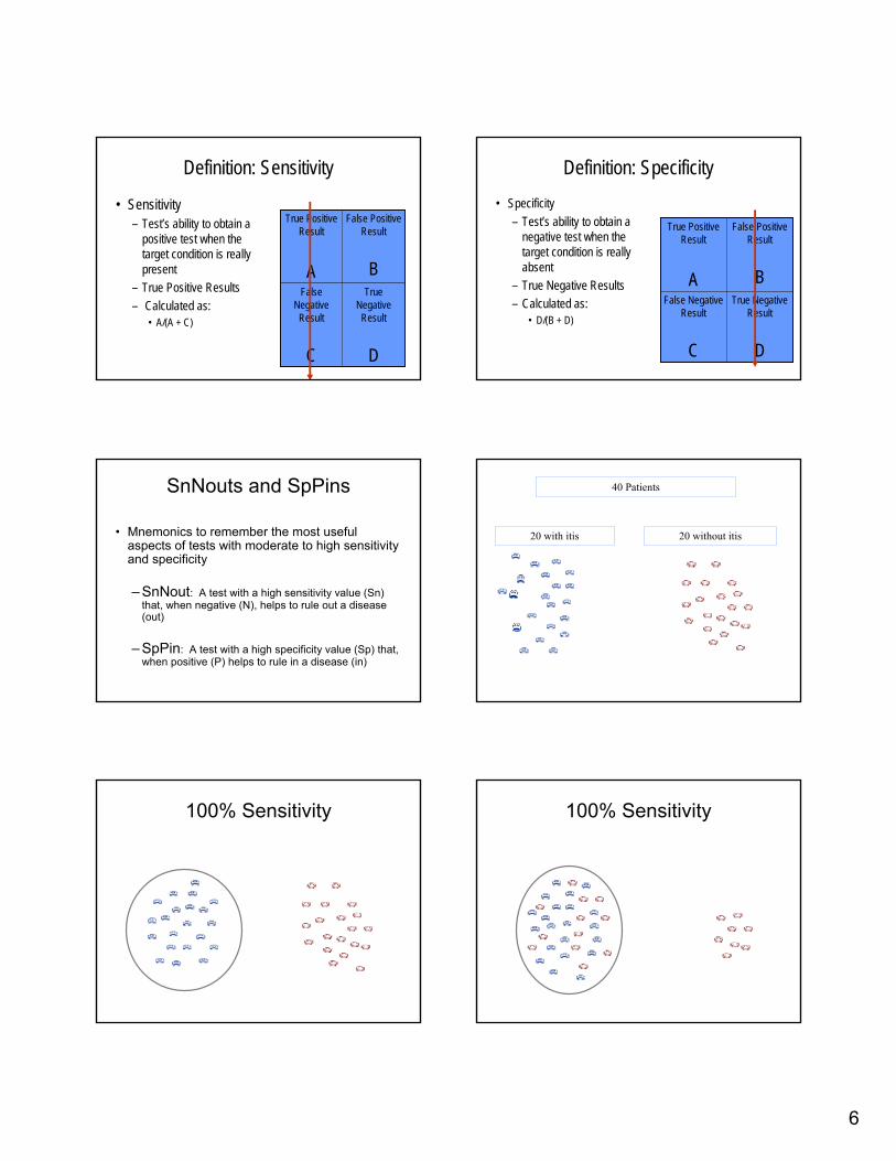
Definition: Sensitivity
• Sensitivity– Test’s ability to obtain a
positive test when the target condition is really present
– True Positive Results– Calculated as:
• A/(A + C)
True Negative
Result
D
False Negative
Result
C
False Positive Result
B
True Positive Result
A
Definition: Specificity• Specificity
– Test’s ability to obtain a negative test when the target condition is really absent
– True Negative Results– Calculated as:
• D/(B + D)
True Negative Result
D
False Negative Result
C
False Positive Result
B
True Positive Result
A
SnNouts and SpPins
• Mnemonics to remember the most useful aspects of tests with moderate to high sensitivity and specificity
– SnNout: A test with a high sensitivity value (Sn)that, when negative (N), helps to rule out a disease (out)
– SpPin: A test with a high specificity value (Sp) that, when positive (P) helps to rule in a disease (in)
40 Patients
20 with itis 20 without itis
100% Sensitivity 100% Sensitivity

7
100% Specificity 100% Specificity
Myelopathy Example
• Sensitivity: = 26%
• Specificity: = 94%
So is this a good test for screening for myelopathy?
D=47(True
Negative)
C=37(False
Negative)
B=3(False
Positive)
A=13(True Positive)
Is it important for a Screening Test to have High Sensitivity or Specificity?
Does This Patient Belong in My Clinic??? Red Flags
•Temperature > 100 F•BP >160/95 mmHg•Resting Pulse >100bpm•Resting Respiration >25bpm•Fatigue
•Drop-attacks •Dizziness (lightheadedness) related to neck movement)•Dysphasia •Dysarthria•Diplopia•Positive cranial nerve signs
•Occipital headache and numbness•Severe limitation during neck AROM in all directions•Signs of cervical myelopathy
•Age over 50 years•Previous history of cancer•Unexplained weight loss•Constant pain, no relief with bed rest•Night pain
•Sensory disturbance of the hands•Muscle wasting of hand intrinsic muscles•Unsteady gait•Hoffman’s reflex•Hyperreflexia•Bowel and bladder disturbances•Multisegmentalweakness and/or sensory changes
Inflammatory or Systemic DiseaseVertebral Artery
Insufficiency
Upper Cervical Ligamentous
InstabilityNeoplastic ConditionsCervical Myelopathy

8
Screening for Ankle Fractures: Example
EXAMPLE: Ankle radiographs
Your patient is a 26 year-old male who turned his ankle playing basketball last night. He has been able to walk on it with a pronounced limp. There is substantial swelling and discoloration. He complains of pain about the lateral malleolus.
EXAMPLE: Ankle radiographs
Do you need to get an x-ray?
100
0
Deg
ree
of C
erta
inty
Decision to Order X-Rays
Action – No X-Ray (how certain would you need to be?)
Ottawa Ankle Rules

9
Do you need to order x-rays?You presume he has a 25% chance of having a fracture.
– The patient is positive on the OAR• Revised probability of fracture: 35.3%
– The patient is negative on the OAR • Revised probability of fracture: 0.99%
SnNout
• 20 year-old male• Left ankle injury sustained during a football game• Moderate edema• Limited ROM• Unable to WB• Tenderness over posterior fibula
Screening for Knee Fractures: Another Example
• March 13, 2004: A 9-year-old girl had injured her left knee in a gymnastics event the evening before. She was performing a front flip and heard a pop in her knee as she landed. She reported immediate pain in the left knee and was unable to walk. She had not been seen by another medical professional.
Clinical Question?
Ligament?Meniscus?Fracture?
Is diagnostic imaging necessary on a 9-year-old female presenting with anterior
knee pain in a direct access setting?

10
The Ottawa Knee Rule
A knee x-ray series is only required for knee injury patients with any of these findings:
1. Age 55 years or olderor
2. Isolated tenderness of patella *or
3. Tenderness at the head of fibulaor
4. Inability to flex to 90 oor
5. Inability to bear weight both immediately and in emergency department (4 steps)**
* No bone tenderness of knee other than patella** Unable to transfer weight twice onto each lower
limb regardless of limping.
Can we apply the OKR to our patient?
• Bachmann et al. Ann Intern Med. 2004. 140:121-124.
• Originally derived by Steil et al (Ann Emerg Med)
• Numerous validation studies have demonstrated the accuracy of the OKR
• The probability of a fracture after a negative OKR was 0.37%
• Of the 4249 patients, 5 had a false negative
OKR in Children
• Bulloch et al (Ann Emerg Med)
• 750 children age 2-16 (Mean 11.8) who had sustained a traumatic knee injury within 7 days
• 70 fractures, all which were detected using the OKR: 100% sensitivity
…back to our patient.
• Patient was tender to palpation at the distal ¼ of the patella.
• Patient was unable to walk secondary to reports of pain.
• X-ray revealed a transverse fracture across the inferior aspect of her patella.
SnNout
So What is all the Talk about Likelihood Ratios?

11
Likelihood Ratios
• Reflects a combination of the information contained in sensitivity and specificity values into a ratio that can be used to quantify shifts in probability once the diagnostic tests results are known.
• Positive likelihood ratio (LR+)
• Negative likelihood ratio (LR–)
Where do likelihood ratios come from?
• In some happy (though rare) instances, investigators or test developers provide them.
• Much more commonly, we have to calculate them for ourselves.
• Luckily, these are very simple calculations when you understand 2X2 tables.
Definition: LR of a positive test result (LR+)
• LR Positive Test Result (LR+)– The ratio of the true positive rate to the false
positive rate– Calculated as: sensitivity
(1 – specificity)- Value: Indicates the increase in odds favoring the
condition given a positive result
Definition: LR of a negative test result (LR-)
• LR Negative Test Result (LR-)– The ratio of the false negative rate to the true
negative rate– Calculated as: (1 – sensitivity)
specificity- Value: Indicates the change in odds favoring the
condition given a negative test result
Rarely alters probability to an important degree
.5-11-2
Small shifts in probability.2-.52-5
Moderate shifts in probability
.1-.25-10
Large and conclusive shifts in probability
< .1> 10
Interpretation-LR+LR Negative Likelihood Ratio
• So let’s say we have a test with a Sensitivity of .85 and a Specificity of .20
• Then the –LR = (1-.85)/.20= .75
• Rarely alters probability to an important degree

12
-LR=.2
-LR=.08
Treatment Threshold
• The point at which the examination and evaluation process stops and treatment begins.
• How do we determine this?
PROBABILITY OF DISEASE
InformationalContribution
Pre-test Probability Post-test
Probability
Treatment Threshold
0% 50% 100%
Screening Tests
SnNout
Part IIIScreening for Red Flags
Chad Cook PT, PhD, MBA, OCS, FAAOMPTDivision of Physical Therapy
Department of Community and Family MedicineDepartment of Surgery
Director of Outcomes Research-CESODuke University
What is a Red Flag?
• Signs and Symptoms found in the patient history and clinical examination that may tie a disorder to a serious pathology.
• < 5% of Primary Care Physicians Routinely Examine for Red Flags during an Initial Screen
Sizer P, Brismee JM, Cook C. Medical screening for red flags in the diagnosis and management of musculoskeletal spine pain. Pain Pract. 2007 (in press).
Bishop PB, Wing PC. Knowledge transfer in family physicians managing patients with acute low back pain: a prospective randomized control trial. Spine J. 2006: 6:282-8.

13
Categorizing Red Flags• Category I: Factors which Require
Immediate Medical Attention• Category II: Factors which Require
Subjective Questioning and Precautionary Examination and Treatment Procedures
• Category III: Factors which Require Further Physical Testing and Differentiation Analysis
Cook C. Orthopedic Manual Therapy. An Evidence Based Approach. Prentice Hall; Upper Saddle River, NJ: 2007.
Category I:• Blood in Sputum• Elevated Sedimentation Rate• Loss of Consciousness or Altered mental
state• Bowel and Bladder Dysfunction• Severe Non-mechanical pain• Progressive Neurological Deficit• Heart-Related Symptoms
Cook C. Orthopedic Manual Therapy. An Evidence Based Approach. Prentice Hall; Upper Saddle River, NJ: 2007.
Category II:• Age > 50• Clonus• Fever• Gait Deficits• History of a disorder with
predilection for infection or hemorrhage
• History of a metabolic bone disorder
• History of cancer
• Impairment precipitated by recent trauma
• Long-term corticosteroid use
• Long-term worker’s compensation
• Non-healing sores or wounds
• Recent history of unexplained weight loss
• Writhing pain
Cook C. Orthopedic Manual Therapy. An Evidence Based Approach. Prentice Hall; Upper Saddle River, NJ: 2007.
Category III:
• Myelopathic Symptoms• Abnormal Reflexes• Bilateral or Unilateral Radiculopathy or
Paresthesia• Unexplained Referred Pain• Unexplained Significant Upper or Lower
Limb Weakness
Cook C. Orthopedic Manual Therapy. An Evidence Based Approach. Prentice Hall; Upper Saddle River, NJ: 2007.
Cervical-Specific Red Flags
• Category I Findings– Head Injury (concussion or altered mental
status)– Cervical spine fractures (Canadian C-Spine
Rules) – Upper Cervical Spine Instability
Sizer P, Brismee JM, Cook C. Medical screening for red flags in the diagnosis and management of musculoskeletal spine pain. Pain Pract. 2007 (in press).
Diagnostic Recommendations for Post-Concussion Disorder—DSM-IV
• A. A history of head trauma that has caused significant cerebral concussion.
• B. Evidence from neuropsychological testing or quantified cognitive assessment of difficulty in attention
• C. Three (or more) of the following occur shortly after the trauma and last at least 3 months:– 1. Becoming fatigued easily.– 2. Disordered sleep.– 3. Headache.– 4. Vertigo or dizziness.– 5. Irritability or aggression on little or no provocation.– 6. Anxiety, depression, or affective lability.– 7. Changes in personality (e.g., social or sexual inappropriateness).– 8. Apathy or lack of spontaneity.

14
Diagnostic Recommendations for Post-Concussion Disorder—DSM-IV
• D. The symptoms in Criteria B and C have their onset following head trauma or else represent a substantial worsening of preexisting symptoms.
• E. The disturbance causes significant impairment in social or occupational functioning and represents a significant decline from a previous level of functioning.
• F. The symptoms do not meet criteria for Dementia Due to Head Trauma and are not better accounted for by another mental disorder
Iverson DL. Misdiagnosis of the persistent postconcussion syndrome in patients with depression.Arch Clin Neuropsychol. 2006 May;21(4):303-10.
ICD-10 Criteria• Require a history of TBI and the presence of
three or more of the following eight symptoms: • 1) headache,• 2) dizziness, • 3) fatigue, • 4) irritability, • 5) insomnia, • 6) concentration• 7) memory difficulty, • 8) intolerance of stress, emotion, or alcohol.
World Health Organization: The ICD-10 Classification of Mental and Behavioural Disorders: Clinical Descriptions and Diagnostic Guidelines. Geneva, World Health Organization, 1992
“or” Sensitive but not Specific
• Often misdiagnosed as – Head injury– Posttraumatic stress– Depression – Whiplash
• Swedish Post-Concussion Symptoms questionnaire
• Rivermead Post-Concussion Questionnaire
Canadian C-Spine Rules
Sensitivity = 99
Stiell et al. Canadian CT head rule study for patients with minor head injury: methodology for phase II (validation and economic analysis). Ann Emerg Med. 2001;38(3):317-22.
Modified Sharp Purser Test
Cook C. Physical Examination Tests for Neurological Screening. In: Cook C, Hegedus E.Physical Examination Tests: An Evidence Based Approach. Upper Saddle River, NJ; Prentice Hall: 2007.
Alar Ligament Stability Test
Cook C. Physical Examination Tests for Neurological Screening. In: Cook C, Hegedus E.Physical Examination Tests: An Evidence Based Approach. Upper Saddle River, NJ; Prentice Hall: 2007.

15
Shear Testing
Cook C. Physical Examination Tests for Neurological Screening. In: Cook C, Hegedus E.Physical Examination Tests: An Evidence Based Approach. Upper Saddle River, NJ; Prentice Hall: 2007.
Cervical-Specific Red Flags
• Category II Findings– VBI– Congenital and Hereditary Conditions
(Maroteux-Lamy Syndrome, MorquioSyndrome, RA, Down Syndrome, Marfan’sSyndrome, Klippel-Feil syndrome
– Gait Dysfunction/Balance (myelopathy)
Sizer P, Brismee JM, Cook C. Medical screening for red flags in the diagnosis and management of musculoskeletal spine pain. Pain Pract. 2007 (in press).
Vertebrobasilar Insufficiency
NTCook C. Physical Examination Tests for Neurological Screening. In: Cook C, Hegedus E.Physical Examination Tests: An Evidence Based Approach. Upper Saddle River, NJ; Prentice Hall: 2007.
Assessment for the presence of symptoms and signs associated with VBI occurs at four stages in themanagement of a patient with an upper quadrant disorder:
1. History (subjective examination)2. Physical (objective) examination3. During treatment of the cervical spine, and4. Following treatment.
Although many VBI tests have shown mixed results in relation to changes in vertebrobasilar arterial blood flow in experimental, it appears that end-range rotation is the most sensitive cervical position. Recent research has also identified blood flow changes in the simulated manipulation position.
Rivett et al. Clinical Guidelines for Assessing Vertebrobasilar Insufficiency in the Management of Cervical Spine Disorders. Australian Physiotherapy Association.
Cervical-Specific Red Flags• Category III Findings
– Myelopathy or Visceral Pain
Sizer P, Brismee JM, Cook C. Medical screening for red flags in the diagnosis and management of musculoskeletal spine pain. Pain Pract. 2007 (in press).
http://www.mona.uwi.edu/fpas/courses/physiology/neurophysiology/ReferredPainPattern.jpg
http://kline18.tripod.com/neck_files/image008.jpg
Hoffmann’s Test
Cook C. Physical Examination Tests for Neurological Screening. In: Cook C, Hegedus E.Physical Examination Tests: An Evidence Based Approach. Upper Saddle River, NJ; Prentice Hall: 2007.
Others-Myelopathy
Cook C. Physical Examination Tests for Neurological Screening. In: Cook C, Hegedus E.Physical Examination Tests: An Evidence Based Approach. Upper Saddle River, NJ; Prentice Hall: 2007.

16
Thoracic-Specific Red Flags• Category I Findings
– Viscerosomatic Pain – Tumors and Fractures
http://www.wehelpwhathurts.homestead.com/visceral-somatic_referral_patterns_resize_smaller.jpg
Thoracic-Specific Red Flags
• Category II Findings– Metabolic Disorders (Osteoporosis)– Long term corticosteroid use– Age greater than 50– Spondylodiscitis
Sizer P, Brismee JM, Cook C. Medical screening for red flags in the diagnosis and management of musculoskeletal spine pain. Pain Pract. 2007 (in press).
Thoracic Kyphosis(Compression Fracture)
• Cook et al. 2002• O’Brien et al. 1999• 48-49 degrees of
kyphosis = compression fracture
http://www.ki.se/odont/cariologi_endodonti/exarb/Marie_Nilsson.html
Heel Drop Test/Percussion(Spondylodiscitis)
• Discitis, Spondylodiscitis, Spondylitis, Vertebral Pyogenic Osteomyelitis, or Epidural Abscess.
• Bacterial infection of the disc and surrounding body and tissues
• Tenderness during spine palpation is most sensitive measure (Deyo & Weinstein 2001)
Sizer P, Brismee JM, Cook C. Medical screening for red flags in the diagnosis and management of musculoskeletal spine pain. Pain Pract. 2007 (in press).
Thoracic-Specific Red Flags• Category III
Findings– Thoracic disc lesions
(T6)– Spinal Cord
Compression Disorders
http://www.eorthopod.com/images/ContentImages/spine/spine_thoracic/herniation/thoracic_herniation_symptom01.jpg
Others-Myelopathy
http://academic.uofs.edu/faculty/kosmahle1/courses/pt351/lab351/babinski.htm
NT
Cook C. Physical Examination Tests for Neurological Screening. In: Cook C, Hegedus E.Physical Examination Tests: An Evidence Based Approach. Upper Saddle River, NJ; Prentice Hall: 2007.
17
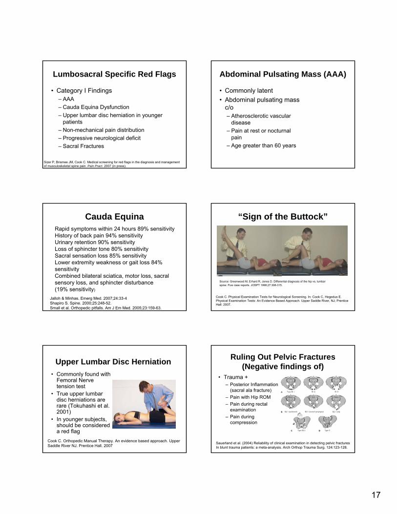
Lumbosacral Specific Red Flags
• Category I Findings– AAA– Cauda Equina Dysfunction– Upper lumbar disc herniation in younger
patients– Non-mechanical pain distribution– Progressive neurological deficit– Sacral Fractures
Sizer P, Brismee JM, Cook C. Medical screening for red flags in the diagnosis and management of musculoskeletal spine pain. Pain Pract. 2007 (in press).
Abdominal Pulsating Mass (AAA)
• Commonly latent• Abdominal pulsating mass
c/o– Atherosclerotic vascular
disease – Pain at rest or nocturnal
pain – Age greater than 60 years
Cauda Equina
Jalloh & Minhas. Emerg Med. 2007;24:33-4Shapiro S. Spine. 2000;25:248-52.Small et al. Orthopedic pitfalls. Am J Em Med. 2005;23:159-63.
Rapid symptoms within 24 hours 89% sensitivityHistory of back pain 94% sensitivityUrinary retention 90% sensitivityLoss of sphincter tone 80% sensitivitySacral sensation loss 85% sensitivityLower extremity weakness or gait loss 84% sensitivityCombined bilateral sciatica, motor loss, sacral sensory loss, and sphincter disturbance (19% sensitivity)
“Sign of the Buttock”
Source: Greenwood M, Erhard R, Jones D. Differential diagnosis of the hip vs. lumbarspine: Five case reports. JOSPT 1998;27:308-315.
Cook C. Physical Examination Tests for Neurological Screening. In: Cook C, Hegedus E.Physical Examination Tests: An Evidence Based Approach. Upper Saddle River, NJ; Prentice Hall: 2007.
Upper Lumbar Disc Herniation• Commonly found with
Femoral Nerve tension test
• True upper lumbar disc herniations are rare (Tokuhashi et al. 2001)
• In younger subjects, should be considered a red flag
Cook C. Orthopedic Manual Therapy. An evidence based approach. Upper Saddle River NJ. Prentice Hall. 2007
Ruling Out Pelvic Fractures(Negative findings of)
• Trauma +– Posterior Inflammation
(sacral ala fracture)– Pain with Hip ROM– Pain during rectal
examination– Pain during
compression
Sauerland et al. (2004) Reliability of clinical examination in detecting pelvic fracturesIn blunt trauma patients: a meta-analysis. Arch Orthop Trauma Surg. 124:123-128.

18
Lumbosacral Specific Red Flags
• Category II Findings– Compression fractures– Pyogenic infections (rapid onset, leading to
fever, malaise, severe low back pain), Osteomyelitis, Spondylodiscitis
– Neurogenic vs. vascular– Non-traumatic, bilateral low back and SIJ pain
Sizer P, Brismee JM, Cook C. Medical screening for red flags in the diagnosis and management of musculoskeletal spine pain. Pain Pract. 2007 (in press).
Lumbar Compression fracture
• History sensitivity specificity– age >50 0.84 0.61– age >70 0.22 0.96– trauma 0.30 0.85– corticosteroid use 0.06 0.995
– in elderly trauma can be minor
Deyo RA, Jarvik JG. Diagnostic evaluation of low back pain with emphasis on imaging. Ann Intern Med. 2002;137:586-97..
Spine Cancer• History sensitivity specificity
– Age > 50 0.77 0.71– previous history 0.31 0.98
of cancer– failure to improve 0.31 0.90
in 1 mo. of therapy– no relief -bed rest >0.90 0.46– duration > 1 mo 0.50 0.81– age >50 or cancer hx or 1.00 0.60
unexplained wt loss or failure of conservative tx.
– Insidious onset– constitutional symptoms
Deyo RA, Jarvik JG. Diagnostic evaluation of low back pain with emphasis on imaging. Ann Intern Med. 2002;137:586-97..
Ankylosing Spondylitis
• History sensitivity specificity– age at onset <40 1.00 0.07– pain not relieved by supine 0.80 0.49– morning back stiffness 0.64 0.59– pain duration >3 months 0.71 0.54– 4 of 5 questions above positive 0.23 0.82
also: improved by exercise
+LR = 1.27
Deyo RA, Jarvik JG. Diagnostic evaluation of low back pain with emphasis on imaging. Ann Intern Med. 2002;137:586-97..
Lumbosacral Specific Red Flags
• Category III Findings– Myelopathy– Radiculopathy– Visceral or Somatic Referred Pain (SIJ vs.
Lumbar origin)
Sizer P, Brismee JM, Cook C. Medical screening for red flags in the diagnosis and management of musculoskeletal spine pain. Pain Pract. 2007 (in press).
Others-Myelopathy
http://academic.uofs.edu/faculty/kosmahle1/courses/pt351/lab351/babinski.htm
NT
Cook C. Physical Examination Tests for Neurological Screening. In: Cook C, Hegedus E.Physical Examination Tests: An Evidence Based Approach. Upper Saddle River, NJ; Prentice Hall: 2007.

19
Lumbar Category III Findings• Bilateral lower
extremity weakness or numbness
• Referred Pain
Visceral Referred PainVisceral and somatic referred pain can refer pain to the legs
Several structures of the lumbar spine and pelvis can refer pain to thelegs
Hip Specific Red Flags
• Category I Red Flags– Hip Fracture– AVN
• Trauma (not always)
• Typically involves groin pain
• X-ray in early stages are normal
Predicting Hip Fracture• Bone DEXA <-2.14 T• And• Age >74 years
• Bone DEXA , -2.14 T• And• Age < 74 years• And• 1 functional difficulty• And• Walking speed ,
0.96m/s
Sensitivity = 70.4%Specificity = 78.7%
Jin et al. Classification algorithms for hip fracture prediction based on recursive partitioning methods. Med Des Making. 2004;24:386.
Ruling Out Hip Fractures(Negative findings of)
• + Pubic Percussion Test (LR+ = 9 to 313)
• ER of one limb versus the other
Hip Specific Red Flags• Category II Findings
– Infection– Total Hip Replacement
Failure• Typical failure occurs at
zones 1 and 7 and involve rocking or pistoning (cement vs. cementless-no difference)
McCaskie et al. Radiological evaluation of the interfaces after cemented totalHip replacement JBJS Br 1996;78:191-4

20
Risk Factor for Infection in THR
• Skin ulcerations / necrosis
• Rheumatoid Arthritis• Previous hip/knee
operation• Recurrent UTI• Oral corticosteroids
• Chronic renal insufficiency
• Diabetes • Neoplasm requiring
chemo• Tooth extraction
Windsor et al JBJS; 1990
Clinical Findings for Infection in THR
??100%Fever
27%?100%Drainage
27%100%?High WBC
77%?100%Swelling
96%100%100%Pain
Later StagesSubacuteAcuteType
Windsor et al JBJS; 1990
OK: So the patient belongs in your clinic- now what?
Differentiation of the Upper Quarter
Josh Cleland, PT, PhD, OCS, FAAOMPTAssistant Professor
Franklin Pierce College 0% Reliable 100% Reliable?
Cervical Radiculopathy
• Wainner et al, Reliability and diagnostic accuracy of the clinical examination and patient self report measures for cervical radiculopathy. Spine, 2003. (QUADAS= 10)
• Lauder et al, Predicting electrodiagnostic outcome in patients with upper limb symptoms: Are the history and physical exam helpful?. Arch Phys Med Rehabil. 2000. (QUADAS = 9)

21
Enter study?
YES
EXITNO
EMG/NCS
PT Hx & UQ/Neck Eval Proc.(Rater 1 &2, Blinded to EMG results & Dx)
YES NO
+ Dx Radic. Or CTS?(Hx & EMG/NCS)
Patient completes NDI, HSFS, VAS, FABQ,& Hand Diagram (EMGer blinded)
Initial: Reliability & Dx accuracy-Clinical Examination -Self-report Instruments (SRI)
(single & in clusters)Metric: Sn/Sp & Likelihood RatiosGold standard: Neural Impairment re: positive EMG/NCS study
Prescribed CR. RxSurg Non-Surg
DATA ANALYSIS
Pt. referred for radiculopathy &/or CTS that meet entry criteria.
*EMG lab Technician, Resident, Staff informs patient of study, invites patient to participate
Referral Sources:10 OrthopedicsNeurosurgeryHand clinic/surgery2 0 FPC, other
Prescribed CTS. RxSurg Non-Surg
• Electrodiagnostic testing used as reference standard
• Raters blinded to patient’s diagnosis and suspected condition
• Patient reports? • Patient reports?
Reflex Testing- Biceps
SensitivityWainner et al: .24Lauder et al: .10
Reflex Testing- Brachioradialis
SensitivityWainner et al: .06Lauder et al: .17

22
Reflex Testing- Triceps
SensitivityWainner et al: .03Lauder et al: .10
Motor Exam
Motor Exam
.93.03First DIWainner et al
.84.06APB
.89.06FCR
.94.12Triceps
.90.12ECR
.94.24Bicep
.89.24DeltoidSpecSensTest
Sensory Examination
Sensory Examination
.46.38Decreased pin prickLauder et al
.81.12C8Wainner et al
.77.18C7
.66.24C6
.86.29C5
SpecSensTest
Cervical Range of Motion
• Cervical flexion < 55: Sens = .89• Cervical rotation ipsilateral < 60: Sens = .89
– Wainner et al, Spine 2003

23
Spurling’s Test
Cleland, Icon Learning Systems, 2005Cook and Hegedus, Prentice Hall, 2007
Cervical Distraction Test
• Sensitivity: .44– Wainner et al, Spine,
2003.
POSITIVE TEST:
Decrease in symptoms
Cleland, Icon Learning Systems, 2005
Shoulder Abduction Test
Cook, Prentice Hall, 2007
Upper Limb Tension Test- Median Nerve Bias
POSITIVE TEST:• Symptom Reproduction• Side-to-side differences
in elbow ext > 10 deg• Contralateral Cx SB
increases symptoms OR
Ipsilateral SB dec sxs
• Sensitivity: .97– Wainner et al, Spine, 2003.
Cleland, Icon Learning Systems, 2005
Upper Limb Tension Test- Radial Nerve Bias
POSITIVE TEST:• Symptom Reproduction• Side-to-side differences
in elbow ext > 10 deg• Contralateral Cx SB
increases symptoms OR
Ipsilateral SB dec sxs
• Sensitivity: .72– Wainner et al, Spine, 2003.

24
Screening for the Presence of Cervicognenic Headaches
• Mean ROM in asymptomatic group was 44◦degrees
• Mean ROM towards the side of the HA in the symptomatic group was 28 ◦
• Posture• Pain pressure threshold• CROM• Manual Assessment• Muscle extensibility• Mechanosensitivity of
neural tissue• Craniocervical flexion
test
• Manual Examination of upper cervical spine
• Length of pectoralismajor muscle
Sensitivity= .80

25
Cervical Rotation Lateral Flexion Test
• The patient is seated and the examiner passively rotates the head away form the affected side.
• The examiner than sidebends the head towards the chest.
• The test is positive if restrictions limits the sidebending.
Lindgren, Muscle and Nerve, 1995
Rib Spring Test
• Smedmark et al, Man Ther, 2000.
• Sensitivity and specificity have not been reported.
Thoracic Outlet Syndrome
www.medicalmultimediagroup.com
Hyper Abduction Test• Patient is seated and
examiner palpates radial pulse
• The patient is told to place the arm in 90 degrees of abduction and full external rotation
• After 60 seconds the radial pulse is palpated
• A positive test: change in radial pulse or reports of parasthesias.
Cook and Hegedus, Prentice Hall, 2007
Roos Test
• The patient is seated and instructed to abduct and externally rotate their arms.
• The patient is then instructed to pump their hands.
• This is repeated for 60 seconds
Cook and Hegedus, Prentice Hall, 2007

26
Adson’s Test• The patient is sitting
and the arms are placed in 15◦ of abd.
• The patient is instructed to inhale and point their chin toward the side being tested.
• The examiner records the radial pulse
Cook and Hegedus, Prentice Hall, 2007
Costoclavicular Test• The patient is seated
with both arms at their side.
• The patient is instructed to retract and depress the shoulders.
• The examiner assesses changes in the radial pulse.
Cook and Hegedus, Prentice Hall, 2007
Wright Test
• Patient is seated and examiner palpates radial pulse.
• The patient is instructed to hyper-abduct his or her arm.
• Position held for 1 to 2 minutes
Cook and Hegedus, Prentice Hall, 2007
Special Tests1. Adson2. Hyperabduction3. Wright4. Roos5. Tinel

27
Physical Examination Shoulder:Impingement
• The examiner brings the patient’s arm into 90 degrees of shoulder/elbow flexion and forcefully internally rotates the arm.
• Test is positive if pain occurs with internal rotation.
The Hawkins Kennedy Test
• Calis et al:– Sens = .92, Spec = .25– +LR = 1.23, -LR of .32
• The examiner stabilizes the scapula while forcing the patient’s arm into maximal elevation.
• Test is positive if pain is reproduced.
The Neer Test
• Calis et al – Sens = .89, Spec = .31– +LR = 1.29, -LR of .35
• Patient’s arm is held in 90 degrees of abduction and 80-85 degrees of ER with the elbow flexed. Examiner applies resistance against ER and then IR in same position.
• Test is positive for intra-articular disease if patient exhibits greater weakness in IR when compared to ER
• Test is positive for impingement syndrome if patient has greater weakness with ER.
Internal Rotation Resisted Strength Test (IRRST)
• Zaslav KR – Sens = .88, Spec = .96– +LR = 22, -LR of .13
Rotator Cuff Tear
28
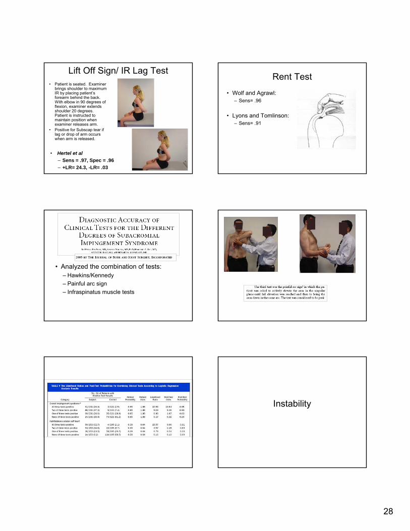
Lift Off Sign/ IR Lag Test• Patient is seated. Examiner
brings shoulder to maximum IR by placing patient’s forearm behind the back. With elbow in 90 degrees of flexion, examiner extends shoulder 20 degrees. Patient is instructed to maintain position when examiner releases arm.
• Positive for Subscap tear if lag or drop of arm occurs when arm is released.
• Hertel et al– Sens = .97, Spec = .96– +LR= 24.3, -LR= .03
Rent Test
• Wolf and Agrawl:– Sens= .96
• Lyons and Tomlinson:– Sens= .91
• Analyzed the combination of tests:– Hawkins/Kennedy– Painful arc sign– Infraspinatus muscle tests
Instability

29
Anterior Release Test• Performed in supine with
affected shoulder over edge of table. Arm is abducted 90 degrees with elbow flexed 90 degrees and a posterior force is placed on the humeral head while the arm is maximally ER. Humeral head is then released.
• Test is positive if a sudden pain or typical sxs are reproduced.
• Gross et al– Sens = .92, Spec = .89
Labral Tear
Biceps Load Test• Patient is supine with
examiner grasping wrist and elbow. Arm is abducted to 90 degrees with elbow flexed to 90 degrees and forearm supinated. Examiner ER arm until patient becomes apprehensive and then the patient is asked to flex the elbow against the examiner’s resistance.
• Test is positive if patient’s apprehension remains or pain is produced.
• Kim et al– Sens = .90, Spec = .97– +LR = 30, -LR of .10
Biceps Load II• Patient is supine with
examiner grasping wrist and elbow. Arm is elevated 120 degrees and fully ER with elbow held in 90 degrees of flexion and forearm supinated. Examiner then resists elbow flexion by the patient.
• Test is positive if resisted elbow flexion causes pain.
• Kim et al– Sens = .90, Spec = .97– +LR = 30, -LR of .10 What about the AC joint?

30
Active Compression Test
• While standing, patient is asked to flex arm to 90 degrees with elbow in full extension. Patient then adducts arm 10 degrees and IR humerus. Examiner applies downward force to arm as patient resists. Patient then fully supinates arm and repeats procedure.
• Positive if pain localized to AC
Cross-Body Abduction Test
• Therapists flexes shoulder to 90 degrees and adducts across body
• Positive if pain is elicited at the AC joint
• Sens= .77• Spec= .79
AC Resisted Extension Test
• Patient is standing with arm flexed to 90 degrees and elbow bent to 90 degrees
• Patient is asked to extend arm against resistance
• Positive if pain is reproduced in the AC joint
• Sens= .72• Spec= .85
Questions?Structural Differentiation Diagnosis of the Upper
and Lower Quarter
Duke UniversityDurham NC, USA
Concord NH, USA

31
Where do we start?Patient One Patient Two
Prevalence of Low Back Pain
• In 2002, point prevalence was 26.4% (Deyo et al. Spine 2006) (lifetime = 70%)
• 45 and older > than 44 and younger• Three year incidence predicted by
– Depression (OR = 2.3)– Nerve root contact on MRI (OR = 1.9)– Central Stenosis (OR = 1.8)
Jarvik et al. Three-year incidence of low back pain in an initially asymptomaticCohort. Spine. 2005;30:1541-8.
Epidemiology• 39% Discogenic Pain (Schwarzer et al.
1995)• 15-40% Zygapopyseal joints (Schwarzer et
al. 1995; Manchikanti et al. 1999)• 6-13% Sacroiliac Pain (Schwarzer et al.
1995; Bogduk 1995)• 2-10% (Deyo et al. 1994; Govind 2004)• 33% Undefined (Bogduk)
Laslett M. In: Movement, stability, & lumbopelvic pain. Churchill Livingstone. 2007
Ruling out the Lumbar Spine
• Should focus here first since the prevalence is high
• Low back pain is not homogenous• Factors outside physical components can
amplify or are associated with LBP
History and Examination Combined?
• Pain not relieved by supine sensitivity = 80 for ankylosing spondylitis
• Morning back stiffness sensitivity = 64 for ankylosing spondylitis
• Pain duration >3 months sensitivity = 71 for ankylosing spondylitis
• Pseudoclaudication sensitivity = 60 for stenosis
Deyo et al. What can history and physical examination tell use about low Back pain. JAMA. 1992;268:760-6.
Discogenic Pain
• Centralization or peripheralization sensitivity = 92-95 (Donelson et al. 1997)
• Sciatica sensitivity = 95• Ankle dorsiflexion weakness sensitivity =
35• Great toe weakness sensitivity = 50• Impaired ankle reflex sensitivity = 50• Plantar flexion weakness sensitivity = 6
Deyo RA, Jarvik JG. Diagnostic evaluation of low back pain with emphasis on imaging. Ann Intern Med. 2002;137:586-97..

32
Straight Leg Raise
Cook C. Physical Examination Tests for Neurological Screening. In: Cook C, Hegedus E.Physical Examination Tests: An Evidence Based Approach. Upper Saddle River, NJ; Prentice Hall: 2007.
The Slump Sit Test
Cook C. Physical Examination Tests for Neurological Screening. In: Cook C, Hegedus E.Physical Examination Tests: An Evidence Based Approach. Upper Saddle River, NJ; Prentice Hall: 2007.
Ruling out the ZygapophysealJoints
5/7 Revel’s Criteria sensitivity = 13• age over 65 years• pain well relieved by recumbencyno exacerbation of pain with: • coughing and sneezing, • forward flexion,• extension, • rising from flexion • extension-rotation test
Laslett et al. BMC Musculoskeletal Disorders 2004, 5:43
Ruling out Stenosis• Age > 65 years sensitivity = 77%• Pain below buttocks sensitivity = 88• Leg symptoms worse with walking, better
sitting sensitivity = 81• Best posture for symptoms is sitting
sensitivity = 89• Worst posture for symptoms is walking or
standing sensitivity = 89
Katz et al. Arth Rheum. 1995;38:1236-41.Fritz et al. J Spinal Disorders. 1997;10:410-6.
Ruling out Stenosis
• Age>65 sensitivity = 77• Severe lower extremity pain sensitivity =
65• Symptoms worsen when walking
sensitivity = 71• Numbness sensitivity = 63
Deyo RA, Jarvik JG. Diagnostic evaluation of low back pain with emphasis on imaging. Ann Intern Med. 2002;137:586-97..
Quadrant?• Used to clear the
lumbar spine• Compresses the
foramen on one side

33
Provocative Mobilization (PA’s)
Cook C. Physical Examination Tests for Neurological Screening. In: Cook C, Hegedus E. Physical Examination Tests: An Evidence Based Approach. Upper Saddle River, NJ; Prentice Hall: 2007.
Overpressures?
SIJ Definition
• SIJ Dysfunction is associated with pain that arises from the sacroiliac joint and is caused by asymmetry, or alteration in the stability of the Sacroiliac joint.
• Damen et al. The prognostic value of asymmetric laxity of the sacroiliac joints in pregnancy related pelvic pain. Spine. 2002;27:2820-2824.
Pelvic Girdle Pain Definition• PGP arises in relation to pregnancy, trauma,
osteo-arthrosis and arthritis. • Pain is experienced between the posterior iliac
crest and gluteal fold, particularly in the vicinity of the SIJ.
• Pain may radiate in the posterior thigh and can also occur in conjunction with/or separately in the symphysis.
• The diagnosis of PGP can be reached after exclusion of lumbar causes.
Vleeming et al. European Guidelines on the Diagnosis and Treatment of Pelvic Girdle Pain. 2005
Pseudodisease Findings• CT scan demonstrates 57.5% sensitivity and
69% specificity (Elgafy et al. CORR 2001)• Does asymmetry truly equate to pathology?• If we look hard enough, can’t we find
something; especially if we know it’s there
Special Tests
• No examination movements are exclusive to the SIJ/Pelvis (Cook 2007)
• Most special tests (when performed in isolation) are poorly diagnostic and lack reliability (Cook 2007a and b)
• For screening, the lower prevalence (6-13%) requires higher sensitivities for better screening accuracy

34
Clinical Examination Findings
Cook et al. 2007. Classification of PGP. JMPT, 2007
.88---1.0.13Sit to Stand
.682.63.83.44Lunge
.971.1.67.35Single Leg Stance
.71---1.0.29Step Up
.921.41.83.24Centralization
LR-LR+SpecSensExamination Finding
Clinical Examination Findings
.821.2.50.59Pubic Symphysis Palp
.881.6.83.27Rot Innom
.76---1.0.24Deep Squat
.45---1.0.55Hip PROM
.82.0.83.33MMT Hip
LR-LR+SpecSensExamination Finding
Cook et al. 2007. Classification of PGP. JMPT, 2007
Thigh Thrust
Cook C. Physical Examination Tests for Neurological Screening. In: Cook C, Hegedus E.Physical Examination Tests: An Evidence Based Approach. Upper Saddle River, NJ; Prentice Hall: 2007.
ASLR
Cook C. Physical Examination Tests for Neurological Screening. In: Cook C, Hegedus E.Physical Examination Tests: An Evidence Based Approach. Upper Saddle River, NJ; Prentice Hall: 2007.
Resisted Hip Abduction
Cook C. Physical Examination Tests for Neurological Screening. In: Cook C, Hegedus E.Physical Examination Tests: An Evidence Based Approach. Upper Saddle River, NJ; Prentice Hall: 2007.
Fortin Finger Test
Cook C. Physical Examination Tests for Neurological Screening. In: Cook C, Hegedus E.Physical Examination Tests: An Evidence Based Approach. Upper Saddle River, NJ; Prentice Hall: 2007.

35
Pubic Symphysis Palpation
Cook C. Physical Examination Tests for Neurological Screening. In: Cook C, Hegedus E.Physical Examination Tests: An Evidence Based Approach. Upper Saddle River, NJ; Prentice Hall: 2007.
Motion Palpation Tests?
Cook C. Physical Examination Tests for Neurological Screening. In: Cook C, Hegedus E.Physical Examination Tests: An Evidence Based Approach. Upper Saddle River, NJ; Prentice Hall: 2007.
Combinations of Findings Prevalence of Hip Pain• Period Prevalence (1 year)
– Men = 7.1%– Women = 12.7%
• Point Prevalence – Men = 4.3%– Women = 10%
• Persistent Chronic Pain– Men = 1.9%– Women = 4.7%
Wijnhoven et al. Prevalence of musculoskeletal disorders is systematicallyHigher in women than men. Clin J Pain. 2006;22:717-24.
Ruling out the Hip• Hip Labrum
– Pain in sitting.– Presence of clicking or popping during gait,
squatting or other activities.– Presence of a click during active or passive motion
of the hip• Femoral Acetabular Impingement
– <IR during hip flexion– Mechanical groin pain (Laude et al. 2007)
Cook C. Physical Examination Tests for Neurological Screening. In: Cook C, Hegedus E.Physical Examination Tests: An Evidence Based Approach. Upper Saddle River, NJ; Prentice Hall: 2007.
Scour/Impingement Test
Cook C. Physical Examination Tests for Neurological Screening. In: Cook C, Hegedus E.Physical Examination Tests: An Evidence Based Approach. Upper Saddle River, NJ; Prentice Hall: 2007.
36
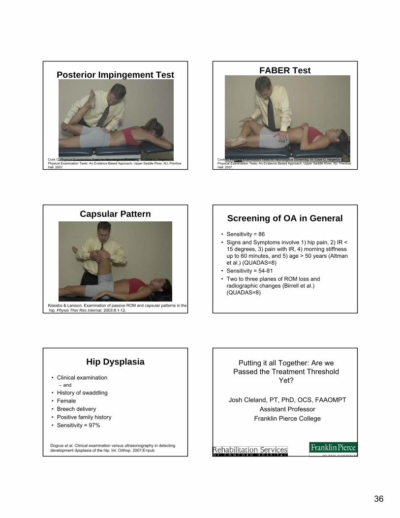
Posterior Impingement Test
Cook C. Physical Examination Tests for Neurological Screening. In: Cook C, Hegedus E.Physical Examination Tests: An Evidence Based Approach. Upper Saddle River, NJ; Prentice Hall: 2007.
FABER Test
Cook C. Physical Examination Tests for Neurological Screening. In: Cook C, Hegedus E.Physical Examination Tests: An Evidence Based Approach. Upper Saddle River, NJ; Prentice Hall: 2007.
Capsular Pattern
Klassbo & Larsson. Examination of passive ROM and capsular patterns in thehip. Physio Ther Res Internat. 2003:8:1-12.
Screening of OA in General• Sensitivity = 86• Signs and Symptoms involve 1) hip pain, 2) IR <
15 degrees, 3) pain with IR, 4) morning stiffness up to 60 minutes, and 5) age > 50 years (Altman et al.) (QUADAS=8)
• Sensitivity = 54-81• Two to three planes of ROM loss and
radiographic changes (Birrell et al.) (QUADAS=8)
Hip Dysplasia• Clinical examination
– and• History of swaddling• Female• Breech delivery• Positive family history• Sensitivity = 97%
Dogrue et al. Clinical examination versus ultrasonography in detecting development dysplasia of the hip. Int. Orthop. 2007;E=pub.
Putting it all Together: Are we Passed the Treatment Threshold
Yet?
Josh Cleland, PT, PhD, OCS, FAAOMPTAssistant Professor
Franklin Pierce College

37
Treatment Threshold
• The point at which the examination and evaluation process stops and treatment begins.
• How do we determine this?
PROBABILITY OF DISEASE
InformationalContribution
Pre-test Probability Post-test
Probability
Treatment Threshold
0% 50% 100%
Likelihood Ratios
• Quantify the direction and magnitude of change in the pretest probability based on the test result.
• Provide the best information needed to select the test that will surpass the threshold for action. Rarely alters
probability to an important degree
.5-11-2
Small shifts in probability
.2-.52-5
Moderate shifts in probability
.1-.25-10
Large and conclusive shifts in probability
< .1> 10
Interpretation-LR+LR
Pretest Probability
Based on a number of factors
• Age• Symptoms• Epidemiological data • Clinical experience

38
Evidence to Practice
• Nomogram– Graphical tool– Estimates overall
probability of disease based on diagnostic testing results
– Pretest probability = 20%– +LR = 8– Posttest probability ???
• A 19 year-old baseball pitcher
• Shoulder pain 3 months duration.
• Symptoms are exacerbated during any overhand throwing motion.
• Reports hearing a “click”in his shoulder during the wind-up phase of pitching.
• 15% pretest probability of a labral tear.
• You select the O’Brien test as described by Guanche and Jones.
• +LR = 2.33.
Case Example
• You decide to select another special test. This time you select to use the Anterior Slide Test as described by Kibler et al which has a +LR of 9.75.
Clinical Prediction Rules
But as clinicians we use combinations of tests and
measures?
Clinical Prediction Rules
• Decision-making tools for clinicians, containing variables from the history, physical exam, and simple diagnostic tests.
• Improves the clinician's accuracy in predicting a diagnosis or expected outcome.
• Combines the diagnostic properties of sensitivity, specificity and likelihood ratios.

39
Developing a CPR
Step 1 DerivationId. Factors with predictive value
Step 2 ValidationEvidence of reproducible accuracy
Narrow validation vs. Broad validationApply to pop. & Multiple settingssimilar setting with varying as step 1 prevalence.
Step 3: Impact analysisEvidence the rule changes behaviors& improves pt outcomes &/or reduces costs.
Can We Use CPRs to Guide Decision-Making in Patients with Upper Quarter Symptoms?
Can we surpass the Treatment Threshold for the Identification of Cervical
Radiculopathy?
Purposes
• To determine the measurement properties of clinical examination items used in the management of CR– Reliability– Validity (concurrent)
• To develop a clinical prediction rule for the diagnosis of CR
Subject Recruitment
• Referred for EMG/NCS testing of suspected CR or CTS
• No prior EMG/NCS test for condition
• Aged 18 – 70 yrs*• Sx duration ≥ 1 month • No condition limiting
UE function
• No prior surgery or fracture of neck or wrist
• No peripheral neuropathy
• Not off work > 6mo• No bilateral radiating
arm pain

40
Enter study?
YES
EXITNO
EMG/NCS
PT Hx & UQ/Neck Eval Proc.(Rater 1 &2, Blinded to EMG results & Dx)
YES NO
+ Dx Radic. Or CTS?(Hx & EMG/NCS)
Patient completes NDI, HSFS, VAS, FABQ,& Hand Diagram (EMGer blinded)
Initial: Reliability & Dx accuracy-Clinical Examination -Self-report Instruments (SRI)
(single & in clusters)Metric: Sn/Sp & Likelihood RatiosGold standard: Neural Impairment re: positive EMG/NCS study
Prescribed CR. RxSurg Non-Surg
DATA ANALYSIS
Pt. referred for radiculopathy &/or CTS that meet entry criteria.
*EMG lab Technician, Resident, Staff informs patient of study, invites patient to participate
Referral Sources:10 OrthopedicsNeurosurgeryHand clinic/surgery2 0 FPC, other
Prescribed CTS. RxSurg Non-Surg
Reference Criterion: EMG/NCS Procedures
• EMG– Needle EMG: selected extremity
– NCS– Median & Ulnar sensory/motor studies
Clinical Examination: Tests & Measures
• Conventional Neurologic Examination of the UE– Muscle-stretch Reflexes – Sensory status (dermatome & median nerve
field, sharp/dull)– Motor status (MMT)
• Scaled Measurements– Cervical ROM and wrist diameter
**.23.49.89Involved side Cervical Rot <60**.27.41.89Cervical Flex <55
1.3.12.22.97+ULTT A4.4**.90.44Distraction
3.5**.94.22Valsalva2.1**.92.17Shld Abd3.5**.86.50Spurling’s A3.7**.94.24MMT Biceps
4.9**.95.24MSR Biceps2.1**.86.29Derm C52.2--.71.65“Neck mvmnt improves”
2.3--.84.38“Where most bothersome”neck/scapula
LR+LR-SpSnTEST
+ ULTT A+ Cervical Rot Involved < 600
+ Spurling’s A+ Neck Distraction
But what is the best combination of tests and measures?
30.3.99.244
6.1.94.393
.88.56.392
+LRSpecificitySensitivityNumber of Positive Findings
41
Decreased Biceps MSR
- ULTT A
+ 4 Items of the TIC
Am I screening out or ruling in the disorder?
Ok- so we are pretty sure this patient has cervical radiculopathy
now what?
•Case Series, N=15, cervical radiculopathy•12 weeks of treatment (mean = 11 sessions)
•Start 18#, increase 1-2 # / session based on response•Duration 15 minutes: Cycle 30 sec on / 10 sec off
•7 of 15 (53%) got complete resolution of symptoms (sxs <12 wks) and 3 no change
6 Patients also received mobilization/manipulation
Dec 2005
Intermittent Cervical Traction Results
• Mean age: – 51.7 (sd 8.2)
• Duration of symptoms:– 20.4 weeks (range 8-
52)• Average number of PT
sessions:– 7.1 (range 6-10)
28 Consecutive PatientsScreened for Eligibility
Satisfied Eligibility Criteria
Yes11 Patients
No17 Patients
11 Patients Agreed to Participate

42
Numeric Pain Rating (0-10)
0123456789
1 2 3 5 6 7 8 9 10 11 12
Baseline Discharge 6-month follow -up
MCID= 2 PointsChilds et al, Spine, 2005
Neck Disability Index
05
101520253035404550
1 2 3 4 5 6 7 8 9 10 11Baseline Discharge 6-month follow -up
MCID= 7 pointsCleland et al, Spine, 2005
Can we Identify Patients with Mechanical Neck Pain?
• Purpose: to determine if a clinician could identify patients with neck pain

43
Clinical Tests
• Manual Procedures:– OA– AA– C2-C7
• Spurling test
• CROM
Results
1.2.979.188.9Total CROM
.293.477.377.8Spurling’s
.3990.972.2MEP
-LR+LRSpecSensTest
Ok- so the patient has mechanical neck pain: Now What?
Can CPRs help to identify patients with neck pain that are likely to
benefit from thrust manipulation?
The Use of Spinal Manipulation in the Management of Neck Pain
thrust & non-thrust techniques
Guide to PT Practice:“A manual therapy technique comprised of a continuum of skilled passive movements to joints and/or related soft tissues that are applied at varying speeds and amplitudes, including a small amplitude/high velocity therapeutic movement”
• 100 patients with neck pain– All received cervical
spine manipulation
• Success defined as– +4 on GROC– 50% pain reduction– Satisfaction
• Mean NPRS 5.4• Mean NDI 11.7
The Cervical Spine Thrust Manipulation Rule
Variables in C-spine CPR
• NDI < 11.5• Bilateral involvement• Not performing
sedentary work > 5h/day
• Feeling better with neck movement
• Extension does not worsen
• No cervical radic
Infinite5+5.3341.933.202.071+LRVariables

44
3/6+LR= 1.9
4/6+LR= 5.3
5/6 and 6/6 +LR Infinite
C-Spine ManipCervical Spine ManipulationCervical Spine Manipulation
• Only 37% of therapists who manipulate target the cervical spine in this patient population?
• (Hurley et al 2002)
The Explanation?• Survey of 129 manipulative therapists in UK
Adams and Sims (Physiother Res Int, 1998)
#1 - Anxiety about possible complications
Perhaps clinicians believe that the potential benefits Perhaps clinicians believe that the potential benefits do not outweigh the risk?do not outweigh the risk?
Thoracic Spinal Manipulation: A Possible Alternative?
• Perhaps disturbances in joint mobility in the thoracic spine contribute to mechanical neck pain.
• (Norlander et al 1995, 1996, 1998)
• Can you obtain similar benefits while reducing the associated risks by directing techniques to the thoracic spine instead of the neck?
19 received thoracic manip
17 received placebo
P < .01Change ScoresChange Scores
Manipulation: 15.5 mm Manipulation: 15.5 mm Placebo group: 4.2 mmPlacebo group: 4.2 mm
2005
Exposure Outcome
Reference Standard for Success
Please rate the overall condition of your neck from the time that you began treatment until now
A very great deal worse About the same A very great deal betterA great deal worse A great deal betterQuite a bit worse Quite a bit betterModerately worse Moderately betterSomewhat worse Somewhat betterA little bit worse A little bit betterA tiny bit worse A tiny bit better
(almost the same) (almost the same)
Jaeschke et al, Controlled Clinical Trials, 1989

45
Informed ConsentExamination
YESSuccess
NO
THORACIC MANIPULATION
THORACIC MANIPULATION
SuccessYESNONon-
Success
Success on GROC
Success on GROC
Visit 1
Visit 2
Visit 3
Baseline Examination
• Numerous self report measures– NDI– NPRS– Body diagram– FABQ– TSK
• Demographics• Extensive Standardized History• Extensive Physical Examination
• 22 patients participated in a second clinical exam – Laupacis et al, JAMA, 1997
First Treatment SessionThoracic Manipulation
12
3
Range of Motion Exercise
• 10 repetitions each side; 3-4 times daily
• Maintain Usual Activity Within Limits of Pain
Erhard RE. The Spinal Exercise Handbook, 1998
Second Treatment
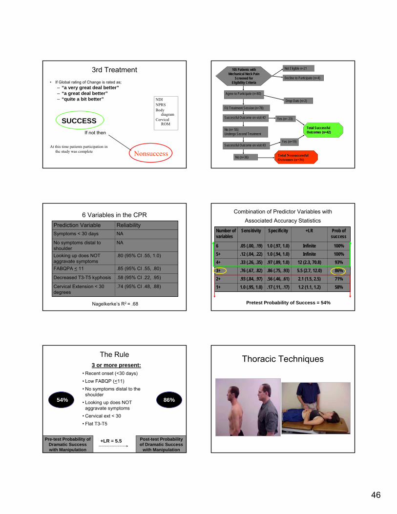
• If Global rating of Change is rated as;– “a very great deal better”– “a great deal better”– “quite a bit better”
SUCCESSIf not then
NDINPRSBody diagramFABQTSKCervical
ROM
2nd Treatment Session
46
3rd Treatment• If Global rating of Change is rated as;
– “a very great deal better”– “a great deal better”– “quite a bit better”
SUCCESS
At this time patients participation in the study was complete
NDINPRSBody
diagramCervical
ROM
Nonsuccess
If not then
105 Patients with Mechanical Neck Pain
Screened for Eligibility Criteria
Not Eligible n=21
Decline to Participate (n=4)
Agree to Participate (n=80)
No (n= 55)Undergo Second Treatment
No (n=36)
FU Treatment Session (n=78)
Drop Outs (n=2)
Successful Outcome on visit #2 Yes (n= 23)
Successful Outcome on visit #3 Yes (n=19)
Total Successful Outcomes (n=42)
Total NonsuccessfulOutcomes (n=36)
6 Variables in the CPR
.74 (95% CI .48, .88)Cervical Extension < 30 degrees
.58 (95% CI .22, .95)Decreased T3-T5 kyphosis
.85 (95% CI .55, .80)FABQPA < 11
.80 (95% CI .55, 1.0)Looking up does NOT aggravate symptoms
NANo symptoms distal to shoulder
NASymptoms < 30 days
ReliabilityPrediction Variable
Nagelkerke’s R2 = .68
Combination of Predictor Variables with Associated Accuracy Statistics
58%1.2 (1.1, 1.2).17 (.11, .17)1.0 (.95, 1.0)1+71%2.1 (1.5, 2.5).56 (.46, .61).93 (.84, .97)2+86%5.5 (2.7, 12.0).86 (.75, .93).76 (.67, .82)3+93%12 (2.3, 70.8).97 (.89, 1.0).33 (.26, .35)4+100%Infinite 1.0 (.94, 1.0).12 (.04, .22)5+100%Infinite 1.0 (.97, 1.0).05 (.00, .19)6
Prob of success
+LR SpecificitySensitivityNumber of variables
Pretest Probability of Success = 54%
The Rule
Pre-test Probability of Dramatic Success with Manipulation
54%
3 or more present:• Recent onset (<30 days)• Low FABQP (<11)• No symptoms distal to the
shoulder• Looking up does NOT
aggravate symptoms• Cervical ext < 30• Flat T3-T5
86%
Post-test Probability of Dramatic Success
with Manipulation
+LR = 5.5
Thoracic Techniques

47
The Cervical Spine Thrust Manipulation Rule
Variables in C-spine CPR• NDI < 11.5• Bilateral involvement• Not performing sedentary work
> 5h/day• Feeling better with neck
movement• Extension does not worsen• No cervical radic
Variables in T-spine CPR• FABQPA < 11• Onset < 30 days• Decreased kyphosis T3-T5• Cervical extension < 30 • Looking up does not
aggravate• No symptoms distal to
shoulder
Can CPRs be useful in determining the prognosis of patients with shoulder pain? • Age
• Gender• Education• Duration • Onset• Precipitating cause• Prior history• Neck pain• Shoulder pain
• Workload• Physical activity• Coping• Fear-avoidance• Kinesiophobia
587 patients with first episode of shoulder pain

48
350 patients workers with first episode of shoulder pain• Age• Gender• Education• Duration • Onset• Precipitating cause• Prior history• Neck pain• Shoulder pain
• Workload• Physical activity• Coping• Fear-avoidance• Kinesiophobia
What about the use of CPRs and a classification system for the
management of LBP?
Low Back Pain ClassificationsOriginal Classification Criteria
Stabilization exercises
Activities to Promote
Centralization
Specific Exercise Stabilization Traction
Centralization phenomenon
Frequent prior episodes,
hypermobility
Leg pain, neurological
signs
Manipulation and exercise
Manipulation
SI special tests“closing”/ ”opening”
Mechanical/ auto-traction

49
Enroll into Study
Examination
50% Reduction in ODQ
YESSuccess
NO
Spinal Manipulation
SI Region Manipulation
50% Reduction in ODQ Success
YESNO
Non-Success
Visit 2
Visit 3
Examination
Visit 1
Flynn et al, Spine, 2002
Spinal Manipulation Intervention
• Translate the pelvis towards you and maximally side-bend the patient’s lower extremities and trunk to the right
• Without losing the right sidebending lift & rotate the trunk so the patient rests on their left shoulder
• Contact the patient’s right ASIS with your left hand
• Grasp the top shoulder and scapula with your right hand and rotate the trunk to the left while maintaining the right side-bending
• Once the right ASIS starts to elevate, perform a smooth thrust in an anterior to posterior direction
Spinal Manipulation CPR
Hip IR > 35 degrees Lumbar hypomobility
Physical Exam
HistoryFABQWK < 19Symptoms < 16 days
No symptoms distal to the knee
Flynn et al, Spine, 2002
Predicting Success with Manipulation
Pre-test Probability of Dramatic Success with Manipulation
45%
4 or more present:• Recent onset (<16
days)• Low FABQ (<19)• No symptoms below
knee• Lumbar stiffness• Good hip IR (>350)
95%
Post-test Probability of Dramatic Success
with Manipulation
+LR = 24.3
Pre-Test Probability of
Success = 45%
(+) LR = 24.4
At least 4/5in CPR
Post-TestProbability of
Success
= 95%Maj. John D. Childs, PT, PhD, MBAJulie M. Fritz, PT, PhDTimothy W. Flynn, PT, PhDJames J. Irrgang, PT, PhDKevin K. Johnson, PTGuy R. Majkowski, PTAnthony Delitto, PT, PhD
Department of Physical TherapyUniversity of Pittsburgh
Pittsburgh, PA
Department of Physical TherapyWilford Hall Medical Center
San Antonio, TX

50
•First two sessions (wk 1):
• Spinal manipulation
• AROM exercise
•Final 3 sessions:
• Stabilization exercises
Manipulation Treatment GroupExercise Stabilization
Validation of the Rule
Pre-test Probability of Dramatic Success with Manipulation
45%
4 or more present:• Recent onset (<16
days)• Low FABQ (<19)• No symptoms below
knee• Lumbar stiffness• Good hip IR (>350)
92%
Post-test Probability of Dramatic Success
with Manipulation
+LR = 13.2
Group*CPR*Time Interaction
05
101520253035404550
Baseline 1-week 4-weeks 6-months
OD
Q S
core + CPR (manip)
- CPR (manip)+ CPR (exercise)- CPR (exercise)
*P<0.001
*Significant differences between +CPR/manip group and +CPR/exercise group and –CPR/manip group. No difference between +CPR/exercise and –CPR/exercise groups
0
1
2
3
4
5
6
7
Baseline One week Four weeks Six months
Time
NPR
S Sc
ore
+ CPR (Manipulation Group)- CPR (Manipulation Group)+ CPR (Exercise Group)- CPR (Exercise Group)
Significant differences between +CPR/manip group and +CPR/exercise group and –CPR/manip group. No difference between +CPR/exercise and –CPR/exercise groups
Group*CPR*Time Interaction Do Subgroups Matter?
Improvement on the ODQ for the 4-week follow-up based on the patient’s status with respect to the spinal manipulation prediction rule.
0
10
20
30
40
50
60
Four-week follow-up period
Manipulation Group (All subjects)+CPR (Manipulation Group)-CPR (Manipulation Group)Exercise Group (All subjects)+CPR (Exercise Group)-CPR (Exercise Group)All
manip All exerc
Got it –needed it
Got it –Didn’t needed it

51
This is way cool!!!
Validation of the Rule
Pre-test Probability of Dramatic Success with Manipulation
45%
2 factors present:• Recent onset (<16
days)• No symptoms below
knee
91%
Post-test Probability of Dramatic Success
with Manipulation+LR = 12.6
Low Back Pain Classifications
Stabilization exercises
Activities to Promote
Centralization
Specific Exercise Stabilization Traction
Centralization phenomenon
Frequent prior episodes,
hypermobility
Leg pain, neurological
signs
Manipulation and exercise
Manipulation
“closing”/ ”opening”
SI special tests
Mechanical/ auto-traction
•No symptoms below the knee
•Recent symptoms
•Hypomobility
•Low Fear-Avoidance
•More hip IR
?
Admission of Eligible PatientChief c/o LBP without signs of radiculopathy or prior fusion surgery
BASELINE EXAMINATIONBaseline Oswestry Assessment
Standardized Stabilization Exercise Program
2x/week – 8 weeks
FOLLOW-UP EXAMINATIONEight-Week Oswestry Assessment
Exercise InterventionTransversus Abdominus
Multifidus/ Erector Spinae
Quadratus LumborumOblique Abdominals

52
Stabilization Treatment
Quadruped Arm Lifts with Bracing
Quadruped Leg Lifts with Bracing
Quadruped Alternate Arm and Leg Lifts with Bracing
Multifidus/ Erector Spinae
Stabilization TreatmentAbdominal Bracing
Bracing with Heel Slides
Bracing With Leg Lifts
Bracing with Bridging
Bracing in Standing
Bracing with Standing Row Exercise
Bracing with Walking
Transversus Abdominus
Stabilization Treatment
Side Support with Knees Flexed
Side Support with Knees Extended
Quadratus Lumborum
Side Support with Knees Flexed
Side Support with Knees Extended
Hanging Leg Lifts
Oblique Abdominals
05
10152025303540
Success (18) Improved (21) Failed (15)
Initial
Final
Average number of visits: 9.7 + 2.2
RESULTS - Change in Oswestry
Predicting Dramatic Success
Pre-test Probability of Dramatic Success with
Stabilization
33%
3 or more present:• Prone instability test
• Aberrant motions
• Average SLR >910
• Age < 40
Post-test Probability of Dramatic Success with
Stabilization
67%
Predicting Improvement
Pre-test Probability of Some Improvement with
Stabilization
72%
2 or more present:• Prone instability test
• Aberrant motions
• Hypermobility
• FABQ-PA ≤ 8
Post-test Probability of Some Improvement with
Stabilization
94%

53
Low Back Pain Classifications
Stabilization exercises
Activities to Promote
Centralization
Specific Exercise Stabilization Traction
Centralization phenomenon
Frequent prior episodes,
hypermobility
Leg pain, neurological
signs
Manipulation and exercise
Manipulation
“closing”/ ”opening”
SI special tests
Mechanical/ auto-traction
•No symptoms below the knee
•Recent symptoms
•Hypomobility
•Low Fear-Avoidance
•More hip IR
• Prone instability test
• Aberrant motions
• Hypermobility
• Younger age
• Greater SLR ROM
RCT
• The role of “patient specific” exercises in managing LBP is controversial.
• Multicenter RCT to determine if LBP subgroups respond differently to contrasting exercise prescriptions– 312 patients with LBP underwent standardized examination to elicit a
“directional preference” (DP)– Subjects randomized to: 1) directional exercises “matching” their DP,
2) exercises directionally “opposite”, or 3) “nondirectional” exercises.• Outcome measures included pain intensity, location,
disability, medication use, degree of recovery, depression, and work interference.
RCT
Significantly greater improvements occurred in matched subjects compared with both other treatment groups in every outcome (P<0.001), including a threefold decrease in medication use.
Browder DA, Childs JD, Cleland JC, Fritz JM. Effectiveness of an extension-oriented treatment approach in a subgroup of
patients with low back pain: a randomized clinical trial.
• Randomized clinical trial of 48 patients with lumbar radiculopathy:– Average age 39 years– Median symptom duration ~60 days– 56% with symptoms distal to the knee– 10% previous back surgery
• Randomized to receive:– Extension exercise and mobilization protocol– Stabilization exercise protocol
0
10
20
30
40
50
Baseline 1 week 4 weeks 6 months
Strengthening
EOTA
*
*
*
Adjusted ODI scores at each assessment point (*indicates significant difference between groups in change from baseline scores (p<0.05))

54
Subjects
• 76 subjects with work-related low back pain:– Less than 3 weeks duration– 30 female, 46 male– Average age 38.0 + 10.1 years– All subjects placed on work modifications and
referred for physical therapy
Patient with acute LBP
Baseline Evaluation
R
AHCPR Group Classification Group
All patients treated based on AHCPR Guidelines
Patients receive treatment specific to classification
Oswestry Scores
43.34
31.67
43.48
22.18
05
1015202530354045
AHCPR Classification
InitialFour Week
ANCOVA
p = 0.031*
Return to Work Status After Four Weeks
Guideline Group (n=36)
Classification Group (n=41)
No Work Restrictions
21 34
Continued Work Restrictions
15 7
** p = 0.017
Purpose
To determine if patients with LBP would demonstrate greater functional improvement based on …
the initial treatment received (regardless of classification),
the classification sub-grouping(regardless of treatment),
or the interaction of the two factors.

55
Classification
RANDOM ASSIGNMENT
Manipulation Specific Exercise Stabilization
Match Match UnmatchUnmatch UnmatchMatch
LBP Classification and Treatment Process: Randomized Trial
Acute Low Back PainDoes the patient:Have a recent onset of symptoms (< 16 days)AND…no symptoms distal to the knee
YES MANIPULATION Classification
Does the patient have at least 3 of the following:Average SLR ROM >910
Positive prone instability testPositive aberrant movementsAge <40 years
YES STABILIZATIONClassification
NO
Does the patient:Centralize with 2 or more movements in the same direction (i.e., flexion or extension)OR…Centralize with a movement in one direction and peripheralize with an opposite movement
SPECIFIC EXERCISE
Classification
NO
NO
YES
Which sub-group does the patient fit best?
• Low back pain only (no distal sx)
• Status Quo with all movements
• Preference for one posture
• Centralization with motion testing
• Peripheraliza-tion in direction opposite centralization
• Discrepancy in SLR ROM (>100)
• Low FABQ scores (activity scale < 9)
• Hypermobility with spring testing
• Positive prone instability test
• Aberrant motions present
• Increasing episode frequency
• Younger age (<40)
• 3 or more prior episodes
• Greater SLR ROM (>910
bil t l)
• Symptoms below the knee
• Increasing episode frequency
• Greater SLR ROM (>810
bilateral)• Peripheraliz
a-tion with motion testing
• No pain with spring testing
• More recent onset (<16 days)
• Hypomobility with spring testing
• Low back pain only (no distal sx)
• Low FABQ scores (work scale <19)
Factors against
Factors favoring
Factors against
Factors favoring
Factors against
Factors favoring
Specific ExerciseStabilizationMobilization / Manipulation Treatment
Patients treated 2x/week for Four Weeks– Stage I randomly assigned Rx (Manip,
SpEx, Stab) – (maximum visits = 8)
Progressed to Stage II if ODI score improved:– to <20% – or improved 1/3 from initial ODI score
Stage II was Multi-modal exercise
3-Way vs. 2-Way Interactions
MatchedUnmatchedUnmatchedSpecific Exercise
UnmatchedMatchedUnmatchedImmobilization
UnmatchedUnmatchedMatchedMobilization
Specific Exercise
ImmobilizationMobilization
Sub-group Classification
Trea
tmen
ts
Classification sub-groups237
Duration of symptoms >90 days
203Age >65 or <18
178Neurological
signs present
119Oswestry < 25%
47Post-surgical
123Patients consenting
145Patients not consenting
50Patients matched to randomized
treatment group
73Patients not matched to
randomized treatment group
1052New patients with LBP referred to
participating clinics during the recruitment period
268Eligible patients
Results

56
Results
0
10
20
30
40
50
Pretest 4-week One-Year
MatchedUnmatched
p=0.029 p=0.006
DiscussionHypothesis supported:
Outcomes improved when initial treatment matched to a patient’s signs and symptoms using this sub-grouping method.
The outcome depended on the 3-way interaction of matching the “right”treatment to the patient’s “right” sub-group over time.
Homogenous vs Heterogeneous
What about CPRs for the Lower Extremity?
Development of a CPR to Identify Patients with Knee OA who Are Likely
to Benefit from Hip Mobilizations
• Physical Therapy; In Press
Patient Admitted
4 Hip Mobilizations
Evaluation, Functional Tests
≥30% in NPRS OR ≥ 3pt GRCS?
SUCCESSFAIL
Visit 1
Visit 2
48hrs f/u
Clinical Prediction Rule
.895.2.98.13Pain with Ipsilateral Hip Distraction
.726.0.95.32Ipsilateral Hip IR <17°
.726.0.95.32Ipsilateral Knee Flex <122°
.828.1.98.20Ipsilateral Pain or Paresthesia in Hip/Groin
.775.1.95.27Ipsilateral Anterior Thigh Pain
-LR+LRSpSnItem
• Combination of any 2 CPR items; +LR=12.9

57
Results
With pretest probability = 68%, post-test probability with positive test (any 2 CPR items) = 97%
Patellofemoral Pain
Taping CPR
• Pre-test/post-test design (n=50)
• Responders to CPR 1. Positive patellar
tilt test2. Tibial varum >5
degrees(+LR 4.4; 1.3, 12.3)
Orthotics and PFPS• OTC and custom orthotics have been found to
relieve anterior knee pain.– Sutlive, Phys Ther. 2004; Johnston, JOSPT, 2004
• Orthotic CPR developed to determine which patients would respond to off the shelf orthotics
1. Forefoot varus of 2 degrees or greater 2. Great toe extension less than 78 degrees3. Navicular drop test 3 mm or less
No combination of factors resulted in +LR >2.0
Adapted from Sutlive, Phys Ther 2004
Manipulation CPR
• Iverson et al (CSM, 2006) prospective study
• May predict which PFPS pts respond to lumbopelvic manipulation– 50 patients with PFPS– Intervention was lumbopelvic manipulation– Outcomes
Manipulation CPR
• Hip internal rotation difference >14o
• No patient reported stiffness sitting > 20 minutes
• Squatting most painful reported activity• Navicular drop > 3 mm• Ankle dorsiflexion > 16
owith knee flexed
Hip internal rotation difference > 14 o
and any of the 3 remaining 4 predictors is 98 % Specific and has a + LR of 10.0.

58
Clinical Decision Making
• Change in pre-test and post-test probability with subgroups of patients – Orthotics– Taping– Manipulation
Wow! That is a Lot of Evidence: Now What?
The Problem?
• Translation of evidence into clinical practice stalls.
How do we improve translation?
• 3 transitional blocks:– Impeded movement from basic science to
clinical studies– Impeded progress of clinical evidence into
practice standards – Progress of clinical evidence into routine
practice stalls
1st Translational Block
Phys Ther, Feb 2003
Khan et al, BMJ, 2002
2nd Translational Block

59
3rd Translational Block
• Translation of evidence and guidelines into clinical practice stalls.
Implementation Research
Innovation Diffusion
TimeTime
Use
rsU
s er s
Innovators 2.5%Innovators 2.5%
Early Adopters 13.5%Early Adopters 13.5%
Early Majority 34%Early Majority 34%
Late Majority 34%Late Majority 34%
Laggards 16%Laggards 16%
Berwick, Disseminating Innovations in Healthcare, JAMA 2003.
TimeTime
Use
rsU
s er s
Innovators 2.5%Innovators 2.5%
Early Adopters 13.5%Early Adopters 13.5%
Early Majority 34%Early Majority 34%
Late Majority 34%Late Majority 34%
Laggards 16%Laggards 16%
SkepticsSkeptics
Trend Trend setterssetters
Opinion leadersOpinion leaders
The Problem?
• The process of implementing research or guidelines into practice requires significant behavior changes.
• Usually those that are required to change are expected to do so without compelling financial or other advantageous.
Two Reasons People Will Change
• 1. They think something really good will happen if they do
• 2. They think something really bad will happen if they don’t

60
Questions?