Structural cavities are critical to balancing stability ...
11
Structural cavities are critical to balancing stability and activity of a membrane-integral enzyme Ruiqiong Guo a , Zixuan Cang b , Jiaqi Yao a , Miyeon Kim a , Erin Deans c,1 , Guowei Wei b , Seung-gu Kang d,2 , and Heedeok Hong a,c,2 a Department of Chemistry, Michigan State University, East Lansing, MI 48824; b Department of Mathematics, Michigan State University, East Lansing, MI 48824; c Department of Biochemistry & Molecular Biology, Michigan State University, East Lansing, MI 48824; and d Computational Biology Center, IBM Thomas J. Watson Research Center, Yorktown Heights, NY 10598 Edited by William F. DeGrado, University of California, San Francisco, CA, and approved July 30, 2020 (received for review October 25, 2019) Packing interaction is a critical driving force in the folding of helical membrane proteins. Despite the importance, packing defects (i.e., cavities including voids, pockets, and pores) are prevalent in membrane-integral enzymes, channels, transporters, and recep- tors, playing essential roles in function. Then, a question arises regarding how the two competing requirements, packing for sta- bility vs. cavities for function, are reconciled in membrane protein structures. Here, using the intramembrane protease GlpG of Escherichia coli as a model and cavity-filling mutation as a probe, we tested the impacts of native cavities on the thermodynamic stability and function of a membrane protein. We find several sta- bilizing mutations which induce substantial activity reduction without distorting the active site. Notably, these mutations are all mapped onto the regions of conformational flexibility and func- tional importance, indicating that the cavities facilitate functional movement of GlpG while compromising the stability. Experiment and molecular dynamics simulation suggest that the stabilization is induced by the coupling between enhanced protein packing and weakly unfavorable lipid desolvation, or solely by favorable lipid solvation on the cavities. Our result suggests that, stabilized by the relatively weak interactions with lipids, cavities are accommo- dated in membrane proteins without severe energetic cost, which, in turn, serve as a platform to fine-tune the balance between sta- bility and flexibility for optimal activity. membrane protein stability | cavity | packing | GlpG | steric trapping T he van der Waals (vdW) packing interaction is one of the key molecular forces that stabilize proteins (1–5). In general, globular proteins are efficiently packed to minimize the size of cavities (i.e., voids, pockets, and pores) (6). The protein interior has a mean packing density (∼0.74) similar to the crystals of small organic molecules (0.70 to 0.78) and the close-packed sphere model (0.74 to 0.76) (6, 7). Proteins also have a liquid- like character. Protein structures are remarkably tolerant to amino acid substitutions, and the size distribution of cavities agrees with that predicted from the random-packed sphere model (7–9). Creating a cavity in the protein interior involves the loss of packing interaction, which incurs the free energy cost of 25 cal/mol to 30 cal/mol per unit cavity volume (Å 3 ) (1, 2, 4). Nonetheless, in globular proteins, ∼15 cavities are found for every additional 100 residues with a broad size distribution from a few to ∼1,000 Å 3 (7). Why are cavities so prevalent despite their unfavorable contribution to protein stability? Since the folding of globular proteins is majorly driven by the hydrophobic effect rather than by vdW packing, cavities may form randomly as a consequence of folding (7). Certain cavities are nevertheless strictly conserved playing a critical role in function such as ca- talysis, ligand binding, allostery, and transport (10–12). Regarding the impact of cavities on protein stability, helical membrane proteins may serve as a counter model because packing interaction is a critical driving force in the folding (3–5, 13). The folding of helical membrane proteins can be divided into two thermodynamically distinct stages (13): In stage I, nonpolar segments in a polypeptide chain insert into the mem- brane as transmembrane (TM) helices, majorly driven by the hydrophobic effect and backbone hydrogen bonding (14, 15). In stage II, the inserted helices associate to form a compact native structure. In the latter stage, the hydrophobic effect cannot drive the compaction of the membrane-embedded structural elements because of the lack of water inside the lipid bilayer. Thus, to stabilize the native structure, attractive packing and polar in- teractions between TM helices should overcome favorable in- teractions between individual TM helices and solvating lipids (3–5, 13, 16–18). With a similar energetic contribution of packing to the sta- bility, globular proteins tend to accommodate large nonpolar and aromatic residues in the interior, while membrane proteins preferentially bury small residues such as Gly, Ala, and Ser (19–21). Hence, membrane proteins can pack closely, burying a larger fraction of residue areas than globular proteins (22). These studies suggest that membrane proteins extensively utilize packing to achieve their stability (4, 20). However, a number of studies suggest that membrane proteins are not tightly packed. In comparison to globular proteins, the average internal packing density of channels and transporters, which require pores and pockets for function, is low, and that of receptors and Significance The physical principles of membrane protein folding are not well understood. Because of the lack of water inside the cell membrane, the hydrophobic effect cannot drive the folding of membrane-embedded structural elements. Therefore, van der Waals packing interaction becomes a crucial driving force, which may imply that the membrane protein interior is tightly packed. Paradoxically, membrane proteins such as channels, transporters, receptors, and enzymes require cavities (i.e., voids, pockets, and pores) for function. Then, how do membrane proteins achieve the stability carrying out function? Using experiment and molecular dynamics simulation, we show that cavities in membrane proteins can be stabilized by favorable interaction with surrounding lipid molecules and play a pivotal role in balancing stability and flexi- bility for function. Author contributions: R.G., Z.C., G.W., S.-g.K., and H.H. designed research; R.G., Z.C., J.Y., M.K., E.D., G.W., S.-g.K., and H.H. performed research; R.G., Z.C., S.-g.K., and H.H. con- tributed new reagents/analytic tools; R.G., Z.C., J.Y., M.K., E.D., G.W., S.-g.K., and H.H. analyzed data; and R.G., Z.C., G.W., S.-g.K., and H.H. wrote the paper. The authors declare no competing interest. This article is a PNAS Direct Submission. Published under the PNAS license. 1 Present address: Graduate Program in Biochemistry and Biophysics, Brandeis University, Waltham, MA 02453. 2 To whom correspondence may be addressed. Email: [email protected] or honghd@ msu.edu. This article contains supporting information online at https://www.pnas.org/lookup/suppl/ doi:10.1073/pnas.1917770117/-/DCSupplemental. First published August 26, 2020. 22146–22156 | PNAS | September 8, 2020 | vol. 117 | no. 36 www.pnas.org/cgi/doi/10.1073/pnas.1917770117 Downloaded by guest on November 28, 2021
Transcript of Structural cavities are critical to balancing stability ...

Structural cavities are critical to balancing stabilityand activity of a membrane-integral enzymeRuiqiong Guoa, Zixuan Cangb
, Jiaqi Yaoa, Miyeon Kima, Erin Deansc,1, Guowei Weib, Seung-gu Kangd,2,
and Heedeok Honga,c,2
aDepartment of Chemistry, Michigan State University, East Lansing, MI 48824; bDepartment of Mathematics, Michigan State University, East Lansing, MI48824; cDepartment of Biochemistry & Molecular Biology, Michigan State University, East Lansing, MI 48824; and dComputational Biology Center, IBMThomas J. Watson Research Center, Yorktown Heights, NY 10598
Edited by William F. DeGrado, University of California, San Francisco, CA, and approved July 30, 2020 (received for review October 25, 2019)
Packing interaction is a critical driving force in the folding of helicalmembrane proteins. Despite the importance, packing defects(i.e., cavities including voids, pockets, and pores) are prevalent inmembrane-integral enzymes, channels, transporters, and recep-tors, playing essential roles in function. Then, a question arisesregarding how the two competing requirements, packing for sta-bility vs. cavities for function, are reconciled in membrane proteinstructures. Here, using the intramembrane protease GlpG ofEscherichia coli as a model and cavity-filling mutation as a probe,we tested the impacts of native cavities on the thermodynamicstability and function of a membrane protein. We find several sta-bilizing mutations which induce substantial activity reductionwithout distorting the active site. Notably, these mutations areall mapped onto the regions of conformational flexibility and func-tional importance, indicating that the cavities facilitate functionalmovement of GlpG while compromising the stability. Experimentand molecular dynamics simulation suggest that the stabilizationis induced by the coupling between enhanced protein packing andweakly unfavorable lipid desolvation, or solely by favorable lipidsolvation on the cavities. Our result suggests that, stabilized bythe relatively weak interactions with lipids, cavities are accommo-dated in membrane proteins without severe energetic cost, which,in turn, serve as a platform to fine-tune the balance between sta-bility and flexibility for optimal activity.
membrane protein stability | cavity | packing | GlpG | steric trapping
The van der Waals (vdW) packing interaction is one of the keymolecular forces that stabilize proteins (1–5). In general,
globular proteins are efficiently packed to minimize the size ofcavities (i.e., voids, pockets, and pores) (6). The protein interiorhas a mean packing density (∼0.74) similar to the crystals ofsmall organic molecules (0.70 to 0.78) and the close-packedsphere model (0.74 to 0.76) (6, 7). Proteins also have a liquid-like character. Protein structures are remarkably tolerant toamino acid substitutions, and the size distribution of cavitiesagrees with that predicted from the random-packed spheremodel (7–9). Creating a cavity in the protein interior involves theloss of packing interaction, which incurs the free energy cost of25 cal/mol to 30 cal/mol per unit cavity volume (Å3) (1, 2, 4).Nonetheless, in globular proteins, ∼15 cavities are found forevery additional 100 residues with a broad size distribution froma few to ∼1,000 Å3 (7). Why are cavities so prevalent despitetheir unfavorable contribution to protein stability? Since thefolding of globular proteins is majorly driven by the hydrophobiceffect rather than by vdW packing, cavities may form randomlyas a consequence of folding (7). Certain cavities are neverthelessstrictly conserved playing a critical role in function such as ca-talysis, ligand binding, allostery, and transport (10–12).Regarding the impact of cavities on protein stability, helical
membrane proteins may serve as a counter model becausepacking interaction is a critical driving force in the folding (3–5,13). The folding of helical membrane proteins can be dividedinto two thermodynamically distinct stages (13): In stage I,
nonpolar segments in a polypeptide chain insert into the mem-brane as transmembrane (TM) helices, majorly driven by thehydrophobic effect and backbone hydrogen bonding (14, 15). Instage II, the inserted helices associate to form a compact nativestructure. In the latter stage, the hydrophobic effect cannot drivethe compaction of the membrane-embedded structural elementsbecause of the lack of water inside the lipid bilayer. Thus, tostabilize the native structure, attractive packing and polar in-teractions between TM helices should overcome favorable in-teractions between individual TM helices and solvating lipids(3–5, 13, 16–18).With a similar energetic contribution of packing to the sta-
bility, globular proteins tend to accommodate large nonpolar andaromatic residues in the interior, while membrane proteinspreferentially bury small residues such as Gly, Ala, and Ser(19–21). Hence, membrane proteins can pack closely, burying alarger fraction of residue areas than globular proteins (22).These studies suggest that membrane proteins extensively utilizepacking to achieve their stability (4, 20). However, a number ofstudies suggest that membrane proteins are not tightly packed. Incomparison to globular proteins, the average internal packingdensity of channels and transporters, which require pores andpockets for function, is low, and that of receptors and
Significance
The physical principles of membrane protein folding are notwell understood. Because of the lack of water inside the cellmembrane, the hydrophobic effect cannot drive the folding ofmembrane-embedded structural elements. Therefore, van derWaals packing interaction becomes a crucial driving force, whichmay imply that the membrane protein interior is tightly packed.Paradoxically, membrane proteins such as channels, transporters,receptors, and enzymes require cavities (i.e., voids, pockets, andpores) for function. Then, how do membrane proteins achieve thestability carrying out function? Using experiment and moleculardynamics simulation, we show that cavities in membrane proteinscan be stabilized by favorable interaction with surrounding lipidmolecules and play a pivotal role in balancing stability and flexi-bility for function.
Author contributions: R.G., Z.C., G.W., S.-g.K., and H.H. designed research; R.G., Z.C., J.Y.,M.K., E.D., G.W., S.-g.K., and H.H. performed research; R.G., Z.C., S.-g.K., and H.H. con-tributed new reagents/analytic tools; R.G., Z.C., J.Y., M.K., E.D., G.W., S.-g.K., and H.H.analyzed data; and R.G., Z.C., G.W., S.-g.K., and H.H. wrote the paper.
The authors declare no competing interest.
This article is a PNAS Direct Submission.
Published under the PNAS license.1Present address: Graduate Program in Biochemistry and Biophysics, Brandeis University,Waltham, MA 02453.
2To whom correspondence may be addressed. Email: [email protected] or [email protected].
This article contains supporting information online at https://www.pnas.org/lookup/suppl/doi:10.1073/pnas.1917770117/-/DCSupplemental.
First published August 26, 2020.
22146–22156 | PNAS | September 8, 2020 | vol. 117 | no. 36 www.pnas.org/cgi/doi/10.1073/pnas.1917770117
Dow
nloa
ded
by g
uest
on
Nov
embe
r 28
, 202
1

photosystems is similar (23). On average, membrane proteinshave a larger fraction of the number of residues which contactcavities than globular proteins (19). A recent NMR relaxationstudy shows that the internal side chains of folded membraneproteins are highly dynamic compared to those of globularproteins (24). This implies that membrane proteins may not be astightly packed as globular proteins, and the resulting low side-chain entropic cost can significantly compensate the lack of thehydrophobic effect as a driving force for membrane proteinfolding (24).Here, we focus on elucidating how the two competing re-
quirements, that is, packing for stability vs. cavities for function,are reconciled in the native structures of membrane proteins.Toward this goal, we test three hypotheses using the intra-membrane protease GlpG of Escherichia coli as a model(Fig. 1A): 1) If cavities compromise the stability, improvingpacking by cavity-filling mutation will generally enhance thestability. 2) If cavities are critical to function, protein confor-mation can be locked into either inactive or active state bymodifying the cavity size. 3) Lipid solvation can reduce the en-ergetic cost of cavity formation such that cavities can be accom-modated despite their unfavorable contribution to the stability.GlpG is a member of the widely conserved rhomboid protease
family. Rhomboids are intramembrane serine proteases with aSer−His catalytic dyad buried in the membrane (25). They reg-ulate diverse biological processes such as epidermal growthfactor signaling, mitochondrial quality control, cell adhesion, andcell-to-cell communication by cleaving a specific peptide bond inmembrane-bound signaling proteins or enzymes (26). Extensiveenzyme kinetic and structural studies have been carried out toelucidate the proteolytic mechanisms (27–36). Recently, GlpGhas emerged as an important model for studying membraneprotein folding (37–42).So far, the contribution of packing to the stability and function
of membrane proteins has been mainly studied using deletion(large-to-small) mutations (4, 37, 40). Here, using experimentand molecular dynamics (MD) simulation, we approach thisproblem in an opposite way; that is, the role of the structuralcavities in GlpG is probed by cavity-filling (small-to-large) mu-tation. We find that, although compromising stability, cavities inmembrane proteins are critical to facilitating conformationalchanges and balancing stability and flexibility for optimal activity.Cavities can be accommodated without severe energetic cost,through weak stabilization by lipid solvation, and this weak in-teraction fine-tunes the stability−flexibility balance.
ResultsStable Membrane Protein GlpG Has Packing Features Similar toGlobular Proteins. GlpG has moderate thermodynamic stability(ΔGo
U = 5 kcal/mol to 6 kcal/mol) and resistance to thermaldenaturation (Tm = ∼70 °C) in mild dodecylmaltoside (DDM)micelles (37, 38). The internal packing density (PD) of GlpG is0.724, which falls within the typical range for globular (0.74 ±0.03) and helical membrane proteins (0.73 ± 0.02; MP:PD,Membrane Protein Packing Database; Fig. 1B) of known struc-ture (7, 43). Using a 1.4-Å-radius probe on the CASTp (Com-puted Atlas of Surface Topography of proteins) server (44),we found a total of 24 cavities with a broad size distribution(molecular surface volume, Vms = 2.0 Å3 to 141.7 Å3) in GlpG(Fig. 1C and SI Appendix, Table S1). This number corresponds to13.4 cavities/100 residues, which is similar to the average fre-quency of cavities in globular proteins (∼15/100 residues) obtainedwith the same method (7). We also analyzed the packing of otherrhomboid proteases. Since only one rhomboid structure is avail-able from a distinct origin other than E. coli (Haemophilus influ-enzae GlpG, 40.1% sequence identity to E. coli GlpG) (32), webuilt a structural model of a distant homolog, human RHBDL2(26.2% sequence identity to E. coli GlpG) (25), using homology
modeling and structural refinement with MD simulation (SI Ap-pendix, Fig. S1). The packing features of the three rhomboids, thatis, the PD (0.702 to 0.737), and the frequency (8.6 to 19.1/100residues) and size distribution (Vms = 3.9 Å3 to 523.6 Å3) ofcavities, are comparable to those of globular proteins (SI Appen-dix, Fig. S2) (7, 43). Among the 24 cavities in E. coli GlpG, 13overlapped with the cavities in the structurally equivalent regionsof the other two rhomboids (SI Appendix, Fig. S3).
A
B
C
Fig. 1. Structure and packing of GlpG. (A) Structural snapshot of the TMdomain of E. coli GlpG (residues 91 to 272) from the MD simulation in thelipid bilayer (POPE:POPG, molar ratio = 3:1). (B) Distribution of the internalPD of membrane proteins of known structure (resolution <2.8 Å; n, thenumber of structures). The PD was calculated on the MP:PD server (http://proteinformatics.charite.de/mppd/links/). (C) Size distribution of the cavitiesin E. coli GlpG (PDB ID code 3B45) (55), H. influenzae GlpG (PDB ID code2NR9) (32), and human RHBDL2 (modeled). The cavity volumes (Vms) wereobtained on the CASTp server (http://sts.bioe.uic.edu/castp/index.html).
Guo et al. PNAS | September 8, 2020 | vol. 117 | no. 36 | 22147
BIOPH
YSICSAND
COMPU
TATIONALBIOLO
GY
Dow
nloa
ded
by g
uest
on
Nov
embe
r 28
, 202
1
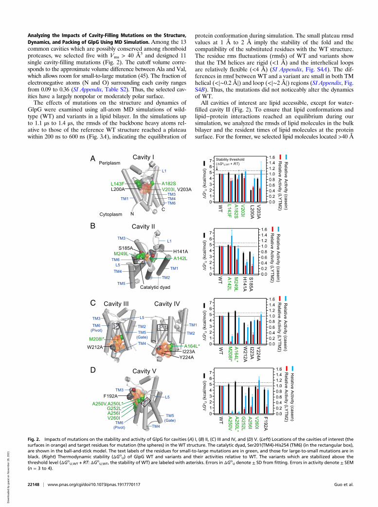
Analyzing the Impacts of Cavity-Filling Mutations on the Structure,Dynamics, and Packing of GlpG Using MD Simulation. Among the 13common cavities which are possibly conserved among rhomboidproteases, we selected five with Vms > 40 Å3 and designed 11single cavity-filling mutations (Fig. 2). The cutoff volume corre-sponds to the approximate volume difference between Ala and Val,which allows room for small-to-large mutation (45). The fraction ofelectronegative atoms (N and O) surrounding each cavity rangesfrom 0.09 to 0.36 (SI Appendix, Table S2). Thus, the selected cav-ities have a largely nonpolar or moderately polar surface.The effects of mutations on the structure and dynamics of
GlpG were examined using all-atom MD simulations of wild-type (WT) and variants in a lipid bilayer. In the simulations upto 1.1 μs to 1.4 μs, the rmsds of the backbone heavy atoms rel-ative to those of the reference WT structure reached a plateauwithin 200 ns to 600 ns (Fig. 3A), indicating the equilibration of
protein conformation during simulation. The small plateau rmsdvalues at 1 Å to 2 Å imply the stability of the fold and thecompatibility of the substituted residues with the WT structure.The residue rms fluctuations (rmsfs) of WT and variants showthat the TM helices are rigid (<1 Å) and the interhelical loopsare relatively flexible (<4 Å) (SI Appendix, Fig. S4A). The dif-ferences in rmsf between WT and a variant are small in both TMhelical (<|∼0.2 Å|) and loop (<|∼2 Å|) regions (SI Appendix, Fig.S4B). Thus, the mutations did not noticeably alter the dynamicsof WT.All cavities of interest are lipid accessible, except for water-
filled cavity II (Fig. 2). To ensure that lipid conformations andlipid−protein interactions reached an equilibrium during oursimulation, we analyzed the rmsds of lipid molecules in the bulkbilayer and the resident times of lipid molecules at the proteinsurface. For the former, we selected lipid molecules located >40 Å
A
B
C
D
Fig. 2. Impacts of mutations on the stability and activity of GlpG for cavities (A) I, (B) II, (C) III and IV, and (D) V. (Left) Locations of the cavities of interest (thesurfaces in orange) and target residues for mutation (the spheres) in the WT structure. The catalytic dyad, Ser201(TM4)-His254 (TM6) (in the rectangular box),are shown in the ball-and-stick model. The text labels of the residues for small-to-large mutations are in green, and those for large-to-small mutations are inblack. (Right) Thermodynamic stability (ΔGo
U) of GlpG WT and variants and their activities relative to WT. The variants which are stabilized above thethreshold level (ΔGo
U,WT + RT: ΔGoU,WT, the stability of WT) are labeled with asterisks. Errors in ΔGo
U denote ± SD from fitting. Errors in activity denote ± SEM(n = 3 to 4).
22148 | www.pnas.org/cgi/doi/10.1073/pnas.1917770117 Guo et al.
Dow
nloa
ded
by g
uest
on
Nov
embe
r 28
, 202
1

B
C
A
D
Fig. 3. All-atom MD simulation of GlpG WT and variants in the lipid bilayer (POPE:POPG, molar ratio = 3:1). (A) The rmsds of the backbone heavy atomsrelative to those in the crystal structure of WT (PDB ID code 2IC8) (35). (B) Equilibration of the lipid conformation in the bulk bilayer region measured by theaverage rmsd as a function of the time lag τ for (Top) POPE and (Bottom) POPG lipids. The color codes of the traces for WT and variants are the same as in A.(C) The lipid exchange at the protein surface measured by the lipid contact autocorrelation function on time. The color codes of the traces for WT and variantsare the same as in A. (D) Impacts of the mutations on the packing in the cavities measured by the differences in cavity volume (<VCav>WT − <VCav>Mut), OSP(<OSPMut> ‒ <OSPWT>, averaged over the residues surrounding each cavity), and BA (BAMut ‒ BAWT, summed over the residues surrounding each cavity).“Control” (red bar) indicates the mean of each difference packing parameter calculated over WT and the variants which do not contain the mutation on thedesignated cavity. The error bar in “Control” corresponds to the SD. When a mutation induces an increase in packing exceeding the upper SD limit of thecorresponding control, the mutation is regarded as “cavity filling” and marked with an asterisk.
Guo et al. PNAS | September 8, 2020 | vol. 117 | no. 36 | 22149
BIOPH
YSICSAND
COMPU
TATIONALBIOLO
GY
Dow
nloa
ded
by g
uest
on
Nov
embe
r 28
, 202
1

away from the protein surface as the bulk lipids (45 ± 2 POPE,1-palmitoyl-2-oleoyl-phosphatidylethanolamine, and 12 ± 1 POPG,1-palmitoyl-2-oleoyl-phosphatidylglycerol). Then, we calculated thermsd for each lipid molecule as a function of time lag, which wasaveraged over all selected lipid molecules (SI Appendix, Materialsand Methods). For WT and variants, the average rmsds of POPEand POPG reached a plateau at 3.6 Å to 3.8 Å within ∼20 ns, in-dicating that our simulation can sample lipid conformations underequilibrium conditions (Fig. 3B). Next, to test the equilibration ofprotein−lipid interactions, we evaluated the autocorrelation func-tion on time for all lipid heavy atoms in contact with the proteins (SIAppendix,Materials and Methods). The characteristic lipid residencetimes at the protein surface, τ1 and τ2 (the times when the corre-lation function decreased by 1/e and 1/e2 from the start, respec-tively), were 83 ± 9 ns and 291 ± 25 ns, respectively, which were 4 to15 times longer than the relaxation of the bulk lipids (Fig. 3C). Thisresult indicates that our simulation is long enough for lipid mole-cules to fully exchange at the protein surface as well as for proteinconformations to equilibrate with the surrounding lipids.Did the small-to-large mutations indeed improve packing in
the targeted cavities? To answer this question, we took threeapproaches from MD simulation of WT and variants. Taking thestructures from all time frames after the equilibration (>500 ns),we measured the volume of each cavity (VCav) using a grid-basedmethod (46). With the equilibrated structures at 1 μs, we eval-uated the occluded surface packing (OSP) (20) and buried sur-face area (BA) for each residue.All cavities displayed a dynamic feature. The volume of each
cavity fluctuated with the SD (σVol) of 20‒150% relative to themean cavity volume (<VCav>; SI Appendix, Figs. S5 and S6).Despite the fluctuation, four mutations, L143F (cavity I), A142L(cavity II), M208I (cavity III), and A164L (cavity IV), effectivelyreduced the volume of the cavities targeted for mutation(i.e., <VCav>WT ‒ <VCav>Mut,targeted is larger than <<VCav>WT ‒<VCav>Mut,not-targeted> + σ<VCav > WT‒ <VCav > Mut,not-targeted)(Fig. 3D and SI Appendix, Fig. S6). The difference in OSP andBA values between WT and a variant fluctuated as a function ofresidue number apparently in a random manner (SI Appendix,Figs. S7 and S8, respectively). Nevertheless, L143F (cavity I),M249L (cavity II), and M208I (cavity III) from the OSP analysis,and L143F (cavity I), V203I (cavity I), A142L (cavity II), A164L(cavity IV), A250L (cavity V), and G252L (cavity V) fromthe BA analysis, improved packing in the respective cavities(Fig. 3D). Overall, 8 out of 11 single small-to-large mutations
improved packing in the targeted cavities by any measure, thusregarded as cavity-filling mutations.
A Benchmark Study to Validate Mutation-Induced Changes in Packingfrom MD Simulation. How do the changes in packing induced bycavity-filling mutations influence the stability and function ofGlpG? Before answering this question, we validated our ap-proach for quantifying mutation-induced changes in packing bycarrying out a benchmark MD simulation using bacteriorho-dopsin (bR) as a model. Joh et al. (4) have measured the changesin cavity volume induced by a series of cavity-creating mutationsfrom the crystal structures of bR WT and variants. From therelationship between the changes in stability and cavity volume,they have determined the contribution of vdW packing to thestability.We constructed all-atomic models of WT and six variants of
bR in DMPC (1,2-dimyristoyl- phosphatidylcholine) bilayers(Fig. 4A) and performed MD simulation up to ∼1.4 μs (Fig. 4B).In each system, the backbone rmsd relative to the correspondingcrystal structure reached a plateau within 200 ns to 600 ns. Onthe basis of the spatial location of mutation, we targeted twocavities for volume analysis (Fig. 4A), that is, cavity IbR createdby the mutations L94A, L111A, I148A, I148V, and L152A, andcavity IIbR created by V49A. Since we were interested in howwell MD simulation can capture the local volume changes in-duced by mutation, the series of the volume changes of cavity IbRwere compared between simulation (the volumes measured bythe grid-based method) and experiment (the volumes measuredusing a 1.0-Å-radius probe) (SI Appendix, Table S3) (4, 46). Wefound a discrepancy which probably stemmed from the differ-ence in the cavity detection method, or, more likely, the differ-ence in the extent of protein flexibility allowed between thecrystal lattices and the bilayers in silico. Nonetheless, the com-parison of the normalized volume changes using Z scores (zi =(xi − μ)/s; xi is the volume of cavity IbR in each variant, and μ ands are the mean and SD of the cavity volumes in the variants,respectively) displayed a reasonable correlation between simu-lation and experiment (R = 0.90; Fig. 4C). Thus, our MD sim-ulation can reliably capture the local volume changes inducedby mutation.Next, we further attempted to predict the mutation-induced
stability changes using the volume changes measured from MDsimulation. Since a given mutation influenced not only the vol-ume of the cavity targeted by the mutation but also that not
B
D
CA
Fig. 4. Benchmark MD simulation of WT and variants of bR in a DMPC bilayer. (A) The structure of WT bR (PDB ID code 1PY6) (68). The major internal cavities(the surfaces), the mutated residues for cavity-creating mutations (the spheres), and bound retinal (the sticks). (B) The rmsds of the backbone heavy atoms ofWT and variants during MD simulation. (C) Correlation between the normalized cavity volumes (Z scores) determined from experiment (4) and simulation. (D)Correlation between the protein stabilities determined from experiment (4) and simulation. The dashed lines indicate the deviation by ±1.0 kcal/mol from thefitted linear line.
22150 | www.pnas.org/cgi/doi/10.1073/pnas.1917770117 Guo et al.
Dow
nloa
ded
by g
uest
on
Nov
embe
r 28
, 202
1

targeted, it was difficult to reliably predict the stability changesonly using the volume changes of a single cavity targeted formutation, which was the approach with the static crystal struc-tures (4). Therefore, at our benchmarking level, we constructed asimple linear response model to predict the stability changes(ΔΔGo
U,WT-Mut,Simul) using the volume changes of both cavities,
ΔΔGoU,WT-Mut,Simul = cIΔVI,WT-Mut + cIIΔVII,WT-Mut + b, [1]
where ΔVI,WT-Mut and ΔVII,WT-Mut denote the mutation-inducedvolume changes of cavities IbR and IIbR, respectively, obtainedfrom MD simulation, the coefficients cI and cII represent thepacking contributions to the stability in the respective cavities,and b is related to the contribution of lipid or water solvation tothe stability changes upon mutation. These coefficients were fit-ted to the model. Intriguingly, the stability changes can be mod-eled within the errors of ±1.0 kcal/mol with a reasonablecorrelation (R = 0.87) (Fig. 4D and SI Appendix, Table S4). Thus,our MD simulation approach can serve not only to evaluate themutation-induced changes in cavity volume but also to build amodel for reliable prediction of the free energy changes of mem-brane proteins (see Lipid Solvation on Cavity Fine-tunes Stability).
Impacts of Native Cavities in the N Subdomain on Stability andActivity. Next, we investigated the impacts of cavity-filling mu-tations on the thermodynamic stability (ΔGo
U) and activity ofGlpG. To measure the stability in DDM micelles, we employedsteric trapping, which couples spontaneous unfolding of a doublybiotinylated protein to competitive binding of bulky monovalentstreptavidin (mSA) (SI Appendix, Fig. S9A) (38). Mutations weremade in the background of the double-biotin variant, 172/267-BtnPyr2 (172/267: the residues for cysteine substitution; BtnPyr: athiol-reactive biotin label with a pyrene fluorophore) (SI Appendix,Fig. S9B). The 172/267-BtnPyr2 possesses the same global stabilityas WT without biotin labels (38). Steric trapping allows for precisedetermination of protein stability (an SE in ΔGo
U ≤ ±0.2 kcal/mol)directly under native conditions (SI Appendix, Fig. S10).Proteolytic activity of GlpG was measured using the second
TM segment of E. coli lactose permease (LYTM2) (47) as asubstrate in DDM micelles and large DMPC:CHAPS(3-((3-cholamidopropyl) dimethylammonio)-1-propanesulfonate) bicelles(SI Appendix, Figs. S11 and S12). Bicelles were used to test GlpGactivity in a bilayer environment. For LYTM2, the activities ofvariants relative to WT in micelles were highly correlated with thosein bicelles (SI Appendix, Fig. S13). We also measured the activity forthe generic water-soluble substrate, casein (SI Appendix, Fig. S14).Casein, which would access the catalytic dyad directly from the bulkwater, served as a probe to test the intactness of the active siteupon mutation.The impact of mutation on the stability and activity of GlpG
exhibited a unique dependence on the targeted cavity. Cavity I issurrounded by the residues mainly from the N-terminal half ofGlpG (TM1–L1–TM2–TM3–L3: N subdomain) and penetratesthe protein interior near the bilayer center. Three small-to-largemutations on cavity I, including the cavity-filling mutationsL143F and V203I, did not significantly alter the stability andactivity relative to WT (Fig. 2A).Next, we targeted the water-filled cavity near the catalytic dyad
(cavity II) also surrounded by the residues mainly from the Nsubdomain (Fig. 2B). This cavity is known as a water retentionsite, providing water molecules required for proteolysis (48).Despite the improved packing in cavity II by the mutationsA142L and M249L, the WT stability level was retained (Fig. 2B).Our MD simulation indicates that the water molecules remainedbound in the cavity upon mutation (SI Appendix, Fig. S15).Probably due to the unperturbed water retention, A142L did notaffect the activity for both TM and water-soluble substrates. Incomparison, M249L at the junction between L5 and TM6
selectively reduced the activity for LYTM2 to 52 ± 3%, whileretaining the activity for casein at 92 ± 10%. It has been suggestedthat the opening of the L5 loop enables the access of the scissilebond in a substrate to the active site (27, 49) (see the next section).Thus, it is likely that the improved packing by M249L at theL5−TM6 junction partially inhibited the opening of L5, leading tothe activity reduction preferentially for the TM substrate.A number of structural and folding studies indicate that the N
subdomain of GlpG serves as a rigid structural template, whilethe C subdomain possesses conformational plasticity undergoingsubglobal unfolding (28, 31, 38, 39, 49). Cavity-filling mutationsin the already stable N subdomain did not increase the stability,probably because the improved packing induced a strain withouta gain of stability. It is also possible that, for lipid-accessiblecavity I, the cavity-filling mutations modified the lipid−cavityinteractions to counteract the stabilization by the improvedpacking (see Lipid Solvation on Cavity Fine-tunes Stability).
Impacts of Native Cavities in the C Subdomain on Stability andActivity. We targeted three cavities (cavities III to V; Fig. 2 Cand D) in the C subdomain of GlpG (TM4–L4–TM5–L5–TM6)harboring the catalytic dyad Ser201/His254. Crystal structuresshow that the C-terminal segment TM5–L5–TM6 is subject toconformational changes. In a majority of apo structures, TM5 ispacked against TM2, and the L5 loop caps the catalytic dyad(Fig. 2C) (35). In comparison, TM5 is tilted away from TM2, orL5 forms an “open cap” or disordered conformation in thestructures bound with several mechanism-based inhibitors andtwo apo structures (SI Appendix, Fig. S16) (27, 28, 31, 49, 50). Onthe basis of the dramatic activation induced by deletion muta-tions at the TM2–TM5 interface and the plasticity of TM5 andL5, it has been suggested that TM substrates bind to theTM2−TM5 interface and that TM5 serves as a “gate” controllingthe access of TM substrates to the catalytic dyad along with theL5 cap (27, 49, 51). However, chemical cross-linking betweenTM2 and TM5 retains activity, and a modeling study predictsthat substrate binding may not require opening of TM5 (34, 50).Thus, the gating role of TM5 has been debated.Interestingly, the mutations targeting the cavities near TM5
(M208I on cavity III and A164L on cavity IV) effectively reducedthe volume of the two cavities by 30 to 60% (SI Appendix, Fig.S6) and enhanced the stability (ΔΔGo
U,WT-Mut > RT, thermalenergy = ∼0.6 kcal/mol; R, gas constant; T, absolute tempera-ture, 298 K). Cavity III deeply intrudes into the TM4−TM5 in-terface (Fig. 2C). M208I on this cavity stabilized GlpG by +0.6 ±0.2 kcal/mol, substantially decreasing the activity to 8 ± 1% forthe TM substrate LYTM2. This inactivation is unexpected be-cause the mutated site is not only distant from the catalytic dyadbut also does not directly interfere with substrate binding at theTM2−TM5 interface. Cavity IV is located at the substrate bindingsite, but distant from the catalytic dyad (Fig. 2C). A164L on thiscavity induced the largest stabilization (+0.9 ± 0.2 kcal/mol)among tested mutations, decreasing the activity to 38 ± 3% forLYTM2. Notably, both M208I and A164L displayed larger rela-tive activity for casein (28 ± 6% and 94 ± 9%, respectively) thanthat for LYTM2. Thus, the activity loss for the TM substrate wasnot caused by the disruption of the active site. From MD simu-lation, all tested mutations did not perturb the hydrogen bondbetween Ser201 and His254 (SI Appendix, Table S5), which acti-vates Ser201 for the nucleophilic attack on the scissile peptidebond (35).The gain of interaction (i.e., the enhanced stability) and loss of
function by the cavity-filling mutations near TM5 implies thestabilization of the gate-closed conformation. Supporting this,MD simulation shows that M208I shrank the substrate bindingsite by tilting of TM5 (SI Appendix, Fig. S17), slightly decreasingthe Cα−Cα distance between Phe153 (TM2) and Trp236 (TM5)by up to ∼0.8 Å (SI Appendix, Fig. S18). These results suggest a
Guo et al. PNAS | September 8, 2020 | vol. 117 | no. 36 | 22151
BIOPH
YSICSAND
COMPU
TATIONALBIOLO
GY
Dow
nloa
ded
by g
uest
on
Nov
embe
r 28
, 202
1

critical role of the cavities near TM5 in mediating the gatingmotion for substrate binding.Finally, we tested mutations on cavity V at the TM3−TM6
interface in the periplasmic side (Fig. 2D). TM6 harboring thecatalytic His254 is also subject to conformational changes (SIAppendix, Fig. S16). Mechanism-based inhibitors covalentlybound to Ser201 induce a slight outward (52) or inward (28, 53)pivot motion at the periplasmic end of TM6. However, the in-volvement of this small-amplitude motion in the catalyticmechanism has been elusive.The stepwise increase of the side-chain volume on cavity V by
A250V and A250L gradually increased the stability by +0.4 ± 0.2and +0.7 ± 0.2 kcal/mol, respectively, dramatically decreasingthe activity for both LYTM2 and casein to <5% (Fig. 2D).G252L and A256I on the same cavity also substantially de-creased the activity for LYTM2 to 15 to 25%, retaining the ac-tivity for casein at 60 to 65%. Thus, the activity loss for the TMsubstrate by these mutations was not necessarily due to the dis-ruption of the active site. V260I at the lipid-exposed positionoutside of cavity V fully restored the activity for both LYTM2and casein (>85%) without altering the stability. In the crystalstructures, the pivot motion of TM6 involves the rotation ofsmall Ala250 and Gly252 on TM6 against bulky Val188, Gln189,and Tyr192 on TM3 (SI Appendix, Fig. S19). Therefore, it islikely that the increases in the side-chain volume on cavity Vinhibit the pivot motion of TM6 and that this motion has a largeimpact on the proteolysis mechanism.As a control, we tested the impacts of cavity-creating muta-
tions on all cavities (Fig. 2). As expected, these mutations de-creased the stability to various extents (ΔΔGo
U,WT-Mut = –0.6kcal/mol to ‒2.7 kcal/mol). H141A (cavity II), which is known todisrupt the water retention and conduction toward the active site(31), similarly reduced the activity for LYTM2 and casein to 30 ±6% and 44 ± 8%, respectively (Fig. 2B). Except for this mutationand Y224A (cavity IV), the latter of which had a minor impacton the stability and activity (Fig. 2C), all other cavity-creatingmutations substantially reduced the activity for LYTM2 to 10 to50% regardless of the degree of destabilization, retaining theactivity for casein at 60 to 120%. Thus, the activity loss for theTM substrate induced by the cavity expansion did not stem fromeither the disruption of the active site or the conformationaldestabilization (see Are All Cavities Critical to Function?).
Additivity, Cooperativity, and Propagation of Stabilizing Mutations.Our finding that all stabilizing mutations are mapped onto the
more flexible C subdomain and substantially reduce the activitysuggests that these mutations stabilize inactive conformations ofGlpG by inhibiting the functionally important “gating” (A164Land M208I) or “pivot” (A250L) motion. Next, we tested whetherthese stabilization effects were additive by measuring the stabilityand activity of the pairwise double and triple variants (Fig. 5A).All of these variants were almost inactive for both substrates.Although some of the variants were stabilized, the individualstabilization effects were not additive.To track down the molecular origin of the nonadditivity, we
carried out a thermodynamic cycle analysis to determine the freeenergy of interaction (ΔΔGInter) between the substituted resi-dues (SI Appendix, Materials and Methods and Fig. 5B) (54).Slight negative cooperativity (ΔΔGInter = +0.6 ± 0.2 kcal/mol)occurred between M208I (cavity III) and A164L (cavity IV),which are spatially close (Fig. 5C). Interestingly, larger negativecooperativity was observed between M208I (cavity III) andA250L (cavity V) (ΔΔGInter = +1.2 ± 0.4 kcal/mol) and betweenA164L (cavity IV) and A250L (cavity V) (ΔΔGInter = +1.6 ± 0.3kcal/mol), which are farther separated. This result indicates thatthe flexible C subdomain of GlpG is an ensemble of multipledisjoint conformations, and stabilizing one destabilizes another.Thus, the conformation stabilized by A250L (the TM6 pivotlocked) is not likely to simultaneously populate with that stabi-lized by M208I or A164L (the TM5 gate locked). The negativecooperativity is further supported by MD simulation showingthat the improved packing by the single cavity-filling mutationswas suppressed in either cavity when these mutations werecombined in the double variants (Fig. 3D).By design, steric trapping captures transient opening of the
native tertiary contacts at a specific biotin pair, allowing mea-surement of the local stability of the region encompassing thebiotin pair (38). We compared the mutation-induced stabilitychanges measured at the biotin pair in the C subdomain (172/267-BtnPyr2) to those measured at the N subdomain (95/172-BtnPyr2). The same mutations that had stabilized the C sub-domain (ΔΔGo
U,WT-Mut = +0.6 kcal/mol to +1.0 kcal/mol) didnot stabilize the N subdomain as much (ΔΔGo
U,WT-Mut = −0.1kcal/mol to +0.3 kcal/mol) (SI Appendix, Fig. S20). Thus, thestabilization by the cavity-filling mutations was only locally ef-fective in the C subdomain, not globally propagated to the Nsubdomain.
Lipid Solvation on Cavity Fine-tunes Stability. The crystal structuresand our MD simulation indicate that all of the cavities of interest
A B C
Fig. 5. Additivity and cooperativity of stabilizing cavity-filling mutations. (A) Impacts of the double and triple stabilizing mutations on the stability andactivity. (B) Thermodynamic cycle analysis describing the stability changes induced by the stabilizing single and combined double mutations. (C) Cooperativitybetween the stabilizing mutations. Pairwise interaction energies (ΔΔGInter) were calculated using Fig. 5B (SI Appendix, Materials and Methods). The mutatedresidues (the spheres) and the residues in the catalytic dyad (the sticks) are shown. Errors denote ± SD from fitting.
22152 | www.pnas.org/cgi/doi/10.1073/pnas.1917770117 Guo et al.
Dow
nloa
ded
by g
uest
on
Nov
embe
r 28
, 202
1

except for water-filled cavity II interact with detergent or lipidmolecules. For WT, the patterns of the cavity−detergent inter-action in the crystal structure are remarkably similar to thecavity−lipid interaction in the simulation (Fig. 6, Left and Mid-dle) (55, 56). For these cavities, we found an interesting corre-lation between the changes in stability upon mutation and theassociated changes in lipid interaction. To quantitatively describethe cavity−lipid interaction, for the time frames from MD sim-ulation (>500 ns), we measured the number of lipid atoms withineach cavity (SI Appendix, Fig. S21) as well as the distance be-tween a specific residue in each cavity and its closest approachinglipid atom (SI Appendix, Fig. S22).For the cavity-filling mutations, L143F (cavity I), M208I
(cavity III), and G252L (cavity V), the increase in packing wascoupled to noticeable displacement of lipid atoms from thecavities targeted for mutation (Fig. 6, Right), while, to less extent,for A164L (cavity IV) and A250L (cavity V). Under the as-sumption that the mutational impacts on the lipid−protein in-teractions are only local near the cavities targeted for mutation,the packing−desolvation coupling offers an opportunity toquantify the strengths of lipid−protein interactions relative tothose of protein packing, which yields various outcomes in thestability (SI Appendix, Fig. S23). This assumption is supported bythe fact that the cavity-filling mutations distinctively improve thepacking in the cavity targeted for mutation (Fig. 3D). Towardthis goal, we employed the formulation suggested by Flemingand Engelman (3) to study TM helix−helix interactions,
–ΔΔGoU,WT-Mut = ΔΔGpacking + ΔΔGPL + ΔΔGLL
= σpacking × Δ<VCav>WT-Mut + ΔΔGEx,PL.[2]
ΔΔGpacking denotes the free energy change induced by the changein protein packing upon mutation (i.e., Δ<VCav>WT-Mut =
<VCav>WT – <VCav>Mut determined fromMD simulation). σpackingis the vdW packing contribution to the protein stability per unitcavity volume (σpacking = –29 cal/mol/Å3) (2–4). ΔΔGPL andΔΔGLL represent the free energy changes in lipid solvation (ordesolvation) upon mutation and the associated lipid reorganiza-tion, respectively. We define their sum (ΔΔGPL + ΔΔGLL) as thefree energy change in “lipid exchange” (ΔΔGEx,PL), which can beinterpreted as the preference (or disfavor) of partitioning a lipidsegment in a cavity relative to the surrounding lipids.On the basis of our validated MD simulation approach for
predicting the free energy changes of membrane proteins (Fig.4), we evaluated the contribution of protein packing vs. lipidsolvation to the stability of GlpG using Eq. 2. The weak stabili-zation by the mutation L143F on cavity I (–ΔΔGo
U,WT-Mut =–0.2 ± 0.2 kcal/mol) stems from the stabilization by the improvedpacking (ΔΔGpacking = –1.7 ± 0.6 kcal/mol), which is canceledout by the unfavorable lipid desolvation (ΔΔGEx,PL = +1.5 ± 0.6kcal/mol) (Fig. 6A). The unfavorable lipid desolvation indicatesthat cavity I in WT has been stabilized by interacting lipids. Thestabilization by M208I on cavity III (–ΔΔGo
U,WT-Mut = –0.6 ± 0.1kcal/mol) is the outcome of the coupling between the improvedpacking (ΔΔGpacking = –1.0 ± 0.4 kcal/mol) and the weakly un-favorable lipid desolvation (ΔΔGEx,PL = +0.4 ± 0.4 kcal/mol)(Fig. 6 B, Right). In contrast, A164L (–ΔΔGo
U,WT-Mut = –0.9 ±0.1 kcal/mol) is mainly stabilized by the improved packing(ΔΔGpacking = –0.8 ± 0.8 kcal/mol; Fig. 6 C, Right) on cavity IVwith a negligible contribution from lipid solvation (ΔΔGEx,PL =–0.1 ± 0.8 kcal/mol). A250L does not improve packing on cavity V(ΔΔGpacking = ∼0 kcal/mol) but induces favorable lipid solvation(ΔΔGEx,PL = –0.7 ± 0.2 kcal/mol), which stabilizes the protein(–ΔΔGo
U,WT-Mut = –0.7 ± 0.2 kcal/mol) (Fig. 6D). AlthoughA250L induces a subtle change in lipid solvation (Fig. 6 D, RightTop), the attraction of lipid atoms to cavity V is observed for the
B
A C
D
Fig. 6. Impacts of cavity-filling mutations on lipid−protein interaction for cavities (A) I, (B) III, (C) IV, and (D) V. (Left) Crystallographic detergent molecules(the sticks in red) bound to the cavities in WT GlpG (PDB ID code 3B45). (Middle) The structural snapshots of WT (orange) and variant (cyan) at 1 μs from MDsimulation are superimposed with the closest approaching lipid molecules in the stick model. The residues surrounding each cavity are shown in the surfaces.The site of each mutation is marked with an asterisk. The movement of lipid chain segments induced by mutation is indicated with black arrows. (Right) Thedistance distributions between a specific residue atom and the closest-approaching lipid heavy atoms in each designated cavity. In D, the structural snapshots(Middle) and cavity–lipid distances (Right) of WT and A250L are shown at the top, and those of WT and G252L are at the bottom.
Guo et al. PNAS | September 8, 2020 | vol. 117 | no. 36 | 22153
BIOPH
YSICSAND
COMPU
TATIONALBIOLO
GY
Dow
nloa
ded
by g
uest
on
Nov
embe
r 28
, 202
1
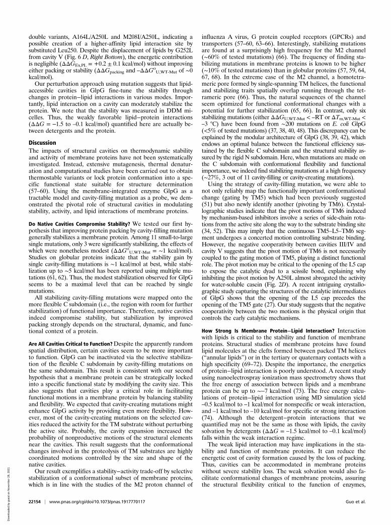
double variants, A164L/A250L and M208I/A250L, indicating apossible creation of a higher-affinity lipid interaction site bysubstituted Leu250. Despite the displacement of lipids by G252Lfrom cavity V (Fig. 6 D, Right Bottom), the energetic contributionis negligible (ΔΔGEx,PL = +0.2 ± 0.1 kcal/mol) without improvingeither packing or stability (ΔΔGpacking and –ΔΔGo
U,WT-Mut of ∼0kcal/mol).Our perturbation approach using mutation suggests that lipid-
accessible cavities in GlpG fine-tune the stability throughchanges in protein−lipid interactions in various modes. Impor-tantly, lipid interaction on a cavity can moderately stabilize theprotein. We note that the stability was measured in DDM mi-celles. Thus, the weakly favorable lipid−protein interactions(ΔΔG = ‒1.5 to –0.1 kcal/mol) quantified here are actually be-tween detergents and the protein.
DiscussionThe impacts of structural cavities on thermodynamic stabilityand activity of membrane proteins have not been systematicallyinvestigated. Instead, extensive mutagenesis, thermal denatur-ation and computational studies have been carried out to obtainthermostable variants or lock protein conformation into a spe-cific functional state suitable for structure determination(57–60). Using the membrane-integrated enzyme GlpG as atractable model and cavity-filling mutation as a probe, we dem-onstrated the pivotal role of structural cavities in modulatingstability, activity, and lipid interactions of membrane proteins.
Do Native Cavities Compromise Stability? We tested our first hy-pothesis that improving protein packing by cavity-filling mutationgenerally stabilizes a membrane protein. Among 11 small-to-largesingle mutations, only 3 were significantly stabilizing, the effects ofwhich were nonetheless modest (ΔΔGo
U,WT-Mut = ∼1 kcal/mol).Studies on globular proteins indicate that the stability gain bysingle cavity-filling mutations is ∼1 kcal/mol at best, while stabi-lization up to ∼5 kcal/mol has been reported using multiple mu-tations (61, 62). Thus, the modest stabilization observed for GlpGseems to be a maximal level that can be reached by singlemutations.All stabilizing cavity-filling mutations were mapped onto the
more flexible C subdomain (i.e., the region with room for furtherstabilization) of functional importance. Therefore, native cavitiesindeed compromise stability, but stabilization by improvedpacking strongly depends on the structural, dynamic, and func-tional context of a protein.
Are All Cavities Critical to Function?Despite the apparently randomspatial distribution, certain cavities seem to be more importantto function. GlpG can be inactivated via the selective stabiliza-tion of the flexible C subdomain by cavity-filling mutations onthe same subdomain. This result is consistent with our secondhypothesis that a membrane protein can be strategically lockedinto a specific functional state by modifying the cavity size. Thisalso suggests that cavities play a critical role in facilitatingfunctional motions in a membrane protein by balancing stabilityand flexibility. We expected that cavity-creating mutations mightenhance GlpG activity by providing even more flexibility. How-ever, most of the cavity-creating mutations on the selected cav-ities reduced the activity for the TM substrate without perturbingthe active site. Probably, the cavity expansion increased theprobability of nonproductive motions of the structural elementsnear the cavities. This result suggests that the conformationalchanges involved in the proteolysis of TM substrates are highlycoordinated motions controlled by the size and shape of thenative cavities.Our result exemplifies a stability−activity trade-off by selective
stabilization of a conformational subset of membrane proteins,which is in line with the studies of the M2 proton channel of
influenza A virus, G protein coupled receptors (GPCRs) andtransporters (57–60, 63–66). Interestingly, stabilizing mutationsare found at a surprisingly high frequency for the M2 channel(∼60% of tested mutations) (66). The frequency of finding sta-bilizing mutations in membrane proteins is known to be higher(∼10% of tested mutations) than in globular proteins (57, 59, 64,67, 68). In the extreme case of the M2 channel, a homotetra-meric pore formed by single-spanning TM helices, the functionaland stabilizing traits spatially overlap running through the tet-rameric pore (66). Thus, the natural sequences of the channelseem optimized for functional conformational changes with apotential for further stabilization (65, 66). In contrast, only sixstabilizing mutations (either ΔΔGU,WT-Mut < –RT or ΔTm,WT-Mut <–3 °C) have been found from ∼200 mutations on E. coli GlpG(<5% of tested mutations) (37, 38, 40, 48). This discrepancy can beexplained by the modular architecture of GlpG (38, 39, 42), whichendows an optimal balance between the functional efficiency sus-tained by the flexible C subdomain and the structural stability as-sured by the rigid N subdomain. Here, when mutations are made onthe C subdomain with conformational flexibility and functionalimportance, we indeed find stabilizing mutations at a high frequency(∼27%, 3 out of 11 cavity-filling or cavity-creating mutations).Using the strategy of cavity-filling mutation, we were able to
not only reliably map the functionally important conformationalchange (gating by TM5) which had been previously suggested(51) but also newly identify another (pivoting by TM6). Crystal-lographic studies indicate that the pivot motions of TM6 inducedby mechanism-based inhibitors involve a series of side-chain rota-tions from the active site along the way to the substrate binding site(34, 52). This may imply that the continuous TM5–L5–TM6 seg-ment undergoes a concerted motion controlling substrate binding.However, the negative cooperativity between cavities III/IV andcavity V suggests that the pivot motion of TM6 is not necessarilycoupled to the gating motion of TM5, playing a distinct functionalrole. The pivot motion may be critical to the opening of the L5 capto expose the catalytic dyad to a scissile bond, explaining whyinhibiting the pivot motion by A250L almost abrogated the activityfor water-soluble casein (Fig. 2D). A recent intriguing crystallo-graphic study capturing the structures of the catalytic intermediatesof GlpG shows that the opening of the L5 cap precedes theopening of the TM5 gate (27). Our study suggests that the negativecooperativity between the two motions is the physical origin thatcontrols the early catalytic mechanisms.
How Strong Is Membrane Protein−Lipid Interaction? Interactionwith lipids is critical to the stability and function of membraneproteins. Structural studies of membrane proteins have foundlipid molecules at the clefts formed between packed TM helices(“annular lipids”) or in the tertiary or quaternary contacts with ahigh specificity (69–72). Despite the importance, the energeticsof protein−lipid interaction is poorly understood. A recent studyusing nanoelectrospray ionization mass spectrometry shows thatthe free energy of association between lipids and a membraneprotein can be up to ∼‒7 kcal/mol (73). The free energy calcu-lations of protein−lipid interaction using MD simulation yield–0.5 kcal/mol to –1 kcal/mol for nonspecific or weak interaction,and –1 kcal/mol to –10 kcal/mol for specific or strong interaction(74). Although the detergent−protein interactions that wequantified may not be the same as those with lipids, the cavitysolvation by detergents (ΔΔG = –1.5 kcal/mol to –0.1 kcal/mol)falls within the weak interaction regime.The weak lipid interaction may have implications in the sta-
bility and function of membrane proteins. It can reduce theenergetic cost of cavity formation caused by the loss of packing.Thus, cavities can be accommodated in membrane proteinswithout severe stability loss. The weak solvation would also fa-cilitate conformational changes of membrane proteins, assuringthe structural flexibility critical to the function of enzymes,
22154 | www.pnas.org/cgi/doi/10.1073/pnas.1917770117 Guo et al.
Dow
nloa
ded
by g
uest
on
Nov
embe
r 28
, 202
1

transporters, and GPCRs. Other MD simulation studies onGlpG suggest that bound lipids near the active site may competewith substrate binding (75). Intriguingly, highly deformed lipidssurrounding GlpG boost its translational diffusion within themembrane, enhancing the activity (76).The cavity stabilization by solvation is not limited to mem-
brane proteins. For globular proteins, theoretical studies predictthat the free energy of transfer of water from the bulk to internalcavities (ΔGhydration) widely varies from 5 kcal/mol to ‒12 kcal/mol,highly depending on the polarity of the cavity surface (77). Anexperimental study has shown that cavity hydration can stabilize aglobular protein by 1.5 kcal/mol to 2.0 kcal/mol (78). Therefore, forboth globular and membrane proteins, cavity solvation by eitherwater or lipids can counterbalance the energetic cost caused by theloss of protein packing (24).
Concluding Remark. Here, we have elucidated a versatile role ofcavities in balancing the stability and flexibility for activity ofmembrane proteins. Our strategy employing cavity-filling muta-tion in combination with experiment and simulation may serve asa tool for mapping conformational changes critical to function andprovide guidance for membrane protein design and engineering.
Materials and MethodsDetailed information on materials, modeling of RHBDL2, MD simulation,expression and purification of GlpG, biotin labeling, stability and activity de-termination, and thermodynamic cycle analysis can be found in SI Appendix.
Structural Modeling of Human Rhomboid Protease RHBDL2. The sequencealignment of RHBDL2 and E. coli GlpG in the predicted TM helices wasobtained from the previous literature and used for the comparative mod-eling of RHBDL2 (the core TM helices, TM1 to TM6) on the Rosetta softwarewith three structural templates of E. coli GlpG (2IC8, 2XOV, and 3B45) (25, 3555, 79, 80). The modeled structure with the lowest energy was chosen as thestarting point of MD simulation for further relaxation in a POPC explicitmembrane and water using the CHARMM (Chemistry at Harvard Macro-molecular Mechanics)-27 force field (81).
MD Simulation of GlpG. We first simulated for GlpG WT, and then moved onfor each variant based on the equilibrated WT conformation. The WT systemwas built with the crystal structure of E. coli GlpG (Protein Data Bank [PDB] IDcode 2IC8; ref. 35) embedded in a lipid bilayer (POPE:POPG = 231:77) con-structed using the web-based CHARMM-GUI (graphical user interface)membrane builder (82). The composite system was immersed in the TIP3Pwater solvent (83), followed by a charge neuralization and ionization with150 mM NaCl in a box of 100 × 100 × 81 Å3. Intermolecular and intra-molecular potential energies were enumerated based on the CHARMM36
force field (84). All simulations were massively parallelized in the GPU(graphics processing unit)-accelerated IBM Power8 machine with a 2-fstime step in the semiisotropic isobaric and isothermal ensemble of 1 atmand 310 K.
Preparation and Biotin Labeling of GlpG. The TM domain of GlpG (residues 87to 276) encoded in pET15b vector was expressed in E. coli BL21(DE3)RP strainwith an N-terminal His6-tag (38). Purified double cysteine variants (172C/267C or 95C/172C) at 0.5 mM Tris-(2-carboxyethyl) phosphine hydrochloride(TCEP, Pierce) was incubated with a 20-fold molar excess of the thiol-reactivebiotin derivative with a pyrene fluorophore (BtnPyr-IA) in the base buffer,50 mM Tris-(hydroxymethyl) aminomethane hydrochloride (TrisHCl), 200 mMNaCl, pH 8.0, and 0.2% DDM overnight in the dark at 25 °C.
Preparation of mSA. WT mSA, and mSA-S45A (Kd,biotin = 9.0 nM) and mSA-S27A (Kd,biotin = 1.4 nM) variants with reduced biotin affinities, were pre-pared as described previously (38). Each variant contained a single cysteinemutation S83C in the active subunit to conjugate the thiol-reactive dabcylquencher (Dabcyl Plus C2 maleimide, Anaspec).
Determining Thermodynamic Stability (ΔGoU) of GlpG. ΔGo
Us of GlpG variantswere determined using steric trapping, that is, by measuring the attenuatedsecond binding of mSA coupled to unfolding. Binding was detected byquenching of pyrene monomer fluorescence (λEx. = 345 nm and λEm. = 390 nm)from BtnPyr labels conjugated to the double-biotin variants of GlpG (172/267-BtnPyr2 or 95/172-BtnPyr2) upon binding of mSA labeled with dabcyl quencher(38); 1 μM GlpG was titrated with an mSA variant, mSA-S45A or mSA-S27A, in5 mM DDM, 0.25 mM TCEP, 20 mM sodium phosphate, and 200 mM NaCl (pH7.5) at 25 °C. The second binding phase in the binding isotherm was fitted tothe equation to obtain ΔGo
U (SI Appendix, Materials and Methods).
Activity Assays of GlpG. Proteolytic activity of GlpG was measured using thecleavage rates of the second TM segment of E. coli lactose permease fusedto staphylococcal nuclease (SN-LYTM2) in DDM micellar or DMPC/CHAPSbicellar solution, or Bodipy-FL labeled casein (Thermo Fisher) in DDM solution.For SN-LYTM2, the position at the five residues upstream from the scissile bondwas mutated to Cys for labeling with thiol-reactive environment-sensitive flu-orophore iodoacetyl-7-nitrobenz-2-oxa-1,3-diazol (IA-NBD, Setareh Biotech).Time-dependent change of NBD or Bodipy fluorescence, the initial slope ofwhich represented the activity, was monitored with λEx. = 485 nm and λEm. =535 nm.
Data Availability. All data supporting the findings of this study are availablewithin this article and SI Appendix.
ACKNOWLEDGMENTS. We thank the H.H. laboratory members for criticalcomments, and Lin Song for helping with the OSP analysis. Special thanks goto anonymous reviewers for constructive comments. This work was sup-ported by NIH Grants R01GM118685 (to H.H.) and R01GM126189 (to G.W.).
1. J. T. Kellis Jr., K. Nyberg, A. R. Fersht, Energetics of complementary side-chain packingin a protein hydrophobic core. Biochemistry 28, 4914–4922 (1989).
2. A. E. Eriksson et al., Response of a protein structure to cavity-creating mutations andits relation to the hydrophobic effect. Science 255, 178–183 (1992).
3. K. G. Fleming, D. M. Engelman, Specificity in transmembrane helix−helix interactionscan define a hierarchy of stability for sequence variants. Proc. Natl. Acad. Sci. U.S.A.98, 14340–14344 (2001).
4. N. H. Joh, A. Oberai, D. Yang, J. P. Whitelegge, J. U. Bowie, Similar energetic con-tributions of packing in the core of membrane and water-soluble proteins. J. Am.Chem. Soc. 131, 10846–10847 (2009).
5. M. Mravic et al., Packing of apolar side chains enables accurate design of highly stablemembrane proteins. Science 363, 1418–1423 (2019).
6. F. M. Richards, Areas, volumes, packing and protein structure. Annu. Rev. Biophys.Bioeng. 6, 151–176 (1977).
7. J. Liang, K. A. Dill, Are proteins well-packed? Biophys. J. 81, 751–766 (2001).8. W. A. Lim, R. T. Sauer, Alternative packing arrangements in the hydrophobic core of
lambda repressor. Nature 339, 31–36 (1989).9. J. Wen, X. Chen, J. U. Bowie, Exploring the allowed sequence space of a membrane
protein. Nat. Struct. Biol. 3, 141–148 (1996).10. R. Kadirvelraj, N. C. Sennett, S. J. Polizzi, S. Weitzel, Z. A. Wood, Role of packing
defects in the evolution of allostery and induced fit in human UDP-glucose dehy-drogenase. Biochemistry 50, 5780–5789 (2011).
11. Q. Kuang, P. Purhonen, H. Hebert, Structure of potassium channels. Cell. Mol. Life Sci.72, 3677–3693 (2015).
12. Y. Y. Tseng, J. Liang, Estimation of amino acid residue substitution rates at localspatial regions and application in protein function inference: A Bayesian Monte Carloapproach. Mol. Biol. Evol. 23, 421–436 (2006).
13. J. L. Popot, D. M. Engelman, Membrane protein folding and oligomerization: Thetwo-stage model. Biochemistry 29, 4031–4037 (1990).
14. T. Hessa et al., Recognition of transmembrane helices by the endoplasmic reticulumtranslocon. Nature 433, 377–381 (2005).
15. Z. Cao, J. M. Hutchison, C. R. Sanders, J. U. Bowie, Backbone hydrogen bond strengthscan vary widely in transmembrane helices. J. Am. Chem. Soc. 139, 10742–10749 (2017).
16. H. Gratkowski, J. D. Lear, W. F. DeGrado, Polar side chains drive the association ofmodel transmembrane peptides. Proc. Natl. Acad. Sci. U.S.A. 98, 880–885 (2001).
17. F. X. Zhou, H. J. Merianos, A. T. Brunger, D. M. Engelman, Polar residues drive asso-ciation of polyleucine transmembrane helices. Proc. Natl. Acad. Sci. U.S.A. 98,2250–2255 (2001).
18. J. U. Bowie, Membrane protein folding: How important are hydrogen bonds? Curr.Opin. Struct. Biol. 21, 42–49 (2011).
19. L. Adamian, J. Liang, Helix-helix packing and interfacial pairwise interactions of res-idues in membrane proteins. J. Mol. Biol. 311, 891–907 (2001).
20. M. Eilers, S. C. Shekar, T. Shieh, S. O. Smith, P. J. Fleming, Internal packing of helicalmembrane proteins. Proc. Natl. Acad. Sci. U.S.A. 97, 5796–5801 (2000).
21. L. Adamian, V. Nanda, W. F. DeGrado, J. Liang, Empirical lipid propensities of aminoacid residues in multispan alpha helical membrane proteins. Proteins 59, 496–509(2005).
22. A. Oberai, N. H. Joh, F. K. Pettit, J. U. Bowie, Structural imperatives impose diverseevolutionary constraints on helical membrane proteins. Proc. Natl. Acad. Sci. U.S.A.106, 17747–17750 (2009).
23. P. W. Hildebrand, K. Rother, A. Goede, R. Preissner, C. Frömmel, Molecular packingand packing defects in helical membrane proteins. Biophys. J. 88, 1970–1977 (2005).
24. E. S. O’Brien et al., Membrane proteins have distinct fast internal motion and residualconformational entropy. Angew. Chem. Int. Ed. Engl. 59, 11108–11114 (2020).
Guo et al. PNAS | September 8, 2020 | vol. 117 | no. 36 | 22155
BIOPH
YSICSAND
COMPU
TATIONALBIOLO
GY
Dow
nloa
ded
by g
uest
on
Nov
embe
r 28
, 202
1
25. M. K. Lemberg, M. Freeman, Functional and evolutionary implications of enhancedgenomic analysis of rhomboid intramembrane proteases. Genome Res. 17, 1634–1646(2007).
26. M. Freeman, The rhomboid-like superfamily: Molecular mechanisms and biologicalroles. Annu. Rev. Cell Dev. Biol. 30, 235–254 (2014).
27. S. Cho, R. P. Baker, M. Ji, S. Urban, Ten catalytic snapshots of rhomboid intra-membrane proteolysis from gate opening to peptide release. Nat. Struct. Mol. Biol.26, 910–918 (2019).
28. S. Cho, S. W. Dickey, S. Urban, Crystal structures and inhibition kinetics reveal a two-stage catalytic mechanism with drug design implications for rhomboid proteolysis.Mol. Cell 61, 329–340 (2016).
29. S. W. Dickey, R. P. Baker, S. Cho, S. Urban, Proteolysis inside the membrane is a rate-governed reaction not driven by substrate affinity. Cell 155, 1270–1281 (2013).
30. S. Urban, J. R. Lee, M. Freeman, Drosophila rhomboid-1 defines a family of putativeintramembrane serine proteases. Cell 107, 173–182 (2001).
31. Z. Wu et al., Structural analysis of a rhomboid family intramembrane protease revealsa gating mechanism for substrate entry. Nat. Struct. Mol. Biol. 13, 1084–1091 (2006).
32. M. J. Lemieux, S. J. Fischer, M. M. Cherney, K. S. Bateman, M. N. James, The crystalstructure of the rhomboid peptidase from Haemophilus influenzae provides insightinto intramembrane proteolysis. Proc. Natl. Acad. Sci. U.S.A. 104, 750–754 (2007).
33. K. Strisovsky, H. J. Sharpe, M. Freeman, Sequence-specific intramembrane proteolysis:Identification of a recognition motif in rhomboid substrates. Mol. Cell 36, 1048–1059(2009).
34. Y. Xue, Y. Ha, Large lateral movement of transmembrane helix S5 is not required forsubstrate access to the active site of rhomboid intramembrane protease. J. Biol.Chem. 288, 16645–16654 (2013).
35. Y. Wang, Y. Zhang, Y. Ha, Crystal structure of a rhomboid family intramembraneprotease. Nature 444, 179–180 (2006).
36. A. Ben-Shem, D. Fass, E. Bibi, Structural basis for intramembrane proteolysis byrhomboid serine proteases. Proc. Natl. Acad. Sci. U.S.A. 104, 462–466 (2007).
37. R. P. Baker, S. Urban, Architectural and thermodynamic principles underlying intra-membrane protease function. Nat. Chem. Biol. 8, 759–768 (2012).
38. R. Guo et al., Steric trapping reveals a cooperativity network in the intramembraneprotease GlpG. Nat. Chem. Biol. 12, 353–360 (2016).
39. D. Min, R. E. Jefferson, J. U. Bowie, T. Y. Yoon, Mapping the energy landscape forsecond-stage folding of a single membrane protein. Nat. Chem. Biol. 11, 981–987(2015).
40. W. Paslawski et al., Cooperative folding of a polytopic α-helical membrane proteininvolves a compact N-terminal nucleus and nonnative loops. Proc. Natl. Acad. Sci.U.S.A. 112, 7978–7983 (2015).
41. Z. Wang, J. M. Jumper, K. F. Freed, T. R. Sosnick, On the interpretation of force-induced unfolding studies of membrane proteins using fast simulations. Biophys. J.117, 1429–1441 (2019).
42. N. P. Schafer, H. H. Truong, D. E. Otzen, K. Lindorff-Larsen, P. G. Wolynes, Topologicalconstraints and modular structure in the folding and functional motions of GlpG, anintramembrane protease. Proc. Natl. Acad. Sci. U.S.A. 113, 2098–2103 (2016).
43. A. Rose, D. Theune, A. Goede, P. W. Hildebrand, MP:PD—A data base of internalpacking densities, internal packing defects and internal waters of helical membraneproteins. Nucleic Acids Res. 42, D347–D351 (2014).
44. J. Dundas et al., CASTp: Computed atlas of surface topography of proteins withstructural and topographical mapping of functionally annotated residues. NucleicAcids Res. 34, W116-8 (2006).
45. A. E. Counterman, D. E. Clemmer, Volumes of individual amino acid residues in gas-phase peptide ions. J. Am. Chem. Soc. 121, 4031–4039 (1999).
46. T. Paramo, A. East, D. Garzón, M. B. Ulmschneider, P. J. Bond, Efficient character-ization of protein cavities within molecular simulation trajectories: trj_cavity. J. Chem.Theory Comput. 10, 2151–2164 (2014).
47. S. Maegawa, K. Ito, Y. Akiyama, Proteolytic action of GlpG, a rhomboid protease inthe Escherichia coli cytoplasmic membrane. Biochemistry 44, 13543–13552 (2005).
48. Y. Zhou, S. M. Moin, S. Urban, Y. Zhang, An internal water-retention site in therhomboid intramembrane protease GlpG ensures catalytic efficiency. Structure 20,1255–1263 (2012).
49. Y. Wang, Y. Ha, Open-cap conformation of intramembrane protease GlpG. Proc. Natl.Acad. Sci. U.S.A. 104, 2098–2102 (2007).
50. S. Zoll et al., Substrate binding and specificity of rhomboid intramembrane proteaserevealed by substrate-peptide complex structures. EMBO J. 33, 2408–2421 (2014).
51. R. P. Baker, K. Young, L. Feng, Y. Shi, S. Urban, Enzymatic analysis of a rhomboidintramembrane protease implicates transmembrane helix 5 as the lateral substrategate. Proc. Natl. Acad. Sci. U.S.A. 104, 8257–8262 (2007).
52. Y. Xue, Y. Ha, Catalytic mechanism of rhomboid protease GlpG probed by 3,4-di-chloroisocoumarin and diisopropyl fluorophosphonate. J. Biol. Chem. 287, 3099–3107(2012).
53. A. Ticha et al., General and modular strategy for designing potent, selective, andpharmacologically compliant inhibitors of rhomboid proteases. Cell. Chem. Biol. 24,1523–1536 e4 (2017).
54. A. Horovitz, Double-mutant cycles: A powerful tool for analyzing protein structureand function. Fold. Des. 1, R121–R126 (1996).
55. Y. Wang, S. Maegawa, Y. Akiyama, Y. Ha, The role of L1 loop in the mechanism ofrhomboid intramembrane protease GlpG. J. Mol. Biol. 374, 1104–1113 (2007).
56. K. R. Vinothkumar, Structure of rhomboid protease in a lipid environment. J. Mol.Biol. 407, 232–247 (2011).
57. M. J. Serrano-Vega, F. Magnani, Y. Shibata, C. G. Tate, Conformational thermo-stabilization of the beta1-adrenergic receptor in a detergent-resistant form. Proc.Natl. Acad. Sci. U.S.A. 105, 877–882 (2008).
58. A. T. Bozzi et al., Crystal structure and conformational change mechanism of a bac-terial Nramp-family divalent metal transporter. Structure 24, 2102–2114 (2016).
59. K. Y. Chen, F. Zhou, B. G. Fryszczyn, P. Barth, Naturally evolved G protein-coupledreceptors adopt metastable conformations. Proc. Natl. Acad. Sci. U.S.A. 109,13284–13289 (2012).
60. I. Smirnova, V. Kasho, J. Sugihara, H. R. Kaback, Trp replacements for tightly inter-acting Gly-Gly pairs in LacY stabilize an outward-facing conformation. Proc. Natl.Acad. Sci. U.S.A. 110, 8876–8881 (2013).
61. B. Borgo, J. J. Havranek, Automated selection of stabilizing mutations in designedand natural proteins. Proc. Natl. Acad. Sci. U.S.A. 109, 1494–1499 (2012).
62. M. Karpusas, W. A. Baase, M. Matsumura, B. W. Matthews, Hydrophobic packing in T4lysozyme probed by cavity-filling mutants. Proc. Natl. Acad. Sci. U.S.A. 86, 8237–8241(1989).
63. K. M. Chen, D. Keri, P. Barth, Computational design of G protein-coupled receptorallosteric signal transductions. Nat. Chem. Biol. 16, 77–86 (2020).
64. D. J. Scott, L. Kummer, D. Tremmel, A. Plückthun, Stabilizing membrane proteinsthrough protein engineering. Curr. Opin. Chem. Biol. 17, 427–435 (2013).
65. A. L. Stouffer et al., The interplay of functional tuning, drug resistance, and ther-modynamic stability in the evolution of the M2 proton channel from the influenza Avirus. Structure 16, 1067–1076 (2008).
66. A. L. Stouffer, V. Nanda, J. D. Lear, W. F. DeGrado, Sequence determinants of atransmembrane proton channel: An inverse relationship between stability andfunction. J. Mol. Biol. 347, 169–179 (2005).
67. J. U. Bowie, Stabilizing membrane proteins. Curr. Opin. Struct. Biol. 11, 397–402(2001).
68. S. Faham et al., Side-chain contributions to membrane protein structure and stability.J. Mol. Biol. 335, 297–305 (2004).
69. T. Gonen et al., Lipid-protein interactions in double-layered two-dimensional AQP0crystals. Nature 438, 633–638 (2005).
70. P. F. Knowles, A. Watts, D. Marsh, Spin-label studies of lipid immobilization indimyristoylphosphatidylcholine-substituted cytochrome oxidase. Biochemistry 18,4480–4487 (1979).
71. A. Laganowsky et al., Membrane proteins bind lipids selectively to modulate theirstructure and function. Nature 510, 172–175 (2014).
72. C. R. Sanders, K. F. Mittendorf, Tolerance to changes in membrane lipid compositionas a selected trait of membrane proteins. Biochemistry 50, 7858–7867 (2011).
73. X. Cong et al., Determining membrane protein-lipid binding thermodynamics usingnative mass spectrometry. J. Am. Chem. Soc. 138, 4346–4349 (2016).
74. G. Hedger, D. Shorthouse, H. Koldsø, M. S. P. Sansom, Free energy landscape of lipidinteractions with regulatory binding sites on the transmembrane domain of the EGFreceptor. J. Phys. Chem. B 120, 8154–8163 (2016).
75. S. H. White, A. N. Bondar, Lipid-mediated helix gating in the GlpG rhomboid proteasefrom Escherichia coli. Biophys. J. 100, 358 (2011).
76. A. J. B. Kreutzberger, M. Ji, J. Aaron, L. Mihaljevi�c, S. Urban, Rhomboid distorts lipidsto break the viscosity-imposed speed limit of membrane diffusion. Science 363,497–504 (2019).
77. L. R. Olano, S. W. Rick, Hydration free energies and entropies for water in proteininteriors. J. Am. Chem. Soc. 126, 7991–8000 (2004).
78. K. Takano, J. Funahashi, Y. Yamagata, S. Fujii, K. Yutani, Contribution of watermolecules in the interior of a protein to the conformational stability. J. Mol. Biol. 274,132–142 (1997).
79. K. R. Vinothkumar et al., The structural basis for catalysis and substrate specificity of arhomboid protease. EMBO J. 29, 3797–3809 (2010).
80. A. Leaver-Fay et al., ROSETTA3: An object-oriented software suite for the simulationand design of macromolecules. Methods Enzymol. 487, 545–574 (2011).
81. B. R. Brooks et al., CHARMM: The biomolecular simulation program. J. Comput. Chem.30, 1545–1614 (2009).
82. S. Jo, T. Kim, V. G. Iyer, W. Im, CHARMM-GUI: A web-based graphical user interfacefor CHARMM. J. Comput. Chem. 29, 1859–1865 (2008).
83. W. L. Jorgensen, J. Chandrasekhar, J. D. Madura, R. W. Impey, M. L. Klein, Comparisonof simple potential functions for simulating liquid wate. J. Chem. Phys. 79, 926–935(1983).
84. R. B. Best et al., Optimization of the additive CHARMM all-atom protein force fieldtargeting improved sampling of the backbone φ, ψ and side-chain χ(1) and χ(2) di-hedral angles. J. Chem. Theory Comput. 8, 3257–3273 (2012).
22156 | www.pnas.org/cgi/doi/10.1073/pnas.1917770117 Guo et al.
Dow
nloa
ded
by g
uest
on
Nov
embe
r 28
, 202
1