Structural and Functional Diversity of Peptide Toxins from ... · in tarantula toxins, and...
33
1 Structural and Functional Diversity of Peptide Toxins from Tarantula Haplopelma hainanum (Ornithoctonus hainana) Venom Revealed by Transcriptomic, Peptidomic and Patch-clamp Approaches Yi-Ya Zhang 1† , Yong Huang 2† , Quan-Ze He 1 , Ji Luo 1 , Li Zhu 1 , Shan-Shan Lu 1 , Jin-Yan Liu 1 , Peng-Fei Huang 1 ,Xiong-Zhi Zeng 1 *, Song-Ping Liang 1 * Address: † These authors contributed equally to this work. 1 The Key Laboratory of Protein Chemistry and Developmental Biology of Ministry of Education, College of Life Sciences, Hunan Normal University, Changsha 410081, P. R. China 2 State Key Laboratory of Pathogen and Biosecurity, Beijing Institute of Microbiology and Epidemiology, 20 Dong-Da Street, Fengtai District, Beijing 100071, China * Corresponding authors. Tel.: +86 731 88872556; fax: +86 731 88861304. E-mail addresses: [email protected] (Zeng, X), [email protected] (S. Liang). Running title: Structural and Functional Diversity of Peptide Toxins from Ornithoctonus hainana Keywords:454 sequencing; Haplopelma hainanum; spider venom; cysteine pattern; molecular diversity; evolution http://www.jbc.org/cgi/doi/10.1074/jbc.M114.635458 The latest version is at JBC Papers in Press. Published on March 13, 2015 as Manuscript M114.635458 Copyright 2015 by The American Society for Biochemistry and Molecular Biology, Inc. by guest on May 24, 2019 http://www.jbc.org/ Downloaded from by guest on May 24, 2019 http://www.jbc.org/ Downloaded from by guest on May 24, 2019 http://www.jbc.org/ Downloaded from by guest on May 24, 2019 http://www.jbc.org/ Downloaded from
Transcript of Structural and Functional Diversity of Peptide Toxins from ... · in tarantula toxins, and...

1
Structural and Functional Diversity of Peptide Toxins from Tarantula Haplopelma hainanum
(Ornithoctonus hainana) Venom Revealed by Transcriptomic, Peptidomic and Patch-clamp Approaches
Yi-Ya Zhang1†, Yong Huang2†, Quan-Ze He1, Ji Luo1, Li Zhu1, Shan-Shan Lu1, Jin-Yan Liu1, Peng-Fei
Huang1,Xiong-Zhi Zeng1*, Song-Ping Liang1*
Address:
†These authors contributed equally to this work.
1The Key Laboratory of Protein Chemistry and Developmental Biology of Ministry of Education, College of
Life Sciences, Hunan Normal University, Changsha 410081, P. R. China
2State Key Laboratory of Pathogen and Biosecurity, Beijing Institute of Microbiology and Epidemiology, 20
Dong-Da Street, Fengtai District, Beijing 100071, China
* Corresponding authors. Tel.: +86 731 88872556; fax: +86 731 88861304.
E-mail addresses: [email protected] (Zeng, X), [email protected] (S. Liang).
Running title: Structural and Functional Diversity of Peptide Toxins from Ornithoctonus hainana
Keywords:454 sequencing; Haplopelma hainanum; spider venom; cysteine pattern; molecular diversity;
evolution
http://www.jbc.org/cgi/doi/10.1074/jbc.M114.635458The latest version is at JBC Papers in Press. Published on March 13, 2015 as Manuscript M114.635458
Copyright 2015 by The American Society for Biochemistry and Molecular Biology, Inc.
by guest on May 24, 2019
http://ww
w.jbc.org/
Dow
nloaded from
by guest on May 24, 2019
http://ww
w.jbc.org/
Dow
nloaded from
by guest on May 24, 2019
http://ww
w.jbc.org/
Dow
nloaded from
by guest on May 24, 2019
http://ww
w.jbc.org/
Dow
nloaded from

2
Background:Spider venom is a complex mixture
and contains over 400 venom peptides per species.
Results: Highly abundant of hypermutation,
fragment insertion/deletion and variable
post-translational modifications were observed in
venom gland, and the highly diverse toxins exhibit
diverse functions.
Conclusion: Spider toxins are abundant and
variable,which work together generating diverse
bioactivities.
Significance: Our study reveals the mechanism of
structural and functional diversity of peptide toxins
from Ornithoctonus hainana.
Abstract
Spider venom is a complex mixture of bioactive
peptides to subdue their prey. Early estimates
suggested that over 400 venom peptides are
produced per species. In order to investigate the
mechanisms responsible for this impressive
diversity, transcriptomics based on
second-generation high-throughput sequencing was
combined with peptidomic assays to characterize the
venom of the tarantula Haplopelma hainanum. The
genes expressed in the venom glands were identified
and the bioactivity of their protein products was
analyzed using the patch-clamp technique. A total of
1,136 potential toxin precursors were identified that
clustered into 90 toxin groups, of which 72 were
novel. The toxin peptides clustered into 20 cysteine
scaffolds that included between 4 to 12 cysteines,
and 14 of these groups were newly identified in this
spider. Highly abundant toxin peptide transcripts
were present and resulted from hypermutation
and/or fragment insertion/deletion. In combination
with variable post-translational modifications, this
genetic variability explained how a limited set of
genes can generate hundreds of toxin peptides in
venom glands. Furthermore, the intraspecies venom
variability illustrated the dynamic nature of spider
venom, and revealed how complex components
work together generate diverse bioactivities that
facilitate adaptation to changing environments, types
of prey, and milking regimes in captivity.
Abbreviations: RP-HPLC, reverse phase
high-performance liquid chromatography;
MALDI-TOF-MS, matrix-assisted laser
desorption/ionization time-of-flight mass
spectrometry; TTX-S, tetrodotoxin-sensitive;
TTX-R, tetrodotoxin-resistant; Nav channels,
voltage-activated sodium channels; Kv channels,
voltage-activated potassium channels; Cav channels,
voltage-activated calcium channels; EGTA, ethylene
glycol-bis (β-aminoethyl
ether)-N,N,N’,N’-tetraacetic acid; TEA,
tetraethylammonium; CCA, a
-cyano-4-hydroxycinnamic acid; DRG, dorsal root
ganglion; HEPES, N-hydroxyethyl
piperazine-N-ethanesulfonic acid.
Introduction
Spiders are one of the most ancient venomous
animals that have been roaming the earth for 300
million years and number nearly 40,000 species.
Tarantulas are a group of often hairy and large
arachnids that comprise more than 860 species. Like
other venomous animals, these predators use a
complex arsenal of venom to paralyze and kill their
prey, and many of these toxins have proved to be
invaluable tools for pharmacological studies of
voltage-sensitive and ligand-gated ion channels
(1-4). The vast majority of spider toxins are cysteine
rich polypeptides and their properties and structures
have been reviewed in detail (4-7). The molecular
diversity of spider toxins has also been investigated
and they appear to be based on a limited set of
structural scaffolds such as the inhibitor cysteine
knot (ICK) and disulfide-directed -hairpin (DDH)
whose cysteines form three to five characteristic
disulfide bonds, which is a much smaller number
than toxins from other species such as marine cone
snails. The lack of a complete genome sequence
may be responsible for our currently limited
knowledge of the cysteine pattern diversity present
in tarantula toxins, and improving this knowledge
by guest on May 24, 2019
http://ww
w.jbc.org/
Dow
nloaded from
3
will be challenging.
Expressed sequence tags (ESTs) are short sequence
reads derived from cDNA libraries that are useful
tools for the identification of transcripts in species
without a fully sequenced genome (8,9). In the past
few years, all reports on tarantula toxin
transcriptomics have utilized classical cloning and
Sanger sequencing strategies (9). In previous work,
420 peptides were detected by mass spectrometry
but few could be paired with peptide precursors
identified from cDNA and genomic DNA
sequencing (10). Furthermore, this limited data
focused mainly on highly abundant and smaller
toxin precursors, while less prevalent and longer
gene sequences were largely ignored, which has
proved a barrier to research on the molecular
diversity and genetic mechanisms of toxin evolution
(11). The relatively new 454 Life Sciences
pyrosequencing technology has been successfully
implemented in a number of species, including
spiders (12-16). This approach provides a more
comprehensive landscape of the transcriptomic
content of venom glands and has improved technical
capabilities that identify longer sequences, a wider
range of sensitivity, and greater accuracy than
traditional Sanger sequencing (17,18). The longer
reads (>300 bp on average) generated with 454
pyrosequencing allows coverage of full-length
(60–120 amino acids) precursors, which affords the
direct identification of spider toxin precursors and
avoids the errors inherent in assembling reads into
contigs as typically required for other
second-generation technologies that generate shorter
read lengths (19). In this study, 1,136 toxin
precursors were identified using 454 Life Sciences
pyrosequencing and classified into 15 Classes (Class
A-O). Sequence analysis revealed that extensive
hypermutation and fragment insertion/deletion
dramatically increased the molecular diversity of
toxin transcripts, and this diversity was further
enhanced by highly variable post-translational
modifications (PTMs).
The high intraspecies variability may explain the
transcriptome differences between this work and
previous work. Analysis of the bioactivity of 14 of
the identified toxins against DRG ion channels using
patch-clamping revealed functional diversity. In
conclusion, variable peptide processing and
selective expression explains how a limited set of
gene transcripts can generate hundreds of toxin
peptides in spider venom that have diverse activities
and cooperate to subdue potential prey species.
Materials and Methods
Materials
Dithiothreitol (DTT), iodoacetamide, and
trifluoroacetic acid (TFA) were obtained from
Sigma (St. Louis, MO, USA). Acetonitrile (ACN)
was a domestic product (chromatogram grade).
Milli-Q H2O was used for the preparation of all
buffers. Other chemicals were analytical grade.
cDNA Library Construction and 454 sequencing
The tarantula spiders were collected from Hainan,
China. Venom glands of Haplopelma hainanum
were obtained 2 days after being milked by
electrical stimulation, and ground to fine powder in
liquid nitrogen. Total RNA was extracted with
TRIzol (Invitrogen, Carlsbad, CA, USA) and used to
construct a cDNA library. 5 μg of full-length
double-stranded cDNA was then processed by the
standard Genome Sequencer library-preparation
method using the 454 DNA Library Preparation Kit
(Titanium chemistry) to generate single-stranded
DNA ready for emulsion PCR (emPCR™). The
cDNA library was sequenced according to GS FLX
technology (454/Roche).
Sequence assembly and aligment
Sequence reads were trimmed by excluding adapters
and low-quality regions using the NGen module of
the DNAStar Lasergene software suite.
Subsequently, assembly was performed with
SeqMan pro (DNASTAR, USA) using high
stringency de novo transcriptome assembly (100%
identity between reads with 50 nucleotide sequence
overlap). Similar reads were assembled into contigs
by guest on May 24, 2019
http://ww
w.jbc.org/
Dow
nloaded from

4
using CLC Genomics Workbench 3 with its default
parameters. All assembled contigs and rest reads
were searched against the NCBI subset of the EST
database with BLASTn. All these sequences were
also searched with BLASTx in all 6 reading frames
to the non-redundant protein database to found out
their correct translation product. For both searches
the blast version 2.2.25+ and an E-value threshold of
e 10-3 with a bit score >40 was considered and
recorded as a significant match for each query
sequence. We analyzed the blast results and used a
home-made PERL script to classify representative
sequences into five categories (‘Toxin-like’,
‘Putative toxins’, ‘Cellular Proteins’, ‘Unknown
function’ and ‘No Hit’).
Gene Ontology (GO) annotations
Functional characteristics of the transcriptome were
predicted using BLAST2GO software (20) with the
NCBI non-redundant protein database (cut-off
e-value of 10-5) using contigs. Each contig with GI
accession (NCBI) of the significant hits retrieved
was assigned GO terms according to molecular
function, biological process and cellular component
ontologies at a level that provides the most abundant
category numbers (21).
Toxin Identification and Evolutionary Analyses
The identification of toxin peptide sequences was
carried out from the raw data using tBlastn and a
signal peptide can be predicted with the SignalP 3.0
program (http://www.cbs.dtu.dk/ services/SignalP/).
The propeptide cleavage site was ascertained from
the known start site of previously characterized
mature toxins. As mentioned previously, such long
sequence reads are likely to contain the full nucleic
sequences of toxin precursors. The ‘Toxin-like’
sequences, the sequences representing ‘no hits’ and
with an abundance of cysteine residues, may encode
new toxin peptides. Toxin precursors were selected
out according to the following four parameters: the
proteins came from a full open reading frame (ORF)
translateing by Geneious software(22); the proteins
containing more than 4 Cys residues (23); the
proteins containing more than 45 amino acids; The
toxin precursors were clustered into toxin groups
according to their sequence similarity. All precursor
sequences were aligned using the ClustalX. The
resulting alignment was imported into MEGA
software to construct phylogenetic tree by the
neighbor-joining method (24), and bootstrap values
were estimated from 500 replicates.
Electrophysiological assays
Toxins were purified and identified as described
previously (10). Rat DRG neurons were acutely
dissociated from 30-day old Sprague–Dawley rats
and maintained in short-term primary culture
according to the method described by Xiao and
Liang (25). Ionic currents were recorded from DRG
cells under whole-cell patch-clamp techniques using
an Axon 700B patch-clamp amplifier (Axon
Instruments, Irvine, CA, USA) at room temperature
(20–25°C). Patch pipettes with DC resistance of 2–3
MΩ were fabricated from borosilicate glass tubing
(VWR micropipettes; VWR Co., West Chester, PA,
USA) using a 2-stage vertical microelectrode puller
(PC-10; Narishige, Tokyo, Japan) and fire-polished
by a heater (Narishige). Experimental data were
acquired and analyzed using the programs Clampfit
10.0 (Axon Instruments) and Sigmaplot 9.0 (Sigma,
St. Louis, MO, USA). For Na+ current recording, the
pipette solution contained (in mM): CsCl 145,
MgCl2 4, HEPES 10, EGTA 10, Glucose 10, ATP 2
(PH 7.2), and the external solution contained (in
mM): NaCl 145, KCl 2.5, CaCl2 1.5, MgCl2 2,
HEPES 10, D-Glucose 10 (pH 7.4). For K+ currents
recording, the pipette solution contained (in mM):
KCl 135, KF 25, NaCl 9, CaCl2 0.1, MgCl2 1, EGTA
1, HEPES 10 and ATP-Na2 3, adjusted to pH 7.4
with 1M KOH, and the external bath solution
contained (in mM): NaCl 150, KCl 30, CaCl2 5,
MgCl2 4, TTX 0.3, HEPES 10 and Dglucose 10,
adjusted to pH 7.4 with 1M NaOH. For Ca2+ current
recording, the internal solution contained (in mM):
Cs-methane sulfonate 110, phosphocreatine 14,
HEPES 10, EGTA 10, ATP-Mg 5, adjusted to pH 7.3
with CsOH, and the external solution contained (in
by guest on May 24, 2019
http://ww
w.jbc.org/
Dow
nloaded from

5
mM): BaCl2 10, tetraethylammonium (TEA)-Cl 125,
TTX 0.3 and HEPES 10, adjusted to pH 7.4 with
TEA-OH.
Results
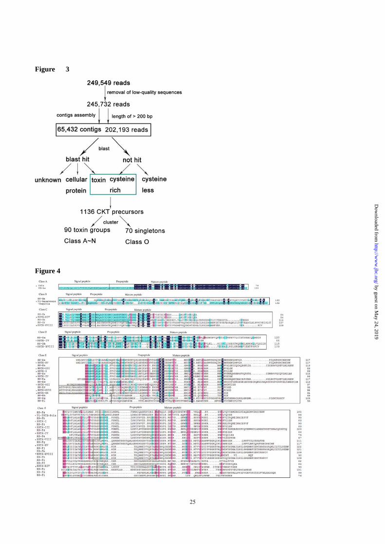
454 Sequencing Statistics and Transcriptome
Assembly
The mRNAs of six venom glands from tarantula
Ornithoctonus hainana were extracted and
sequenced using GS FLX technology (454/Roche)
following the manufacturer’s protocol. Sequencing
revealed a total of 249,549 reads (amounting to
~757 Mb) with an average length of ~328 bases per
read (max. 830 bp, min. 40 bp, 6.72% of reads <100
bp, 84.6% of reads 100–500 bp, 8.63% of
reads >500 bp). The raw sequencing data can be
downloaded from SRA (NCBI) using accession
number SRP040123. After removing sequences of
low quality, a total of 215,640 reads were assembled
into 65,432 contiguous DNA sequences (contigs)
with an average length of 625 bp (36.3 reads/contig),
with the rest remaining as single reads. Whereas this
study focused mainly on toxin peptides, numerous
other protein sequences were identified and will be
described elsewhere. As outlined in the experimental
procedures section, we searched for toxin peptide
sequences directly from the sequencing reads, as the
average read length of >200 bp allowed the
identification of full-length toxin precursors. Toxin
peptides were also searched for in the contigs, and
no additional toxin peptide sequences were found. In
total, 52,570 reads displayed similarity to known
peptide toxins or ‘Toxin-Like’ sequences; the
‘Putative Toxins’ category includes sequences rich
in cysteine residues and sharing sequence identity
with toxins or proteins including the ICK motif (5%)
that were not identified by a BLAST search; ‘the
‘Cellular Proteins’ category includes transcripts
coding for proteins involved in cellular processes
(44%); The ‘Unknown Function’ category includes
reads that shared sequence identity with previously
described sequences with no functional assessment
or hypothetical genes; The ‘No Hit’ category
indicates no match with currently known sequences.
The results are summarized in Figure 1.
Functional Annotation
A search against publicly available databases
(nr/NCBI, Swiss-Prot + TREMBL/EMBL) revealed
that 8,773 high-confidence proteins were associated
with GO terms and further grouped into Molecular
Functions (MF), Biological Process (BP) and
Cellular Components (CC) at the second level
according to standard gene ontology terms
(http://www.geneontology.org). Based on
annotations from gene ontology (GO) analysis
(Figure 2), transcripts were categorized into 2,610
groups with 80% identity threshold. These included
functional annotations for 816 Biological Process
(BP), 798 Molecular Function (MF), and 996
Cellular Component (CC) categories (Figure 2).
Highly expressed transcripts were enriched in
metabolism and translation processes, indicating that
venom glands are metabolically active and engaging
in the intensive protein synthesis and processing
required for venom production. Transcripts
associated with binding, catalysis and channel
regulation were also highly represented, indicating
that venom peptides may play an important role in
prey inactivation by binding to ion channels. GO
terms related to redox homeostasis and proteolysis
were also enriched, which may be related to the
extensive post-translational modification of spider
toxins that was reported previously (9).
Spider Toxin Transcript Identification and
Classification
Although spider toxins contain diverse disulfide
bridge patterns and fold into a variety of
three-dimensional structures, cysteine-rich domains
are a common feature shared by many toxin
sequences (26,27). Overall, 1,136 toxin precursors
were identified in the venom gland transcriptome,
and 65.8% of the mature peptides included two
adjacent cysteine residues. From previous
proteomics results (10), we were able to predict
precursor endoproteolytic and amidation sites with
high confidence, and 90 toxins and variants
accounted for 95% of all toxin precursors. These
by guest on May 24, 2019
http://ww
w.jbc.org/
Dow
nloaded from

6
were divided into 14 classes (Class A-N) based on
the number of cysteine residues present (Table 1),
and 70 sequences did not fall into these categories in
Class O (Figure 3). Of the 18 Haplopelma hainanum
peptide toxins previously characterized, 11 belonged
to Class G along with 252 variants, four belonged to
Class E along with 39 variants, and the other three
belonged to Class C, L and M along with 19, 25 and
4 variants, respectively.
Class A
Class A contained seven novel transcripts, including
HN-Aa and its variants (Figure 4). The signal
peptide and propeptide of these precursors are
identical, and their cleavage sites are LFA/ED and
LESEK, respectively. The mature peptide has twelve
Cys residues, which is the highest number in a toxin
peptide scaffold in H. hainanum reported to date,
although two other spider toxins share this cysteine
pattern (28,29). TX-L precursors from Lycosa
singoriensis superfamily VI have the cysteines
arranged C-CC-C-C-C-C-C-C-C-C-C, whereas
HN-Aa has the double cysteine shifted to the left.
The double knot toxin (DkTx) from the Earth Tiger
tarantula (Selenocosmia huwena) has the pattern
C-C-CC-C-C-C-C-CC-C-C, and this sequence
shares 87% similarity with HN-Aa. DkTx
selectively and irreversibly activates the capsaicin-
and heat-sensitive channel TRPV1 (29). This
cycteine pattern therefore indicates that HN-Aa may
be a TRPV1 channel activator. The six variants
identified showed high similarity with the mature
peptide, and the presence of different lengths of
mature peptide results in great potential for toxin
variability.
Class B
Class B contains only four members, the fewest of
all classes (Figure 4). HN-Ba and its three variants
include eleven Cys residues, and HN-Ba precursors
contain over 100 residues that includes the
consensus VIAYA cleavage signal, while the
propeptide numbered only four residues. A large
number of Gln residues were found in the mature
peptide, and the C-terminus is rich in Thr and Ser
residues, which is strikingly different from other
toxins present in this species. The cysteine pattern is
shares homology with the U1-hexatoxin-Hsp201a
from the Funnel-web spider (30). Other than this
highly conserved cysteine pattern, HN-Ba showed
no obvious sequence similarity with other peptide
toxins.
Class C
Class C contains 29 transcripts that clustered into
the known toxin HN-Ca (HNTX-XIV) and two
novel toxins (HN-Cb and HN-Cc; Figure 4), all of
which included 10 Cys residues, but Class C toxins
were further divided into two clades based on
cysteine pattern. HN-Ca and HN-Cb shared the
pattern -C-C-CC-C-C-C-C-C-C- and formed the
first clade, and these toxins do not have a propeptide
but do shared a signal peptide containing the
cleavage site ELVSC. The mature HN-Cb peptide is
rich in Val residues and has a C-terminus that is
approximately 20 longer than HN-Ca (HNTX-XIV),
indicating diverse functions. Further research is
needed to investigate whether these additional
residues are cleaved during post-translational
modification or remain and have some additional
functional significance. HN-Cc exhibited the
cysteine pattern -C-C-C-CC-C-C-C-C-C-, and the
signal peptide, propeptide and mature peptide were
distinct from HN-C[a-b], although HN-Cc peptides
shared sequence similarity and included an extended
cysteine variant not present in HNTX-XVIII.
Class D
The four toxin precursors in Class D contain nine
cysteine residues and exhibited two different
cysteine patterns (Figure 4). HN-Da shares
significant similarity with HNTX-IV (31) but has the
cysteine pattern -C-C-CC-C-C-C-C-C- with an
additional three cysteines at the C-terminal region
that also included a particularly large number of Ser
residues. HN-Db has the cysteine pattern
-C-C-C-CC-C-C-C-C- and shared an identical signal
peptide and propeptide with HNTX-XVIII, but the
mature peptide was different, in particular due to the
additional cysteine. The additional C-terminal
by guest on May 24, 2019
http://ww
w.jbc.org/
Dow
nloaded from

7
residues could be removed to produce a more
canonical spider toxin during maturation.
Class E
The mature peptides of Class E displayed two
distinct cysteine patterns and the precursors were
classified into nine groups (Figure 4). HN-E[a-d, f,
h-i] shared the cysteine pattern -C-C-CC-C-C-C-C-
and the other two toxins shared the cysteine pattern
and -C-C-C-CC-C-C-C- (Table 1). HN-E[a-c] share
high sequence similarity with members of groups
HNTX-I, HNTX-III and HNTX-IV, respectively,
and include a 30 residue C-terminal extension.
Interestingly, the CIC motif was present in the
majority of these sequences, which is typical of
µ-AGTXs from the American funnel web spider
(Agelenopsis aperta) and PnTx3-3 from the
Brazilian armed spider (32-35). We speculated that
the cysteines in this motif are not involved in
disulfide bond formation because PnTx3-3 only has
two disulfide bonds, with the fourth and fifth Cys
residues excluded (32). Members of HN-Ed share
sequence similarity with HNTX-VII, with the
propeptide lost and a longer mature peptide. Toxin
precursors lacking propeptides are commonly
observed in scorpions but rarely in spiders. Four
known toxins, HN-Ef (HNTX-XVII), HN-Eg
(HNTX-XVIII), HN-Ee (HNTX-XV) and HN-Eh
(HNTX-XX) have been chemically and functionally
characterized in our previous work (10). HN-Ef
(HNTX-XVII) and HN-Eh (HNTX-XX) exhibited
the shortest mature peptides in this Class. The
typical motif CVX1CVIX5CVIIX1CVIII, where X is
any residue except cysteine was present in
precursors, and was first identified in Mu-agatoxins
from Agelenopsis aperta (32). Numerous variants
differed by only one or two amino acids, suggesting
these may be ideal for studying structure-function
relationships. Moreover, the HN-Ei mature peptide
is identical with HWTX-XV (Table 2). Whilst
interesting, it is not unprecedented for toxins from
different venomous animals to share identical
sequences.
Class F
Class F includes 15 toxins (149 precursors) and
divided into three clades based on the seven cysteine
pattern present in these peptides (Figure 4). Ten
(HN-F[a-g, k, n-o]) share the
-C-C-CC-C-C-C-,pattern, and four (HN-F[f-j, m])
exhibited the -C-C-C-CC-C-C- pattern, while only
HN-Fl had the pattern -C-CC-C-C-C-C-. The pattern
in HN-Fa is shared with that in
U1-theraphotoxin-Pc1a, a toxin that possesses
strong in vitro anti-plasmodial activity against the
intra-erythrocyte stage of P. falciparum (33,36).
HN-F[b-c] share high homology with HNTX-I and
HNTX-III, including the conservative sequence
NEINACSPVF in the C-terminal region. HN-Fd
shares high sequence identity with HNTX-IV, but
includes the much longer conserved C-terminal
sequence
“MRSMYPVQFSNWMYLANGGIMSSTSSACQL
MSINK” that is enriched with Ser. While HN-Fe has
the same signal peptide as HNTX-VIII, the
C-terminus is much longer. The mature peptide of
HN-Ff shares minimal sequence identity with
HNTX-XIV, and the key Cys residues are changed
into other amino acids. HN-F[g-j] shares high
sequence similarity with HNTX-XV and
HNTX-XVIII, but can not form four disulfide bonds
as there is a cysteine missing in the C-terminal
region. We speculated that despite the high identity
with known toxins, these peptides must display a
novel cysteine pattern. HN-F[k-m] shares sequence
similarity with toxins from Ornithoctonus huwena,
and HN-Fk shares sequence similarity with
HWTX-VIII, including a high proportion of basic
residues in the mature peptides. HN-Fm shares
similarity with HWTX-XVIII, and HN-Fl also
includes a large number of basic residues, but no
homologs were identified from a BLAST search,
therefore this appears to be a novel toxin.
Class G
Class G includes the greatest number of peptides,
accounting for 36% of all toxins, and 10 of these
(HN-Ga (HNTX-I), HN-Gc (HNTX-III), HN-Ge
by guest on May 24, 2019
http://ww
w.jbc.org/
Dow
nloaded from

8
(HNTX-IV), HN-Gg (HNTX-IX), HN-Gj
(HNTX-VII), HN-Gm (HNTX-XIII), HN-Gn
(HNTX-X), HN-Gr (HNTX-XIII), HN-Gs
(HNTX-XIX), HN-Gu (HNTX-XVI), HN-Go
(HNTX-XII)) have been chemically and
functionally characterized in our previous work.
These ten toxins contain six cysteine residues
forming three disulfide bonds. Based on sequence
similarity, Class G peptides were further divided
into 25 distinct toxin groups (Figure 5). HN-G [b, d,
f, i, l, q] are similar to the known toxins HNTX-I,
HNTX-III, HNTX-IV, HNTX-IX, HNTX-VII, and
HNTX-XII, but with a longer C-terminal region.
The three disulfides have a 1-4, 2-5 and 3-6
connectivity that form the distinctive inhibitor
cysteine knot (ICK) motif. The majority of members
in this class are variants that differ by only one or
two amino acids, implying that they are natural
mutants. In contrast to the traditional
ICK-motif-containing spider toxins, HN-Gt and
HN-Gv are similar to HNTX-XVI and HNTX-XVIII,
and adopt a -C-C-C-CC-C- cysteine pattern. Toxins
HN-G[w-y] share high sequence similarity with
HWT-Xa, HWTX-XIII, and HWTX-XVI,
respectively, while HN-Gw, HN-Gx and HN-Gy
differ from HWTX-Xa, HWTX-XIII and
HWTX-XVI respectively by only two amino acid
residues. The signal peptides and propeptides of
these toxins included a signal-peptide cleavage site
similar to the CYASE sequence.
Classes H and I
Although most of the known spider toxins contain
more than six cysteine residues, the members of
classes H and may include four to five cysteines.
Class H included five transcripts with five cysteines
in a distinctive -CC-CC-C- pattern comprising two
contiguous Cys pairs (Figure 5). This cysteine
pattern is novel, and homology between HN-Ha
with other toxins is very low. HN-Ha groups only
accounted for about 0.2 % of the toxin transcripts,
and are therefore unlikely to play a key role in the
venom function of H. hainanum.
All of the 28 transcripts were classified into six
toxin groups according to sequence homology.
Three toxins, accounting for a quarter of the Class I
sequences, did not generate hits in a BLAST search
(Figure 8). HN-Id shares homology with
HWTX-XV from O. huwena, which is a
tetrodotoxin (TTX)-sensitive voltage-gated sodium
channel (VGSC) inhibitor. The signal peptide and
propeptide of HN-Id are highly variable, but HN-Id
has a highly conserved mature peptide and shares
the -C-CC-C- cysteine arrangement with HN-Ic.
HN-I[a-b] also share this pattern, but also did not
generate hits in a BLAST search. HN-Ie differs from
HNTX-III, having a C-terminal mutation and
lacking Cys 5 and Cys 6. The first and second Cys
are replaced by other residues in the mature peptide
of HN-If.
Classes J and K
Class J contains a set of eight homologous
transcripts (HN-Ja) with the cysteine pattern
x3Cx3Cx8Cx4Cx5Cx4Cx5Cx1Cxn, where x is any
amino acid and n is an undefined number (Figure 6).
In contrast to some other classes, these peptides do
not contain the double-cysteine (-CC-) motif, and
this cysteine pattern is consistent with that of
LSTX-M1 and Omega-agatoxin-1A (28,37), in
which ten Cys residues form four intra-chain and
one inter-chain disulfide bonds. In contrast, HN-Ja
did not show sequence similarity with LSTX-M1 or
Omega-agatoxin-1A, but is similar to HNTX-II.
Class K contains a set of six transcripts which
share the cysteine pattern -C-C-C-C-C-C-C-. The
C-terminal region is elongated compared with
HNTX-II family peptides and includes an additional
Cys residue (Figure 9). The signal peptide and
propeptide was the same for all members of these
classes, but with C-terminal regions of the of mature
peptides were variable.
Class L
Class L comprises 100 transcripts that share the
cysteine pattern -C-C-C-C-C-C- (Figure 6). HN-La
(HNTX-II) was the most significant component in
this class. The signal-peptide and propeptide of
by guest on May 24, 2019
http://ww
w.jbc.org/
Dow
nloaded from

9
HN-L[b-d] were identical with that of HNTX-II, and
the mature peptides were also very similar, and all
included the conserved NHHDKIRNRKV sequence
motif in the C-terminal region. HN-Le shares high
sequence similarity with HWTX-VII, but the third
Cys in the mature peptide is replaced by Leu, and
the conservative sequence VLKCR is added to the
C-terminal region. HN-L[f-i] may be narrowly
distributed or even species-specific, since no
homologs from other spiders have been reported to
date. HN-Lg and HN-Li have identical signal
peptides, both lack a propeptide, and both have
similar mature peptides that include the Cys pattern
x2Cx5CxCx12Cx11Cxn. A Lys-rich motif is also
present in the mature peptide of HN-L (Figure 6).
Classes M and N
Mature peptides of Class M shared the cysteine
pattern -C-C-C-C-C-, which is characteristic of
kunitz-type sequences and is found in HWTX-XI
and HWTX-VII from O. huwena (38). HN-M[a-b]
share high sequence similarity with HNTX-II, with
the third Cys replaced by Asp, Leu or Ile in HN-Ma.
Similarly, the fifth cysteine in mature HN-Mb
peptides is replaced by other residues, and there are
several additional Cys residues in the C-terminal
region. HN-M [c, h] share sequence homology with
HNTX-VIII, with a double cysteine missing from
the cysteine pattern (Figure 10), whereas HN-Md
shares high sequence similarity with HNTX-XI,
except the HN-Md3 variant has an extended
C-terminal region enriched with basic residues. The
three toxins HN-M[e-g] were novel and generated
no hits in a BLAST search (Figure 6).
Class N toxins exhibited a -C-C-C-C- cysteine
pattern, and HN-N [a-h] shared some similarity with
HNTX-II, huwentoxin II and HWTX-VII. HN-Ni
exhibited no obvious sequence similarity with any
known sequences (Figure 6).
Toxin Abundance Among Classes
The abundance of toxin ESTs is reflective of venom
composition and provides information on their
diversity and evolution. Major differences in
transcription levels between different toxin groups
and between toxin classes were investigated (Figure
7). Unsurprisingly, most venom peptides were
expressed at relatively high levels and were present
in a variety of forms at the transcriptional level. This
suggests that the more abundant toxin transcripts are
mirrored by a greater number of peptide toxins.
Moreover, gene classes containing larger numbers of
precursors also tended to produce the highest
number of total reads. Overall, Class G produced the
highest number of reads and the largest number of
precursors, while classes E, F, L, M and N were of
intermediate abundance (Figure 7A). Analysis of the
data showed that the 30 most abundant toxin
peptides accounted for 84.7% of the total toxin
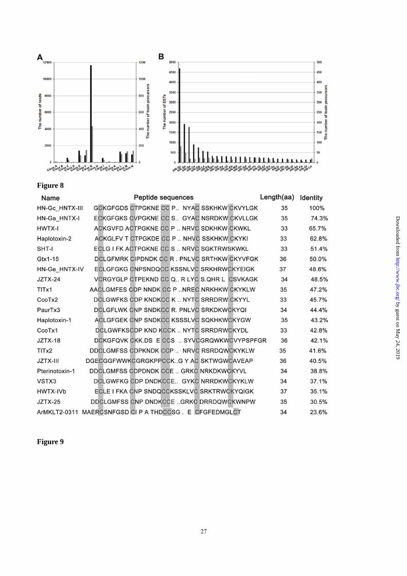
transcripts (Figure 7B). HN-Gc (HNTX-III) had the
highest number of transcripts, which were translated
into 82 different HN-Gc variants. A BLAST search
revealed that HN-Gc shares considerable sequence
identity with 20 toxins from spiders and cone snails
(Figure 8), indicating that HN-Gc may be an
ancient ancestral toxin from which others evolved.
Most toxin precursors included a number of variants
that enriches toxin diversity, although there exist
only three mutations between HN-Ee, HN-Md and
HN-Gn, suggesting that spiders could further
improve venom selectively through greater sequence
diversification.
Variation in Toxin Precursor Sequence and
Expression
Classes A–N included multiple variants for each
toxin sequence, due to random mutations in both
prepro and mature toxin regions. This led to broad
distributions in the chromatographic fractions due to
the close molecular weights of the variants, and is
reflected in the diversity of structures and
physicochemical properties. Rapid gene duplication
accompanied by focal hypermutation of residues
encoded in the mature peptide sequence, and the
high conservation of cysteine residues due to high
codon bias can explain the diversity of the variants
(10,11,39). Alignment of the amino acid sequences
of HN-Ge and 18 homologous peptides identified
by guest on May 24, 2019
http://ww
w.jbc.org/
Dow
nloaded from

10
several mutations (Figure 9). All variants except
HN-Ge18 and Ge-15 share the same propeptide
sequence, and some sequences are highly similar.
HN-Gb and HN-Gd have an extended C-terminal
region compared with HN-Gc (Figure 5). Alteration
of the termination codon, point mutations, insertion
and deletions, and shuffling of sections of
oligonucleotide sequence are responsible for the
diversification of peptides (Figure 10), and is
common to the transcriptome of both snakes and
spiders (40-42). Mutations appear to occur randomly
and display evident sequence diversity without
disrupting the ICK scaffold. Unsurprisingly, 321
precursor variants resulted from 18 known toxins,
but only 23 of these overlapped. The significance of
the molecular evolutionary process is two-fold:
most residues mutate randomly, which generates
numerous variants, but at the same time, the
molecular mechanisms of transcription preserve
cysteine residues, resulting in a high conservation of
the molecular scaffold (43). Although a high number
of toxin variants were present in the venom gland
transcriptome of H. hainanum, many of the gene
products may not be functionally active, but may
play a role in the evolution of future toxin variants.
Comparison of proteomic and transcriptomic data
revealed that only 15 fully sequenced and 9 partially
sequenced venom peptides were identified using the
454 transcriptome approach (Figure 11), and the vast
majority of transcripts coding for CKTs did not
appear as translated peptides in the venom proteome.
The combination of a targeted mutagenic
mechanism to generate high variability with the
subsequent action of diversifying selection on highly
expressed variants might explain the
hypervariability (44). Most transcripts are perhaps
unlikely to be translated, apart from critically
important components. This mechanism might
prevent the venom gland from going to the expense
of producing toxins unnecessarily, which makes the
process more systematic and efficient(45). Studying
toxin diversity is of importance for the discovery of
novel toxins, and will help to explain toxin
evolution.
Modification and Processing of Multiple Peptide
Products
The variety of toxin variants in spider venom partly
results from the diversity of toxin gene products and
partly from post-translational modifications,
sequence homologues, and protein degradation
(38,41,46,47). For example, F1-29.54-4123 and
F1-30.82-4112.8 share the same precursor, but
several residues are modified in the C-terminal
region of the mature peptides (Figure 11), and
amidation provides further diversity. A single
glycine is a common amidation signal, as is Gly
followed by a single or dibasic endoproteolytic site
at the C-terminus (48). Amidation signals were
identified at the C-terminus of three toxins (G-K in
HN-Ga, Gc, Ge). During post-translational
processing, precursors are processed to yield mature
truncated toxins. A single peptide (K) or tripeptide
(R-M-D) was removed from the C-terminus of the
deduced peptides by the precursor processing
enzyme in HN-Gg (HNTX-IX) and HN-Go
(F3-24.71-4057.6). F3-30.36-3538.0 and
F3-25.85-3351.9 are truncated forms of HN-Gm,
with a dipeptide (RR) or tripeptide (WRR) deleted
from the C-terminus, while F6-25.12-3998.8 and
F3-24.71-4057.6 are truncated forms of HN-Go with
a tetrapeptide (GRMD) or tripeptide (RMD) deleted
from the C-terminus. We speculate that precursors
were alternatively spliced and modified during
posttranslational processing, as was observed in
venoms from O. huwena(41). HNTX-IX shares the
same amino acid sequence with HNTX-IX-2, but the
molecular weight is higher by 10 Da, and
preliminary sequence analysis showed that this may
be due to an unknown modification, as observed in
O. huwena (49). Despite the very low expression
levels of most of these variants, they contributed a
great deal to the overall diversity of spider toxins.
Effect of Functional Diversity on Rat DRG
Neurons
To explore the functions of these novel spider toxins,
we investigated the activities of 14 representative
by guest on May 24, 2019
http://ww
w.jbc.org/
Dow
nloaded from

11
toxins on Nav, Kv and Cav channels in rat DRG
neurons using the whole-cell patch-clamp technique.
In total, 11 six cysteine-containing toxins were
chosen from Class G. HN-Ef (HNTX-XVII) has four
disulfide bonds, HN-La (F8_17.06) includes the
DHH motif, and HN-O38 has three cysteine residues
that can only form one disulfide. All 14 tested toxins
show very weak or lack inhibition on voltage-gated
potassium channels, although they exhibits variable
inhibitory effects on sodium or calcium channels.
Some toxins with high sequence similarity exhibited
similar bioactivity against the ion channels (Figure
12). HN-Gc (HNTX-III), HN-Ge (HNTX-IV) and
HNTX-V all contain the ICK motif, and all inhibited
tetrodotoxin-sensitive (TTX-sensitive (TTX-S)
VGSCs by >70%. However, HN-Ga (HNTX-I) also
shares this motif but did not affect TTX-S VGSCs.
The hydrophilic Asn19 and basic His27 are
conserved among the first three toxins, but are
replaced by Gly and acidic Asp in HN-Ga, which
may be responsible for the different channel
inhibition activity. This result indicates that the
mutation of key residues could profoundly affect
toxin activity. HN-Gu (HN-XVI) showed high
inhibition against TTX-R VGSCs, and this toxin has
an ICK motif, but there are 14 residues between the
fourth and fifth cysteines, which is much longer than
in other ICK-containing toxins. Moreover, toxins
with diverse cysteine patterns could have similar
functions. For example, at a concentration of 100
nM, HN-Ge (HNTX-IV) inhibited calcium channels
by >50% and TTX-S sodium channels by >30%.
Similarly, HN-O38 has only three cysteine residues
while F8_17.06 has a complete DDH motif, and
both of these toxins have a cysteine pattern distinct
from ICK motif toxins, yet all three inhibited
calcium currents by >30%.
In summary, the activity of spider toxins on ion
channels was highly variable. Toxins with conserved
sequence patterns can share similar functions or
behave very differently. Toxins exhibiting low
activity against DRG ion channels may be important
for inactivating prey, but further functional studies
are needed.
Discussion
Spider toxins form a huge reservoir of
chromosomally-encoded short peptides that exhibit
remarkable structural diversity. Many of the more
highly abundant toxins have been studied, but the
mechanisms of toxin diversity and evolution are far
from being well understood. In this work, a high
throughput 454 pyrosequencing approach was
deployed as it offers the high-throughput, accuracy
and relatively long reads (>300 bp on average)
required to cover the full length of spider toxin
precursors (60–120 amino acids). A total of 1,136
toxin precursors were detected that clustered into 90
toxin peptides, 18 of which have been previously
reported, 44 of which were novel but shared
sequence similarity with previously reported H.
hainanum toxins, 14 of which shared sequence
similarity with known toxins from other spiders, and
14 that were novel and did not retrieve any hits
following a BLAST search (Figure 14). A total of 20
different cysteine scaffolds were present that
contained between four and twelve Cys residues. Six
of these cysteine scaffolds have been presented in
previous work, but 14 were novel. The cysteine
pattern -C-C-CC-C-C- that folds into the highly
stable cysteine knot (ICK) motif was the most
abundant. This toxin scaffold is found in a wide
range of bioactive peptides in spiders, snakes or
other venomous animals (4,50-54), suggesting this
structure evolved in an ancient venomous ancestor.
Many toxin variants were identified at the
transcriptional level which arose from a limited set
of genes through hypermutation and fragment
insertion/deletion. Although the mutations occurred
randomly, in general sequences diverged but the
common cysteine scaffold was conserved. Moreover,
alternative cleavage sites, heterogeneous
post-translational modifications, and highly variable
N- and C-terminal truncations further increase
peptide diversity (10).
The abundance of different toxin transcripts
was highly variable and reflected in the composition
of venom peptides, and his is important for
by guest on May 24, 2019
http://ww
w.jbc.org/
Dow
nloaded from

12
understanding of toxin diversity and evolution. In
general there was a positive correlation between
mRNA levels and peptide abundance. Class G
contained both the highest number of transcripts and
the largest number of peptide precursors. This
important toxin class may target channels conserved
in various types of prey animal. Toxins of
intermediate abundance also may also be related to
hunting. A surprisingly large number of transcript
sequences were expressed at relatively low levels,
many of which differed from more abundant
peptides by single amino acid changes, deletions,
and frame and stop codon shifts. Some toxin
variants were generated from alternative cleavage
sites, interrupted or elongated cysteine patterns. The
toxins in low abundance might be considered as the
track of toxin evolution, namely, natural selection
might drive the spider to retain abundant fractions of
the venom, which are applied by the spider in prey
capture and/or defense.
Only 18 of the identified toxin genes have been
described in previous work. We hypothesize that
toxin variants arose by random mutations and focal
hypermutations from rapid gene duplication, and
post-translational modifications provide further
structural diversity. These processes allow the
venom transcriptome to rapidly adapt to changes in
the environment or type of prey (55,56). It has been
suggested that it is the cumulative effect of
individual spider toxins that is responsible for the
lethality, and different toxin groups exist in different
spiders.
Analysis of toxin activity on DRG ion channels
showed that some of the toxins with similar
sequence patterns generally exhibited similar
functions, however, with some exceptions, some of
such toxins alternatively displayed distinct functions.
Moreover, some of the peptides with more divergent
sequences can also share similar functions if key
residues are conserved. The diversity of venom
toxins results in many peptides working together to
improve the efficiency by which prey are
incapacitated.
In conclusion, a combination of transcriptomics,
peptidomics and electrophysiology revealed the
impressive diversity of spider toxin peptides at the
transcriptional and peptide structural and functional
levels.
Acknowledgements
We are thankful to Dr. Zhonghua Liu and Ying
Wang for knowledge support. We thank The
Cooperative Innovation Center of Engineering and
New Products for Developmental Biology of Hunan
Province (20134486). This work was supported by a
grant of the National Basic Research Program of
China (973), 2010CB529801.
by guest on May 24, 2019
http://ww
w.jbc.org/
Dow
nloaded from

13
Reference
1. Binford, G. J., Bodner, M. R., Cordes, M. H., Baldwin, K. L., Rynerson, M. R., Burns, S. N., and Zobel-Thropp,
P. A. (2009) Molecular evolution, functional variation, and proposed nomenclature of the gene family that
includes sphingomyelinase D in sicariid spider venoms. Mol Biol Evol 26, 547-566
2. Liang, S. (2008) Proteome and peptidome profiling of spider venoms. Expert review of proteomics 5, 731-746
3. Grishin, E. (1999) Polypeptide neurotoxins from spider venoms. Eur J Biochem 264, 276-280
4. Liang, S. (2004) An overview of peptide toxins from the venom of the Chinese bird spider Selenocosmia
huwena Wang [=Ornithoctonus huwena (Wang)]. Toxicon 43, 575-585
5. Escoubas, P., and Rash, L. (2004) Tarantulas: eight-legged pharmacists and combinatorial chemists. Toxicon 43,
555-574
6. Corzo, G., and Escoubas, P. (2003) Pharmacologically active spider peptide toxins. Cell Mol Life Sci 60,
2409-2426
7. Rash, L. D., and Hodgson, W. C. (2002) Pharmacology and biochemistry of spider venoms. Toxicon 40, 225-254
8. Fernandes-Pedrosa Mde, F., Junqueira-de-Azevedo Ide, L., Goncalves-de-Andrade, R. M., Kobashi, L. S.,
Almeida, D. D., Ho, P. L., and Tambourgi, D. V. (2008) Transcriptome analysis of Loxosceles laeta (Araneae,
Sicariidae) spider venomous gland using expressed sequence tags. BMC Genomics 9, 279
9. Chen, J., Zhao, L., Jiang, L., Meng, E., Zhang, Y., Xiong, X., and Liang, S. (2008) Transcriptome analysis
revealed novel possible venom components and cellular processes of the tarantula Chilobrachys jingzhao venom
gland. Toxicon 52, 794-806
10. Tang, X., Zhang, Y., Hu, W., Xu, D., Tao, H., Yang, X., Li, Y., Jiang, L., and Liang, S. (2010) Molecular
diversification of peptide toxins from the tarantula Haplopelma hainanum (Ornithoctonus hainana) venom based
on transcriptomic, peptidomic, and genomic analyses. J Proteome Res 9, 2550-2564
11. Escoubas, P. (2006) Molecular diversification in spider venoms: a web of combinatorial peptide libraries. Mol
Divers 10, 545-554
12. Durban, J., Juarez, P., Angulo, Y., Lomonte, B., Flores-Diaz, M., Alape-Giron, A., Sasa, M., Sanz, L., Gutierrez,
J. M., Dopazo, J., Conesa, A., and Calvete, J. J. (2011) Profiling the venom gland transcriptomes of Costa Rican
snakes by 454 pyrosequencing. BMC Genomics 12, 259
13. Terrat, Y., Biass, D., Dutertre, S., Favreau, P., Remm, M., Stocklin, R., Piquemal, D., and Ducancel, F. (2012)
High-resolution picture of a venom gland transcriptome: case study with the marine snail Conus consors.
Toxicon 59, 34-46
14. Rokyta, D. R., Lemmon, A. R., Margres, M. J., and Aronow, K. (2012) The venom-gland transcriptome of the
eastern diamondback rattlesnake (Crotalus adamanteus). BMC Genomics 13, 312
15. Lluisma, A. O., Milash, B. A., Moore, B., Olivera, B. M., and Bandyopadhyay, P. K. (2012) Novel venom
peptides from the cone snail Conus pulicarius discovered through next-generation sequencing of its venom duct
transcriptome. Marine genomics 5, 43-51
16. Prosdocimi, F., Bittencourt, D., da Silva, F. R., Kirst, M., Motta, P. C., and Rech, E. L. (2011) Spinning gland
transcriptomics from two main clades of spiders (order: Araneae)--insights on their molecular, anatomical and
behavioral evolution. PLoS ONE 6, e21634
17. Zhang, J., Chiodini, R., Badr, A., and Zhang, G. (2011) The impact of next-generation sequencing on genomics.
Journal of genetics and genomics = Yi chuan xue bao 38, 95-109
18. Liu, L., Li, Y., Li, S., Hu, N., He, Y., Pong, R., Lin, D., Lu, L., and Law, M. (2012) Comparison of
next-generation sequencing systems. J Biomed Biotechnol 2012, 251364
19. Zheng, Y., Zhao, L., Gao, J., and Fei, Z. (2011) iAssembler: a package for de novo assembly of
by guest on May 24, 2019
http://ww
w.jbc.org/
Dow
nloaded from

14
Roche-454/Sanger transcriptome sequences. BMC Bioinformatics 12, 453
20. Conesa, A., Gotz, S., Garcia-Gomez, J. M., Terol, J., Talon, M., and Robles, M. (2005) Blast2GO: a universal
tool for annotation, visualization and analysis in functional genomics research. Bioinformatics 21, 3674-3676
21. Ashburner, M., Ball, C. A., Blake, J. A., Botstein, D., Butler, H., Cherry, J. M., Davis, A. P., Dolinski, K.,
Dwight, S. S., Eppig, J. T., Harris, M. A., Hill, D. P., Issel-Tarver, L., Kasarskis, A., Lewis, S., Matese, J. C.,
Richardson, J. E., Ringwald, M., Rubin, G. M., and Sherlock, G. (2000) Gene ontology: tool for the unification
of biology. The Gene Ontology Consortium. Nature genetics 25, 25-29
22. Kearse, M., Moir, R., Wilson, A., Stones-Havas, S., Cheung, M., Sturrock, S., Buxton, S., Cooper, A.,
Markowitz, S., Duran, C., Thierer, T., Ashton, B., Meintjes, P., and Drummond, A. (2012) Geneious Basic: an
integrated and extendable desktop software platform for the organization and analysis of sequence data.
Bioinformatics 28, 1647-1649
23. Duan, D., Rong, M., Zeng, Y., Teng, X., Zhao, Z., Liu, B., Tao, X., Zhou, R., Fan, M., Peng, C., Chen, P., Liang,
S., and Lu, M. (2011) Electrophysiological characterization of NSCs after differentiation induced by OEC
conditioned medium. Acta neurochirurgica 153, 2085-2090
24. Kumar, S., Nei, M., Dudley, J., and Tamura, K. (2008) MEGA: a biologist-centric software for evolutionary
analysis of DNA and protein sequences. Briefings in bioinformatics 9, 299-306
25. Xiao, Y., and Liang, S. (2003) Inhibition of neuronal tetrodotoxin-sensitive Na+ channels by two spider toxins:
hainantoxin-III and hainantoxin-IV. Eur J Pharmacol 477, 1-7
26. Naamati, G., Askenazi, M., and Linial, M. (2009) ClanTox: a classifier of short animal toxins. Nucleic Acids Res
37, W363-368
27. Kuzmenkov, A. I., Fedorova, I. M., Vassilevski, A. A., and Grishin, E. V. (2012) Cysteine-rich toxins from
Lachesana tarabaevi spider venom with amphiphilic C-terminal segments. Biochim Biophys Acta 1828, 724-731
28. Zhang, Y., Chen, J., Tang, X., Wang, F., Jiang, L., Xiong, X., Wang, M., Rong, M., Liu, Z., and Liang, S. (2010)
Transcriptome analysis of the venom glands of the Chinese wolf spider Lycosa singoriensis. Zoology (Jena) 113, 10-18
29. Bohlen, C. J., Priel, A., Zhou, S., King, D., Siemens, J., and Julius, D. (2010) A bivalent tarantula toxin activates
the capsaicin receptor, TRPV1, by targeting the outer pore domain. cell 141, 834-845
30. Wen, S., Wilson, D. T., Kuruppu, S., Korsinczky, M. L., Hedrick, J., Pang, L., Szeto, T., Hodgson, W. C.,
Alewood, P. F., and Nicholson, G. M. (2005) Discovery of an MIT-like atracotoxin family: spider venom
peptides that share sequence homology but not pharmacological properties with AVIT family proteins. Peptides
26, 2412-2426
31. Li, D., Xiao, Y., Xu, X., Xiong, X., Lu, S., Liu, Z., Zhu, Q., Wang, M., Gu, X., and Liang, S. (2004)
Structure--activity relationships of hainantoxin-IV and structure determination of active and inactive sodium
channel blockers. J Biol Chem 279, 37734-37740
32. Adams, M. E. (2004) Agatoxins: ion channel specific toxins from the American funnel web spider, Agelenopsis
aperta. Toxicon 43, 509-525
33. Pimentel, C., Choi, S. J., Chagot, B., Guette, C., Camadro, J. M., and Darbon, H. (2006) Solution structure of
PcFK1, a spider peptide active against Plasmodium falciparum. Protein Sci 15, 628-634
34. Miranda, D. M., Romano-Silva, M. A., Kalapothakis, E., Diniz, C. R., Cordeiro, M. N., Moraes-Santos, T., De
Marco, L., Prado, M. A., and Gomez, M. V. (2001) Spider neurotoxins block the beta scorpion toxin-induced
calcium uptake in rat brain cortical synaptosomes. Brain Res Bull 54, 533-536
35. Cordeiro Mdo, N., de Figueiredo, S. G., Valentim Ado, C., Diniz, C. R., von Eickstedt, V. R., Gilroy, J., and
Richardson, M. (1993) Purification and amino acid sequences of six Tx3 type neurotoxins from the venom of
the Brazilian 'armed' spider Phoneutria nigriventer (Keys). Toxicon 31, 35-42
36. Choi, S. J., Parent, R., Guillaume, C., Deregnaucourt, C., Delarbre, C., Ojcius, D. M., Montagne, J. J., Celerier,
by guest on May 24, 2019
http://ww
w.jbc.org/
Dow
nloaded from

15
M. L., Phelipot, A., Amiche, M., Molgo, J., Camadro, J. M., and Guette, C. (2004) Isolation and characterization
of Psalmopeotoxin I and II: two novel antimalarial peptides from the venom of the tarantula Psalmopoeus
cambridgei. FEBS Lett 572, 109-117
37. Bindokas, V. P., Venema, V. J., and Adams, M. E. (1991) Differential antagonism of transmitter release by
subtypes of omega-agatoxins. J Neurophysiol 66, 590-601
38. Jiang, L., Peng, L., Chen, J., Zhang, Y., Xiong, X., and Liang, S. (2008) Molecular diversification based on
analysis of expressed sequence tags from the venom glands of the Chinese bird spider Ornithoctonus huwena.
Toxicon 51, 1479-1489
39. Wong, E. S., and Belov, K. (2012) Venom evolution through gene duplications. Gene 496, 1-7
40. Ingrao, F., Rauw, F., Lambrecht, B., and van den Berg, T. (2013) Infectious Bursal Disease: A complex
host-pathogen interaction. Developmental and comparative immunology 41, 429-438
41. Jiang, L., Zhang, D., Zhang, Y., Peng, L., Chen, J., and Liang, S. (2010) Venomics of the spider Ornithoctonus
huwena based on transcriptomic versus proteomic analysis. Comp Biochem Physiol Part D Genomics
Proteomics 5, 81-88
42. Park, S., Park, M., and Rafii, F. (2013) Comparative transcription analysis and toxin production of two
fluoroquinolone-resistant mutants of Clostridium perfringens. BMC Microbiol 13, 50
43. Yuan, C., Jin, Q., Tang, X., Hu, W., Cao, R., Yang, S., Xiong, J., Xie, C., Xie, J., and Liang, S. (2007) Proteomic
and peptidomic characterization of the venom from the Chinese bird spider, Ornithoctonus huwena Wang. J
Proteome Res 6, 2792-2801
44. Conticello, S. G., Gilad, Y., Avidan, N., Ben-Asher, E., Levy, Z., and Fainzilber, M. (2001) Mechanisms for
evolving hypervariability: the case of conopeptides. Mol Biol Evol 18, 120-131
45. Zhang, Y., Huang, Y., He, Q., Liu, J., Luo, J., Zhu, L., Lu, S., Huang, P., Chen, X., Zeng, X., and Liang, S. (2014)
Toxin diversity revealed by a transcriptomic study of Ornithoctonus huwena. PLoS One 9, e100682
46. Dutertre, S., Jin, A. H., Kaas, Q., Jones, A., Alewood, P. F., and Lewis, R. J. (2013) Deep venomics reveals the
mechanism for expanded peptide diversity in cone snail venom. Mol Cell Proteomics 12, 312-329
47. Violette, A., Biass, D., Dutertre, S., Koua, D., Piquemal, D., Pierrat, F., Stocklin, R., and Favreau, P. (2012)
Large-scale discovery of conopeptides and conoproteins in the injectable venom of a fish-hunting cone snail
using a combined proteomic and transcriptomic approach. J Proteomics
48. Urbarova, I., Karlsen, B. O., Okkenhaug, S., Seternes, O. M., Johansen, S. D., and Emblem, A. (2012) Digital
marine bioprospecting: mining new neurotoxin drug candidates from the transcriptomes of cold-water sea
anemones. Mar Drugs 10, 2265-2279
49. Rong, M., Duan, Z., Chen, J., Li, J., Xiao, Y., and Liang, S. (2013) Native pyroglutamation of huwentoxin-IV: a
post-translational modification that increases the trapping ability to the sodium channel. PLoS ONE 8, e65984
50. Peng, K., Shu, Q., Liu, Z., and Liang, S. (2002) Function and solution structure of huwentoxin-IV, a potent
neuronal tetrodotoxin (TTX)-sensitive sodium channel antagonist from Chinese bird spider Selenocosmia
huwena. J Biol Chem 277, 47564-47571
51. Zhang, D., and Liang, S. (1993) Assignment of the three disulfide bridges of huwentoxin-I, a neurotoxin from
the spider selenocosmia huwena. Journal of protein chemistry 12, 735-740
52. Lu, S., Liang, S., and Gu, X. (1999) Three-dimensional structure of Selenocosmia huwena lectin-I (SHL-I) from
the venom of the spider Selenocosmia huwena by 2D-NMR. Journal of protein chemistry 18, 609-617
53. Huang, R. H., Liu, Z. H., and Liang, S. P. (2003) Purification and characterization of a neurotoxic peptide
huwentoxin-III and a natural inactive mutant from the venom of the spider Selenocosmia huwena Wang
(Ornithoctonus huwena Wang). Sheng Wu Hua Xue Yu Sheng Wu Wu Li Xue Bao (Shanghai) 35, 976-980
54. Liu, Z., Dai, J., Dai, L., Deng, M., Hu, Z., Hu, W., and Liang, S. (2006) Function and solution structure of
by guest on May 24, 2019
http://ww
w.jbc.org/
Dow
nloaded from

16
Huwentoxin-X, a specific blocker of N-type calcium channels, from the Chinese bird spider Ornithoctonus
huwena. J Biol Chem 281, 8628-8635
55. Morgenstern, D., Rohde, B. H., King, G. F., Tal, T., Sher, D., and Zlotkin, E. (2011) The tale of a resting gland:
transcriptome of a replete venom gland from the scorpion Hottentotta judaicus. Toxicon 57, 695-703
56. Zelanis, A., Andrade-Silva, D., Rocha, M. M., Furtado, M. F., Serrano, S. M., Junqueira-de-Azevedo, I. L., and
Ho, P. L. (2012) A transcriptomic view of the proteome variability of newborn and adult Bothrops jararaca snake
venoms. PLoS neglected tropical diseases 6, e1554
57. Diao, J., Lin, Y., Tang, J., and Liang, S. (2003) cDNA sequence analysis of seven peptide toxins from the spider
Selenocosmia huwena. Toxicon 42, 715-723
58. Zhang, P. F., Chen, P., Hu, W. J., and Liang, S. P. (2003) Huwentoxin-V, a novel insecticidal peptide toxin from
the spider Selenocosmia huwena, and a natural mutant of the toxin: indicates the key amino acid residues related
to the biological activity. Toxicon 42, 15-20
59. Deng, M., Luo, X., Meng, E., Xiao, Y., and Liang, S. (2008) Inhibition of insect calcium channels by
huwentoxin-V, a neurotoxin from Chinese tarantula Ornithoctonus huwena venom. Eur J Pharmacol 582, 12-16
60. Kaiser, II, Griffin, P. R., Aird, S. D., Hudiburg, S., Shabanowitz, J., Francis, B., John, T. R., Hunt, D. F., and
Odell, G. V. (1994) Primary structures of two proteins from the venom of the Mexican red knee tarantula
(Brachypelma smithii). Toxicon 32, 1083-1093
61. Savel-Niemann, A. (1989) Tarantula (Eurypelma californicum) venom, a multicomponent system. Biological
chemistry Hoppe-Seyler 370, 485-498
62. Oswald, R. E., Suchyna, T. M., McFeeters, R., Gottlieb, P., and Sachs, F. (2002) Solution structure of peptide
toxins that block mechanosensitive ion channels. J Biol Chem 277, 34443-34450
63. Diochot, S., Drici, M. D., Moinier, D., Fink, M., and Lazdunski, M. (1999) Effects of phrixotoxins on the Kv4
family of potassium channels and implications for the role of Ito1 in cardiac electrogenesis. Br J Pharmacol 126,
251-263
64. Liao, Z., Cao, J., Li, S., Yan, X., Hu, W., He, Q., Chen, J., Tang, J., Xie, J., and Liang, S. (2007) Proteomic and
peptidomic analysis of the venom from Chinese tarantula Chilobrachys jingzhao. Proteomics 7, 1892-1907
65. Stapleton, A., Blankenship, D. T., Ackermann, B. L., Chen, T. M., Gorder, G. W., Manley, G. D., Palfreyman, M.
G., Coutant, J. E., and Cardin, A. D. (1990) Curtatoxins. Neurotoxic insecticidal polypeptides isolated from the
funnel-web spider Hololena curta. J Biol Chem 265, 2054-2059
66. Skinner, W. S., Adams, M. E., Quistad, G. B., Kataoka, H., Cesarin, B. J., Enderlin, F. E., and Schooley, D. A.
(1989) Purification and characterization of two classes of neurotoxins from the funnel web spider, Agelenopsis
aperta. J Biol Chem 264, 2150-2155
Figure legends
Figure 1. Functional classification of Ornithoctonus hainana venom gland transcripts. The graph shows the
relative proportion of different types of transcripts belonging to the categories Toxin-like, Putative toxin,
Cellular Proteins, Unknown and non-matched sequences, and those that generated no hits following a BLAST
search.
Figure 2. Gene ontology-based annotations. Results are split into Biological process, Molecular function and
Cellular component based on standard gene ontology terms. The different ontology categories are represented
on the X-axis, and the number of ESTs matching GO annotation terms (prior to clustering) is represented on
the Y-axis.
by guest on May 24, 2019
http://ww
w.jbc.org/
Dow
nloaded from

17
Figure 3. EST processing, toxin identification and classification.
Figure 4. Alignment of Ornithoctonus hainana venom gland toxin precursors from Classes A-F. Signal
peptides and propeptides are shown in framed boxes. Identical residues are shaded. Homology with other
spider toxins is shown with black dots.
Figure 5. Alignment of Ornithoctonus hainana venom gland toxin precursors from Classes G-I. Signal
peptides and propeptides are shown in framed boxes. Identical residues are shaded. Homology with other
spider toxins is shown with black dots.
Figure 6. Alignment of Ornithoctonus hainana venom gland toxin precursors from Classes J-N. Signal
peptides and propeptides are shown in framed boxes. Identical residues are shaded. Homology with other
spider toxins is shown with black dots.
Figure 7. Transcriptional profiling of Ornithoctonus hainana toxins. ESTs and toxin precursors are shown in
black and grey, respectively. (A) Different transcriptional levels between Classes. (B) The 30 most abundant
toxins (X-axis) account for 84.7% of total toxins.
Figure 8. Comparison of 21 selected spider toxin amino acid sequences. HN-Gc, HN-Ga and HN-Ge are from
Haplopelma hainanum. HWTX-I, HWTX-IVb and SHT-I are from Haplopelma schmidti. Haplotoxin-2 and
Haplotoxin-1 are from Haplopelma lividum. Gtx1-15 is from Grammostola rosea. JZTX-24, JZTX-18,
JZTX-III and JZTX-25 are from Chilobrachys guangxiensis. TlTx1 is from Theraphosa blond. CcoTx1 and
CcoTx2 are from Ceratogyrus marshalli. PaurTx3 is from Paraphysa scrofa. Pterinotoxin-1 is from
Pterinochilus murinus. VSTX3 is from Grammostola rosea. ArMKLT2-0311 is from Conus arenatus.
Figure 9. Alignment of HN-Ge group toxin precursors.
Figure 10. Comparison of cDNA and protein sequences of HN-Gc and HN-Gd. A C-terminal extension in
HN-Gd caused by a cytosine insertion in the cDNA sequence leads to the delayed termination of the coding
region.
Figure 11. Full and partial sequences in the Ornithoctonus hainana venom transcriptome and proteome.
Figure 12. Functional diversity of Ornithoctonus hainana the CKTs. (A) Effect of 14 selected toxins on
tetrodotoxin-sensitive (TTX-S) voltage-gated sodium channels (VGSCs), TTX-resistant (TTX-R) VGSCs,
voltage-gated calcium channels and voltage-gated potassium channels in rat dorsal root ganglion (DRG)
neurons. Sodium current traces were evoked by a 50 ms depolarization to –10 mV from a holding potential of
–80 mV. Calcium currents were evoked by a 100 ms depolarization to 0 mV from a holding potential of –40
mV. Potassium currents were elicited at +30 mV from a holding potential of –90 mV. (B) Sequence alignment
of 14 representative cysteine knot toxin (CKT) precursors.
Figure 13. Phylogenetic analysis of toxins sharing high homology with known Haplopelma hainanum toxins
using the neighbor-joining method.
by guest on May 24, 2019
http://ww
w.jbc.org/
Dow
nloaded from
18
Figure 14. Comparison of toxins identified in this and previous studies. Previous studies include 16 toxin
families from 11 superfamilies.
Table 1. The main features of Haplopelma hainanum venom gland toxins
Table 2. Mature peptides from different tarantula species
by guest on May 24, 2019
http://ww
w.jbc.org/
Dow
nloaded from

19
Table 1
Class cysteine precursors Name similar sequences scaffold(s)
A 12(CC) 7 HN-Aa double-knot toxin C-C-CC-C-C-C-C-CC-C-C [1]
B 11(CC) 4 HN-Ba U34-TRTX C-C-CC-C-C-C-C-C-C-C [2]
C 10(CC) 29 HN-C[a~c] HNTX-XIV, HNTX-XVIII C-C-CC-C-C-C-C-C-C [3], C-C-C-CC-C-C-C-C-C
[4]
D 9(CC) 4 HN-D[a~b] HNTX-XVIII, HNTX-IV C-C-CC-C-C-C-C-C [5], C-C-C-CC-C-C-C-C [6]
E 8(CC) 84 HN-E[a~i]
HNTX-I, HNTX-III, HNTX-IV, HNTX-XII,
HNTX-XV, HNTX-XVII, HNTX-XVIII,
HNTX-XX, HWTX-XV,
C-C-CC-C-C-C-C [7], C-C-C-CC-C-C-C [8]
F 7(CC) 150 HN-F[a~l]
U1-TRTX-Pc1a, HNTX-I, HNTX-III, HNTX-IV,
HNTX-VIII, HNTX-XIV, HNTX-XV,
HNTX-XVIII, HWTX-XIII, HWTX-XVIII
C-C-CC-C-C-C [9], C-C-C-CC-C-C [10]
G 6(CC) 397 HN-G[a~y]
HNTX-I, HNTX-III, HNTX-IV, HNTX-IX,,
HNTX-VII, HNTX-VIII, HNTX-X, HNTX-XII,
HNTX-XIII, HNTX-XIX, HNTX-XV, HNTX-XVI,
HNTX-XVIII, HWTX-Xa, HWTX-XIII,
HWTX-XVI
C-C-CC-C-C [11], C-C-CC-C-C [12]
by guest on May 24, 2019 http://www.jbc.org/ Downloaded from

20
H 5(CC) 2 HN-Ha non CC-CC-C [13]
I 4(CC) 28 HN-I[a~f] HWTX-XV, HNTX-III C-C-CC [14], C-CC-C [15]
J 8 8 HN-Ja HNTX-II C-C-C-C-C-C-C-C [16]
K 7 6 HN-Ka HNTX-II C-C-C-C-C-C-C [17]
L 6 94 HN-L[a~i] HNTX-II, HWTX-VII C-C-C-C-C-C [18]
M 5 132 HN-M[a~h] HNTX-II, HNTX-VIII, HNTX-XI C-C-C-C-C [19]
N 4 134 HN-N[a~i] HNTX-II, HNTX-VII, HNTX-VIII, HWTX-VII,
HWTX-VII C-C-C-C [20]
by guest on May 24, 2019 http://www.jbc.org/ Downloaded from

21
Table 2
Amino acid sequences aa Peptide toxin species Reference
CIGEGVPCDENDPRCCSGLVCLKPTLHGIWYKSYYCYKK 39 HWTX-XVIa1 Haplopelma schmidti (38)
HNTX-XVI Haplopelma hainanum (10)
CIGEGVPRDENDPRCCSGLVCLKPTLHGIWYKSYYCYKK 39 HWTX-XVIa5 Haplopelma schmidti (38)
HNTX-XVI-5 Haplopelma hainanum (10)
CIGEGVPCDENDPRCCSGLVCLKPALHGIWYKSYYCYKK 39 HWTX-XVIa10 Haplopelma schmidti (38)
HNTX-XVI-16 Haplopelma hainanum (10)
ECRWYLGGCSQDGDCCKHLQCHSNYEWCVWDGTFS 36 HWTX-V Haplopelma schmidti (57-59)
HNTX-IX-2 Haplopelma hainanum (10)
DCAGYMRECKEKLCCSGYVCSSRWKWCVLPAPW 33 HWTX-III Haplopelma schmidti (53)
HNTX-VIII Haplopelma hainanum (10)
SCAKPGEMCMRIKCCDGQCGCNRGTGRCFCK 31 HWTX-XVIIb1 Haplopelma schmidti (38)
HNTX-XX Haplopelma hainanum (10)
IFECVFSCDIEKEGKPCKPKGEKKCSGGWKCKIKLCLKI 39 TXP1 Brachypelma smithi (60)
ESTx2 Aphonopelma californicum (61)
YCQKWMWTCDEERKCCEGLVCRLWCKRIINM 31 MTx2 Grammostola rosea (62)
PaTX2 Paraphysa scrofa (63)
by guest on May 24, 2019 http://www.jbc.org/ Downloaded from

22
CGGWMAKCADSDDCCETFHCTRFNVCGK 28 HWTX
CM3-42.8
Haplopelma schmidti (43)
JZTX F3-11.69 Chilobrachys guangxiensis (64)
ADCVGDGQRCADWAGPYCCSGYYCSCRSMPYCRCRSDS 38 CT-II Hololena curta (65)
Mu-agatoxin-3 Agelenopsis aperta (66)
EYCYIPRRRCVTTEQCCKPYDTVNNFAACGMAWPEDKKRKVNKCYICD
NELTLCTR
57
HN-Ei4 Haplopelma hainanum This work
HWTX-XV Haplopelma schmidti (38)
GCFKEGKWCPKSAPCCAPLKCKGPSIKQQKCVRE 35HN-Gw2 Haplopelma hainanum This work
HWTX-Xa Haplopelma schmidti (38)
KCKPNGQKCESPTECCSMACSSDKSCEEVEHTHLHFG 38
HN-Gx4 Haplopelma hainanum This work
KCKPNGQKCESPAECCSMACSSDKSCEEVEHTHLHFG HWTX-XIII Haplopelma schmidti (38)
CNGRDVPCDPDPAKNRRCCSGLECLKPYLHGIWYQDYYCYVEKSGR
HN-Gy3 Haplopelma hainanum This work
CNGKDVPCDPDPAKNRRCCSGLECLKPYLHGIWYQDYYCYVEKSGR HWTX-XVI Haplopelma schmidti (38)
by guest on May 24, 2019 http://www.jbc.org/ Downloaded from
25
Figure 3
Figure 4
by guest on May 24, 2019
http://ww
w.jbc.org/
Dow
nloaded from

26
Figure 5
Figure 6
Figure 7
by guest on May 24, 2019
http://ww
w.jbc.org/
Dow
nloaded from
27
Figure 8
Figure 9
by guest on May 24, 2019
http://ww
w.jbc.org/
Dow
nloaded from

28
Figure 10
Figure 11
Figure 12
by guest on May 24, 2019
http://ww
w.jbc.org/
Dow
nloaded from
Peng-Fei Huang, Xiong-Zhi Zeng and Song-Ping LiangYi-Ya Zhang, Yong Huang, Quan-Ze He, Ji Luo, Li Zhu, Shan-Shan Lu, Jin-Yan Liu,
Peptidomic and Patch-clamp Approacheshainanum (Ornithoctonus hainana) Venom Revealed by Transcriptomic,
Structural and Functional Diversity of Peptide Toxins from Tarantula Haplopelma
published online March 13, 2015J. Biol. Chem.
10.1074/jbc.M114.635458Access the most updated version of this article at doi:
Alerts:
When a correction for this article is posted•
When this article is cited•
to choose from all of JBC's e-mail alertsClick here
by guest on May 24, 2019
http://ww
w.jbc.org/
Dow
nloaded from
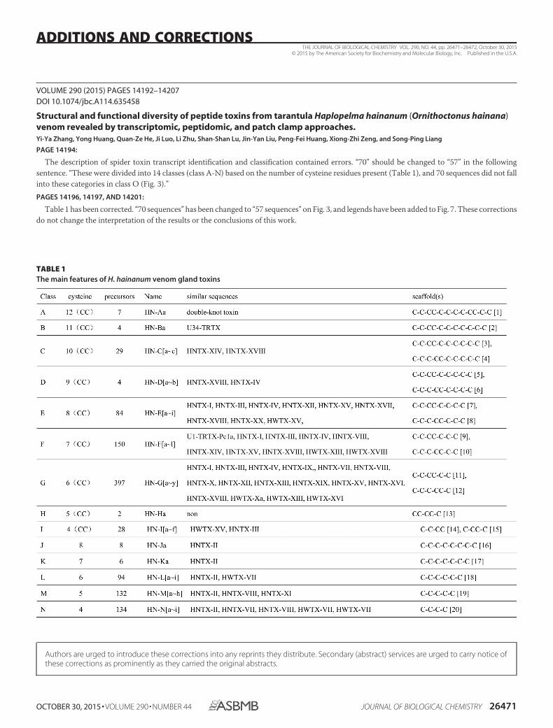
VOLUME 290 (2015) PAGES 14192–14207DOI 10.1074/jbc.A114.635458
Structural and functional diversity of peptide toxins from tarantula Haplopelma hainanum (Ornithoctonus hainana)venom revealed by transcriptomic, peptidomic, and patch clamp approaches.Yi-Ya Zhang, Yong Huang, Quan-Ze He, Ji Luo, Li Zhu, Shan-Shan Lu, Jin-Yan Liu, Peng-Fei Huang, Xiong-Zhi Zeng, and Song-Ping Liang
PAGE 14194:
The description of spider toxin transcript identification and classification contained errors. “70” should be changed to “57” in the followingsentence. “These were divided into 14 classes (class A-N) based on the number of cysteine residues present (Table 1), and 70 sequences did not fallinto these categories in class O (Fig. 3).”PAGES 14196, 14197, AND 14201:
Table 1 has been corrected. “70 sequences” has been changed to “57 sequences” on Fig. 3, and legends have been added to Fig. 7. These correctionsdo not change the interpretation of the results or the conclusions of this work.
TABLE 1The main features of H. hainanum venom gland toxins
THE JOURNAL OF BIOLOGICAL CHEMISTRY VOL. 290, NO. 44, pp. 26471–26472, October 30, 2015© 2015 by The American Society for Biochemistry and Molecular Biology, Inc. Published in the U.S.A.
OCTOBER 30, 2015 • VOLUME 290 • NUMBER 44 JOURNAL OF BIOLOGICAL CHEMISTRY 26471
ADDITIONS AND CORRECTIONS
Authors are urged to introduce these corrections into any reprints they distribute. Secondary (abstract) services are urged to carry notice ofthese corrections as prominently as they carried the original abstracts.

Additions and Corrections
26472 JOURNAL OF BIOLOGICAL CHEMISTRY VOLUME 290 • NUMBER 44 • OCTOBER 30, 2015


![INSTRUCTION MODEL MF-M - Department of Physics · 2014-09-19 · INSTRUCTION MODEL MF-M NARISHIGE] NARISHIGE SCIENTIFIC INSTRUMENT LAB. JAPAN , 9-28 KASUYA 4-CHOME SETAGAYA-KU. ...](https://static.fdocuments.us/doc/165x107/5e7478e8b2dd1b5bea6906a7/instruction-model-mf-m-department-of-physics-2014-09-19-instruction-model-mf-m.jpg)