Stroke - Overview · Stroke - Overview Dr Natasha Gerbis Neurology Registrar Royal North Shore...
66
Stroke - Overview Dr Natasha Gerbis Neurology Registrar Royal North Shore Hospital
Transcript of Stroke - Overview · Stroke - Overview Dr Natasha Gerbis Neurology Registrar Royal North Shore...

Stroke - Overview Dr Natasha Gerbis
Neurology Registrar Royal North Shore Hospital

Acknowledgement
Thank you Dr Jason Gu

Overview
● Anatomy ○ The CNS: an overview
○ Cortical function
○ Cerebral vasculature
○ Motor and sensory pathways
○ Skull compartments
○ The ventricular system
○ Vision pathways
● Pathophysiology ○ Ischaemic stroke
○ Embolic stroke
○ Haemorrhagic Stroke
○ Young stroke
● Neuro-imaging

Anatomy 1) The CNS

Nervous System - Central Nervous System
(CNS): consists of brain and spinal cord
- Peripheral Nervous System (PNS): nerves joining CNS to peripheral structures

The Brain Grey Matter - dense in nerve cell bodies eg central part of spinal cord and surface of cerebral hemispheres. White Matter - contains nerve processes, often myelinated.

Embryonic development
Primary Brain Vesicles Secondary Brain vesicles (wk 7)
Mature Brain
Prosencephalon (forebrain)
Telencephalon Cerebral hemisphere
Diencephalon Thalamus
Mesencephalon Mesencephalon Midbrain
Rhombencephalon (hindbrain)
Metencephalon Pons, cerebellum
Myelencephalon Medulla oblongata

Anatomy 2) Cortical Function

Frontal Lobe - Lies anterior to central sulcus - Precentral gyrus: primary motor cortex - Inferior frontal gyrus of the dominant
hemisphere is the motor speech area - Broca’s area.
- Middle frontal gyrus: Frontal eye field, controls voluntary conjugate deviation of the eyes when scanning, causes eye deviation towards the side of lesion.
- Higher functions: intellectual, judgemental, predictive faculties, planning of behaviour

The clinical picture
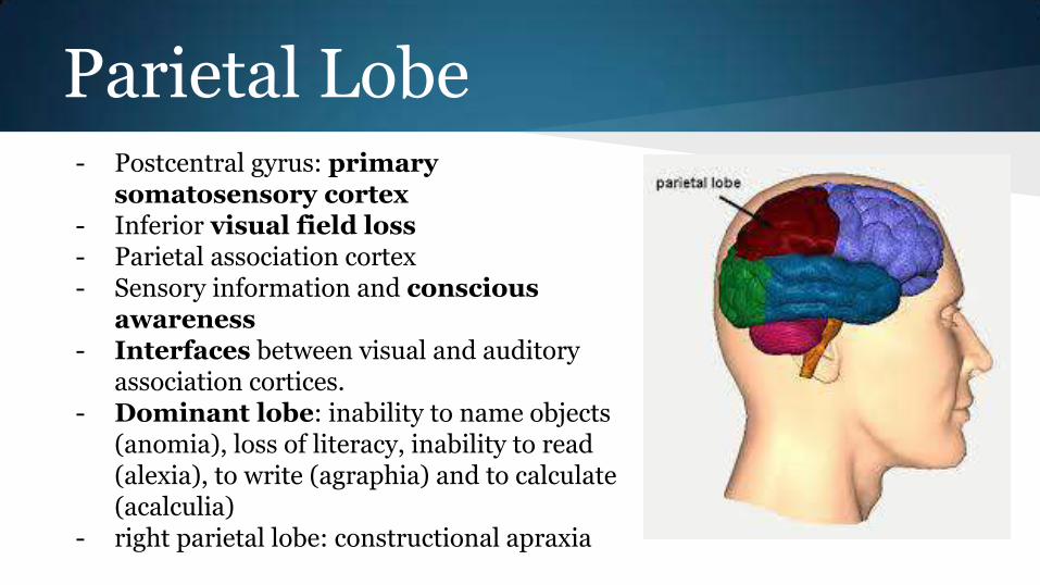
Parietal Lobe - Postcentral gyrus: primary
somatosensory cortex - Inferior visual field loss - Parietal association cortex - Sensory information and conscious
awareness - Interfaces between visual and auditory
association cortices. - Dominant lobe: inability to name objects
(anomia), loss of literacy, inability to read (alexia), to write (agraphia) and to calculate (acalculia)
- right parietal lobe: constructional apraxia

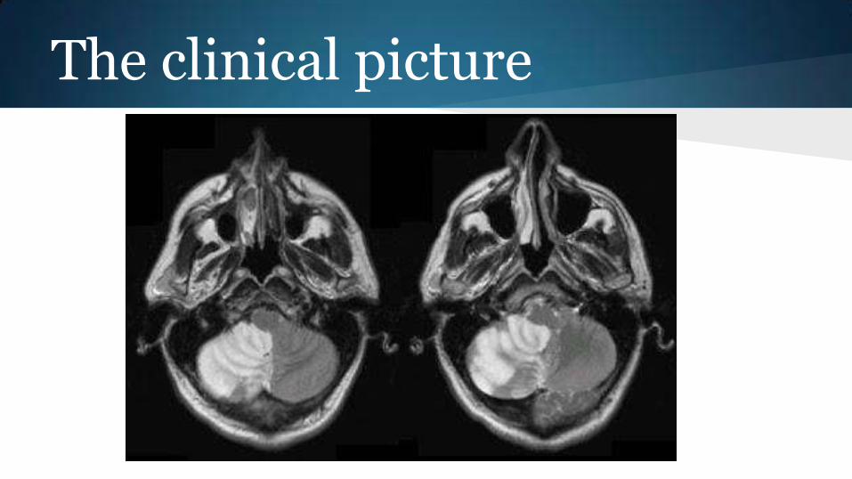
The clinical picture
Mrs Z, inability finding her jeans pocket, bumping into doors around corners and some right arm weakness. Difficulty with her speech, in particular with naming things.

Temporal Lobe - Primary auditory cortex:
conscious perception of sound. Bilaterally represented.
- Dominant hemisphere: Wernicke’s area.
- Inferior medial part is curled to form the hippocampus: memory and emotional aspects of behaviour.
- Amygdala:receive from olfactory tract.

The clinical picture
Aphasia. Word finding difficulty
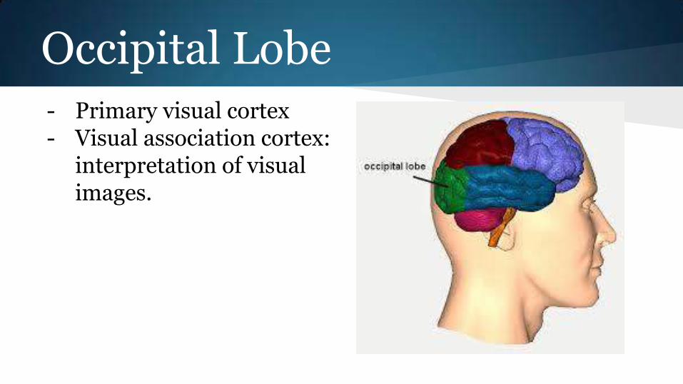
Occipital Lobe - Primary visual cortex - Visual association cortex:
interpretation of visual images.

The clinical picture
Aphasia, ataxia, arm and leg drift Homonymous hemi

Cerebellum - Midline: postural control - Ipsilateral fibres - Dysarthria - Nystagmus - Ataxia
The clinical picture

Basal ganglia - Striatum, pallidum, substantia
nigra, subthalamic nucleus, circuit connections
- Eye movements, in conjunction with the superior colliculus (midbrain)
- Motivation - Movement disorders - Ascending and descending tracts
(internal capsule)

Brainstem
- Midbrain: nerve nuclei (3rd, 4th), substantia nigra, reticular formation (arousal and consciousness), central tegmental tract
- Pons: tracts between cerebrum, medulla, cerebellum. Sensory signals to the thalamus.
- Medulla oblongata: continuous with the pons. Contains cardiac, respiratory, vomiting and vasomotor centres (Heart rate, breathing and blood pressure)

The clinical picture

Anatomy 3) Cerebral vasculature

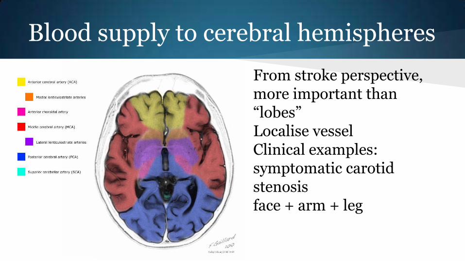
Blood supply to cerebral hemispheres
From stroke perspective, more important than “lobes” Localise vessel Clinical examples: symptomatic carotid stenosis face + arm + leg

MCA

Lateral Medullary Syndrome (Aka Wallenberg Syndrome)
- PICA - vestibular nuclei - vomiting, vertigo, nystagmus - inferior cerebellar peduncle: ipsilateral
cerebellar signs - central tegmental tract: palatal myoclonus - lateral spinothalamic tract: contralateral pain
and temp (body) - Spinal trigeminal nucleus and tract:
ipsilateral pain and temp (face) - nucleus ambiguus: ipsi laryngeal, pharyngeal
and palatal hemiparalysis. dysphasia, hoarseness and diminished gag
- sympathetic fibres: ipsilateral horner’s syndrome


Watershed infarct
Hypoperfusion → Watershed Infarct
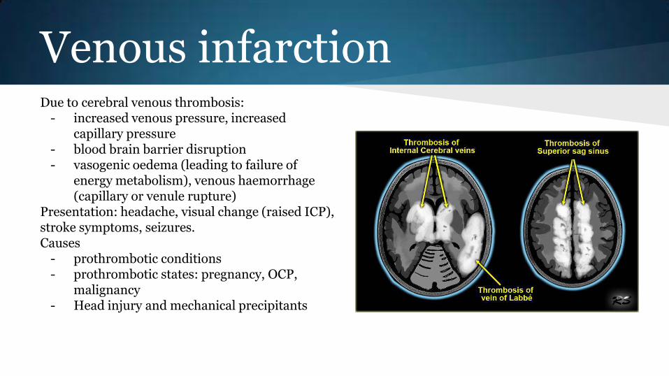
Due to cerebral venous thrombosis: - increased venous pressure, increased
capillary pressure - blood brain barrier disruption - vasogenic oedema (leading to failure of
energy metabolism), venous haemorrhage (capillary or venule rupture)
Presentation: headache, visual change (raised ICP), stroke symptoms, seizures. Causes
- prothrombotic conditions - prothrombotic states: pregnancy, OCP,
malignancy - Head injury and mechanical precipitants
Venous infarction


Anatomy 4) Motor and Sensory Pathways

Motor and Sensory pathways
Anatomy 5) Skull compartments

Skull and compartments


Anatomy 6) The Ventricular System

Ventricular system


Anatomy 7) Visual pathways

Optic radiation

Visual Field Testing

Pathophysiology 1) Ischaemic stroke
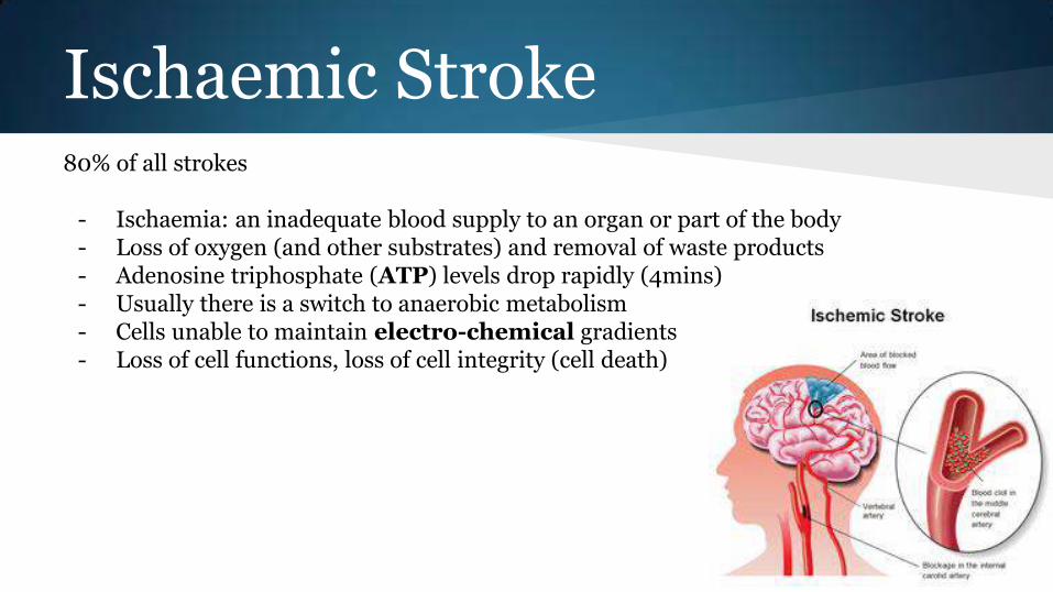
Ischaemic Stroke 80% of all strokes
- Ischaemia: an inadequate blood supply to an organ or part of the body - Loss of oxygen (and other substrates) and removal of waste products - Adenosine triphosphate (ATP) levels drop rapidly (4mins) - Usually there is a switch to anaerobic metabolism - Cells unable to maintain electro-chemical gradients - Loss of cell functions, loss of cell integrity (cell death)

Ischaemic Stroke 3 main types of ischaemia:
- Thrombosis: in situ (within vessel) obstruction of an artery
- Embolism: particle of debris originating elsewhere that blocks arterial access to a particular brain region
- Systemic hypoperfusion: more general circulatory problem

Embolic stroke 1. Cardiac source definite:
a. LA thrombus, LV thrombus, AF and pAF, myocardial infarction, rheumatic MV or AV disease, bioprosthetic heart valve, dilated cardiomyopathy
2. Cardiac sources possible a. PFO, atrial septal aneurysm, LV aneurysm
3. Ascending aortic atheromatous disease 4. True unknown source

Thrombotic stroke Large vessel (common and internal carotids, vertebral, circle of willis, proximal branches)
- Atherosclerosis - Dissection - Arteritis/vasculitis - Fibromuscular dysplasia - Vasoconstriction - Aortic disease *
Small Vessel disease (intracerebral arterial system ie penetrating arteries from the distal vertebral, basilar, MCA, CoW):
- Lipohyalinosis (secondary to hypertension) and fibrinoid degeneration
- Atheroma formation

Pathophysiology 2) Haemorrhagic stroke

Haemorrhagic stroke Intracerebral vs subarachnoid haemorrhage Common causes for spontaneous intracerebral haemorrhage:
- Primary: Hypertension (lacunar), trauma, bleeding diatheses, amyloid angiopathy, illicit drug use (amphetamines and cocaine), vascular malformation.
- Secondary: bleeding into tumours, aneurysmal rupture, vasculitis
- consider age, size, location, past history, imaging
Pathophysiology 3) Young stroke

Young stroke - Often still atherosclerosis or AF - Dissection - Cardiac defects - Vasospasm - Hypercoagulable state - Vasculitis - Migraine and other headache disorders - Metabolic (CADASIL, Fabry disease, MELAS)

Should especially be considered in young patients eg <45yrs of age, history of clotting dysfunction, history of cryptogenic stroke:
- sickle cell anaemia, polycythaemia rubra vera, essential thrombocytosis, HITS, Protein C or S deficiency, Prothrombin gene mutation, Factor V Leiden (APC resistance), AT III defciency, Anti-phospholipid syndrome, hyperhomocysteinaemia
Infectious and inflammatory disorders - cause a rise in acute phase reactants such as fibrinogen, CRP, coagulation
factors VII and VIII
Blood disorders

Neuro-imaging

CT - Exclude haemorrhage - Immediate
- hyperdense vessel - Early (1-3 hrs or hyperacute stage)
- loss of grey-white differentiation and hypoattenuation of deep nuclei
- cortical hypodensity with associated parenchymal swelling and gyral effacement
- first week: - more attenuation - More swelling
- Onwards: - swelling subsides, gliosis begins to
occur - eventually results in low density
lesion with negative mass effect

CT: Advanced techniques 1. CT Angiography 2. CT Perfusion
a. Cerebral blood volume b. Cerebral blood flow c. Mean transit time d. Time to peak e. Penumbra: MTT - CBV

Perfusion MTT vs CBV

MRI Greater sensitivity for acute ischaemic infarction in the first few hours
1. T1: a. Water/CSF is dark b. Sensitive for tissue c. Low density
2. T2/FLAIR: a. Water/CSF is bright b. Less immediately sensitive, but does show high signal by 6-12hrs c. S ulcal effacement, mass effect
3. DWI/ADC: correlates with infarct core 4. GE/SWI: sensitive for haemorrhage


Key points

1) Where is it?

2) Does it fit the vasculature?

3) What is the mechanism?
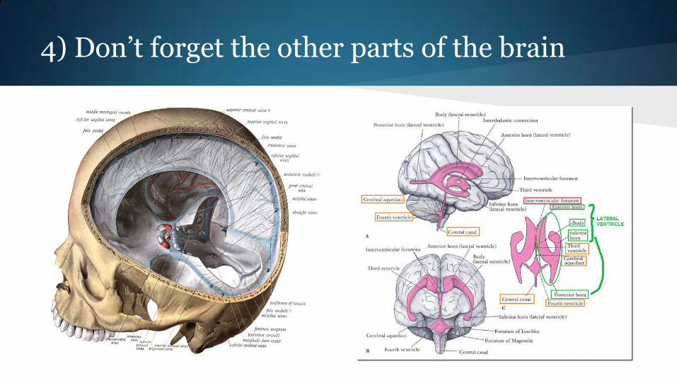
4) Don’t forget the other parts of the brain
5) Don’t forget the rest of the patient

Thank you
Any questions?

Resources:
- Illustrated atlas neuroanatomy - Nolte, neuroanatomy - Uptodate - Radiopedia