STROKE IN CHILDREN - asmn.re.it IN CHILDREN_180117.pdf · STROKE IN CHILDREN Dr. Carlotta Spagnoli...
63
STROKE IN CHILDREN Dr. Carlotta Spagnoli Child Neurology Unit Head of the Unit: Dr. Carlo Fusco ASMN IRCCS Reggio Emilia
Transcript of STROKE IN CHILDREN - asmn.re.it IN CHILDREN_180117.pdf · STROKE IN CHILDREN Dr. Carlotta Spagnoli...

STROKE IN CHILDREN
Dr. Carlotta SpagnoliChild Neurology Unit
Head of the Unit: Dr. Carlo FuscoASMN IRCCS Reggio Emilia

PRESENTATION OUTLINE STROKE EPIDEMIOLOGY NEWBORN CHILDREN > 1 mo CEREBRAL SINOVENOUS THROMBOSIS
Risk factorsTherapy
ARTERIAL ISCHAEMIC STROKERisk factorsTherapy
HAEMORRHAGIC STROKERisk factorsTherapy
CONCLUSIONS

EPIDEMIOLOGY
1 month-19 yo2.3 /100,000/year:1.2/100,000/year for ischemic lesions1.1/100,000/year for haemorrhagic lesionsBoys > girls
NewbornsIncidence of perinatal stroke: 1 in 1600-5000 births.
Fullerton et al., 2003
Perlman et al., 1994Schulzke et al., 2005
INCIDENCE

EPIDEMIOLOGYMORTALITY20-40% overall30% of haemorrhagic strokes20% of ischaemic strokes (50% due to underlying condition)6-15% have recurrent strokes(mortality is higher in this group)ADVERSE PREDICTORSintractable intracranial hypertension
NEUROLOGICAL PROGNOSIS50-80% have neurological sequelae (> hemiparesis)Neuropsychological deficitsSymptomatic epilepsy

• group of heterogeneous conditions • focal disruption of cerebral blood flow secondary to• arterial or cerebral venous thrombosis or embolization, • 20 weeks of foetal life - 28th post-natal day,• confirmed by neuroimaging or neuropathological studies
Raju et al., 2007
Most common subtypes:• arterial ischemic stroke (AIS) • cerebral sinovenous thrombosis (CSVT)
Approximately 40% unrecognized in the neonatal period.Chabrier et al., 2011
PERINATAL ISCHAEMIC STROKE


Lynch, 2009


Male,10 m.
Born at term from CS.SGA.
Uneventful neonatal period.No known maternal risk factors.R hand fisted.Asimmetry of use of upper limbs.
Coagulation and thrombophilia investigations: unremarkable.
COL4A1 genetic testing pending.

Incidence: 0.7/100,000/year

RISK FACTORS
•Dehydration•Prothrombotic disorders (1/3-2/3)•Systemic diseases, sepsis•Anemia (iron deficiency, SCD, thalassemia)•Malignancies•Head & Neck infections (meningitis, mastoiditis, ear infections, tonsillitis, sinusitis)•Head injury•Brain tumours•In the neonatal age also adverse perinatal factors.
Many have multiple risk factors.
TREATMENT•Supportive/symptomatic treatment
•UFH, LMWH•Monitoring of aPTT (UFH) and anti-factor Xa levels (LMWH)
•Lower risk of recurrence in the treatment group•Newborns: 3-6 weeks treatment (3 mo max): 5-10 d UFH followed by LMWH.
•If relevant haemorrhagic component: repeat neuroimaging < 1 week continue therapy only if thrombosis has extended.
•Long-term treatment with warfarin in older children without haemorrhage •3-6 months
OUTCOMEMortality < 5%40% long-term neurological impairment (> motor). Predictors of adverse outcome: seizures, coma, presence of infarcts.Predictors of good outcome: older age, involvement of lateral/sigmoid sinus, absence of parenchymal involvement and anticoagulation. Chronic symptoms of idiopathic intracranial hypertension in > 25%.
ARTERIAL ISCHAEMIC STROKE

Mallick et al., 2014
Mackay et al., 2011
AGE DISTRIBUTIONinPAEDIATRICARTERIALISCHEMIC STROKE

CLINICAL PRESENTATION


Arteriopathy 53%Cardiac disorders 31%Infection 24%Acute head and neck disorders 23%Acute systemic conditions 22%Chronic systemic conditions 19%Prothrombotic states 13%Chronic head and neck disorders 10%Risk factors for atherosclerosis 2%Other 22%
Examples: pulmonary hypertension, hemolytic uremic syndrome, nephrotic syndrome, protein-losing enteropathy, autoimmune aplastic or hemolytic anemia, idiopathic thrombocytopenic purpura, Kasabach-Merritt syndrome, acute renal failure, Kartagener syndrome, asplenia, pancreatitis, and ulcerative colitis.

Arteriopathy is defined as the imaging appearance of an in situ arterial abnormality (stenosis, irregularity, occlusion, banding, pseudoaneurysm, dissection flap) not attributable to an exogenous thrombus (eg, cardioembolism) and not considered a normal developmental variant.
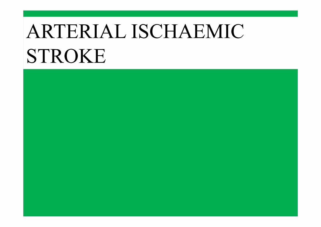
ARTERIOPATHIESFocal Cerebral Arteriopathy of Childhood— «idiopathic » focal intracranial arterial stenosis. Mostly precipitated by a preceding viral infection ( > upper respiratory). Transient Cerebral Arteriopathy Monophasic: no additional stenoses nor progression of the original stenosis in the follow-up imaging.Post varicella angiopathy—
Moyamoya disease and Moyamoya syndrome —
Sickle cell arteriopathy— approximately 100-fold increased risk of stroke. Clinically silent strokes (usually deep WM small arteries occlusion, accumulation of injury neurocognitive dysfunction).Symptomatic strokes (usually large vessels internal carotid system occlusive disease moyamoya syndrome)
Vasculitis —
Cervicocephalic Arterial Dissection—

Diagnostic criteria for TIA/AIS due to PVA in children: 1) first-ever AIS/TIA ≤1 y after varicella with exanthema;2) no possible AIS/TIA etiology other than PVA after investigations (angiitis of the CNS, infectious agents, cardiac , metabolic and prothrombotic conditions); 3) AIS/TIA manifestations and cerebral infarct location consistent with unilateral vascular disease affecting the supraclinoid ICA, A1 or A2 segments of the ACA, or M1 or M2 segments of the MCA.
Diagnosis supported by cerebral vessel imaging showing vascular stenoses of these arterial segments. In children with normal vessel imaging: cerebral infarct(s) limited to the vascular territory of the lenticulostriate arteries.
Lanthier et al., 2005
POST-VARICELLA ANGIOPATHY

VZV vasculopathy is caused by productive viral infection in arteries.
Rash or CSF pleocytosis not required.
CSF:30% VZV DNA +, 93% anti-VZV IgG + (reduced serum/CSF ratio= intrathecal synthesis).
PATHOGENESIS
Nagel et al., 2008Gilden et al., 2009
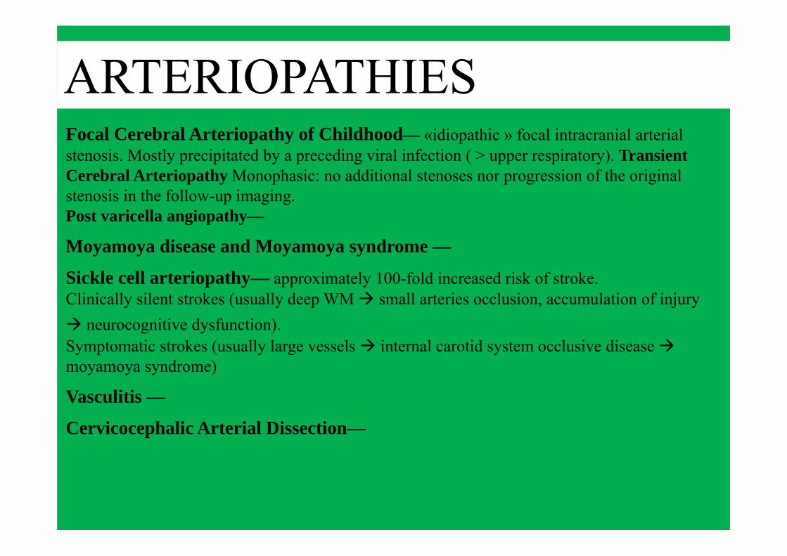
UNILATERAL ARTERIOPATHY: PATIENTS CHARACTERISTICS
Estimated absolute risk of varicella-associated stroke: 1:15,000 children.Askalan et al., 2001
Main infarct localization: basal ganglia (78%)
CLINICAL PRESENTATION:HemiparesisHemichoreaAphasia Bartolini et al., 2011
Slight male predominanceAge range: median age at first stroke 4.8 y (range 0.3–16.3).

59% permanent neurological deficits.77% residual arterial lesions (improved 45%, stabilized 32%),23% complete normalization.Trend towards better functional outcome if complete normalization.Cortical infarction poor functional outcome.Possible worsening in the first months increased unilateral arterial abnormalities (median interval: 3.5 mo, range 1–14).
Initial worsening does not predict recurrence.No difference in recurrence rate between normalizing and residual arteriopathy at final imaging
OUTCOME - TCA

Female 3y.Unremarkable past history.Acute onset L side hemiplegia + R facial nerve paralysis.Legs pain in the previous week.
Acute otitis one month before.Varicella primary infection 3 months before.
CASE PRESENTATION


Female 3y.
Therapy:ev methylprednisolone 30 mg/kg for 5 days followed by oral deflazacortASA 50 mg/dayCoQ10 45 mgAlready on aciclovir + ceftriaxone
ASA continued for 6 mo.Very good neurological recovery.
CASE PRESENTATION

ARTERIOPATHY AND OTHER INFECTIONSSmall case reports and case series on AIS following other infections:

MOYAMOYA DISEASE/SYNDROME
RISK FACTORS for MOYAMOYA SYNDROME:
• Neurofibromatosis type I• Asian heritage• Radiotherapy• Intrathecal chemotherapy• Congenital cardiac abnormality• Haemoglobinopathy (SCD)• Shunted hydrocephalus• Trisomy 21, Williams syndrome
etc.
Moyamoya disease/syndrome:Progressive occlusive disease of the distal internal carotid arteries resulting in collaterals formation.Ischemic >> haemorrhagic strokes

After a median follow-up of 1.4 y, monophasic strictly unilateral arteriopathy (PVA/TCA) in 94%
Braun et al., 2009 PVA/TCA worsening possible during the first months
Sébire et al., 2004; Sébire, 2006
Median time of established diagnosis of progressive arteriopathy (Moyamoya/vasculitis): 1 year, (range: 6.5 mo-2.7 y).
Braun et al., 2009
FOLLOW-UP RECOMMENDATIONSMRA to be repeated 3–6 months after diagnosis, and again at 6–12 months
Sébire et al., 2004; Sébire, 2006
UNILATERAL ARTERIOPATHY (FOLLOW-UP)

VZV infection only in TCA (44%)
No chickenpox in the year prior to stroke in progressive arteriopathies.
Patients with progressive arteriopathy more often show:
•arterial occlusion,
•ACA involvement,
•abnormal collateral moyamoya vessels.
Moyamoya vessels remain a significant predictor even after accounting for arterial
occlusion.
DIFFERENTIAL DIAGNOSIS PROGRESSIVE vs. TCA/PVA

CEREBRAL VASCULITIS• Infectious vasculitis• Necrotizing vasculitides• Vasculitis associated with
collagen vascular disease• Vasculitis associated with
other systemic disease(Ulcerative colitis, Behcet’s, sarcoidosis
etc)• Takayasu arteritis• Hypersensitivity vasculitides• Miscellaneous(secondary to neoplasia/radiation,
Kawasaki disease, dermatomyositis-polymyositis etc.)
• Primary CNS vasculitis

Male, 8 y
Movement disorder (choreoathetosis + ballismus, R>> L) in the last month
Axial hypotoniaMotor impersistenceGrimacingDisarthria
Elevated anti-Streptolysine titre.Normal EEG, EKG, Ecocardiogram
Rheumatic choreaFrontal WM + L nucleus caudatus(dis-immune vasculitis)

7.5-20% of pediatric stroke cases. Anterior > posterior circulation.Male predominance. Traumatic (blunt/penetrating, also trivial), non-traumatic, physical exertion.Headache/Cervicalgia.10% recurrent. 14% have multiple ischemic events prior to definitive diagnosis, and 9% afterwards.
Roach et al.,2008Chabrier et al., 2003Fullerton et al., 2001
CERVICOCEPHALIC ARTERIAL DISSECTION
RISK FACTORSMarfan syndromeEhlers-Danlos type IVCoarctation of the aortaCystic medial necrosisAD polycystic kidney diseaseOsteogenesis imperfectaAtherosclerosisMoyamoya syndromePharyngeal infectionsRoach et al.,2008
TREATMENTControversial.Endovascular treatment.Anticoagulation (risk of haemorrhage) vs. antiplatelet (risk of recurrence).Warfarin or LMWHNo consensus as to which patients will most likely benefit fromantiplatelet vs. anticoagulation therapy.
Pandey et al., 2015


CARDIAC DISORDERS
Congenital heart malformations (> if complex, R to L shunt, cyanosis)Valvular heart diseases Cardiac arrhythmiasCardiomyopathy (Muscular dystrophy, Kearns-Sayre syndrome, Friedreich ataxia)Cardiac surgery and catheterisationExtracorporeal Membrane Oxygenation (ECMO)Kawasaki disease

RIS.MAGN.CERVELLO E TRONCO ENCEFALICO ANGIO RM DISTRETTO VASCOLARE INTRACRANICO 03/09 05
Esame limitato da artefatti da movimento. Segnale alterato della testa del nucleo caudato, del lenticolare e della corona radiata a sinistra. Segnale alterato in regione temporo insulare sinistra. I reperti allo stato attuale depongono per edema emisferico sinistro. Lieve effetto massa sul corno anteriore del ventricolo laterale sx. Dubbia alterazione della testa del caudato di destra. Lo studio vascolare limitatamente agli artefatti da movimento evidenzia scarsa visualizzazione dei rami distali della art. cerebrale media di sinistra. Carotidi interne, vertebrali (V4) e basilare nei limiti. Arterie cerebrali posteriori visualizzabili.

Cardiac embolism
Duchenne muscular dystrophy (deletion on exon 53) right hemiplegia (L hemispheric stroke)


Chronic systemic disorders• SCD• Indwelling catheter• Trisomy 21• Other genetic disorders• Haematological malignancy• Iron deficiency• Oral contraceptive pill• Connective tissue disorders• Solid extracranial tumours• L-asparaginase therapy
Acute systemic disorders• Fever >48 h• Sepsis• Shock• Dehydration• Acidosis• Hypoxia• Viral gastroenteritis
Chronic head and neck disorders• Migraine• Brain tumour• Other cranial or neck tumour• Ventricular shunt• PHACES syndrome
Acute head and neck disorders• Head or neck trauma• Pharyngitis• Meningitis• Recent intracranial surgery• Otitis media• Sinusitis• Mastoiditis
ACUTE/CHRONIC SYSTEMIC ANDHEAD&NECK DISORDERS

METABOLIC DISORDERS• ORGANIC ACIDAEMIAS• MITOCHONDRIAL
ENCEPHALOMYOPATHIES• FABRY DISEASE• ORNITHINE TRANSCARBAMYLASE
DEFICIENCY• HOMOCYSTINURIA

HOMOCYSTEINE TO BE TESTED
LACUNAR, SMALL VESSELSINVOLVEMENT


2007 2011
Onset in the neonatal periodPartial, early‐onset epilepsyProgressive microcephaly, pyramidal and extrapyramidal signs in all for limbs, nystagmus RC Complex I deficiencyDeceased aged 8 y


HYPERCOAGULABLE STATES

Diplegic Spastic CP, microcephaly, symptomatic epilepsy. Hyperhomocysteinemia. Heterozygous for the C677T mutation on the MTHFR gene.On folinate + multivitamins (B group) N homocysteinemia

RECURRENCE RISK AFTER FIRST ISCHEMIC STROKE
Fullerton et al., 2016
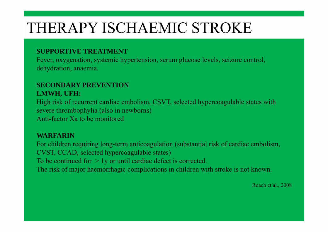
THERAPY ISCHAEMIC STROKESUPPORTIVE TREATMENTFever, oxygenation, systemic hypertension, serum glucose levels, seizure control, dehydration, anaemia.
SECONDARY PREVENTIONLMWH, UFH:High risk of recurrent cardiac embolism, CSVT, selected hypercoagulable states with severe thrombophylia (also in newborns)Anti-factor Xa to be monitored
WARFARINFor children requiring long-term anticoagulation (substantial risk of cardiac embolism, CVST, CCAD, selected hypercoagulable states)To be continued for > 1y or until cardiac defect is corrected.The risk of major haemorrhagic complications in children with stroke is not known.
Roach et al., 2008

THERAPY ISCHAEMIC STROKESECONDARY PREVENTIONASPIRIN3-5 mg/kg/d (to be reduced to 1-3 mg/kg/d in case of side effects)3-5 y or longer if ongoing risk of recurrence.In acute AIS (first week) of unknown cause, after exclusion of SCD, high risk of recurrent cardiac embolism, severe hypercoagulable state.(Transfusion required in SCD)Annual influenza immunization, immunization for VZV.ASA therapy should be suspended during Influenza and VZV infection.
THROMBOLYTIC THERAPY (rt-PA)Safety and efficacy data are scarce. High complication rate in systemic thromboses.Indicated in case of occlusion of large intracranial vessel, with high risk of severe disability and death.It may be considered in selected cases of CSVT.
Roach et al., 2008

THERAPY MOYAMOYADIRECT (connection between the internal and external carotid systems)or INDIRECT (with subpial compensatory circulation) BYPASS if:•inadequate cerebral perfusion•recurrent ischaemic lesions
TCA/PVASteroids + antivirals if recent VZV infection

NON-TRAUMATIC HAEMORRHAGIC STROKE
Specific risk factors (vascular malformations):AVM (33-47%)Intracranial aneurysms
Other risk factors:Hematological and coagulation disorders (26.5%)Bleeding from a brain tumour (13.2%)
Clinical presentationOlder children: acute headache, vomiting, rapid neurological deteriorationInfants/toddlers: irritability, headache, vomitingSeizures (37%)Hemiparesis (16%)Coma (3%)
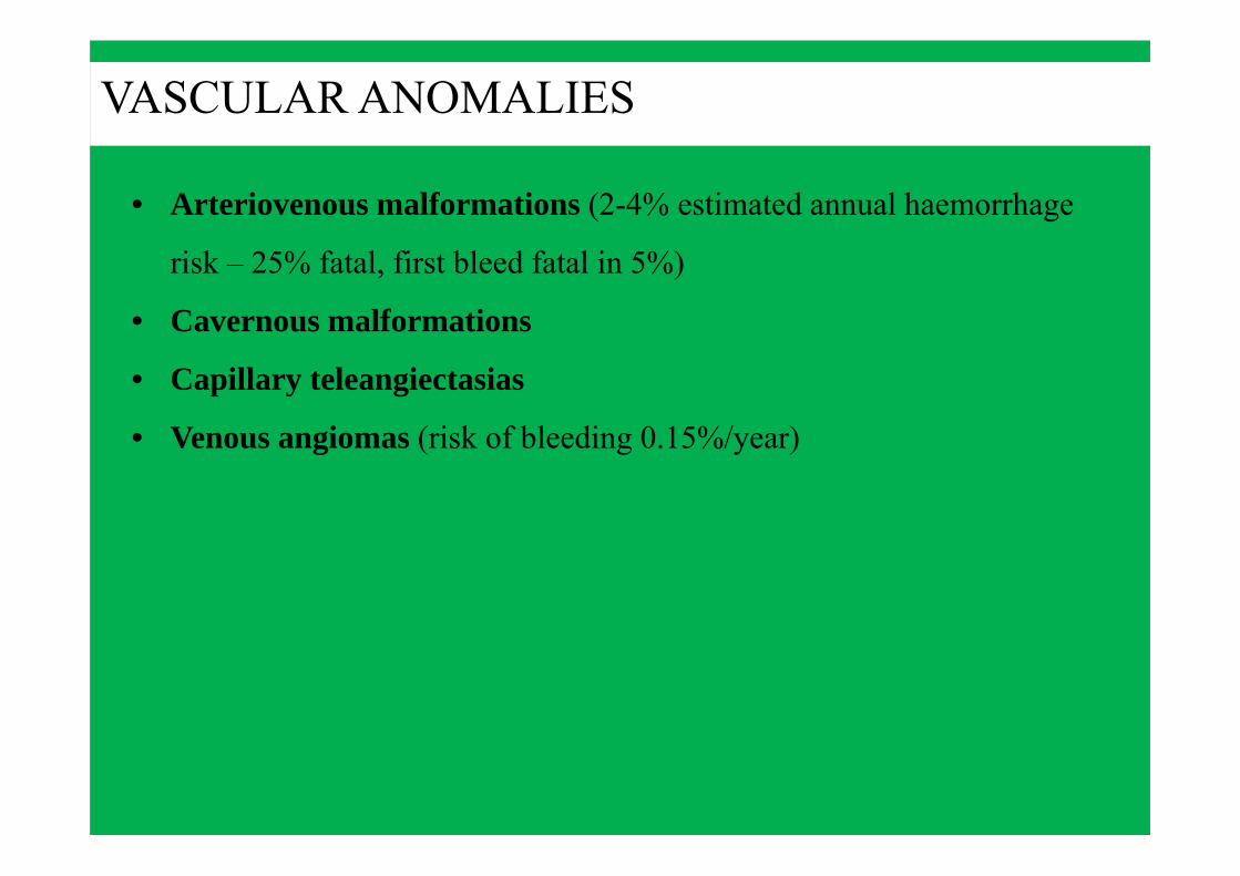
VASCULAR ANOMALIES
• Arteriovenous malformations (2-4% estimated annual haemorrhage
risk – 25% fatal, first bleed fatal in 5%)
• Cavernous malformations
• Capillary teleangiectasias
• Venous angiomas (risk of bleeding 0.15%/year)

ARTERIOVENOUS MALFORMATIONS• 80% supratentorial.• 75% of AVM presents with haemorrhage.• Less frequently: seizures, headache, focal neurological
deficits, isolated intracranial hypertension.• Hydrocephalus (> in VoGAM)
• In symptomatic newborns: high output cardiac failure
• Consider hereditary haemorrhagic teleangectasia (Rendu-Osler-Weber) if multiple CNS AVMs
Koach et al., 2008
• Alternative syndromic diagnoses (PHACES, Williams, etc.)

• Risk of rebleeding: 2-4%, >0-5 y after diagnosis.
• Risk factors for rupture: size, previous haemorrhage, deep-seated/infratentorial location, deep venous drainage, female sex, associated aneurysms, diffuse AVM morphology.
El-Ghanem et al., 2016
ARTERIOVENOUS MALFORMATIONS
Spetzler-Martin grading system


ANEURYSMS
Biphasic presentation: < 2 y or > 10 y> Subaracnoid Haemorrhage,Intracranial Haemorrhage,Intraventricular Haemorrhage.
Compared to adults:> posterior circulation,> giant,less have > 1 (5% vs. 20%)
Infectious aneurysms:Infective endocarditisStructural heart lesionsChronic pulmonary infectionsDrug abuse
Risk factors:Coarctation of the aortaAD polycystic kidneyFMDEhlers-Danlos (IV)Pseudoxanthoma elasticum

Girl, aged 13 y at the time.
Tuberous sclerosis complex.
Symptomatic partial epilepsy.

ANEURYSM AT THE BIFURCATION OF THE RIGHT EXTERNAL CAROTID ARTERY

THERAPY• Whenever possible, a non-urgent procedure is performed in the
2-6 weeks after haemorrhage.• The risk of malignant endocranic hypertension is higher than
in the elderly.
• Neurosurgery• Interventional radiology

CONCLUSIONS• Perinatal stroke often underrecognised in the neonatal period.
• Traditional stroke risk factors uncommon in children.
• Numerous infectious, post-infectious, systemic, metabolic and genetic causes
need to be considered.
• VZV.
• Multiple risk factors → need for thorough investigations.
• Arteriopathy increases the risk of recurrent strokes and poor outcome.
• AVM risk of rebleeding / death.
• Better evidence for medical, neurosurgical and interventional radiology
options in the older age group.