Stridor, Stertor and Snoring- Common Pediatric Airway Problems
87
Stridor, Stertor and Snoring- Common Pediatric Airway Problems Melissa G. Kress, DO Chief, Section of Pediatric Otolaryngology- Head & Neck Surgery
Transcript of Stridor, Stertor and Snoring- Common Pediatric Airway Problems

Stridor, Stertor and Snoring-Common Pediatric Airway
ProblemsMelissa G. Kress, DO
Chief, Section of Pediatric Otolaryngology- Head & Neck Surgery

I have no financial relationships to disclose.

Pediatric Airway Problems
• Stridor• Stertor• Snoring

Stridor
• Definition– sound produced by turbulent airflow from
obstruction in the upper aerodigestive tract• laryngeal• tracheobronchial
– sound and pitch dependent on air flow dynamics

Stridor
• Timing in respiratory cycle determines anatomic location of airway lesion– Inspiratory– Biphasic– Expiratory

Inspiratory Stridor
• Partial supraglottic airway obstruction
• Other aerodigestive tract symptoms– suprasternal and
intercostal retractions
– feeding difficulties– muffled cry

Biphasic Stridor
• Partial obstruction at the level of the glottis
• Primarily inspiratory stridor
• Other aerodigestive tract symptoms– hoarseness– aphonia– nasal flaring– retractions

Expiratory Stridor
• Partial obstruction at the level of the subglottis or proximal trachea
• Other aerodigestive tract symptoms– xiphoid retractions– barking cough– nasal flaring

Stridor
• Congenital–85%– anatomical anomalies– present by 4 months of age
• Acquired– 15%

Stridor: Diagnosis
• History• Flexible fiberoptic laryngoscopy• Direct laryngoscopy with rigid
bronchoscopy• Barium esophagram• CT neck and chest

Congenital Stridor
• Laryngeal webs• Laryngotracheal
esophageal clefts• Tracheoesophageal
fistulae• Laryngeal and subglottic
cysts• Subglottic hemangioma
• Tracheomalacia• Tracheal stenosis• Tracheal compression• Complete vascular rings• Anomalous innominate
artery• Pulmonary artery sling
#1 Laryngomalacia#2 Bilateral True Vocal Fold (Cord) Paralysis#3 Laryngotracheal Stenosis (Subglottic Stenosis)

Laryngomalacia• #1 cause of newborn
stridor• 58% congenital
laryngeal anomalies• males 2:1• inspiratory stridor• presents shortly after
birth• worsens by 6-8 weeks
of age• “omega-shaped”
epiglottis• usually self-limited
– resolves by 12-15 months of age

Laryngomalacia
• Symptoms:– stridor with:
• agitation• feeding• supine position
– feeding difficulties• failure to thrive
– rare:• cyanosis• pectus excavatum

Laryngomalacia
• Etiology

Laryngomalacia
Type 1 prolapse of mucosa
overlying the arytenoid cartilages
Type 2foreshortened
aryepiglottic folds
Type 3posterior
displacement of the epiglottis
Olney: Laryngoscope, 109(11) Nov 1999

Laryngomalacia

Laryngomalacia
Supraglottoplasty

True Vocal Fold Paralysis
• #2 Etiology of newborn stridor
• 10% all congenital laryngeal lesions
• no gender predilection
• biphasic stridor• 58% present within
first 12 hours of birth• unilateral or bilateral
– unilateral: left > right

True Vocal Fold Paralysis
• Idiopathic• Anatomic• Neurologic– congenital– acquired

True Vocal Fold Paralysis
• Congenital– CNS
• Arnold-Chiari malformation I & II
• Leukodystrophy• Encephalocele• Hydrocephalus• Cerebral or nuclear
dysgenesis• Perinatal asphyxia

True Vocal Fold Paralysis
• Acquired– Thoracic surgery• PDA ligation
– ETT trauma• Recurrent laryngeal nerve compression
– LMA• Arytenoid dislocation• Recurrent laryngeal nerve compression

True Vocal Fold Paralysis
• Bilateral– biphasic stridor– apnea– cyanosis– aspiration– dysphagia– ineffective cough
• Unilateral– stridor– hoarseness– abnormal cry– aspiration– dysphagia– ineffective cough
Signs and Symptoms

True Vocal Fold ParalysisManagement- Airway
• Respiratory distress– Endotracheal
intubation– Laryngoscopy
• Direct
– Tracheostomy– Posterior
cordotomy– Swallowing
evaluation
• Stable– Flexible fiberoptic
laryngoscopy– Swallowing evaluation

True Vocal Fold Paralysis Management- Feeding
• Normal MBSS– Oral feeding
• Aspiration– NG tube feeding for 6 weeks– Repeat MBSS• Normal
– Oral feeding
• Aspiration– G-tube

Subglottic Stenosis
• #3 most common congenital laryngeal anomaly
• no gender predilection
• congenital or acquired
• narrowed airway 2-3mm below true vocal folds
• subglottic diameter of 4mm or less in a full-term neonate

Subglottic Stenosis
• Symptoms– stridor–may be biphasic– dyspnea– cough• brassy or barky
– hoarseness– weak or unusual cry

Subglottic Stenosis
• Congenital– Stenosis without
prior intubation, or extrinsic compression
– Presentation• after upper
respiratory infection
• “recurrent croup”– Age• birth to few
months
• Acquired– Prior intubation or
trauma– Presentation• failed extubation• recurrent croup
after extubation
– Age• 2wks - 10 yrs• Majority < 1

Subglottic Stenosis
• Radiographic findings– long segment of
subglottic narrowing

Subglottic Stenosis
Meyer-Cotton Grading System
-most frequently used system
-grade correlates with prognosis
Myer, et al. (1994)

Subglottic StenosisGrading
Grade 1

Subglottic StenosisGrading
Grade 2 Grade 3

Subglottic Stenosis- Management of suspected SGS
• Stable airway– History– Flexible fiberoptic
laryngoscopy– Direct
laryngoscopy with rigid bronchoscopy
• Unstable/unsafe airway
– Intubation or tracheostomy
– Direct laryngoscopy with rigid bronchoscopy

Subglottic Stenosis-Surgical Management
• Evaluate overall medical condition– BPD, poor neurologic status, LBW
• Tracheostomy• Airway reconstruction– Open surgical management
• LTR- laryngeal tracheal reconstruction– Single or double stage
– Endoscopic surgical management• Balloon dilation

Subglottic StenosisLaryngeal tracheal reconstruction
Willging and Cotton (1995)
Anterior and posterior cartilage
grafts
Posterior cricoidotomy
Anterior costal cartilage graft
Posterior cricoidotomy
Four quadrant cartilage division
Anterior costal cartilage graft
Posterior costal cartilage graft

Subglottic StenosisEndoscopic management
• Balloon Dilation

Acquired Stridor
• Croup– Laryngotracheobronchitis
• Epiglottitis– Supraglottitis
• Foreign body– Laryngeal or tracheal– Esophageal
• Laryngeal papilloma

Laryngotracheobronchitis- Croup
• Most common cause of upper airway obstruction– 6 months to 6
years of age– Most commonly 1-2
years of age– Barky cough– Biphasic stridor– Viral prodrome

Laryngotracheobronchitis- Croup
• Causes– Parainfluenza– Influenza
• Diagnosis– History– AP and lateral
neck x-rays• Steeple sign
– Flexible laryngoscopy
– DLB

Laryngotracheobronchitis- Croup
• Management– Oral
corticosteroids– Inhaled racemic
epinephrine– Possible
intubation

Epiglottitis- Supraglottitis
• Swelling of epiglottis and supraglottic structures• Presentation– Triad of drooling,
dysphagia and distress
– Fever– Tripod position

Epiglottitis• Causes– Haemophilus
influenza type B• Blood cultures
• Diagnosis– History– Lateral neck x-
ray• Thumb print sign

Epiglottitis- Supraglottitis
• Management– Intubation in the
OR– PICU• 24-72 hours
– ceftriaxone, cefuroxime or cefotaxime

Laryngeal Foreign Body
• Biphasic stridor• Flexible
laryngoscopy• Direct
laryngoscopy with removal

Esophageal Foreign Body
• Drooling• Stridor• X-ray– AP chest

Esophageal Foreign Body
• Endoscopic esophagoscopy for removal

Laryngeal papilloma-Juvenile Recurrent Respiratory Papillomas
• JRRP• HPV
– Types 6,11• 2-5 years of age
– Most commonly diagnosed by age 5
• Inspiratory stridor• Hoarseness• Diagnosis
– Flexible laryngoscopy

JRRP
• Management– Surgical• Microlaryngoscop
y– CO2 laser– Microdebrider – Intralesional
cidofovir

JRRP
• Management–Medical• Alfa-interferon• Indole-3-carbinol• Proton pump
inhibitors• Beta mannan• Celebrex-
celecoxib

Stertor
• Airway obstruction above the level of the larynx
• Snoring-like noise from nasopharyngeal or oropharyngeal obstruction
• Awake patient

Stertor
• Congenital – Choanal atresia– Congenital pyriform aperture stenosis– Nasal lacrimal duct cyst(s)– Juvenile nasopharyngeal angiofibroma– Craniofacial abnormalities
• Pierre Robin Syndrome• Down Syndrome• Treacher Collins Syndrome• Crouzon Syndrome

Choanal Atresia• Persistence of the
buccopharyngeal membrane
• 1 in 5000-8000 live births
• Respiratory distress at birth relieved by crying– Paradoxical cyanosis
• Feeding difficulties• Unilateral > bilateral• 29% bony• 71% mixed bony and
membranous• Males < females

Choanal Atresia
• CHARGE Syndrome– C Coloboma of eye– H Heart abnormality– A Atresia of choanae– R Retarded growth/development– G Genital hypoplasia– E Ear anomalies/deafness
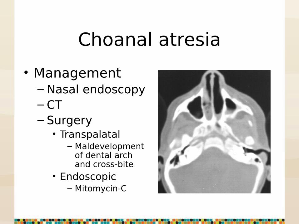
Choanal atresia
• Management– Nasal endoscopy– CT– Surgery• Transpalatal
– Maldevelopment of dental arch and cross-bite
• Endoscopic– Mitomycin-C

Congenital Pyriform Aperture Stenosis
• Bony overgrowth of the nasal process of the maxilla at occurs at 4 months gestation
• Pyriform aperture with <11mm
• Newborn with respiratory distress, poor feeding, FTT and recurrent cycles of cyanosis and apnea

Congenital Pyriform Aperture Stenosis
• Associates abnormalities
– Holoprosencephaly– Midline nasal cavity
defects– Microcephaly– Cleft lip/palate– Hypopituitarism– Hypotelorism– Esophageal or
duodenal atresia– Central megaincisor

Congenital Pyriform Aperture Stenosis
• Management– Nasal endoscopy– CT–Medical• Steroid drops
and topical decongestants

Congenital Pyriform Aperture Stenosis
• Management– Surgical• Bony removal
floor of nose- sublabial approach with nasal stents

Nasolacrimal Duct Cysts
• Distal nasolacrimal duct obstruction– Incomplete
canalization– Obstruction of the
valves of Hasner
• Endoscopic resection or fenestration

JNA
• Benign vascular tumor
• Arises from pterygopalatine fossa
• Presentation– Unilateral epistaxis– Nasal obstruction– Adolescent male
• Nasal endoscopy for diagnosis

JNA
• Management– CT for extent of
bony destruction and involvement of adjacent structures
–MRI size of tumor
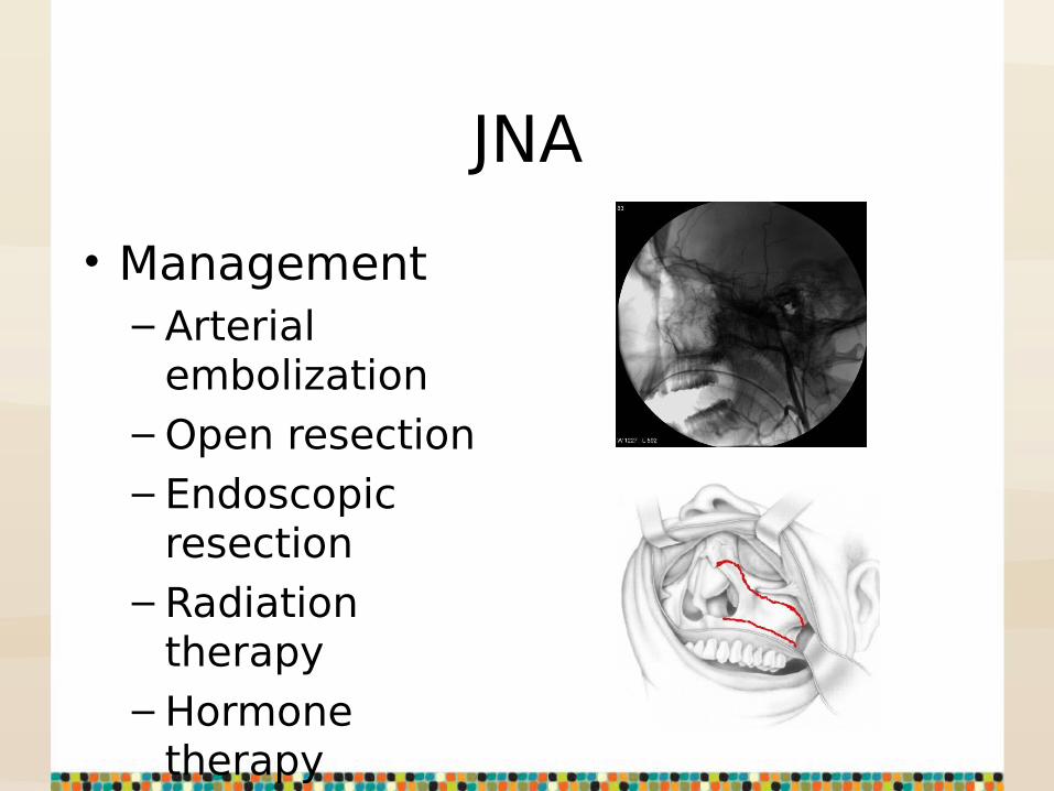
JNA
• Management– Arterial
embolization– Open resection– Endoscopic
resection– Radiation
therapy– Hormone
therapy

Pierre Robin Syndrome
• Retrognathia• Micrognathia• Glossoptosis• Management– Airway– Feeding– Surgery
• Tongue-lip adhesion• Mandibular
distraction osteogenesis

Down Syndrome
• Midface hypoplasia• Relative
macroglossia• Hypotonia• High incidence
of OSA

Treacher Collins Syndrome
• Midface abnormalities– Maxillary
hypoplasia– Choanal
atresia/stenosis• Mandibular
hypoplasia• Tongue base
abnormalities

Crouzon Syndrome
• Midface hypoplasia– Choanal
atresia/stenosis
• Tongue base abnormalities

Stertor
• Acquired– Swollen turbinates– Adenoid
hypertrophy– Foreign body– Nasal polyps– Peritonsillar
abscess– Retropharyngeal
abscess

Swollen Turbinates
• Inferior turbinates– Allergy– Infection
• Treatment– Nasal steroids– Antihistamines
• Oral• Topical
– Surgery• Usually reserved for
adults

Adenoid Hypertrophy
• Nasal obstruction–Mouthbreathing• Darth Vader
– Hyponasal speech

Adenoid Hypertrophy
• Evaluation– Nasal endoscopy– CT- only when
evaluating chronic sinusitis
– Lateral neck x-ray not useful
• Treatment– Adenoidectomy– Nasal steroids

Nasal Foreign Body
• Persistent unilateral nasal drainage and obstruction

Nasal Polyps
• Cystic fibrosis• Antrochoanal
polyp• Severe allergic
rhinitis• Samter's triad– Nasal polyps– Aspirin sensitivity– Asthma

Peritonsillar Abscess
• Throat pain• Dysphagia• Trismus• Uvular deviation• Palatal edema

Peritonsillar Abscess
• Diagnosis– Physical exam– CT neck

Peritonsillar Abscess
• Management– Needle
aspiration– Incision and
drainage– IV antibiotics
and steroids without surgery if only peritonsillar cellulitis
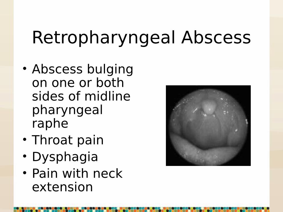
Retropharyngeal Abscess
• Abscess bulging on one or both sides of midline pharyngeal raphe• Throat pain• Dysphagia• Pain with neck
extension

Retropharyngeal Abscess
• Diagnosis– History– Physical Exam– Soft tissue neck
x-ray• Widening of
prevertebral space
– CT neck

Retropharyngeal Abscess
• Management– Incision and
drainage• Danger space 5
– IV antibiotics

Snoring
• Turbulent airflow through the nasopharynx and oropharynx while sleeping.
Snoring
• Common pediatric causes– Adenoid hypertrophy– Tonsillar hypertrophy– Palatal abnormalities

Adenoid Hypertrophy
• Snoring• Hyponasal voice• Mouth breathing

Tonsillar Hypertrophy
• “Kissing tonsils”• Snoring• Muffled voice• Drooling• Sleep disordered
breathing• Dysphagia

Tonsillar Hypertrophy

Tonsillar Hypertrophy
• Acute strep pharyngitis
• Infectious mononucleosis
• Management– CPAP– Tonsillectomy if
persistent upper airway obstruction or recurrent infection

Palatal Abnormalities
• Elongated uvula• Redundancy of
soft palate• Narrowing of the
oropharynx• Treatment– Tonsillectomy– CPAP– UP3

Pediatric Airway Problems
• Stridor• Stertor• Snoring

Thank you!

Questions?