STRIA VASCULARIS AS SOURCE OF ENDOCOCHLEAR POTENTIAL …
7
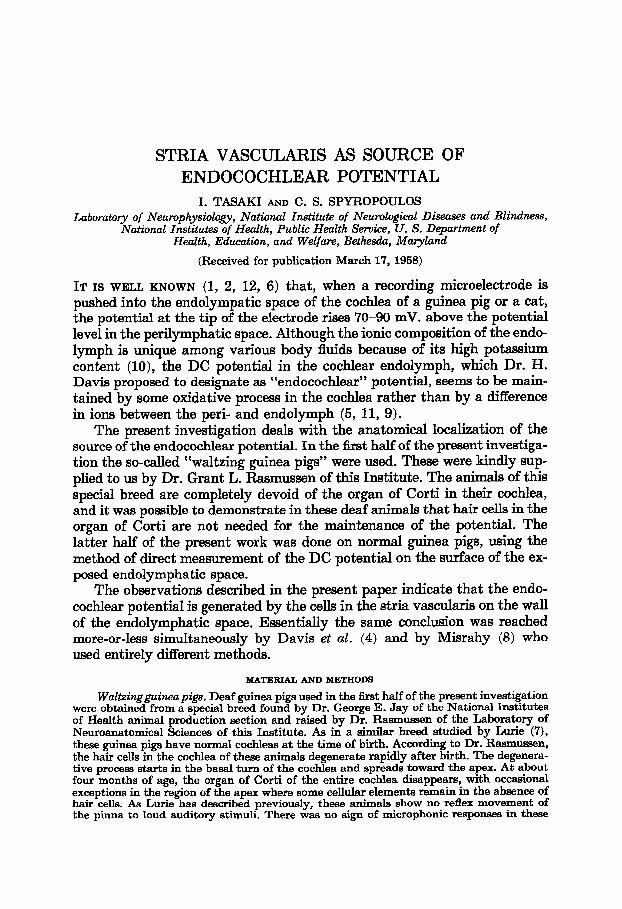
STRIA VASCULARIS AS SOURCE OF ENDOCOCHLEAR POTENTIAL I. TASAKI AND C. S. SPYROPOULOS Laboratory of Neurophysiology, National Institute of Neurological Diseases and Blindness, National Institutes of Health, PubZic Health Service, U. S. Department of Health, Education, and Welfare, Bethesda, Maryland (Received for publication March 17, 1958) IT IS WELL KNOWN (1, 2, 12, 6) that, when a recording microelectrode is pushed into the endolympatic space of the cochlea of a guinea pig or a cat, the potential at the tip of the electrode rises 70-90 mV. above the potential level in the perilymphatic space. Although the ionic composition of the endo- lymph is unique among various body fluids because of its high potassium content (lo), the DC potential in the cochlear endolymph, which Dr. H. Davis proposed to designate as “endocochlear” potential, seems to be main- tained by some oxidative process in the cochlea rather than by a difference in ions between the peri- and endolymph (5, 11, 9). The present investigation deals with the anatomical localization of the source of the endocochlear potential. In the first half of the present investiga- tion the so-called “waltzing guinea pigs” were used. These were kindly sup- plied to us by Dr. Grant L. Rasmussen of this Institute. The animals of this special breed are completely devoid of the organ of Corti in their cochlea, and it was possible to demonstrate in these deaf animals that hair cells in the organ of Corti are not needed for the maintenance of the potential. The latter half of the present work was done on normal guinea pigs, using the method of direct measurement of the DC potential on the surface of the ex- posed endolympha tic space. The observations described in the present paper indicate that the endo- cochlear potential is generated by the cells in the stria vascularis on the wall of the endolymphatic space. Essentially the same conclusion was reached more-or-less simultaneously by Davis et al. (4) and by Misrahy (8) who used entirely different methods. MATERIAL AND METHODS Waltzingguineapigs. Deaf guinea pigs used in the first half of the present investigation were obtained from a special breed found by Dr. George E. Jay of the National Institutes of Health animal production section and raised by Dr. Rasmussen of the Laboratory of Neuroanatomical Sciences of this Institute. As in a similar breed studied by Lurie (7), these guinea pigs have normal co&leas at the time of birth. According to Dr. Rasmussen, the hair cells in the cochlea of these animals degenerate rapidly after birth. The degenera- tive process starts in the basal turn of the cochlea and spreads toward the apex. At about four months of age, the organ of Corti of the entire cochlea disappears, with occasional exceptions in the region of the apex where some cellular elements remain in the absence of hair cells. As Lurie has described previously, these animals show no reflex movement of the pinna to loud auditory stimuli. There was no sign of microphonic responses in these
Transcript of STRIA VASCULARIS AS SOURCE OF ENDOCOCHLEAR POTENTIAL …
STRIA VASCULARIS AS SOURCE OF ENDOCOCHLEAR POTENTIAL
I. TASAKI AND C. S. SPYROPOULOS
Laboratory of Neurophysiology, National Institute of Neurological Diseases and Blindness, National Institutes of Health, PubZic Health Service, U. S. Department of
Health, Education, and Welfare, Bethesda, Maryland
(Received for publication March 17, 1958)
IT IS WELL KNOWN (1, 2, 12, 6) that, when a recording microelectrode is pushed into the endolympatic space of the cochlea of a guinea pig or a cat, the potential at the tip of the electrode rises 70-90 mV. above the potential level in the perilymphatic space. Although the ionic composition of the endo- lymph is unique among various body fluids because of its high potassium content (lo), the DC potential in the cochlear endolymph, which Dr. H. Davis proposed to designate as “endocochlear” potential, seems to be main- tained by some oxidative process in the cochlea rather than by a difference in ions between the peri- and endolymph (5, 11, 9).
The present investigation deals with the anatomical localization of the source of the endocochlear potential. In the first half of the present investiga- tion the so-called “waltzing guinea pigs” were used. These were kindly sup- plied to us by Dr. Grant L. Rasmussen of this Institute. The animals of this special breed are completely devoid of the organ of Corti in their cochlea, and it was possible to demonstrate in these deaf animals that hair cells in the organ of Corti are not needed for the maintenance of the potential. The latter half of the present work was done on normal guinea pigs, using the method of direct measurement of the DC potential on the surface of the ex- posed endolympha tic space.
The observations described in the present paper indicate that the endo- cochlear potential is generated by the cells in the stria vascularis on the wall of the endolymphatic space. Essentially the same conclusion was reached more-or-less simultaneously by Davis et al. (4) and by Misrahy (8) who used entirely different methods.
MATERIAL AND METHODS
Waltzingguineapigs. Deaf guinea pigs used in the first half of the present investigation were obtained from a special breed found by Dr. George E. Jay of the National Institutes of Health animal production section and raised by Dr. Rasmussen of the Laboratory of Neuroanatomical Sciences of this Institute. As in a similar breed studied by Lurie (7), these guinea pigs have normal co&leas at the time of birth. According to Dr. Rasmussen, the hair cells in the cochlea of these animals degenerate rapidly after birth. The degenera- tive process starts in the basal turn of the cochlea and spreads toward the apex. At about four months of age, the organ of Corti of the entire cochlea disappears, with occasional exceptions in the region of the apex where some cellular elements remain in the absence of hair cells. As Lurie has described previously, these animals show no reflex movement of the pinna to loud auditory stimuli. There was no sign of microphonic responses in these
150 I. TASAKI AND C. S. SPYROPOULOS
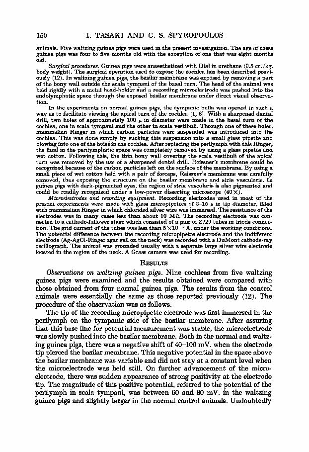
animals. Five waltzing guinea pigs were used in the present investigation. The age of these guinea pigs was four to five months old with the exception of one that was eight months old.
Surgical procecEures. Guinea pigs were anaesthetized with Dial in urethane (0.5 cc. /kg. body weight). The surgical operation used to expose the cochlea has been described previ- ously (12). In waltzing guinea pigs, the basilar membrane was exposed by removing a part of the bony wall outside the Scala tympani of the basal turn. The head of the animal was held rigidly with a metal head-holder and a recording microelectrode was pushed into the endolymphatic space through the exposed basilar membrane under direct visual observa- tion.
In the experiments on normal guinea pigs, the tympanic bulla was opened in such a way as to facilitate viewing the apical turn of the cochlea (1, 6). With a sharpened dental drill, two holes of approximately 100 p in diameter were made in the basal turn of the cochlea, one in Scala tympani and the other in Scala vestibuli. Through one of these holes, mammalian Ringer in which carbon particles were suspended was introduced into the cochlea. This was done simply by sucking this suspension into a small glass pipette and blowing into one of the holes in the cochlea. After replacing the perilymph with this Ringer, the fluid in the perilymphatic space was completely removed by using a glass pipette and wet cotton. Following this, the thin bony wall covering the Scala vestibuli of the apical turn was removed by the use of a sharpened dental drill. Reissner’s membrane could be recognized because of the carbon particles left on the surface of the membrane. By using a small piece of wet cotton held with a pair of forceps, Reissner’s membrane was carefully removed, thus exposing the structure on the basilar membrane and stria vascularis. In guinea pigs with dark-pigmented eyes, the region of stria vascularis is also pigmented and could be readily recognized under a low-power dissecting microscope (40 X).
2MicroeZectrodes and recording equipment. Recording electrodes used in most of the present experiments were made with glass micropipettes of 3-15 p in tip diameter, filled with mammalian Ringer in which chlorided silver wire was immersed. The resistance of the electrodes was in many cases less than about 10 MQ. The recording electrode was con- nected to a cathode-follower stage which consisted of a pair of 2729 tubes in triode connec- tion. The grid current of the tubes was less than 5 Xl0 -l2 A. under the working conditions. The potential difference between the recording micropipette electrode and the indifferent electrode (Ag-AgCl-Ringer agar gel1 on the neck) was recorded with a DuMont cathode-ray oscillograph. The animal was grounded usually with a separate large silver wire electrode located in the region of the neck. A Grass camera was used for recording.
RESULTS Observations on waltzing guinea pigs. Nine co&leas from five waltzing
guinea pigs were examined and the results obtained were compared with those obtained from four normal guinea pigs. The results from the control animals were essentially the same as those reported previously (12). The procedure of the observation was as follows. A The tip of the recording micropipette electrode was first immersed in the perilymph on the tympanic side of the basilar membrane. After assuring that this base line for potential measurement was stable, the microelectrode was slowly pushed into the basilar membrane. Both in the normal a nd waltz- ing guinea pigs, there was a negative shift of 40-100 mV. when the electrode tip pierced the basilar membrane. This negative potential in the space above the basilar membrane was variable and did not stay at a constant level when the microelectrode was held still. On further advancement of the micro- electrode, there was sudden appearance of strong positivity at the electrode tip. The magnitude of this positive potential, referred to the potential of the perilymph in Scala tympani, was between 60 and 80 mV. in the waltzing guinea pigs and slightly larger in the normal control animals. Undoubtedly

COCHLEAR DC POTENTIAL 151
this positive potential is the endocochlear potential discovered by Bekesy (1, 2).
Figure 1 shows two records of the cochlear DC potentials obtained from two successive penetrations of the same basilar membrane. The presence of the initial negative shift prior to the appearance of the endocochlear po- tential is evident. It is also clear that the potential in the endolymph did not vary when the electrode was moved within the endolymphatic space. On withdrawal of the electrode, the potential variation was gradual because of the hold made by the relatively large recording electrode used in these ex-
FIG. 1. Continuous recording of DC potential observed when a glass pipette electrode was slowly pushed into Scala media of “waltzing guinea pig.” Arrows indicate start of endocochlear potential. No microphonic response was observed in this animal. Voltage and time calibrations are common for the two penetrations into same cochlea.
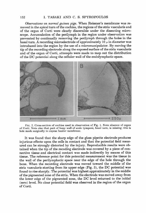
periments. The microphotographs in Fig. 2 show the absence of hair cells in the cochlea from which the records of Fig. 1 were taken. It is evident that the whole organ of Corti has disappeared in this cochlea. The blood vessels in the stria vascularis are almost intact. The marginal cells and other cells in the stria vascularis are present but appear to be slightly atrophic.
Since the entire organ of Corti is missing in these co&leas, it is evident that hair cells are not responsible for generation of the endocochlear potential in these animals. The occasional presence of some cellular elements of the organ of Corti in the apical turn does not affect the present conclusion be- cause the potential variation does not spread from the apex to the basal turn of the cochlea (11). Furthermore, there are no hair cells in this de- generating organ of Corti in the apex.
152 I. TASAKI AND C. S. SPYROPOULOS
Observations on normal guinea pigs. When Reissner’s membrane was re- moved in the apical turn of the cochlea, the regions of the stria vascularis and of the organ of Corti were clearly discernible under the dissecting micro- scope. Accumulation of the perilymph in the region under observation was prevented by continually removing the perilymph through the holes in the basal turn. A recording microelectrode of approximately 10 p in diameter was introduced into the region by the use of a micromanipulator. By moving the tip of the recording electrode along the exposed surface of the stria vascularis and of the organ of Corti, attempts were made to map out the distribution of the DC potential along the cellular wall of the endolymphatic space.
FIG. 2. Cross-section of cochlea used in observation of Fig. 1. Note absence of organ of Corti. Note also that part of bony wall of Scala tympani, basal turn, is missing; this is hole made surgically to expose basilar membrane.
It was found that the sharp edge of the glass pipette electrode produces injurious effects upon the cells in contact and that the potential field meas- ured can be strongly distorted by the injury. Reproducible results were ob- tained when the tip of the recoding electrode was covered by a piece of con- nective tissue and electrical contact was made indirectly by means of this tissue. The reference point for this potential measurement was the tissue in the wall of the perilymphatic space near the edge of the hole through the bone. When the recording electrode was moved toward the middle of the stria vascularis starting from its upper edge (Fig. 3), the DC potential was found to rise sharply. The potential was highest approximately in the middle of the pigmented zone of the stria. When the electrode was moved away from the lower edge of the pigmented zone, the DC level returned to the initial (zero) level. No clear potential field was observed in the region of the organ of Corti.

COCHLEAR DC POTENTIAL 153
The maximum positivity observed was in many cases of the order of 10 mV., but on a few occasions a positive potential of 30-55 mV. was observed. These observations were highly reproducible. When the recording electrode was introduced in the proper position in the cochlea, measurements of the DC potential could be repeated as many times as was desired, yielding nearly constant readings every time. We made at least five measurements on each of approximately 10 co&leas examined.
FIG. 3. Diagrammatic illustration of experiment demonstrating positive poten- tial on surface of stria vascularis relative to potential of other surfaces in Scala media. Perilymph, Reissner’s membrane and endo- lymph were removed when tip of recording electrode was moved along surface.
STRIA VA SCULARIS
: RECORDING I - I ELECTRODE
DISCUSSION
It had been suggested previously by Davis (3) that the source of the cochlear DC potential might be in the region of stria vascularis. The ground for this suggestion is that this region is highly vascularized. Since the DC potential falls when the oxygen supply to the cochlea is suspended, it is inferred that the source of the potential should be closely related to the blood supply to the cochlea.
The experimental findings described under Results indicates in the first place that the organ of Corti is not essential for the maintenance of the endo- cochlear potential. In the second place, it was shown that, in the endo- lymphatic space from which the endolymph had been drained, the region of the stria vascularis exhibits a high DC potential. Undoubtedly, these findings are clear indication that the source of the DC potential is in the stria vascular-is, although they do not show how the DC potential is produced.
The evidence presented by Davis et al. (4) in support of the view that the stria vascularis is the source of the endocochlear potential is similar to our experiments on the waltzing guinea pigs; they tried to destroy hair cells by injecting streptomycin without damaging the stria vascularis. In several
154 I. TASAKI AND C. S. SPYROPOULOS
animals which showed no microphonic responses, they could demonstrate normal DC potentials in the endolymphatic space. In this connection it may also be mentioned that in the waltzing guinea pigs used in the present in- vestigation there was no sign of a microphonic response to loud auditory stimuli. (This conems Lurie’s original observation.) The evidence presented by Misrahy (8) is as follows: When a solution of glucose-glucose oxidase was injected into the various scalae in order to render the region of the basilar membrane hypoxic without affecting the O2 tension in the stria vascularis, the microphonics response disappeared or greatly diminished without being accompanied by any significant loss in the DC potential. This period was interpreted as the time during which the function of the hair cells are im- paired, whereas the stria vascularis is still functioning.
It has been pointed out in the section on Results that, both in normal and waltzing guinea pigs, a strong negative potential can be observed when the recording electrode is penetrating the structure above the basilar membrane. A similar observation was reported previously (12); but there is a significant difference in the size of the recording electrode between the present and the previous experiments. The negativity was ascribed previously to the intra- cellular (resting) potential picked up by submicroscopic recording electrodes. Since the diameter of the recording electrode was, in many of the present experiments, as large as 15 p, it is unlikely that the negativity represents an intracellular potential. It is possible that the potential of the fluid in the space between the reticular lamina and the basilar membrane is strongly negative to the perilymph.
In a previous experiment (l2), it was shown that the phase of the micro- phonic response reverses at the boundary between the space showing the strong negative potential mentioned above and the space with the positive endocochlear potential. Since we have enough evidence to suppose that the hair cells are responsible for generation of microphonic response, it follows that the negative potential exists in the space around the hair cells. The possibility that the negative potential may be within the tectorial membrane is excluded for the same reason.
SUMMARY
1. In waltzing guinea pigs which have no organ of Corti in the cochlea (Fig. 2), the DC endolymphatic potential of approximately normal magni- tude was observed (Fig. 1).
2. In normal co&leas from which both the peri- and endolymph were drained, the DC potential of the surface of the stria vascularis was found to be strongly positive to that of other surface areas (Fig. 3).
3. It was concluded that the DC potential in the cochlear endolymph is produced by the cells in the stria vascularis.
ACKNOWLEDGEMENTS
We are deeply indebted to Dr. Grant L. Rasmussen who kindly supplied us with waltzing guinea pigs. The histological specimen presented in Fig. 2 was prepared in Dr. Rasmussen’s section by Miss Edna P. McCrane.
COCHLEAR DC POTENTIAL 155
REFERENCES
1. BEKESY, G. v. D.C. resting potentials inside the cochlear partition. J. acaust. Sac. Amer., 1952,24: 72-76.
2. BEKESY, G. v. Gross localization of the place of origin of the co&ear microphonics. J. acaust. Sac. Amer., 1952, 24: 399-409.
3. DAVIS, H. Nerve impulse. New York, Josiah Macy, Jr. Foundation, 1953, 224 pp. 4. DAVIS, H., DEATHERAGE, B. H., ROSENBLIJT, B., FERNANDEZ, C., KIMURA, R., AND
SMITH, C. A. Modification of cochlear potentials produced by streptomycin poisoning and by extensive venous obstruction. Laryngascape, 1958, 68: 596-627.
5. DAVIS, H., TASAKI, I., SMITH, C. A., AND DEATHERAGE, B. H. Cochlear potentials after intracochlear injections and anoxia. Fed. Proc., 1955,14: 112.
6. GISSELSSON, L. Neuere Probleme des Cochleaeffektes. Arch. Ohr.-, Nas.-u. Kehlk- Heilk., 1955, 167: 274-282.
7. LURIE, M. H. The waltzing (circling) guinea pig. Ann. OtaZ., St. Louis, 1941, 50: 113-128.
8. MISRAHY, G. Effects of intracochlear injections of glucose-glucose oxidase on the DC potential, microphonics and action potential of the cochlea of guinea pigs. J. acaust. Sac. Amer., 1958, 30:688-689.
9. SMITH, A. S., GESSERT, C. F., DAVIS, H., AND DEATHERAGE, B. H. DC potential of the membraneous labyrinth. Lalyngoscope, 1958, 68: 595-596.
10. SMITH, C. A., LOWRY, 0. H., AND Wu, M. L. The electrolytes of the labyrinthine fluids. Laryngascape, 1954, 64: 141-153.
11. TASAKI, I. Hearing. Ann. Rev. Physial. 1957, 19: 417-438. 12. TASAKI, I., DAVIS, H., AND ELDRIDGE, D. H. Exploration of cochlear potentials in
guinea pig with a microelectrode. J. acaust. Sac. Amer., 1954,26: 765-773.