Stressful Military Training: Endocrine Reactivity ...
9
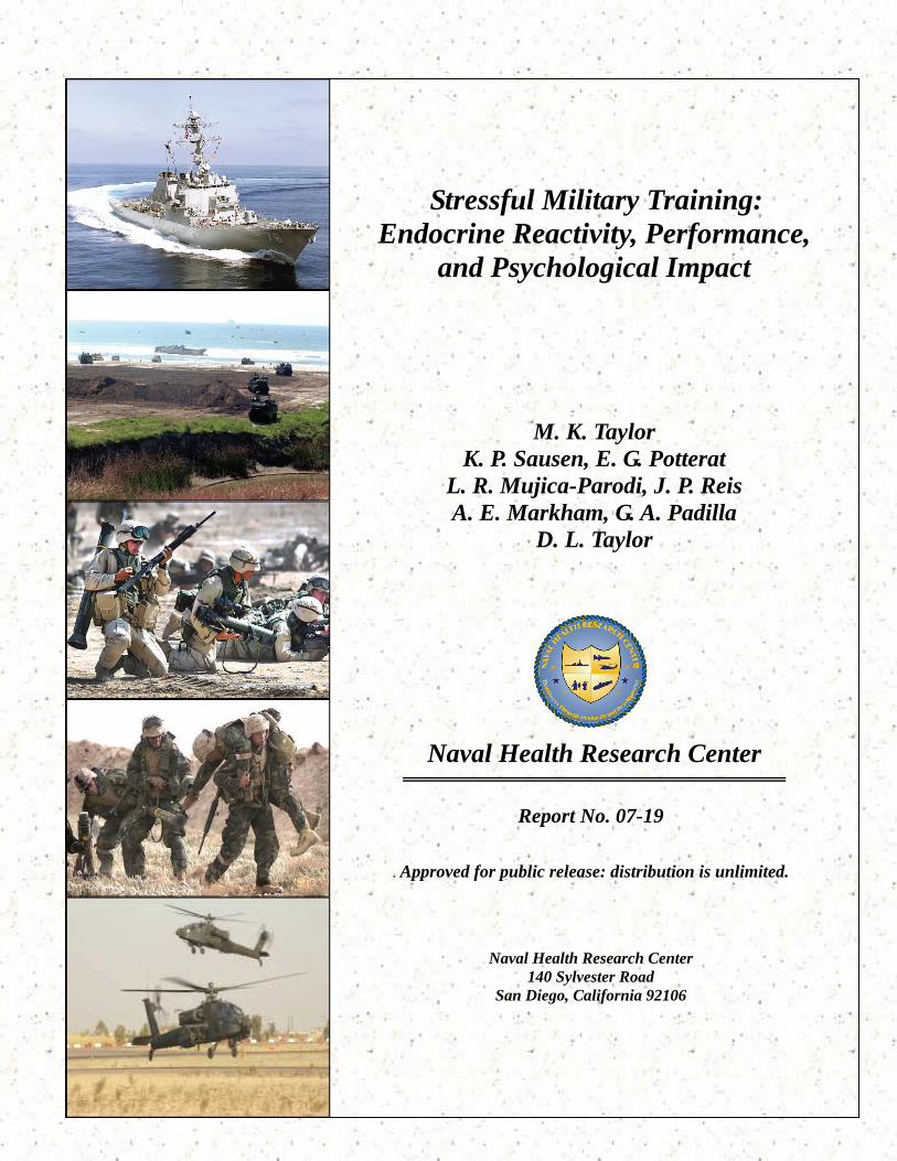
Naval Health Research Center Stressful Military Training: Endocrine Reactivity, Performance, and Psychological Impact M. K. Taylor K. P. Sausen, E. G. Potterat L. R. Mujica-Parodi, J. P. Reis A. E. Markham, G. A. Padilla D. L. Taylor Report No. 07-19 . Approved for public release: distribution is unlimited. Naval Health Research Center 140 Sylvester Road San Diego, California 92106
Transcript of Stressful Military Training: Endocrine Reactivity ...
Naval Health Research Center
Stressful Military Training: Endocrine Reactivity, Performance,
and Psychological Impact
M. K. Taylor K. P. Sausen, E. G. Potterat
L. R. Mujica-Parodi, J. P. Reis A. E. Markham, G. A. Padilla
D. L. Taylor
Report No. 07-19
. Approved for public release: distribution is unlimited.
Naval Health Research Center 140 Sylvester Road
San Diego, California 92106
Delivered by Ingenta to:Naval Medical Center, San Diego
IP : 159.71.185.141Wed, 19 Dec 2007 20:52:00
Aviation, Space, and Environmental Medicine x Vol. 78, No. 12 x December 2007 1143
RESEARCH ARTICLE
Stressful Military Training: Endocrine Reactivity, Performance, and Psychological Impact
Marcus K. Taylor , Kenneth P. Sausen , Eric G. Potterat , Lilianne R. Mujica-Parodi , Jared P. Reis , Amanda E. Markham , Genieleah A. Padilla , and Deborah L. Taylor
T AYLOR MK, S AUSEN KP, P OTTERAT EG, M UJICA -P ARODI LR, R EIS JP, M ARKHAM AE, P ADILLA GA, T AYLOR DL. Stressful military training: endocrine reactivity, performance, and psychological impact. Aviat Space Environ Med 2007; 78:1143 – 9.
Introduction: We examined the responsiveness of both cortisol and dehydroepiandrosterone sulfate (DHEAS) to the stress of survival training in military men and evaluated relationships to performance, peritraumatic dissociation, and the subsequent impact of stressful events. Methods: Baseline salivary cortisol samples were self-collected by 19 men at 0900 and 1930 in a free-living (FL) environment. DHEAS samples were also collected in a subset of this sample ( N 5 12). Samples were subsequently taken at similar time points during a stressful captivity (SC) phase of training. Repeated-measures analyses of variance with follow-up paired t -tests examined differences across time and conditions. Results: Signifi -cant increases were observed at both time points (0900 and 1930) from FL to SC in both cortisol (0900: 9.2 6 3.4 nmol z L 2 1 vs. 18.4 6 10.5 nmol z L 2 1 ; 1930: 3.5 6 3.0 nmol z L 2 1 vs. 27.7 6 10.9 nmol z L 2 1 ) and DHEAS (0900: 1.7 6 1.3 ng z ml 2 1 vs. 6.7 6 3.5 ng z ml 2 1 ; 1930: 1.5 6 0.84 ng z ml 2 1 vs. 4.5 6 3.0 ng z ml 2 1 ). Also, overall performance during a high-intensity captivity-related challenge was inversely related to the DHEAS – cortisol ratio; conversely, overall performance during a low-intensity captivity-related challenge was positively related to DHEAS at the 0900 time point during SC. Dissociation was unrelated to endocrine indices measured during SC, while total impact of events was inversely related to percent change in DHEAS from FL to SC. Conclusions: Corti-sol and DHEAS increase in response to allostatic load, and may relate to human performance during SC as well as PTSD symptoms. Keywords: cortisol , DHEAS , hormones , survival .
CORTISOL IS the end product of hypothalamic- pituitary-adrenal stimulation and is responsible for
stimulating blood glucose for energy and increasing blood pressure in support of the stress response, and has known relationships to physical as well as mental stress. Acutely, increased cortisol release can degrade percep-tual processes important for encoding information (e.g., tunnel vision) and may even enhance memory for par-ticularly salient information ( 28 ). Excessive or chroni-cally high cortisol levels, however, have adverse neural consequences characterized by reduced hippocampal volume and function ( 27,28 ), decreased neurogenesis, decreased brain-derived neurotrophic factor, and im-paired memory ( 3,4 ). The hippocampus is populated with cortisol receptors, and it is through this mechanism that extreme and/or extended cortisol secretion may re-sult in neurotoxicity and degraded cognitive function.
Cortisol is the most widely studied marker of the endo-crine stress response.
Morgan and colleagues have pioneered endocrine re-search in the military survival context. Their recent work has demonstrated robust increases in cortisol concentra-tions in response to Survival, Evasion, Resistance, and Escape training (SERE) — a harsh and realistic environ-ment in which students are taught to survive, evade en-emy captors, resist exploitation, and escape during cap-tivity ( 19,20,22 ). In one study, Morgan and colleagues found inverse relationships between percentage of free (plasma) cortisol and dissociative symptoms during stress (retrospectively recalled at conclusion of training, with specifi c reference to the stressful event), but posi-tive relationships between the retrospectively recalled dissociative and percentage of free cortisol values ob-tained after the conclusion of training ( 22 ). Another study yielded positive relationships between salivary cortisol and dissociative symptoms (retrospectively re-called in reference to the entire captivity experience) ( 20 ). Subtle methodological differences, then, may ex-plain these discrepant fi ndings. With respect to human performance, these researchers have observed positive relationships between percentage of free (plasma) corti-sol and military performance during captivity-related challenges ( 22 ) and, by contrast, inverse relationships between salivary cortisol and performance in a different sample ( 20 ). Salivary cortisol concentrations have been shown to closely approximate plasma values ( 8 ). The re-lationships between cortisol, dissociation, and human
From the Naval Health Research Center, San Diego, CA (M. K. Taylor, K. P. Sausen, J. P. Reis, A. E. Markham, and G. A. Padilla); Naval Special Warfare Center, San Diego, CA (E. G. Potterat); State University of New York, Stony Brook (L. R. Mujica-Parodi); and San Diego State University, CA (D. L. Taylor).
This manuscript was received for review in June 2007 . It was ac-cepted for publication in September 2007 .
Address reprints to: Marcus K. Taylor, Ph.D., Naval Health Research Center, Dept. 162, P.O. Box 85122, San Diego, CA 92186-5122; [email protected]
Reprint & Copyright © by Aerospace Medical Association, Alexan-dria, VA.
DOI: 10.3357/ASEM.2151.2007
Delivered by Ingenta to:Naval Medical Center, San Diego
IP : 159.71.185.141Wed, 19 Dec 2007 20:52:00
1144 Aviation, Space, and Environmental Medicine x Vol. 78, No. 12 x December 2007
ENDOCRINE REACTIVITY TO STRESS — TAYLOR ET AL.
performance during military stress remain unclear and may be sensitive to subtle methodological differences. Additionally, the relationship of cortisol reactivity to the subsequent impact of captivity-related events has not been explored.
Dehydroepiandrosterone (DHEA) and its sulfated version, dehydroepiandrosterone sulfate (DHEAS) are cosecreted with cortisol from the adrenal gland ( 1 ), serve as precursors for androgenic and estrogenic steroids ( 11 ), and have also been used as markers of the endo-crine stress response. DHEAS is considered the most abundant steroid in the body ( 1 ). Both substances peak at 20-25 yr of age in humans and then decline to reach values of 20 – 30% at 70-80 yr ( 23 ).
Findings from animal studies suggest that DHEA and DHEAS confer neurogenesis ( 25 ) as well as neuropro-tection during stress ( 14,15,17 ), although the precise mechanisms of action remain to be confi rmed ( 1 ). Roberts et al. ( 25 ) fi rst reported the neurogenic effects of DHEA and DHEAS. In that animal study, when cultures of dissociated brain cells of mouse embryos were cul-tured with either DHEA or DHEAS, increases were found in the numbers of two specifi c types of nerve cells, neurofi lament-positive neurons and glial fi brillary acid protein-positive astrocytes, along with extensions of the processes of both types of cells. Additionally, Kimonides et al. ( 15 ) found that both DHEA and DHEAS prevent or reduce the neurotoxic actions in the hippocampus of the glutamate agonists N -methyl-D-aspartate (NMDA) and a -amino-3-hydroxy-5-methyl-4-isoxazolepropionic acid although DHEA was more effective. More recently, Kurata ( 17 ) examined possible neuroprotective effects of b -estradiol, DHEA, and DHEAS against NMDA-induced neurotoxicity in rat hippocampal neurons, observing that all three exerted neuroprotective effects, albeit via different mechanisms. In other work, Hajszan et al. ( 12 ) showed that DHEA induces hippocampal spine synapse changes (i.e., remodeling), with DHEA treatment result-ing in a signifi cant decrease in the number of GABAergic axosomatic terminals while non-GABAergic terminals were unaffected, thus suggesting a protective effect against neuroexcitability. Further work has confi rmed that DHEA and DHEAS regulate neuronal function via infl uence on neuronal excitability ( 26 ), and both of these neurosteroids may also confer protection against the neurotoxic effects of corticosterone ( 14 ). Further, DHEA has been identifi ed as a direct buffer against the neuro-toxic effects of cortisol ( 13 ).
Human studies have examined the clinical and thera-peutic applications of these neurosteroids ( 16,24 ), their possible stress-buffering roles during stressful military training ( 20 ), as well as possible relationships to post-traumatic stress disorder (PTSD). Clinically oriented work has examined the role of endogenous and exoge-nous (administered) DHEA and DHEAS in depression, mood regulation, and stress reactivity. Rasmusson and colleagues ( 24 ), for example, studied the effects of adre-nocorticotropic hormone administration on the DHEA responses of women with PTSD, and found that in-creased adrenal DHEA release is associated with de-
creased avoidance and negative mood in this popula-tion. Other work has examined the stress-buffering role of DHEAS during acute military stress. Morgan et al. ( 20 ), for instance, examined relationships between plasma DHEAS, cortisol, dissociation, and performance during military survival training. In this sample of 25 military personnel, DHEAS was increased by opera-tional stress, and the DHEAS-cortisol ratio was higher in participants who reported fewer symptoms of disso-ciation and demonstrated superior military perfor-mance. Finally, cortisol, DHEA, and DHEAS are thought to contribute to the neurobiological dysregulations un-derlying PTSD. Notably, PTSD patients tend to exhibit lower levels of cortisol, increased responsiveness of glu-cocorticoid receptors, and enhanced cortisol-induced negative feedback ( 33 ). Additionally, Charney ( 9 ) sug-gested a negative correlation between DHEA reactivity to adrenal activation and the severity of PTSD. Taken to-gether, the current literature suggesting the clinical and therapeutic applications of these neurosteroids, their possible stress-buffering roles during military stress, and possible relationships to PTSD provide a clear ratio-nale for further clarifying the endocrine responses to military stress as well as relationships to human perfor-mance and PTSD risk factors.
The present study was initiated to extend our under-standing of cortisol and DHEAS responses to the stress of harsh military training, and to evaluate relationships to human performance as well as key risk factors for PTSD (peritraumatic dissociative symptoms and subsequent im-pact of events). Based on previous research, we hypothe-sized that cortisol and DHEAS concentrations would sub-stantially increase in response to the allostatic load imposed by survival training; we also expected that cortisol would relate positively to symptoms of dissociation during stress. Although the assessment of the effects of endocrine re-sponses on subsequent impact of events is an unprece-dented area of inquiry, it was expected that cortisol re-sponses would exacerbate, and DHEAS responses would ameliorate, the subsequent impact of stressful events.
METHODS
Participants
As part of a program of research examining individ-ual differences in stress resilience during military sur-vival training, the participants in this study included 19 healthy, male, active-duty Navy personnel whose mean age was 21.5 yr 6 1.7 SD. Participants were recruited from a secure list of incoming trainees who were sched-uled to report to survival training in the San Diego area. Participation was voluntary and prospective partici-pants were notifi ed that the study was being performed independent of the survival training curriculum. Those who expressed a desire to participate and reported no history of head trauma or PTSD, or addiction to any substances (e.g., tobacco products) were scheduled for an in-person meeting to review the details of the study and provide written informed consent approved by the Naval Health Research Center Institutional Review
Delivered by Ingenta to:Naval Medical Center, San Diego
IP : 159.71.185.141Wed, 19 Dec 2007 20:52:00
Aviation, Space, and Environmental Medicine x Vol. 78, No. 12 x December 2007 1145
ENDOCRINE REACTIVITY TO STRESS — TAYLOR ET AL.
Board. All participants were subjected to rigorous medi-cal and psychological reviews by a medical offi cer and psychologist prior to enrollment in survival training.
Survival, Evasion, Resistance, and Escape (SERE) Training
We have described SERE training in a previous report ( 31 ). Briefl y, United States military members at high risk of capture (e.g., aircrew, special operations personnel) are required to attend the 12-d SERE course, which in-cludes a period of confi nement in a Resistance Training Laboratory (RTL). After an initial phase of classroom-based didactic training, students are taken to the fi eld where they receive applied training in survival, evasion, resistance, and escape techniques. Students are then re-leased into the fi eld and tasked with the goal of evading enemy captors. Upon eventual capture, students are taken to the RTL, where they are expected to apply their recently learned skills of resistance to political indoctri-nation and captivity-related problems. The structured, choreographed nature of this training platform provides a unique and unprecedented medium in which to sys-tematically and scientifi cally examine human stress and performance in a realistic military context. Although the participants were exposed to a broad spectrum of environmental and psychological stressors throughout training, they were given continuous access to water and were encouraged to remain hydrated throughout all phases of the curriculum.
DHEAS and Cortisol Assessment
Baseline salivary samples were obtained over the course of 2 consecutive days, 1 – 3 wk prior to the start of survival training. The saliva samples were self-collected by participants in a free-living (FL) setting and collected in Salivette tubes (Sarstedt, Inc., Newton, NC), which were individually labeled with the date and time of sampling. Specifi cally, the samples were collected dur-ing the workweek (either Mon-Tues or Wed-Thur), and all participants participated in typical daily work rou-tines (approximately 0730 to 1630). On each of the 2 d, samples were obtained at 0900 and 1930. Participants were instructed not to brush or fl oss their teeth, eat, or drink, and to rinse their mouth with cool water immedi-ately prior to sampling. They were also asked not to en-gage in physical activity during the 2-d sampling period. Participants signed a statement of compliance during this period stating that they had not used any substances or medications 12 h prior to or during the data collection period. Salivary cortisol was measured using a Coat-A-Count radioimmunoassay kit (Diagnostic Products Corporation, Los Angeles, CA). It was measured with sensitivity (detectability) of 0.7-55.2 nmol z L 2 1 and intra and interassay coeffi cients of variation of 3 – 7% and 5%, respectively. Additionally, salivary DHEAS was measured using a radioimmunoassay kit (Diagnostic Systems Laboratories, Inc., Webster, TX). It was mea-sured with sensitivity of 0.1-30.0 ng z ml 2 1 and intra and interassay coeffi cients of variation of 5 – 7% and 6%, re-spectively. The assessment of DHEAS was added to our
protocol later in our study with the intent of more fully characterizing the endocrine response. Therefore, DHEAS is analyzed in only a subset of the sample. Average base-line values were calculated for salivary cortisol ( N 5 19), DHEAS ( N 5 12), and the DHEAS-cortisol ratio ( N 5 12) [(Day 1 1 Day 2)/2] at 0900 and 1930, respectively [i.e., free-living condition (FL)]. Subsequently, during the captivity phase of survival training [i.e., stressful captivity (SC)], salivary samples were obtained approximately at the corresponding time points (0900 and 1930). DHEAS-cortisol ratios [DHEAS (ng/ml)/cortisol (nmol/L)] were calculated at both time points, during both conditions (FL and SC).
Clinician-Administered Dissociative States Scale (CADSS)
The 19 self-report items from the CADSS ( 5 ) were used to assess the frequency and intensity of state symp-toms of dissociation. Although the CADSS includes additional items used for clinical observation of the par-ticipant, the 19 self-report items have been shown to comprise a valid, reliable, and independent indicator of dissociative state symptoms ( 20 ). The scale was admin-istered to participants immediately after a high- intensity, captivity-related challenge during SC. This scale is designed to assess how perceptually connected or dis-connected an individual is relative to the individual’s environment. Examples of items include: “ Did you feel as if you were watching the situation as an observer or spectator? ” ; “ Did you space out or in some way lose track of what is going on? ” ; and “ Did you feel as if you were looking at things outside of your body? ” . The self-report scale is composed of 19 items, rated on a Likert scale of 0 (not at all) to 4 (extremely), with a possible to-tal score of 76. Participants were asked to respond to the questions relative to the high-intensity captivity-related challenge. Cronbach’s alpha reliability for CADSS in the present study was 0.83, and the mean CADSS score was 28.5 (SD 5 10.7).
Military Performance
During SC, students were graded on several observed survival target skills during both high- and low- intensity captivity-related challenges. As reported elsewhere, these skills enhance the likelihood of survival ( 18 ). Scores reported here refl ect the sum of the target skills that stu-dents are expected to demonstrate during the captivity-related challenge. Additionally, because of the focus of the current research on identifying individual differ-ences in stress resilience, an individual “ resilience ” score is also reported. Overall performance scores and resil-ience scores are reported for both a low- (OP-Low) and high-intensity captivity-related challenge (OP-High). These performance scores were developed by military experts, independent of this research. Higher scores are indicative of better performance.
Impact of Events Scale-Revised (IES-R)
The IES-R ( 32 ) is a self-report measure designed to as-sess current subjective distress for any specifi c life event.
Delivered by Ingenta to:Naval Medical Center, San Diego
IP : 159.71.185.141Wed, 19 Dec 2007 20:52:00
1146 Aviation, Space, and Environmental Medicine x Vol. 78, No. 12 x December 2007
ENDOCRINE REACTIVITY TO STRESS — TAYLOR ET AL.
It has 22 items, comprising 3 subscales corresponding to DSM-IV-specifi ed PTSD symptoms: avoidance (IES-Avoid; mean of 8 items measuring the extent to which the respondent avoids situations that remind him or her of the stressful or traumatic event), intrusion (IES-Intrusion; mean of 8 items assessing the extent to which one experiences intrusive thoughts), and hyperarousal (IES-Arousal; mean of 6 items measuring anger, irrita-bility, heightened startle response, and hypervigilance). The total impact of events score (IES-Total) is composed of the sum of the three subscales. In the present study, at the conclusion of survival training, respondents were shown a list of diffi culties people sometimes have after stressful life events, and were asked to indicate how distressing each diffi culty has been with respect to the high-intensity situation encountered during captivity on a scale of 0 (not at all) to 4 (extremely). Adequate re liability and predictive validity have been shown for this scale ( 6,29 ) and Cronbach’s alpha reliabilities in the present sample were 0.75, 0.81, and 0.79 for IES-Arousal, IES-Avoidance, and IES-Intrusion, respectively. Cronbach’s alpha reliability for IES-Total was 0.91.
Data Analysis
Data were analyzed using SPSS software, version 15 (SPSS, Inc., Chicago, IL). Analyses included descriptive statistics, analysis of variance (ANOVA) with repeated measures, and bivariate correlations. Descriptive statis-tics were used to summarize the physical and demo-graphic characteristics of the sample. A 2 (0900 vs. 1930) 3 2 (FL vs. SC) repeated-measures ANOVA with follow-up t -tests examined differences in cortisol between FL and SC conditions. Percent change scores from FL to SC [(SC – FL)/FL ] were also calculated for each participant at each time point (0900 and 1930, respectively). These analyses were repeated with DHEAS and DHEAS-cortisol ratios as the dependent variables. Pearson product-moment correlations were performed to examine rela-tionships among endocrine responses, objectively mea-sured performance, peritraumatic dissociative symptoms, and subsequent impact of events. All hypothesis tests were two-sided and the probability of committing a type I error was set at 0.05.
RESULTS
Mean age, body mass index, and years of military ser-vice for this sample were 21.5 yr (SD 5 1.7), 24.2 kg z m 2 2 (SD 5 1.6), and 1.5 yr (SD 5 0.9), respectively. Of the 19 subjects, 14 subjects (73.7%) had received a high school education and 5 (26.3%) were college educated. All sub-jects were Caucasian. Regarding military occupational specialties, 14 subjects (73.7%) were students under in-struction to become Aviation Warfare Specialists/Rescue Swimmers, while the remaining 5 (26.3%) were students under instruction to become Special Warfare/SEAL Of-fi cers. Specifi cally, the Aviation Warfare members had completed the majority of their coursework to receive their warfare designation and were awaiting graduation and subsequent assignment to SERE training. Similarly,
the Special Warfare members had graduated from Basic Underwater Demolition/SEAL training and were awaiting enrollment in SERE school as a component of SEAL Qualifying Training – a 6-mo series of advanced training experiences prior to receiving their warfare designation.
Mean cortisol values during FL were 9.2 nmol z L 2 1 (SD 5 3.4) at 0900 and 3.5 nmol z L 2 1 (SD 5 3.0) at 1930, while mean cortisol values during SC were 18.4 (SD 5 10.5) at 0900 and 27.7 nmol z L 2 1 (SD 5 10.9) at 1930. A repeated-measures ANOVA yielded a signifi cant inter-action (F[1,18] 5 42.9, P , 0.001, observed power 5 1.0), such that cortisol concentrations were higher in the morning than in the evening during FL (indicative of the normal diurnal pattern), but increased substantially from morning to evening during SC. Mean cortisol val-ues increased signifi cantly in response to SC at both time points [0900: t 5 4.1, P , 0.001, mean percent D 5 1 113% (SD 5 120); 1930: t 5 10.2, P , 0.001, mean percent D 5 1 1388% (SD 5 1335)] ( Fig. 1A ).
Mean DHEAS values during FL were 1.7 ng z ml 2 1 (SD 5 1.3) at 0900 and 1.5 ng z ml 2 1 (SD 5 0.8) at 1930; during SC, DHEAS values were 6.7 ng z ml 2 1 (SD 5 3.5) at 0900 and 4.5 ng z ml 2 1 (SD 5 3.0) at 1930. A repeated- measures ANOVA revealed a signifi cant overall effect of SC on DHEAS concentrations [F(1,11) 5 18.3, P , 0.001; ob-served power 5 0.97]. As shown in Fig. 1B , mean DHEAS values increased signifi cantly in response to SC at both time points [0900: t 5 5.4, P , 0.001, mean percent D 5 1 381% (SD 5 284); 1930: t 5 4.1, P , 0.01, mean per-cent D 5 1 207% (SD 5 138)]. DHEAS values were sig-nifi cantly higher in the morning compared with evening hours during SC, but this downward shift was not evi-dent during the FL condition.
To further explore DHEAS responses in relation to cortisol responses, a repeated-measures ANOVA was also used to determine whether the DHEAS-cortisol ratio differed signifi cantly from FL to SC. A signifi cant interaction was observed [F(1,11) 5 18.1, P , 0.001, observed power 5 0.97]. Mean DHEAS-cortisol ratios increased signifi cantly from FL to SC at 0900 [t 5 2 3.0, P , 0.01, mean percent D 5 1 130 (SD 5 159)]; conversely, DHEAS-cortisol ratio scores decreased signi fi cantly from FL to SC at 1930 [t 5 3.2, P , 0.01, mean percent D 5 2 69 (SD 5 20)] ( Fig. 1C ).
A nonsignifi cant inverse trend was observed between overall performance during a high-intensity, captivity-related challenge (OP-High) and DHEAS at 0900 during SC (r 5 2 0.51, P 5 0.10). Inverse relationships emerged between OP-High and the DHEAS-cortisol ratio (r 5 2 0.60, P , 0.05) at the 0900 time point during SC ( N 5 12). Also, inverse relationships were also observed be-tween OP-High and percent change (FL to SC) in DHEAS (r 5 2 0.65, P , 0.05) and DHEAS-cortisol ratio (r 5 2 0.61, P , 0.05) measured at the 1930 time point ( N 5 12). An objectively measured score of resilience during the same challenge was positively associated with corti-sol concentrations (r 5 0.56, P , 0.01) and inversely re-lated to DHEAS-cortisol ratios at 0900 during SC (r 5 2 0.70, P , 0.01) ( N 5 12). Overall performance during

Delivered by Ingenta to:Naval Medical Center, San Diego
IP : 159.71.185.141Wed, 19 Dec 2007 20:52:00
Aviation, Space, and Environmental Medicine x Vol. 78, No. 12 x December 2007 1147
ENDOCRINE REACTIVITY TO STRESS — TAYLOR ET AL.
a low-intensity, captivity-related challenge (OP-Low) was positively associated with DHEAS during SC at the 0900 time point (r 5 0.59, P , 0.05) and a signifi cant trend emerged between OP-Low and the DHEAS-cortisol ratio at the same time point (r 5 0.55, P 5 0.06) ( N 5 12).
Dissociative symptoms (as measured by the CADSS) during the high-intensity captivity-related challenge did not relate to any of the endocrine indices during SC or percent change of the endocrine indices from FL to SC. There was also no relationship between dissociative symptoms and OP-High. IES-Avoid was positively as-sociated with cortisol concentrations during SC at 0900 (r 5 0.55, P , 0.05) as well as percent change in cortisol (FL to SC) at 0900 (r 5 0.52, P , 0.05). Similarly, IES-
Fig. 1. FL vs. SC comparisons for: A) cortisol; B) DHEAS; and C) DHEAS-Cortisol ratio. All values are mean 6 SD. P-values are provided for post hoc comparisons across conditions at each time point: * P , 0.01; ** P , 0.001.
Intrusion was associated with cortisol concentrations during SC at 0900 (r 5 0.58, P , 0.05) as well as percent change in cortisol at this same time point (r 5 0.52, P , 0.05). IES-Arousal was inversely associated with the percent change in DHEAS-cortisol ratio at the 0900 time point (r 5 2 0.71, P , 0.05) and the 1930 time point (r 5 2 0.74, P , 0.05). Finally, IES-Total demonstrated inverse associations with percent change in DHEAS at the 1930 time point (r 5 2 0.69, P , 0.05).
Dissociative symptoms measured immediately after the high-intensity, captivity-related challenge predicted the subsequent impact of events. Specifi cally, positive associations were observed between dissociative symp-toms and IES-Avoid (r 5 0.57, P , 0.01), IES-Intrusion (r 5 0.48, P 5 0.06), and IES-Total (r 5 0.53, P , 0.05).
DISCUSSION
The present study was initiated to extend our under-standing of cortisol and DHEAS responses to the stress of military survival training, and to evaluate the rela-tionships to human performance as well as key symp-toms of PTSD. We demonstrated robust increases in both cortisol and DHEAS in response to the stress of survival training. Additionally, we noted possible intensity-dependent relationships between endocrine indices and performance during SC. Specifi cally, DHEAS and the DHEAS-cortisol ratio were inversely related to overall performance during the high-intensity challenge, but both were positively associated with performance dur-ing the low-intensity challenge. Finally, as hypothesized, cortisol reactivity to SC tended to exacerbate, and DHEAS tended to ameliorate, the subsequent impact of stressful, captivity-related events.
As expected, we observed robust increases in both cortisol and DHEAS in response to SC, evidenced by 1.1- to 13.8-fold increases in cortisol and 2.1- to 3.8-fold increases in DHEAS. These results are in general agree-ment with prior fi ndings ( 20 – 22 ). Specifi cally, Morgan and colleagues examined cortisol reactivity to survival training in 72 Army personnel ( 21,22 ), and showed that cortisol increased substantially during captivity, and peaked directly after stressful, captivity-related chal-lenges. These fi ndings were subsequently replicated in a sample of 25 Navy personnel at a separate survival training facility ( 19 ). Similarly, these same researchers showed that DHEAS levels increased substantially in the Navy personnel during the captivity phase of sur-vival training ( 20 ).
We also observed intensity-dependent relationships between endocrine indices and military performance dur-ing captivity-related problems. During a high-intensity challenge, overall performance scores were inversely associated with DHEAS as well as the DHEAS-cortisol ratio. During the low-intensity challenge, however, positive relationships were observed. Although not ex-amining differences between high- and low-intensity challenges, previous research has shown positive relation-ships between percentage of free cortisol and military performance during captivity challenges ( 22 ) and, by
Delivered by Ingenta to:Naval Medical Center, San Diego
IP : 159.71.185.141Wed, 19 Dec 2007 20:52:00
1148 Aviation, Space, and Environmental Medicine x Vol. 78, No. 12 x December 2007
ENDOCRINE REACTIVITY TO STRESS — TAYLOR ET AL.
contrast, inverse relationships between salivary cortisol and performance ( 20 ). These researchers have also observed positive relationships between the DHEAS-cortisol ratio and performance ( 20 ). Comparison of the present fi ndings with past research is complicated by the fact that different captivity-related challenges may have been examined, and different performance metrics may have been implemented, neither of which is publicly available. These fi ndings may be considered in light of two well-known models of stress and performance: the inverted U (Yerkes-Dodson) hypothesis, which states that an optimal task-specifi c level of arousal is conducive to optimal performance ( 34 ) and the General Adaptation Syndrome, which states that one’s capacity to mount a response in light of continued application of stress even-tually erodes, leading to exhaustion ( 30 ). Studies with larger samples specifi cally designed to examine the endocrinological bases of the stress-performance rela-tionship are needed to adequately test these relation-ships, which exceeds the scope of the present study.
In the present study, dissociative symptoms during the high-intensity, captivity-related challenge did not re-late to any of the endocrine indices during SC or percent change from baseline to SC. In recent survival training research, Morgan et al. ( 20 ) noted positive relationships between dissociative symptoms reported after a captivity-related challenge and salivary cortisol, as well as inverse relationships with DHEAS values during SC. In that study, the dissociation measure was administered after the conclusion of survival training rather than directly after the stressful event, and participants were instructed to report dissociative symptoms with respect to the en-tire captivity experience. Another study by Morgan and colleagues ( 22 ) reported an inverse relationship between salivary cortisol and dissociative symptoms during a captivity-related challenge. In that study, participants completed the questionnaire after the captivity experi-ence but were instructed to answer the questions specifi -cally with respect to the captivity-related challenge. These subtle methodological differences may account for dis-crepancies across these studies.
We identifi ed no relationship between dissociation and overall performance during the high-intensity chal-lenge. In previous studies ( 10,20,22 ), dissociation has been linked with poor military performance during captivity-related challenges. As noted earlier, compar-ing present fi ndings and past research is complicated by the fact that different captivity-related challenges and different performance metrics might have been implemented.
To our knowledge, the assessment of endocrine indi-ces during acute military stress and subsequent assess-ment of impact of events is unprecedented. These pre-liminary fi ndings support our hypothesis that both cortisol and DHEAS responses during SC may infl uence subsequent psychological impact, albeit in contrasting ways. Specifi cally, cortisol may play a more central role in exacerbating intrusive thoughts and avoidance of sit-uations that trigger memories of the stressful event. By contrast, the DHEAS-cortisol ratio may buffer against
heightened physiological arousal (e.g., hypervigilance, increased startle refl exes) occurring subsequent to stress-ful or traumatic events. More research is needed to con-fi rm and extend these fi ndings.
Finally, we observed links between dissociative symp-toms during SC and the subsequent impact of events, which may provide operationally relevant support for the relationship between peritraumatic dissociation and subsequent development of PTSD ( 2,7 ). Current litera-ture across a variety of nonmilitary traumatic contexts suggests that participants reporting more peritraumatic dissociation are at greater risk for developing PTSD ( 29 ), although this notion has been challenged ( 7 ).
Limitations and Future Research Directions
There are several limitations of this study. To begin, this study has a relatively small sample size. Another limitation is that subjects self-collected the salivary sam-ples during the free-living component of the study and were not subject to direct oversight from the research team. To address this, participants were given clear and concise directions regarding administration, and the im-portance of punctuality was expressed repeatedly by the research staff. A further limitation is that the survival-training curriculum is fl uid, possibly limiting the extent to which participants are exposed to identical types and order of stressors. However, the operationally relevant testing environment of SERE dramatically enhances the generalizability of our fi ndings and counterbalances this limitation to a certain extent. Finally, logistical require-ments occasionally resulted in delayed salivary sampling (up to 90 min) during the captivity phase of survival training. Although cortisol concentrations would be sensitive to such a delay during FL conditions, during the SC condition diurnal patterns were overridden by the intensity of the survival training stressor. Finally, logistical limitations prevented the collection of more endocrine data points across the day, during both the FL and SC conditions.
Additional research is needed to clarify the relation-ships of endocrine indices to military performance dur-ing SERE training. Also, it would be of interest to build upon the current body of knowledge in this area by examining the usefulness of interventions to enhance re-silience and performance during training. For example, nutritional and/or pharmacological supplementation may be explored. Additionally, it is likely that there is an optimal set of coping skills that can improve perfor-mance during training, particularly during the captivity-related challenges. These coping skills, once identifi ed, could be taught during initial didactic phases of training so that students can apply them during the practical phase. Moreover, other psychophysiologic and neuro-scientifi c predictors of stress reactivity and performance during SERE training should be studied. Functional magnetic resonance imaging technology, for example, holds great promise for identifying brain regions that may have functional signifi cance with respect to stress responses, performance, dissociation, impact of events,
Delivered by Ingenta to:Naval Medical Center, San Diego
IP : 159.71.185.141Wed, 19 Dec 2007 20:52:00
Aviation, Space, and Environmental Medicine x Vol. 78, No. 12 x December 2007 1149
ENDOCRINE REACTIVITY TO STRESS — TAYLOR ET AL.
and subsequent memory of events. Heart rate variabil-ity is another useful technology that has received some attention as a predictor of performance during SERE training ( 18 ). On a fi nal note, the limited diurnal vari-ation in DHEAS suggests that this ligand may be a better proxy of physiologic stress than cortisol; stress researchers may consider using it as an alternative marker of physiologic stress in operational research.
ACKNOWLEDGMENTS This study was supported by a grant from the Offi ce of Naval
Research, Award No. N0001406WX20141. The views expressed in this article are those of the authors and do not refl ect the offi cial policy or position of the Navy, Department of Defense, or the U.S. Government. Approved for public release; distribution is unlimited. This research has been conducted in compliance with all applicable federal regula-tions governing the protection of human subjects in research.
Appreciation is extended to Michelle Stoia, Sue Sobanski, and Dawn Sasvary for technical assistance. Appreciation is also extended to the students and staff at HS10 Helicopter Squadron, San Diego, CA; HSM41 Helicopter Squadron, San Diego, CA; and Naval Special Warfare Center, San Diego, CA.
REFERENCES 1. Baulieu EE , Robel P. Dehydroepiandrosterone (DHEA) and dehy-
droepiandrosterone sulfate (DHEAS) as neuroactive neuro-steroids . Proc Natl Acad Sci USA 1998 ; 95(8) : 4089 – 91 .
2. Birmes P , Brunet A , Carreras D , Ducasse JL , Charlet JP, et al. The predictive power of peritraumatic dissociation and acute stress for posttraumatic stress: a three-month prospective study . Am J Psychiatry 2003 ; 160 : 1337 – 9 .
3. Bremner JD. Stress and brain atrophy . CNS Neurol Disord Drug Targets 2006 ; 5 : 503 – 12 .
4. Bremner JD. Traumatic stress: effects on the brain . Dialogues Clin Neurosci 2006 ; 8 : 445 – 61 .
5. Bremner JD , Krystal JH , Putnam FW , Southwick SM , Marmar C, et al. Measurement of dissociative states with the Clinician-Administered Dissociative States Scale (CADSS) . J Trauma Stress 1998 ; 11 : 125 – 36 .
6. Briere J . Psychological assessment of adult posttraumatic states 1997 . Washington, DC : American Psychological Association .
7. Bryant RA. Does dissociation further our understanding of PTSD? J Anxiety Disord 2007 ; 21 : 183 – 91 .
8. Cetinkaya S , Ozon A , Yordam N. Diagnostic value of salivary cortisol in children with abnormal adrenal cortex functions . Horm Res 2007 ; 67 : 301 – 6 .
9. Charney DS. Psychobiological mechanisms of resilience and vulnerability: implications for successful adaptation to extreme stress . Am J Psychiatry 2004 ; 161 : 195 – 216 .
10. Eid J , Morgan CA. Dissociation, hardiness, and performance in military cadets participating in survival training . Mil Med 2006 ; 171 : 436 – 42 .
11. Friess E , Schiffelholz T , Steckler T , Steiger A. Dehydro-epiandrosterone – a neurosteroid . Eur J Clin Invest 2000 ; 30 ( 3 ): 46 – 50 .
12. Hajszan T , MacLusky NJ , Leranth C. Dehydroepiandrosterone increases hippocampal spine synapse density in ovariectomized female rats . Endocrinology 2004 ; 145 : 1042 – 5 .
13. Kalimi M , Shafagoj Y , Loria R , Padgett D , Regelson W. Anti-glucocorticoid effects of dehydroepiandrosterone . Mol Cell Biochem 1994 ; 131 : 99 – 104 .
14. Karishma KK , Herbert J. Dehydroepiandrosterone (DHEA) stim-ulates neurogenesis in the hippocampus of the rat, promotes survival of newly formed neurons and prevents corticosterone-induced suppression . Eur J Neurosci 2002 ; 16 : 445 – 53 .
15. Kimonides VG , Khatibi NH , Svendsen CN , Sofroniew MV , Herbert J. Dehydroepiandrosterone (DHEA) and dehydro-
epiandrosterone-sulfate (DHEAS) protect hippocampal neurons against excit atory amino acid-induced neurotoxicity . Proc Natl Acad Sci USA 1998 ; 95 : 1852 – 7 .
16. Kudielka BM , Hellhammer J , Hellhammer DH , Wolf OT , Pirke KM, et al. Sex differences in endocrine and psychological re-sponses to psychosocial stress in healthy elderly subjects and the impact of a 2-week dehydroepiandrosterone treatment . J Clin Endocrinol Metab 1998 ; 83 : 1756 – 61 .
17. Kurata K , Takebayashi M , Morinobu S , Yamakawi S. b -estradiol, dehydroepiandrosterone, and dehydroepiandrosterone sulfate protect against n-methyl-D-aspartate – induced neurotoxicity in rat hippocampal neurons by different mechanisms . J Phar-macol Exp Ther 2004 ; 311 : 237 – 45 .
18. Morgan III CA , Aikins DE , Steffi an G , Coric V , Southwick S. Relation between cardiac vagal tone and performance in male military personnel exposed to high stress: three prospective studies . Psychophysiology 2007 ; 44 : 120 – 7 .
19. Morgan III CA , Rasmusson AM , Wang S , Hoyt G , Hauger RL , Hazlett G. Neuropeptide Y, cortisol, and subjective distress in humans exposed to acute stress: replication and extension of previous report . Biol Psychiatry 2002 ; 52 : 136 – 42 .
20. Morgan III CA , Southwick S , Hazlett G , Rasmusson A , Hoyt G , et al. Relationships among plasma dehydroepiandrosterone sulfate and cortisol levels, symptoms of dissociation, and objective performance in humans exposed to acute stress . Arch Gen Psychiatry 2004 ; 61 : 819 – 25 .
21. Morgan III CA , Wang S , Mason J , Southwick S , Fox P , et al. Hormone profi les in humans experiencing military survival training . Biol Psychiatry 2000 ; 47 : 891 – 901 .
22. Morgan III CA , Wang S , Rasmusson A , Hazlett G , Anderson G , Charney DS. Relationship among plasma cortisol, catechol-amines, neuropeptide Y, and human performance during exposure to uncontrollable stress . Psychosom Med 2001 ; 63 : 412 – 22 .
23. Orentreich N , Brind JL , Vogelman JH. Age changes and sex differences in serum dehydroepiandrosterone sulfate concen-trations throughout adulthood . J Clin Endocrin Metab 1992 ; 1002 - 4 .
24. Rasmusson AM , Vasek J , Lipschitz DS , Vojvoda D , Mustone ME, et al. An increased capacity for adrenal DHEA release is associ-ated with decreased avoidance and negative mood in women with PTSD . Neuropsychopharmacology 2004 ; 29 : 1546 – 57 .
25. Roberts E , Bologa L , Flood JF , Smith GE. Effects of dehydro-epiandrosterone and its sulfate on brain tissue in culture and on memory in mice . Brain Res 1987 ; 406 ( 1-2 ): 357 – 62 .
26. Rupprecht R , di Michele F , Hermann B , Strohle A , Lancel M, et al. Neuroactive steroids: molecular mechanisms of action and implications for neuropsychopharmacology . Brain Res Rev 2001 ; 37 ( 1-3 ): 59 – 67 .
27. Sapolsky RM. Glucocorticoids and hippocampal atrophy in neu-ropsychiatric disorders . Arch Gen Psychiatry 2000 ; 57 : 925 – 35 .
28. Sapolsky RM. Stress and plasticity in the limbic system . Neuro-chem Res 2003 ; 28 : 1735 – 42 .
29. Shalev AY , Peri T , Canetti L , Schreiber S. Predictors of PTSD in injured trauma survivors: a prospective study . Am J Psychiatry 1996 ; 153 : 219 – 25 .
30. Selye H. A syndrome produced by diverse nocuous agents . Nature 1936 ; 138 : 32 .
31. Taylor MK , Sausen KP , Mujica-Parodi LR , Potterat EG , Yanagi MA , Kim H. Neurophysiologic methods to measure stress during Survival, Evasion, Resistance, And Escape training . Aviat Space Environ Med 2007 ; 78 ( 5 ): B224 – 30 .
32. Weiss D , Marmar C. The impact of event scale-revised . In: Wilson J , Keane T , eds. Assessing psychological trauma and PTSD . New York : Guildford ; 1997 .
33. Yehuda R. Biology of posttraumatic stress disorder . J Clin Psy-chiatry 2001 ; 62 ( Suppl 17 ): 41 – 6 .
34. Yerkes RM , Dodson JD. The relation of strength of stimulus to rapidity of habit-formation . J Comparative Neurol Psychol 1908 ; 18 : 459 – 82 .
REPORT DOCUMENTATION PAGE
The public reporting burden for this collection of information is estimated to average 1 hour per response, including the time for reviewing instructions, searching existing data sources, gathering and maintaining the data needed, and completing and reviewing the collection of information. Send comments regarding this burden estimate or any other aspect of this collection of information, including suggestions for reducing the burden, to Washington Headquarters Services, Directorate for Information Operations and Reports, 1215 Jefferson Davis Highway, Suite 1204, Arlington, VA 22202-4302, Respondents should be aware that notwithstanding any other provision of law, no person shall be subject to any penalty for failing to comply with a collection of information if it does not display a currently valid OMB Control number. PLEASE DO NOT RETURN YOUR FORM TO THE ABOVE ADDRESS.
1. Report Date (DD MM YY) 16 04 07
T2. Report Type New
3. DATES COVERED (from - to) DEC 2006 – APR 2007
4. TITLE AND SUBTITLE Stressful Military Training: Endocrine reactivity, Performance, and Psychological Impact
6. AUTHORS Taylor, Marcus K.; Sausen, Kenneth P.; Potterat, Eric, G.; Mujica-Parodi, Lilianne R.; Reis, Jared P.; Miller, Amanda E.; Padilla, Genieleah A.; Taylor, Deborah L.
5a. Contract Number: n/a 5b. Grant Number: 5c. Program Element: 5d. Project Number: 5e. Task Number: 5f. Work Unit Number: 60631/20141
7. PERFORMING ORGANIZATION NAME(S) AND ADDRESS(ES) Naval Health Research Center P.O. Box 85122 San Diego, CA 92186-5122 9. PERFORMING ORGANIZATION REPORT
NUMBER Report No. 07-19
10. Sponsor/Monitor's Acronyms(s) NMRC/NMSC
8. SPONSORING/MONITORING AGENCY NAMES(S) AND ADDRESS(ES) Commanding Officer Commander Naval Medical Research Center Navy Medicine Support Command 503 Robert Grant Ave P.O. Box 240 Silver Spring, MD 20910-7500 Jacksonville, FL 33212-0140
11. Sponsor/Monitor's Report Number(s)
12 DISTRIBUTION/AVAILABILITY STATEMENT
13. SUPPLEMENTARY NOTES Published in: Aviation, Space & Environmental Medicine, 2007, 78(12), 11143-49
14. ABSTRACT (maximum 200 words) Introduction: Cortisol, the primary glucocorticoid, is the end product of hypothalamic-pituitary-adrenal activation and is responsible for stimulating blood glucose for energy and increasing blood pressure in support of the stress response. However, extreme and/or chronically high cortisol levels can have neurotoxic effects. By contrast, dehydroepiandrosterone sulfate (DHEAS), a precursor to the male sex hormone testosterone, is cosecreted with cortisol and is thought to confer neuroprotection during stress. Here we document the responsiveness of both cortisol and DHEAS to the stress of survival training in military men and evaluate relationships to performance and the subsequent impact of events that occurred during training. Methods: Baseline salivary cortisol samples were self-administered by 19 men (mean age = 21.5 years [SD = 1.7]) at 0900 and 1930 in a free-living environment and were averaged across 2 days to estimate the diurnal pattern. DHEAS samples were also collected in a subset of this sample (n = 12). Samples were subsequently taken at similar time points during a captivity phase of survival training. Repeated-measures analyses of variance with follow-up paired t tests examined differences across time and conditions. Results: Significant increases were observed at both time points (0900 and 1930) in response to acute stress in both cortisol (0900: mean = +113% [SD = 120], p < .001; 1930: mean = +1388% [SD = 1335], p < .001) and DHEAS (0900: mean = +381% [SD = 284]; 1930: mean = +207% [SD = 138], p < .01). Also, overall performance during a high-intensity captivity-related problem was inversely related to the DHEAS–cortisol ratio (r = -.61, p < .05); conversely, overall performance during a low-intensity captivity-related problem was positively related to DHEAS during captivity at the 0900 time point (r = .59, p < .05). Dissociation was unrelated to neuroendocrine indices measured during acute stress, while total impact of events was inversely related to percent change in DHEAS from free living to acute stress (r = -.69, p < .05). Conclusions: Cortisol and DHEAS increase in response to allostatic load, and may relate to human performance during acute stress as well as to key symptoms of posttraumatic stress disorder. 15. SUBJECT TERMS stress resilience, coping strategies, cortisol 16. SECURITY CLASSIFICATION OF: 19a. NAME OF RESPONSIBLE PERSON
Commanding Officer a. REPORT UNCL
b.ABSTRACT UNCL
b. THIS PAGE UNCL
17. LIMITATION OF ABSTRACT
UNCL
18. NUMBER OF PAGES
7 19b. TELEPHONE NUMBER (INCLUDING AREA CODE) COMM/DSN: (619) 553-8429
Standard Form 298 (Rev. 8-98)Prescribed by ANSI Std. Z39-18