Strategies to facilitate the development of uncloned or cloned
12
RESEARCH Open Access Strategies to facilitate the development of uncloned or cloned infectious full-length viral cDNAs: Apple chlorotic leaf spot virus as a case study Fater Youssef 1,2 , Armelle Marais 1,2 , Chantal Faure 1,2 , Pascal Gentit 3 and Thierry Candresse 1,2* Abstract Background: Approaches to simplify and streamline the construction of full-length infectious cDNA clones (FL- cDNAs) are needed. Among desirable improvements are the ability to use total nucleic acids (TNA) extracts from infected hosts (to bypass viral purification limitations) for the direct one-step amplification of large FL-cDNAs, the possibility to inoculate plants with uncloned FL-cDNAs and the simplified cloning of these large molecules. Results: Using the 7.55 kb genome of Apple chlorotic leaf spot trichovirus (ACLSV) approaches allowing the rapid generation from TNA extracts of FL-cDNAs under the control of the T7 promoter and the successful inoculation of plants using in vitro transcripts obtained from these uncloned amplification products have been developed. We also show that the yeast homologous recombination system permits efficient cloning of FL-cDNAs and the simultaneous one-step tailoring of a ternary Yeast-Escherichia coli-Agrobacterium tumefaciens shuttle vector allowing efficient inoculation of both herbaceous and woody host plants by agroinfiltration. Conclusions: The fast and efficient strategies described here should have broad applications, in particular for the study of “difficult” plant viruses, such as those infecting woody hosts, and potentially for other, non plant-infecting viral agents. Background Over the past 25 years, our ability to discover and char- acterize viral agents has steadily improved, leading to a constant flow of discovery of novel plant viruses as testi- fied by the literature and by the constant increase in the number of viral species described in successive reports of the International Committee for the Taxonomy of Viruses [1]. The development of next generation sequencing (NGS) techniques promises to increase the rate at which novel plant viruses will be discovered in coming years [2,3]. At the same time, our ability to unambiguously establish the contribution of newly char- acterized viral agents to particular plant diseases has not improved. The fulfilling of Koch’ s postulates has been modified by L. Bos to be adapted to viruses, and represents a fundamental point in plant virology [4]. With the application of these postulates, the role of many viruses in diseases has been deciphered. But for many other plant viruses, technical problems in the identification of alternative herbaceous hosts, in purifi- cation or in experimental transmission have prevented the analysis of their contribution to particular diseases [4]. This is especially true for viruses affecting vegeta- tively propagated crops [5-7], which often have the added disadvantage of being frequently mixed infections [8]. Thus, for many viruses, the demonstration of their involvement in a given disease has not been completed, but has only been postulated on the basis of an associa- tion with symptomatic plants [see for example [9,10]]. One strategy to bypass the problems encountered with fulfilling of Koch’ s postulates involves the use of full- length cDNAs clones (FL-cDNAs) (or DNA clones in the case of DNA viruses) from which infectious RNA transcripts can be obtained in vitro or in vivo [11]. * Correspondence: [email protected] 1 Equipe de Virologie, INRA, UMR 1332 Biologie du Fruit et Pathologie, BP81, 33883 Villenave d’Ornon cedex, France Full list of author information is available at the end of the article Youssef et al. Virology Journal 2011, 8:488 http://www.virologyj.com/content/8/1/488 © 2011 Youssef et al; licensee BioMed Central Ltd. This is an Open Access article distributed under the terms of the Creative Commons Attribution License (http://creativecommons.org/licenses/by/2.0), which permits unrestricted use, distribution, and reproduction in any medium, provided the original work is properly cited.
Transcript of Strategies to facilitate the development of uncloned or cloned
RESEARCH Open Access
Strategies to facilitate the development ofuncloned or cloned infectious full-length viralcDNAs: Apple chlorotic leaf spot virus as a casestudyFater Youssef1,2, Armelle Marais1,2, Chantal Faure1,2, Pascal Gentit3 and Thierry Candresse1,2*
Abstract
Background: Approaches to simplify and streamline the construction of full-length infectious cDNA clones (FL-cDNAs) are needed. Among desirable improvements are the ability to use total nucleic acids (TNA) extracts frominfected hosts (to bypass viral purification limitations) for the direct one-step amplification of large FL-cDNAs, thepossibility to inoculate plants with uncloned FL-cDNAs and the simplified cloning of these large molecules.
Results: Using the 7.55 kb genome of Apple chlorotic leaf spot trichovirus (ACLSV) approaches allowing the rapidgeneration from TNA extracts of FL-cDNAs under the control of the T7 promoter and the successful inoculation ofplants using in vitro transcripts obtained from these uncloned amplification products have been developed. Wealso show that the yeast homologous recombination system permits efficient cloning of FL-cDNAs and thesimultaneous one-step tailoring of a ternary Yeast-Escherichia coli-Agrobacterium tumefaciens shuttle vector allowingefficient inoculation of both herbaceous and woody host plants by agroinfiltration.
Conclusions: The fast and efficient strategies described here should have broad applications, in particular for thestudy of “difficult” plant viruses, such as those infecting woody hosts, and potentially for other, non plant-infectingviral agents.
BackgroundOver the past 25 years, our ability to discover and char-acterize viral agents has steadily improved, leading to aconstant flow of discovery of novel plant viruses as testi-fied by the literature and by the constant increase in thenumber of viral species described in successive reportsof the International Committee for the Taxonomy ofViruses [1]. The development of next generationsequencing (NGS) techniques promises to increase therate at which novel plant viruses will be discovered incoming years [2,3]. At the same time, our ability tounambiguously establish the contribution of newly char-acterized viral agents to particular plant diseases has notimproved. The fulfilling of Koch’s postulates has beenmodified by L. Bos to be adapted to viruses, and
represents a fundamental point in plant virology [4].With the application of these postulates, the role ofmany viruses in diseases has been deciphered. But formany other plant viruses, technical problems in theidentification of alternative herbaceous hosts, in purifi-cation or in experimental transmission have preventedthe analysis of their contribution to particular diseases[4]. This is especially true for viruses affecting vegeta-tively propagated crops [5-7], which often have theadded disadvantage of being frequently mixed infections[8]. Thus, for many viruses, the demonstration of theirinvolvement in a given disease has not been completed,but has only been postulated on the basis of an associa-tion with symptomatic plants [see for example [9,10]].One strategy to bypass the problems encountered with
fulfilling of Koch’s postulates involves the use of full-length cDNAs clones (FL-cDNAs) (or DNA clones inthe case of DNA viruses) from which infectious RNAtranscripts can be obtained in vitro or in vivo [11].
* Correspondence: [email protected] de Virologie, INRA, UMR 1332 Biologie du Fruit et Pathologie, BP81,33883 Villenave d’Ornon cedex, FranceFull list of author information is available at the end of the article
Youssef et al. Virology Journal 2011, 8:488http://www.virologyj.com/content/8/1/488
© 2011 Youssef et al; licensee BioMed Central Ltd. This is an Open Access article distributed under the terms of the Creative CommonsAttribution License (http://creativecommons.org/licenses/by/2.0), which permits unrestricted use, distribution, and reproduction inany medium, provided the original work is properly cited.
However, the construction of infectious FL-cDNAs isstill often complicated and time-consuming for manyreasons: the difficulty to optimize a standardized proto-col for all viruses, the necessity of a perfect junction ofthe promoter and 5’ end of the viral sequence, the diffi-culty to clone large cDNA molecules and the frequentinstability of such clones [11].These difficulties have largely limited the use of FL-
cDNAs to studies on reverse genetics of well character-ized viruses, which have provided access to valuableinformation on the expression of viral genomes, theirreplication and mechanisms involved in the infectioncycle. They also provided further insight on the func-tions of different viral proteins or the mechanisms ofinteraction between viruses and their host plant(s) orvector(s).However, despite their potential to address such ques-
tions, the use of infectious FL-cDNAs to confirm orrefute etiological hypotheses has been rather limited[12-15]. In a recent example, the construction of anagroinfiltrable FL-cDNA clone of Citrus leaf blotch virus(CLBV) allowed the demonstration that CLBV is thecausal agent of the Dweet mottle disease and that in sin-gle infections it does not cause the bud union creasedisease [16]. An example of the widespread use of infec-tious constructs for etiology studies is in the Geminiviri-dae family, for which efficient techniques exist for thedevelopment of both cloned or uncloned infectiousDNA constructs [17,18]. However, there are additionaltechnical difficulties when working with RNA virusesthat are responsible for the limited use of FL-cDNAs inetiology studies of RNA plant viruses.Simplified strategies for the easier and faster develop-
ment of infectious FL-cDNA for etiology studies ofplant viruses should have a number of desirable proper-ties. First, is the ability to use total nucleic acids (TNA)extracts from infected plants as template for cDNAsynthesis [12,19], rather than purified viral RNA as thiswould bypass the need to propagate and purify thevirus. Second, is the ability to use long distance PCR[20] to amplify the viral genomes as single, large PCRfragments, a technique that has been used rarely forgenomes longer than 7 kb [12,19,21-23]. In a number ofsituations, cloning of the infectious FL-cDNA may notbe necessary to validate an etiology hypothesis, so thatthe ability to infect plants using uncloned PCR productsis also of potential interest [24-26]. Lastly, when cloningof FL-cDNAs is used, techniques that facilitate the clon-ing of long PCR fragments or the one-step assembly ofcomplex constructions would be of great interest. Onelittle used strategy with such a potential is the use ofthe efficient homologous recombination machinery ofthe yeast Saccharomyces cerevisiae. Until recently, theapplication of this system has been limited to yeast
genetics and to the construction of plasmids and yeastartificial chromosomes (YACs) [27]. The full power ofthis approach has been demonstrated recently by theassembly of 25 overlapping DNA fragments to generatea synthetic mycoplasma genome in a single step [28]. Invirology, the application of this strategy has been usedas an alternative to difficult classical cloning in Escheri-chia coli, as in the case of Dengue virus type 2, wherethree cDNA fragments of the virus were assembled byhomologous recombination in yeast to generate aninfectious FL-cDNA [29]. The fact that recombination isvery efficient, even with short, 20-30 nucleotides-longoverlap regions between fragments created using PCRprimers has facilitated the construction of recombinantviral genomes [30,31].In the present study, Apple chlorotic leaf spot virus
(ACLSV), the type species of the genus Trichoviruswithin the family Betaflexiviridae [1,32,33], was used asa model system for the development of approaches thatfulfill some of the above criteria for improved prepara-tion of infectious FL-cDNAs. The genomic RNA ofACLSV is about 7.55 kb in length [34,35] and an infec-tious FL-cDNA for a Japanese isolate from apple (P-205) under the control of the CaMV 35S promoter hasbeen constructed [36]. We report on the long distancePCR amplification from TNA extracts of infectious FL-cDNAs under the control of the T7 promoter. We alsoshow that the yeast homologous recombination systempermitted the efficient cloning of such large FL-cDNAsand the simultaneous one-step tailoring of a ternaryyeast-E. coli-Agrobacterium tumefaciens shuttle vectorsallowing efficient infection of plants by agroinfiltration.
ResultsLong distance RT-PCR amplification of ACLSV FL-cDNAsunder the control of the T7 promoter and infectivity invarious hosts of the transcripts synthesized in vitro fromthe uncloned PCR productsThe feasibility of amplification of ACLSV FL-cDNAsunder the control of the T7 promoter from crudenucleic acids extracts obtained from infected plants wasevaluated. The amplification was performed using pri-mers FLAC3 and FLAC5 (Table 1), the later integratinga 18 bp sequence corresponding to the T7 promoter,including the G corresponding to the transcription startsite, fused to the 25 nt 5’ terminal ACLSV genomicsequence. As a consequence, transcripts synthesized ona PCR product integrating this primer are expected tohave a single 5’ extra G as compared to the wild typeviral sequence. A good yield of a PCR product of theexpected ~7.6 kbp size was obtained will all three poly-merases evaluated (Figure 1a and results not shown) butamplification results using the Phusion® High FidelityDNA Polymerase (Finnzyme) proved somewhat erratic
Youssef et al. Virology Journal 2011, 8:488http://www.virologyj.com/content/8/1/488
Page 2 of 12

and difficult to reproduce. Both yield of the correct sizeproduct and limitation of unwanted, shorter productswere improved by using 10-fold diluted first-strandcDNAs (Result not shown).
Full length RNA molecules of ~7.6 kbp were tran-scribed readily in vitro using the T7 RNA polymeraseand the purified RT-PCR amplicons (Figure 1b). A highyield of transcripts, up to 30 μg from about 200 ng of
Table 1 Primers used for the amplification of either ACLSV FL-cDNA or the different PCR fragments used for ACLSVFL-cDNA cloning by homologous recombination in Saccharomyces cerevisiae
Primername
Sequence 5’-3’a Amplified fragment Size(kbp)
FLAC5FLAC3
TAATACGACTCACTATAGTGATACTGATACAGTGTACACTCACGT(30)GTAGTAAAATATTTAAAAGTCTACAG
T7-FL-cDNAb 7.5
30ANotvecT7ACR
A(30)GCGGCCGCTCTAGCTAGAAGCTTTTGTTCCCTTTAGTGGTATCAGTATCACTATAGTGAGTCGTATTAAGATCGGACCCTGGCGTAATAGC
Yeast-pBS70Tb 5.2
ACLSVFFLAC3
TGATACTGATACAGTGTACACTCACGTCGTGAGT(30)GTAGTAAAATATTTAAAAGTCTACAG
FL-cDNAc 7.5
30ACNOSFAC35SR
TAAATATTTTACTACA(30)CGGGTACCGAGCCGACGTGAGTGTACACTGTATCAGTATCACCTCTCCAAATGAAATGAACTTCCTTATA
Yeast-pBS70Tc 5.2
ACPCR1FFLAC3
AGAGGTGATACTGATACAGTGTACACTCACGT(30)GTAGTAAAATATTTAAAAGTCTACAG
FL-cDNA (PCR1)d 7.5
ACPCR2FPCR2
ACA(30)GAGCTCGAATTCGCTGAAATCACC TCGAGTCGTATCGGGCTACCTAGCA Fragment of pBIN61 (PCR2)d 10.7
PCR3FPCR3R
TGCTAGGTAGCCCGATACGACTCGAGGGGGTGGAGCTTCCCATTGCGATCAACTCGAGTCGGTCGAAAAAAG
Fragment of Yeast-pBS70T (PCR3)d 2
PCR4FACPCR4R
CTTTTTTCGACCGACTCGAGTTGATGGCGGTCCTGGGGGCTATGTACACTGTATCAGTATCACCTCTCCAAATGAAATGAACTT
Fragment of Yeast-pBS70T (PCR4)d 2
LEV-RAC-F2
CGGCTCGTATGTTGTGTGGATTTCTACTACGCCTGAAGTGG
Junction between 35S promoter and ACLSVFL-cDNA
0.5
a: The regions of homology introduced in the various primers to allow homologous recombination between fragments are underlined while ACLSV sequencesare indicated in bold.
b: The amplified fragments have been used for cloning the ACLSV T7-FL-cDNA in Yeast-pBS70T vector
c: The amplified fragments have been used for cloning ACLSV 70S-FL-cDNA in Yeast-pBS70T shuttle vector
d: The amplified fragments have been used for cloning ACLSV 35S-FL-cDNA in ternary Yeast-E. coli-A. tumefaciens shuttle vector
Figure 1 Agarose gel electrophoresis analysis of ACLSV full length amplification products and of in vitro transcription productsderived from them. (a) Analysis on a 0.8% non-denaturing agarose gel of the T7-FL-cDNA LD-RT-PCR amplification product (lane 1) and ofmolecular mass markers (Invitrogen 1 kb ladder, lane M); (b) Analysis on a 1% denaturing agarose gel of the T7-FL-cDNA transcription products(lane 2) compared with the input purified T7-FL-cDNA LD-RT-PCR amplification product (lane 1).
Youssef et al. Virology Journal 2011, 8:488http://www.virologyj.com/content/8/1/488
Page 3 of 12
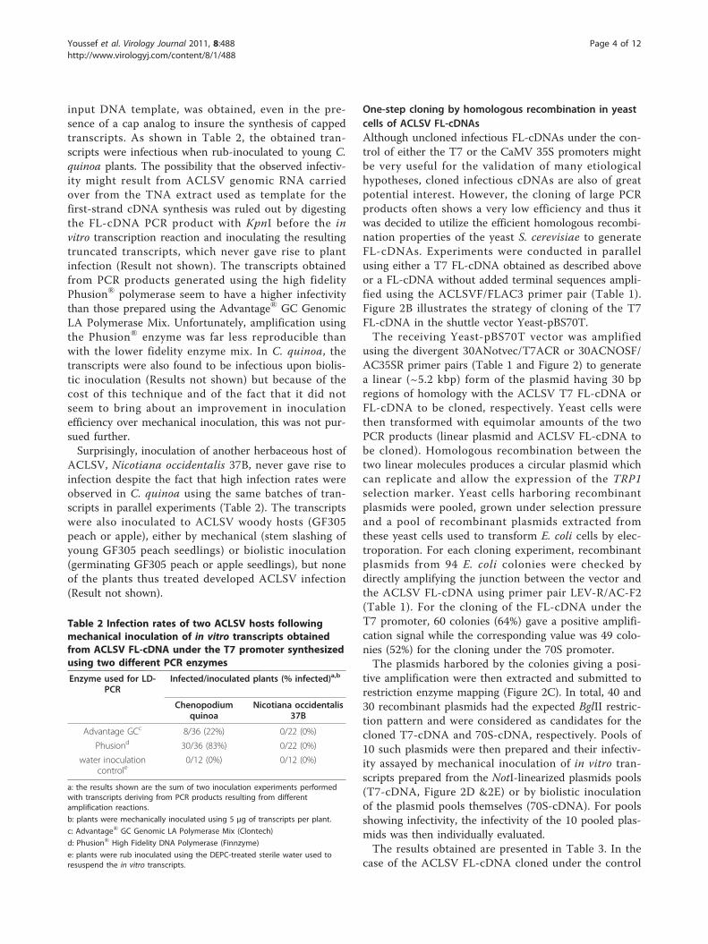
input DNA template, was obtained, even in the pre-sence of a cap analog to insure the synthesis of cappedtranscripts. As shown in Table 2, the obtained tran-scripts were infectious when rub-inoculated to young C.quinoa plants. The possibility that the observed infectiv-ity might result from ACLSV genomic RNA carriedover from the TNA extract used as template for thefirst-strand cDNA synthesis was ruled out by digestingthe FL-cDNA PCR product with KpnI before the invitro transcription reaction and inoculating the resultingtruncated transcripts, which never gave rise to plantinfection (Result not shown). The transcripts obtainedfrom PCR products generated using the high fidelityPhusion® polymerase seem to have a higher infectivitythan those prepared using the Advantage® GC GenomicLA Polymerase Mix. Unfortunately, amplification usingthe Phusion® enzyme was far less reproducible thanwith the lower fidelity enzyme mix. In C. quinoa, thetranscripts were also found to be infectious upon biolis-tic inoculation (Results not shown) but because of thecost of this technique and of the fact that it did notseem to bring about an improvement in inoculationefficiency over mechanical inoculation, this was not pur-sued further.Surprisingly, inoculation of another herbaceous host of
ACLSV, Nicotiana occidentalis 37B, never gave rise toinfection despite the fact that high infection rates wereobserved in C. quinoa using the same batches of tran-scripts in parallel experiments (Table 2). The transcriptswere also inoculated to ACLSV woody hosts (GF305peach or apple), either by mechanical (stem slashing ofyoung GF305 peach seedlings) or biolistic inoculation(germinating GF305 peach or apple seedlings), but noneof the plants thus treated developed ACLSV infection(Result not shown).
One-step cloning by homologous recombination in yeastcells of ACLSV FL-cDNAsAlthough uncloned infectious FL-cDNAs under the con-trol of either the T7 or the CaMV 35S promoters mightbe very useful for the validation of many etiologicalhypotheses, cloned infectious cDNAs are also of greatpotential interest. However, the cloning of large PCRproducts often shows a very low efficiency and thus itwas decided to utilize the efficient homologous recombi-nation properties of the yeast S. cerevisiae to generateFL-cDNAs. Experiments were conducted in parallelusing either a T7 FL-cDNA obtained as described aboveor a FL-cDNA without added terminal sequences ampli-fied using the ACLSVF/FLAC3 primer pair (Table 1).Figure 2B illustrates the strategy of cloning of the T7FL-cDNA in the shuttle vector Yeast-pBS70T.The receiving Yeast-pBS70T vector was amplified
using the divergent 30ANotvec/T7ACR or 30ACNOSF/AC35SR primer pairs (Table 1 and Figure 2) to generatea linear (~5.2 kbp) form of the plasmid having 30 bpregions of homology with the ACLSV T7 FL-cDNA orFL-cDNA to be cloned, respectively. Yeast cells werethen transformed with equimolar amounts of the twoPCR products (linear plasmid and ACLSV FL-cDNA tobe cloned). Homologous recombination between thetwo linear molecules produces a circular plasmid whichcan replicate and allow the expression of the TRP1selection marker. Yeast cells harboring recombinantplasmids were pooled, grown under selection pressureand a pool of recombinant plasmids extracted fromthese yeast cells used to transform E. coli cells by elec-troporation. For each cloning experiment, recombinantplasmids from 94 E. coli colonies were checked bydirectly amplifying the junction between the vector andthe ACLSV FL-cDNA using primer pair LEV-R/AC-F2(Table 1). For the cloning of the FL-cDNA under theT7 promoter, 60 colonies (64%) gave a positive amplifi-cation signal while the corresponding value was 49 colo-nies (52%) for the cloning under the 70S promoter.The plasmids harbored by the colonies giving a posi-
tive amplification were then extracted and submitted torestriction enzyme mapping (Figure 2C). In total, 40 and30 recombinant plasmids had the expected BglII restric-tion pattern and were considered as candidates for thecloned T7-cDNA and 70S-cDNA, respectively. Pools of10 such plasmids were then prepared and their infectiv-ity assayed by mechanical inoculation of in vitro tran-scripts prepared from the NotI-linearized plasmids pools(T7-cDNA, Figure 2D &2E) or by biolistic inoculationof the plasmid pools themselves (70S-cDNA). For poolsshowing infectivity, the infectivity of the 10 pooled plas-mids was then individually evaluated.The results obtained are presented in Table 3. In the
case of the ACLSV FL-cDNA cloned under the control
Table 2 Infection rates of two ACLSV hosts followingmechanical inoculation of in vitro transcripts obtainedfrom ACLSV FL-cDNA under the T7 promoter synthesizedusing two different PCR enzymes
Enzyme used for LD-PCR
Infected/inoculated plants (% infected)a,b
Chenopodiumquinoa
Nicotiana occidentalis37B
Advantage GCc 8/36 (22%) 0/22 (0%)
Phusiond 30/36 (83%) 0/22 (0%)
water inoculationcontrole
0/12 (0%) 0/12 (0%)
a: the results shown are the sum of two inoculation experiments performedwith transcripts deriving from PCR products resulting from differentamplification reactions.
b: plants were mechanically inoculated using 5 μg of transcripts per plant.
c: Advantage® GC Genomic LA Polymerase Mix (Clontech)
d: Phusion® High Fidelity DNA Polymerase (Finnzyme)
e: plants were rub inoculated using the DEPC-treated sterile water used toresuspend the in vitro transcripts.
Youssef et al. Virology Journal 2011, 8:488http://www.virologyj.com/content/8/1/488
Page 4 of 12

Yeast-pBS70T (5.2 kbp)
T7-ACLSV-FL (7.6 kbp)
Figure 2 One-step cloning by homologous recombination in yeast of an ACLSV FL-cDNA under the control of T7 promoter. (A) 0.8%non-denaturing agarose gel electrophoresis of the two PCR fragments used to transform yeast cells. Lane 1: PCR product corresponding to theYeast-PBS70T amplified using the divergent T7ACR/30Anotvec primer pair; Lane 2: ACLSV T7-FL-cDNA amplified using the FLAC5/FLAC3 primerpair; (M): Molecular mass marker (Invitrogen 1 kb ladder). (B) Schematic representation of the cloning by homologous recombination strategy.The 30 bp overlapping regions between the two PCR fragments in which homologous recombination takes place are indicated. (C) BglIIrestriction analysis (lanes 1-4) of four pools of 10 independent recombinant plasmids recovered following retransformation of E. coli cells usingthe plasmid preparation purified from the bulked transformant yeast cells, (M): Molecular mass markers (Invitrogen1 kb ladder). (D) Analysis bynon-denaturing 1% agarose gel electrophoresis of the same four pools of plasmids, following linearization with NotI (lanes 1-4), (M): Molecularmass markers (Invitrogen 1 kb ladder). (E) Analysis by 1% denaturing agarose gel electrophoresis of the in vitro RNA transcripts synthesized fromthe NotI-linearized plasmid pools, (M): 0.5-10 kb RNA ladder (Sigma).
Table 3 Infectivity on Chenopodium quinoa plants and number of infectious plasmids in plasmid pools obtained uponcloning by homologous recombination in yeast cells of ACLSV FL-cDNA under the control of the T7 promoter (T7-cDNA) or of the duplicated 35S promoter (70S-cDNA)
Plasmid pools Infected/inoculated C. quinoa plantsa Infectious plasmids per pool
T7-cDNA Pool 1 0/10 0
T7-cDNA Pool 2 0/26 0
T7-cDNA Pool 3 10/16 1
T7-cDNA Pool 4 0/26 0
70S-cDNA Pool 1 5/6 1
70S-cDNA Pool 2 4/6 1
70S-cDNA Pool 3 0/12 0
a: 3 μg of capped transcripts per C. quinoa plant were used for inoculation of transcripts from T7-cDNA plasmid pools while about 160 ng of plasmids were usedper plant for biolistic inoculation of 70S-cDNA pools (2 shots per plant).
Youssef et al. Virology Journal 2011, 8:488http://www.virologyj.com/content/8/1/488
Page 5 of 12

of the T7 promoter (T7-cDNA), a single pool (Pool 3)showed infectivity and deconvolution of that poolallowed the identification of a single plasmid fromwhich infectious in vitro transcripts could be produced.As expected from the experiments with the unclonedT7-cDNA products, no infectivity was observed uponinoculation of the transcripts derived from this recombi-nant plasmid to N. occidentalis 37B (Result not shown).In the case of the ACLSV FL-cDNA cloned under
the control of the 70S promoter (70S-cDNA), two ofthe 3 tested pools led to infection of the inoculatedplants (Pools 1 and 2) and, upon deconvolution, eachpool was shown to contain a single infectious plasmid(Table 3). For the two ACLSV FL-cDNA cloningattempts the rate of recovery of infectious clones istherefore of about 1-2%.
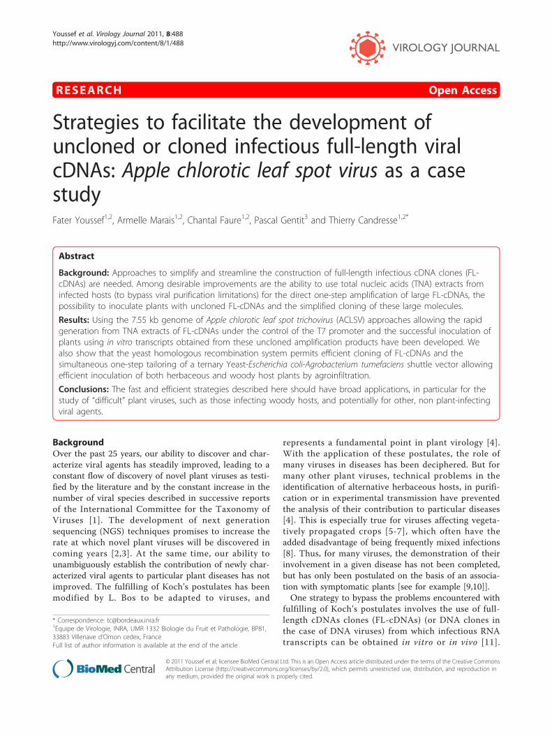
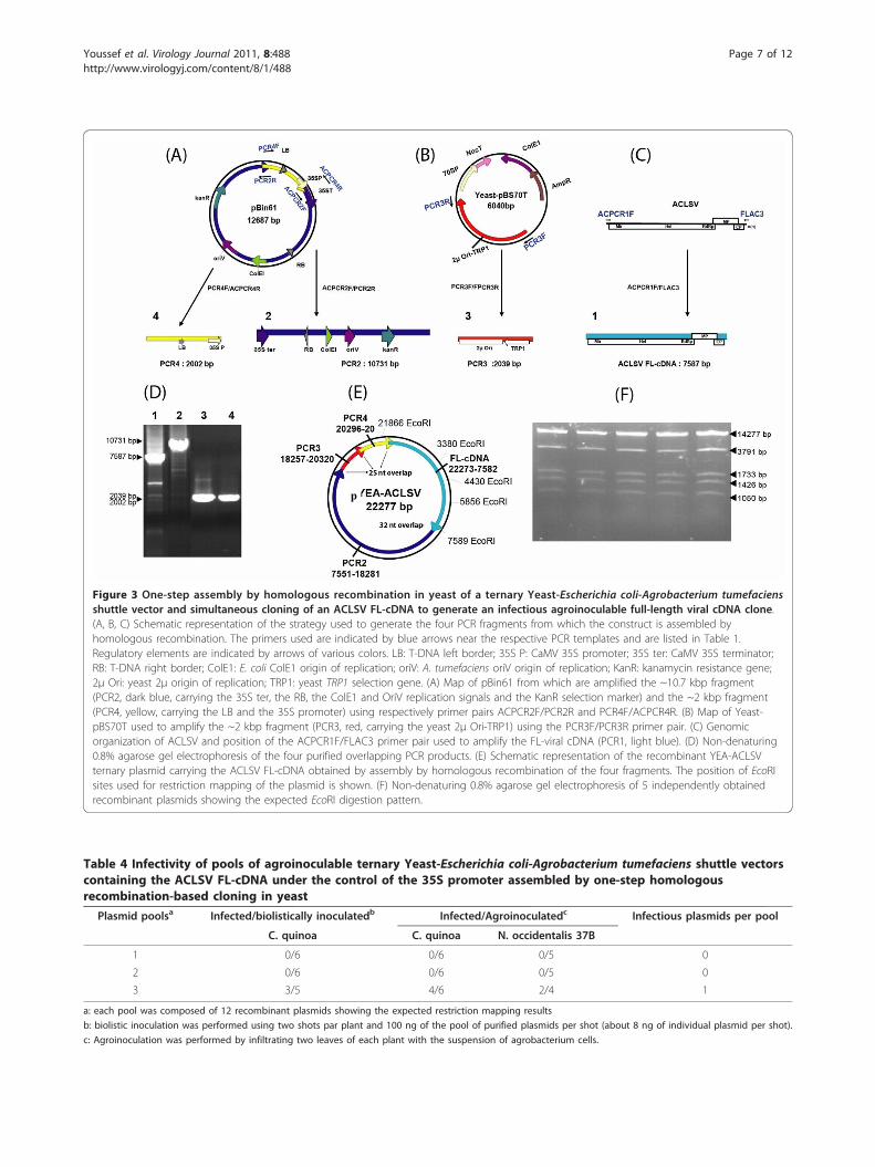
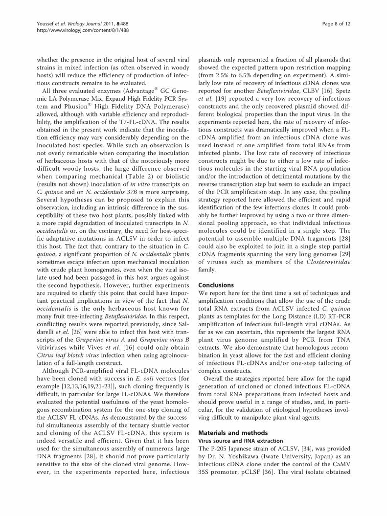
One-step cloning of ACLSV FL-cDNA and construction ofa ternary Yeast-E. coli-A. tumefaciens shuttle vector togenerate an agroinoculable ACLSV infectious cDNA cloneThe use of infectious FL-cDNA constructs that can beinoculated by agroinfection or by agroinfiltration oftenresults in a higher rate of infected plants or may allowthe successful inoculation of hosts that could not beinfected upon mechanical or biolistic inoculation [seefor example [16]]. We therefore decided to try to obtainan infectious agroinoculable ACLSV FL-cDNA con-struct. This was achieved in a single step of simulta-neously assembling a ternary Yeast-E. coli-A.tumefaciens shuttle vector and cloning the ACLSV FL-cDNA in this vector. For this purpose, four overlappingPCR fragments were assembled by homologous recom-bination in yeast (Figure 3). Following transfer to E. coliof the recombinant plasmids isolated from yeast cellsgrowing under selection, restriction mapping with EcoRIshowed that 36 of the 40 tested E. coli colonies con-tained a plasmid with the expected restriction pattern(Figure 3E and 3F). These 36 plasmids were pooled intothree pools of 12 plasmids and the infectivity of thesethree pools was evaluated. Given that the ACLSV FL-cDNA is cloned in the ternary vector under the controlof the 35S promoter, this construct is expected to beinfectious either as purified plasmid inoculated by biolis-tic bombardment or by agroinoculation of A. tumefa-ciens cells harboring it. Both inoculation strategies weretherefore evaluated for each pool of recombinant plas-mids: biolistic inoculation of C. quinoa plants or agroin-filtration of C. quinoa or N. occidentalis 37B plants. Theresults are presented in Table 4.Of the three pools tested, one gave successful inocula-
tion of C. quinoa plants by either of the inoculationtechniques used or of N. occidentalis 37B plants byagroinfiltration. Deconvolution of this pool allowed theidentification of a single infectious recombinant ternary
vector (pYEA-ACLSV). Biolistic bombardment of C. qui-noa with purified YEA-ACLSV resulted in a 60% infec-tion rate, which is to be compared with the 90%infection rate observed using plasmid pCLSF undersimilar inoculation conditions (2 shots per plant, 50 ngpurified plasmid per plant). Inoculation by agroinfiltra-tion with A. tumefaciens cells carrying pYEA-ACLSVresulted in infection rates of 91% and 67% for C. quinoa(a total of 33 inoculated plants in 3 experiments) or N.occidentalis 37B (a total of 18 inoculated plants in 3experiments), respectively.Inoculation by agroinfiltration of pYEA-ACLSV to
young GF305 peach seedlings demonstrated that thisconstruct was also able to generate infection in thiswoody host since 2 out of the 7 inoculated plants werelater shown to be infected (result not shown).As a control, a FL-cDNA was amplified directly from
pCLSF and similarly cloned in a ternary Yeast-E. coli-A.tumefaciens shuttle vector. Out of 64 analyzed recombi-nant plasmids, 50 had the expected EcoRI restrictionpattern and were divided into 5 pools of 10 plasmids.All pools proved infectious when inoculated either bybiolistic or by agroinfiltration, in either C. quinoa or N.occidentalis. Individual testing of 3 randomly selectedplasmids from each of those five pools revealed all ofthem to be infectious (Results not shown).
DiscussionLong Distance (LD) RT-PCR amplification had beenused successfully to generate infectious uncloned PCRproducts [24,26] but starting with purified viral RNApreparations obtained from purified viral particles. Theneed for such purified viral RNA preparations constitu-tes a severe limitation, in particular for viruses infectingwoody hosts and for which alternative herbaceous hostsand/or purification techniques may not be available.Although the results presented here were obtained withtotal extracts from C. quinoa, recently the same techni-que was used successfully to amplify the complete gen-ome of several isolates of Apricot latent virus (ApLV,9.2-9.3 kb) from total RNA extracts obtained frominfected GF305 peach seedlings, which indicates that theprotocol developed is not limited to herbaceous hostsextracts or to the 7.5 kb length of the ACLSV genome(F. Youssef et al., unpublished). On the other hand, itwas not possible to obtain similar results when usingviral double-stranded RNA (dsRNAs) preparations orimmunocaptured virions. Given the infectivity of thepYEA-ACLSV agroinoculable construct to GF305 peachseedlings, all the steps seem today at hand for goingfrom TNA extracts obtained from woody hosts to suc-cessful inoculation of such woody hosts using unclonedor cloned constructs. The efficiency of such a processremains, however, to be directly evaluated. Similarly,
Youssef et al. Virology Journal 2011, 8:488http://www.virologyj.com/content/8/1/488
Page 6 of 12
p
Figure 3 One-step assembly by homologous recombination in yeast of a ternary Yeast-Escherichia coli-Agrobacterium tumefaciensshuttle vector and simultaneous cloning of an ACLSV FL-cDNA to generate an infectious agroinoculable full-length viral cDNA clone.(A, B, C) Schematic representation of the strategy used to generate the four PCR fragments from which the construct is assembled byhomologous recombination. The primers used are indicated by blue arrows near the respective PCR templates and are listed in Table 1.Regulatory elements are indicated by arrows of various colors. LB: T-DNA left border; 35S P: CaMV 35S promoter; 35S ter: CaMV 35S terminator;RB: T-DNA right border; ColE1: E. coli ColE1 origin of replication; oriV: A. tumefaciens oriV origin of replication; KanR: kanamycin resistance gene;2μ Ori: yeast 2μ origin of replication; TRP1: yeast TRP1 selection gene. (A) Map of pBin61 from which are amplified the ~10.7 kbp fragment(PCR2, dark blue, carrying the 35S ter, the RB, the ColE1 and OriV replication signals and the KanR selection marker) and the ~2 kbp fragment(PCR4, yellow, carrying the LB and the 35S promoter) using respectively primer pairs ACPCR2F/PCR2R and PCR4F/ACPCR4R. (B) Map of Yeast-pBS70T used to amplify the ~2 kbp fragment (PCR3, red, carrying the yeast 2μ Ori-TRP1) using the PCR3F/PCR3R primer pair. (C) Genomicorganization of ACLSV and position of the ACPCR1F/FLAC3 primer pair used to amplify the FL-viral cDNA (PCR1, light blue). (D) Non-denaturing0.8% agarose gel electrophoresis of the four purified overlapping PCR products. (E) Schematic representation of the recombinant YEA-ACLSVternary plasmid carrying the ACLSV FL-cDNA obtained by assembly by homologous recombination of the four fragments. The position of EcoRIsites used for restriction mapping of the plasmid is shown. (F) Non-denaturing 0.8% agarose gel electrophoresis of 5 independently obtainedrecombinant plasmids showing the expected EcoRI digestion pattern.
Table 4 Infectivity of pools of agroinoculable ternary Yeast-Escherichia coli-Agrobacterium tumefaciens shuttle vectorscontaining the ACLSV FL-cDNA under the control of the 35S promoter assembled by one-step homologousrecombination-based cloning in yeast
Plasmid poolsa Infected/biolistically inoculatedb Infected/Agroinoculatedc Infectious plasmids per pool
C. quinoa C. quinoa N. occidentalis 37B
1 0/6 0/6 0/5 0
2 0/6 0/6 0/5 0
3 3/5 4/6 2/4 1
a: each pool was composed of 12 recombinant plasmids showing the expected restriction mapping results
b: biolistic inoculation was performed using two shots par plant and 100 ng of the pool of purified plasmids per shot (about 8 ng of individual plasmid per shot).
c: Agroinoculation was performed by infiltrating two leaves of each plant with the suspension of agrobacterium cells.
Youssef et al. Virology Journal 2011, 8:488http://www.virologyj.com/content/8/1/488
Page 7 of 12
whether the presence in the original host of several viralstrains in mixed infection (as often observed in woodyhosts) will reduce the efficiency of production of infec-tious constructs remains to be evaluated.All three evaluated enzymes (Advantage® GC Geno-
mic LA Polymerase Mix, Expand High Fidelity PCR Sys-tem and Phusion® High Fidelity DNA Polymerase)allowed, although with variable efficiency and reproduci-bility, the amplification of the T7-FL-cDNA. The resultsobtained in the present work indicate that the inocula-tion efficiency may vary considerably depending on theinoculated host species. While such an observation isnot overly remarkable when comparing the inoculationof herbaceous hosts with that of the notoriously moredifficult woody hosts, the large difference observedwhen comparing mechanical (Table 2) or biolistic(results not shown) inoculation of in vitro transcripts onC. quinoa and on N. occidentalis 37B is more surprising.Several hypotheses can be proposed to explain thisobservation, including an intrinsic difference in the sus-ceptibility of these two host plants, possibly linked witha more rapid degradation of inoculated transcripts in N.occidentalis or, on the contrary, the need for host-speci-fic adaptative mutations in ACLSV in order to infectthis host. The fact that, contrary to the situation in C.quinoa, a significant proportion of N. occidentalis plantssometimes escape infection upon mechanical inoculationwith crude plant homogenates, even when the viral iso-late used had been passaged in this host argues againstthe second hypothesis. However, further experimentsare required to clarify this point that could have impor-tant practical implications in view of the fact that N.occidentalis is the only herbaceous host known formany fruit tree-infecting Betaflexiviridae. In this respect,conflicting results were reported previously, since Sal-darelli et al. [26] were able to infect this host with tran-scripts of the Grapevine virus A and Grapevine virus Bvitiviruses while Vives et al. [16] could only obtainCitrus leaf blotch virus infection when using agroinocu-lation of a full-length construct.Although PCR-amplified viral FL-cDNA molecules
have been cloned with success in E. coli vectors [forexample [12,13,16,19,21-23]], such cloning frequently isdifficult, in particular for large FL-cDNAs. We thereforeevaluated the potential usefulness of the yeast homolo-gous recombination system for the one-step cloning ofthe ACLSV FL-cDNAs. As demonstrated by the success-ful simultaneous assembly of the ternary shuttle vectorand cloning of the ACLSV FL-cDNA, this system isindeed versatile and efficient. Given that it has beenused for the simultaneous assembly of numerous largeDNA fragments [28], it should not prove particularlysensitive to the size of the cloned viral genome. How-ever, in the experiments reported here, infectious
plasmids only represented a fraction of all plasmids thatshowed the expected pattern upon restriction mapping(from 2.5% to 6.5% depending on experiment). A simi-larly low rate of recovery of infectious cDNA clones wasreported for another Betaflexiviridae, CLBV [16]. Spetzet al. [19] reported a very low recovery of infectiousconstructs and the only recovered plasmid showed dif-ferent biological properties than the input virus. In theexperiments reported here, the rate of recovery of infec-tious constructs was dramatically improved when a FL-cDNA amplified from an infectious cDNA clone wasused instead of one amplified from total RNAs frominfected plants. The low rate of recovery of infectiousconstructs might be due to either a low rate of infec-tious molecules in the starting viral RNA populationand/or the introduction of detrimental mutations by thereverse transcription step but seem to exclude an impactof the PCR amplification step. In any case, the poolingstrategy reported here allowed the efficient and rapididentification of the few infectious clones. It could prob-ably be further improved by using a two or three dimen-sional pooling approach, so that individual infectiousmolecules could be identified in a single step. Thepotential to assemble multiple DNA fragments [28]could also be exploited to join in a single step partialcDNA fragments spanning the very long genomes [29]of viruses such as members of the Closteroviridaefamily.
ConclusionsWe report here for the first time a set of techniques andamplification conditions that allow the use of the crudetotal RNA extracts from ACLSV infected C. quinoaplants as templates for the Long Distance (LD) RT-PCRamplification of infectious full-length viral cDNAs. Asfar as we can ascertain, this represents the largest RNAplant virus genome amplified by PCR from TNAextracts. We also demonstrate that homologous recom-bination in yeast allows for the fast and efficient cloningof infectious FL-cDNAs and/or one-step tailoring ofcomplex constructs.Overall the strategies reported here allow for the rapid
generation of uncloned or cloned infectious FL-cDNAfrom total RNA preparations from infected hosts andshould prove useful in a range of studies, and, in parti-cular, for the validation of etiological hypotheses invol-ving difficult to manipulate plant viral agents.
Materials and methodsVirus source and RNA extractionThe P-205 Japanese strain of ACLSV, [34], was providedby Dr. N. Yoshikawa (Iwate University, Japan) as aninfectious cDNA clone under the control of the CaMV35S promoter, pCLSF [36]. The viral isolate obtained
Youssef et al. Virology Journal 2011, 8:488http://www.virologyj.com/content/8/1/488
Page 8 of 12
upon biolistic inoculation of pCLSF to C. quinoa waspropagated in this host by mechanical inoculation andused as source of virus or of viral nucleic acids. Totalnucleic acids were extracted with the SV Total RNAIsolation System (Promega, Lyon, France) from 30 mgof leaf tissue from symptomatic C. quinoa plants, fol-lowing the manufacturer’s instructions. The yield wasapproximately 15 μg of RNAs in 60 μl of sterile water.
Full-length ACLSV cDNA amplification using LongDistance (LD) RT-PCRSynthesis of the first-strand cDNA was primed with oligo-nucleotide FLAC3: 5’ T(30)GTAGTAAAATATT-TAAAAGTCTACAG 3’, which is complementary to the3’-terminal nucleotides (positions 7552-7527) of the geno-mic RNA of ACLSV-P205 (GenBank D14996). The synth-esis was performed using about 1 μg of TNA and thePrimeScript™ Reverse Transcriptase (Clontech/TaKaRaBio Europe, Saint-Germain en Laye, France) or the ExpandReverse Transcriptase (Roche Diagnostics, Meylan, France),following the manufacturer instructions. In order to obtaina FL-cDNA under the control of the T7 promoter, primerFLAC5 (5’ TAATACGACTCACTATAGTGATACTGA-TACAGTGTACACTCACG 3’) was used in combinationwith FLAC3 in a LD-PCR experiment. FLAC5 contains theT7 promoter (in bold and italic) and the first 26 5’ nucleo-tides of the genome of ACLSV-P205. Three commercialDNA polymerases or DNA polymerases mixes were com-pared for their efficiency in this study: the Advantage® GCGenomic LA Polymerase Mix (Clontech), the Expand HighFidelity PCR System (Roche) and the Phusion® High Fide-lity DNA Polymerase (Finnzyme/Fischer Scientific, Illkirch,France). All enzymes were used according to their supplierrecommendations, using as template 3 μl from the firststrand cDNA product either undiluted or diluted 10-fold.All PCRs were performed in a 25 μl final reaction volume.The PCR cycling parameters recommended by the suppli-ers were used, with the exception of the annealing tempera-ture which was fixed at 60°C for all experiments.Amplicons were purified using the MSB® Spin PCRapacekit (Invitek, les Ullis, France) and eluted in Diethylpyrocar-bonate (DEPC) treated, distilled water. They were thenquantified by electrophoresis on non-denaturing agarosegels and image analysis using the Image J 1.42q software(W. Rasband, NIH, USA). As a control in some experi-ments, LD-PCR amplification was performed directly onthe pCLSF infectious plasmid instead of on cDNAsobtained from TNA extracts.
T7 RNA polymerase in vitro transcription of ACLSV FL-cDNAsTwo hundred nanograms of purified ACLSV FL-cDNAamplicons were used in in vitro transcription reactionsperformed using the phage T7 RNA polymerase and the
mMESSAGE mMACHINE kit (Ambion, Courtaboeuf,France) in the presence of the cap analogue m7G(5’)ppp(5’)G (Ambion). At the end of the synthesis, which wasperformed as recommended by the kit supplier, tran-scripts were treated with 1 μl of TURBO DNase(Ambion) and then used either directly or after purifica-tion on Macro SpinColumn™G-50 (Harvard Apparatus,Les Ullis, France) following the manufacturer’s instruc-tions. Transcripts were quantified as described above forthe PCR products and, if necessary, were stored at -80°Cuntil use in plant inoculation assays.
Inoculation of herbaceous or woody ACLSV host plantsThe ACLSV FL-RNA transcripts were inoculated onyoung (four leaf stage) plants of C. quinoa or N. occi-dentalis cv. 37B by gently rubbing 5 μl of in vitro tran-scripts (adjusted to the desired concentration withDEPC-treated distilled water) on two celite-dustedleaves of each plant (Celite 545, 0,01-0,04 mm, Merck/VWR, Fontenay-sous-Bois, France). Following inocula-tion, leaves were rinsed briefly with distilled water. Ascontrol, plants were inoculated using DEPC-treated dis-tilled water without RNA transcripts. All plants weregrown (16 hours day/8 hours night) and observedweekly for symptoms development.Alternatively, the ACLSV FL-RNA transcripts were bio-
listically inoculated on leaves of young C. quinoa plantsusing the Helios Gene Gun (Bio-Rad/Marnes-La-Coquette,France). For inoculation, 5 μg of transcripts were used toprepare 10 cartridges as recommended by Bio-Rad, using 1μm gold particles. Each plant was bombarded on twoleaves, using a 200 psi pressure. For the inoculation ofwoody host plants, young germinating peach (Prunus persi-cae, cv. GF305) or apple (Malus domestica) seedlings wereinoculated when the growing radicle had reached a lengthof about 0.5-1 cm. Briefly, the envelopes of germinatingseeds were gently removed and the cotyledons were theninoculated by biolistic bombardment (1 shot on each of thecotyledons for the GF305 and 3 shots for groups of 10 ger-minating seeds for M. domestica. After inoculation, seedswere rinsed with distilled water and then placed on wet tis-sue paper in Petri dishes at 4°C in the dark for 24 hours.The next day, they were treated by a broad spectrum fungi-cide by soaking for 30 min in a 0.5% solution of Propamo-carb-HCl followed by rinsing with water. The germinatingembryos were finally transplanted to sterile moist sand andleft to develop in the greenhouse up to the 4-leaf stagebefore being transplanted into pots containing potting soil.As control, plants were biolistically inoculated using theinfectious pCLSF plasmid.
ACLSV detection in inoculated plantsACLSV was detected as described [37], using double-antibody sandwich enzyme-linked immunosorbent assay
Youssef et al. Virology Journal 2011, 8:488http://www.virologyj.com/content/8/1/488
Page 9 of 12

(DAS-ELISA) and immunoglobulins from the P2 polyclo-nal antiserum raised against the P863 ACLSV isolate(INRA, Bordeaux, France). Alternatively, ACLSV infectionwas detected using the A52-A53 ACLSV-specific RT-PCRassay described [38]. Identity of the amplified virus wasconfirmed by direct sequencing of the amplification pro-ducts (Beckman Coulter Genomics, Meylan, France).
One step cloning of ACLSV FL-cDNAs by homologousrecombination in Saccharomyces cerevisiaeThe yeast (S. cerevisiae) strain YPH501 (MATa/MATaura3-52 lys2-801amber ade2-101ochre trp1-Δ63 his3-Δ200 leu2-Δ1) was used [39]. The yeast-E. coli vectorused in these experiments is a pBS70T plasmid [40] inwhich a DNA fragment containing a yeast 2μ origin ofreplication and the yeast TRP1 gene as a selection markerhas been inserted (Yeast-pBS70T). The pBS70T back-bone provides an E. coli ColE1 origin of replication andan ampicillin resistance gene for selection in E. coli, inaddition to signals for transcription in planta: duplicatedCaMV 35S promoter (70SP) and Nopaline synthase geneterminator (NosT) of the cloned FL-cDNA. The com-plete vector was amplified using the Phusion® High Fide-lity DNA Polymerase (Finnzyme) and either one of thedivergent primer pairs shown in Table 1. This strategyproduces PCR products that represent a linear copy ofthe vector with terminal regions of 30 nt overlappingwith the ends of the FL-cDNA PCR product to be cloned.All PCR products were purified using the MSB® SpinPCRapace kit before use.Yeast cultures were grown at 30°C in YPD medium (1%
yeast extract, 2% peptone, 2% glucose) prior to transfor-mation and in SD (Minimal synthetic defined Base) selec-tive medium with the mixture of the amino acid withouttryptophan (-Try DO, Dropout) after transformation.Yeast was transformed using a lithium acetate method anddenatured carrier DNA as described [41], following themodifications of [30]. Approximately 1-2 μg in total of themixture of the PCR fragments in equimolar amounts wereused per transformation. All yeast colonies growing on theselection plates following transformation were collected in3 ml of SD liquid medium and grown for 12 h at 30°C.Plasmid DNA was isolated from these cultures accordingto [42] and used for transformation of E. coli cells (XL1)by electroporation. Further characterization of the recom-binant plasmids and their large-scale purification were car-ried out using standard protocols [43]. All junctions in therecombinant plasmids obtained were confirmed bysequencing (Beckman Coulter Genomics).
One step construction of an agroinoculable ACLSV FL-cDNA in a ternary yeast-E. coli-A. tumefaciens vectorACLSV FL-cDNA was cloned in a de novo constructedternary yeast-E. coli-A. tumefaciens vector by
homologous recombination in yeast cells. This wasachieved by simultaneous transformation of yeast cellsusing 4 PCR products having 25-32 bp overlapping endsderived from the PCR primers (Figure 3 and Table 1).Besides the ACLSV FL-cDNA, the three fragments usedwere: (1) a 10.7 kbp product covering the 35S termina-tor, the T-DNA right border (RB), the E. coli ColE1 ori-gin of replication, the A. tumefaciens OriV origin ofreplication and finally the kanamycin resistance genefrom the pBin61 binary vector [44], (2) a ~2 kbp frag-ment containing the yeast 2μ origin of replication andthe yeast TRP1 gene derived from the Yeast-pBS70Tvector described above and (3) a ~2 kbp fragment con-taining the T-DNA left border (LB) and the CaMV 35Spromoter of pBin61. The long fragment was amplifiedusing the Advantage® GC Genomic LA Polymerase Mixkit while the two shorter ones were amplified with thePhusion® High Fidelity DNA Polymerase. After purifica-tion of all fragments using the MSB® Spin PCRapacekit, yeast cells were transformed with a total of about 3μg of the four fragments in equimolecular amounts. Thevarious steps following transformation were carried outas described above and the recombinant plasmids har-bored by E. coli colonies finally verified by restrictionanalysis. Plasmids showing the expected EcoRI restric-tion pattern were divided into pools and used to trans-form by electroporation (with ~100 ng of purifiedplasmid pools) A. tumefaciens C58C1 cells carrying thepG90 helper plasmid (provided by S. Vernhettes, INRA-Versailles). Agrobacterium transformants were selectedon LB medium plates supplemented with 50 mg/l rifam-picin, 50 mg/l kanamycin and 20 mg/l gentamycin. Alltransformed bacteria growing on the Petri dishes werecollected and grown as pools in LB liquid medium withthe same antibiotics and this culture was then used asthe starter culture to prepare Agrobacterium cells foragroinfiltration.
Inoculation of plants using agroinfiltrationFor agroinfiltration of plants, A. tumefaciens cells carry-ing the relevant plasmid(s) were first grown overnight at28°C in 5 ml of LB medium plus selection antibiotics(see above). These pre-cultures were then used to inocu-late 25 ml cultures of induction medium [LB mediumsupplemented with selection antibiotics, 10 mM of 2N-morpholino-ethane sulfonic acid (MES) and 20 μM ofacetosyringone]. Following incubation overnight at 28°Cunder agitation, bacteria were collected by centrifugationat 6000 g for 15 min at room temperature, re-suspendedin infiltration medium (10 mM MgCl2, 10 mM MES, pH5.6, and 150 μM acetosyringone) and the bacterial sus-pension adjusted to an optical density of 0.6 at 600 nm.The suspension was then incubated for 3 h at roomtemperature before being infiltrated in the intercellular
Youssef et al. Virology Journal 2011, 8:488http://www.virologyj.com/content/8/1/488
Page 10 of 12

spaces of young C. quinoa or N. occidentalis leaves,using a syringe directly placed on the lower leaf surface.Alternatively, young GF305 peach seedlings were inocu-lated by injections of the Agrobacterium cells suspen-sion in their stems using a syringe and a small gaugeneedle. Following their inoculation, plants were moni-tored weekly for symptoms appearance and ACLSVinfection was confirmed as described above.
List of abbreviations70SP: duplicated CaMV 35S promoter; ACLSV: Apple chlorotic leaf spot virus;ApLV: Apricot latent virus; CLBV: Citrus leaf blotch virus; DAS-ELISA: double-antibody sandwich enzyme-linked immunosorbent assay; DEPC:diethylpyrocarbonate; dsRNA: double stranded RNA; FL-cDNA: full-lengthinfectious cDNA clone; LB: T-DNA left border; LD-PCR: long distance PCR;MES: 2N-morpholino-ethane sulfonic acid; NGS: next generation sequencing;NosT: Nopaline synthase gene terminator; RB: T-DNA right border; SD:minimal synthetic defined base; TNA: total nucleic acids; YAC: yeast artificialchromosome
AcknowledgementsFY was supported by a fellowship from the Syrian Ministry of HigherEducation. The Apple chlorotic leaf spot virus infectious cDNA clone pCLSFwas a generous gift of Dr. N. Yoshikawa (Iwate University, Japan), who alsoshared with us prior to publication his protocol for the biolistic inoculationof apple seedlings. The Agrobacterium C58C1 strain carrying the pG90 helperplasmid was provided by Dr. S. Vernhettes (INRA Versailles, France) and theYPH501 yeast strain by Dr Benoit Moury (INRA Avignon, France). We alsowish to thank M. Roncoroni, T. Mauduit, and A. Bailly for taking excellentcare of all the plants used in this work, Dr. Emmanuel Jacquot (UMR Bio3P,Rennes, France) for valuable discussions and suggestions and Anas Abdul-Razzak (UMR GDPP, Bordeaux, France) for sharing with us his skills oncloning by homologous recombination in yeast.
Author details1Equipe de Virologie, INRA, UMR 1332 Biologie du Fruit et Pathologie, BP81,33883 Villenave d’Ornon cedex, France. 2Equipe de Virologie, Université deBordeaux, UMR 1332 Biologie du Fruit et Pathologie, BP81, 33883 Villenaved’Ornon cedex, France. 3Laboratoire de Virologie, Ctifl, Centre de Lanxade,24130 La Force, France.
Authors’ contributionsAll authors contributed to the results presented. FY, AM, PG and TCcontributed to and edited the manuscript. All authors read and approvedthe final manuscript.
Competing interestsThe authors declare that they have no competing interests.
Received: 8 July 2011 Accepted: 31 October 2011Published: 31 October 2011
References1. Fauquet CM, Mayo MA, Maniloff J, Desselberger U, Ball LA: Virus Taxonomy:
Eighth Report of the International Committee of Taxonomy of Viruses London:Elsevier; 2005.
2. Al Rwahnih M, Daubert S, Golino D, Rowhani A: Deep sequencing analysisof RNAs from a grapevine showing Syrah decline symptoms reveals amultiple virus infection that includes a novel virus. Virology 2009,387:395-401.
3. Kreuze JF, Perez A, Untiveros M, Quispe D, Fuentes S, Barker I, Simon R:Complete viral genome sequence and discovery of novel viruses bydeep sequencing of small RNAs: A generic method for diagnosis,discovery and sequencing of viruses. Virology 2009, 388:1-7.
4. Matthews REF: Diagnosis of Plant Virus Disease CRC Press; 1993.
5. Choi YG, Croft BJ, Randles JW: Identification of sugarcane striate mosaic-associated virus by partial characterization of its double-stranded RNA.Phytopathology 1999, 89:877-883.
6. Jelkmann W, Fechtner B, Agranovsky AA: Complete genome structure andphylogenetic analysis of little cherry virus, a mealybug-transmissibleclosterovirus. J Gen Virol 1997, 78:2067-2071.
7. Meng B, Meng B, Pang SZ, Forsline PL, McFerson JR, Gonsalves D:Nucleotide sequence and genome structure of Grapevine rupestris stempitting associated virus-1 reveal similarities to apple stem pitting virus. JGen Virol 1998, 79:2059-2069.
8. Hull R: Matthews’ Plant Virology New York: Academic Press; 2002.9. Jones A, McGavin W: Improved PCR detection of Blackcurrant reversion
virus in Ribes and further evidence that it is the causal agent ofreversion disease. Plant Dis 2002, 86:1333-1338.
10. Meng B, Zhu HY, Gonsalves D: Rupestris stem pitting associated virus-1consists of a family of sequence variants. Arch Virol 1999, 144:2071-2085.
11. Boyer JC, Haenni AL: Infectious transcripts and cDNA clones of RNAviruses. Virology 1994, 198:415-426.
12. Crutzen F, Mehrvar M, Gilmer D, Bragard C: A full-length infectious cloneof Beet soil-borne virus indicates the dispensability of the RNA-2 for virussurvival in planta and symptom expression on Chenopodium quinoaleaves. J Gen Virol 2009, 90:3051-3056.
13. Dizadji A, Koohi-Habibi M, Izadpanah K, Dietrich C, Mossahebi GH, Winter S:Characterization of Lettuce virus X, a new potexvirus infecting lettuce inIran. Arch Virol 2008, 153:1867-1875.
14. Lamprecht S, Jelkmann W: Infectious cDNA clone used to identifyStrawberry mild yellow edge-associated potexvirus as causal agent ofthe disease. J Gen Virol 1997, 78:2347-2353.
15. Satyanarayana T, Bar-Joseph M, Mawassi M, Albiach-Marti MR, Ayllon MA,Gowda S, Hilf ME, Moreno P, Garnsey SM, Dawson WO: Amplification ofCitrus tristeza virus from a cDNA clone and infection of citrus trees.Virology 2001, 280:87-96.
16. Vives MC, Martín S, Ambrós S, Renovell A, Navarro L, Pina JA, Moreno P,Guerri J: Development of a full-genome cDNA clone of Citrus leaf blotchvirus and infection of citrus plants. Mol Plant Pathol 2008, 9:787-797.
17. Guenoune-Gelbart D, Sufrin-Ringwald T, Capobianco H, Gaba V, Polston JE,Lapidot M: Inoculation of plants with begomoviruses by particlebombardment without cloning: Using rolling circle amplification of totalDNA from infected plants and whiteflies. J Virol Methods 2010, 168:87-93.
18. Wege C, Gotthardt RD, Frischmuth T, Jeske H: Fulfilling Koch’s postulatesfor Abutilon mosaic virus. Arch Virol 2000, 145:2217-2225.
19. Spetz C, Moe R, Blystad DR: Symptomless infectious cDNA clone of aNorwegian isolate of Poinsettia mosaic virus. Arch Virol 2008,153:1347-1351.
20. Barnes WM: PCR amplification of up to 35-kb DNA with high fidelity andhigh yield from lambda bacteriophage templates. Proc Natl Acad Sci USA1994, 91:2216-2220.
21. Hasiów-Jaroszewska B, Borodynko N, Popieszny H: Infectious RNAtranscript derived from cloned cDNA of a Pepino mosaic virus isolate.Arch Virol 2009, 154:853-856.
22. Song YS, Min BE, Hong JS, Rhie MJ, Kim MJ, Ryu KH: Molecular evidencesupporting the confirmation of Maracuja mosaic virus as a species ofthe genus Tobamovirus and production of an infectious transcript. ArchVirol 2006, 151:2337-2348.
23. You Y, Shirako Y: Bymovirus reverse genetics: requirement for RNA2-encoded proteins in systemic infection. Mol Plant Pathol 2010, 11:383-394.
24. Choi SH, Ryu KH: Generation of infectious transcripts from Korean strainand mild mottle strain of Potato virus X. J Microbiol 2008, 46:502-507.
25. Fakhfakh H, Vilaine F, Makni M, Robaglia C: Cell-free cloning and biolisticinoculation of an infectious cDNA of Potato virus Y. J Gen Virol 1996,77:519-523.
26. Saldarelli P, Dell’Orco M, Minafra A: Infectious cDNA clones of twograpevine viruses. Arch Virol 2000, 145:397-405.
27. Marykwas DL, Passmore SE: Mapping by multifragment cloning in vivo.Proc Natl Acad Sci USA 1995, 92:11701-1175.
28. Gibson DG, Benders GA, Axelrod KC, Zaveri J, Algire MA, Moodie M,Montague MG, Venter JC, Smith HO, Hutchison CA: One-step assembly inyeast of 25 overlapping DNA fragments to form a complete syntheticMycoplasma genitalium genome. Proc Natl Acad Sci USA 2008,105:20404-2049.
Youssef et al. Virology Journal 2011, 8:488http://www.virologyj.com/content/8/1/488
Page 11 of 12
29. Polo S, Ketner G, Levis R, Falgout B: Infectious RNA transcripts from full-length Dengue virus type 2 cDNA clones made in yeast. J Virol 1997,71:5366-5374.
30. Fernandez-Delmond I, Pierrugues O, de Wispelaere M, Guilbaud L,Gaubert S, Diveki Z, Godon C, Tepfer M, Jacquemond M: A novel strategyfor creating recombinant infectious RNA virus genomes. J Virol Methods2004, 121:247-257.
31. Liang D, Gray SM, Kaplan I, Palukaitis P: Site-directed mutagenesis andgeneration of chimeric viruses by homologous recombination in yeastto facilitate analysis of plant-virus interactions. Mol Plant Microbe Interact2004, 17:571-576.
32. Martelli GP, Candresse T, Namba S: Trichovirus, a new genus of plantviruses. Arch Virol 1994, 134:451-455.
33. Yoshikawa N: Apple chlorotic leaf spot virus. CMI AAB Description of PlantViruses, No. 386 (No.30 revised) 2001.
34. German S, Candresse T, Lanneau M, Huet JC, Pernollet JC, Dunez J:Nucleotide sequence and genomic organization of Apple chlorotic leafspot closterovirus. Virology 1990, 179:104-112.
35. Sato K, Yoshikawa N, Takahashi T: Complete nucleotide sequence of thegenome of an Apple isolate of apple chlorotic leaf spot virus. J Gen Virol1993, 74:1927-1931.
36. Satoh H, Yoshikawa N, Takahashi T: Construction and biolistic inoculationof an infectious cDNA clone of Apple chlorotic leaf spot Trichovirus. AnnPhytopathol Soc Jpn 1999, 65:301-304.
37. Foissac X, Svanella-Dumas L, Gentit P, Dulucq MJ, Marais A, Candresse T:Polyvalent degenerate oligonucleotides reverse transcription-polymerasechain reaction: a polyvalent detection and characterization tool fortrichoviruses, capilloviruses, and foveaviruses. Phytopathology 2005,95:617-625.
38. Candresse T, Lanneau M, Revers F, Grasseau N, Macquaire G, German S,Malinowsky T, Dunez J: An immunocapture PCR assay adapted to thedetection and the analysis of the molecular variability of the Applechlorotic leafspot virus. Acta Hortic 1995, 386:136-147.
39. Sikorski RS, Hieter P: A system of shuttle vectors and yeast host strainsdesigned for efficient manipulation of DNA in Saccharomyces cerevisiae.Genetics 1989, 122:19-27.
40. Yang SJ, Revers F, Souche S, Lot H, Le Gall O, Candresse T, Dunez J:Construction of full-length cDNA clones of Lettuce mosaic virus (LMV)and the effects of intron-insertion on their viability in Escherichia coliand on their infectivity to plants. Arch Virol 1998, 143:2443-2451.
41. Gietz RD, Schiestl RH: Applications of high efficiency lithium acetatetransformation of intact yeast cells using single-stranded nucleic acidsas carrier. Yeast 1991, 7:253-263.
42. Hofmann CS, Winston F: A ten-minute DNA preparation from yeastefficiently releases autonomous plasmid for transformation ofEscherichia coli. Gene 1987, 57:267-272.
43. Sambrook J, Fritsch EF, Maniatis T: Molecular Cloning - A LaboratoryManual. Cold Spring Harbor, New York: Cold Spring Harbor Press;, 2 1989.
44. Bendahmane A, Kanyuka K, Baulcombe DC: The Rx gene from potatocontrols separate virus resistance and cell death responses. Plant Cell1999, 11:781-92.
doi:10.1186/1743-422X-8-488Cite this article as: Youssef et al.: Strategies to facilitate thedevelopment of uncloned or cloned infectious full-length viral cDNAs:Apple chlorotic leaf spot virus as a case study. Virology Journal 2011 8:488.
Submit your next manuscript to BioMed Centraland take full advantage of:
• Convenient online submission
• Thorough peer review
• No space constraints or color figure charges
• Immediate publication on acceptance
• Inclusion in PubMed, CAS, Scopus and Google Scholar
• Research which is freely available for redistribution
Submit your manuscript at www.biomedcentral.com/submit
Youssef et al. Virology Journal 2011, 8:488http://www.virologyj.com/content/8/1/488
Page 12 of 12