
Field Sampling Principles and Practices in Environmental Analysis

Nervous System Sampling Practices, p. 1
1 2
3 STP Position Paper: 4
Recommended Practices for Sampling, Processing and Analysis of the 5
Peripheral Nervous System (Nerves, Somatic and Autonomic Ganglia) 6
during Nonclinical Toxicity Studies1 7
8 9 10 11 12
STP Working Group on Peripheral Nervous System Sampling: 13
14 Brad Bolon (chair), Georg Krinke, Mark T. Butt, Deepa B. Rao, Ingrid D. Pardo, 15
Bernard S. Jortner, Karl Jensen, Robert H. Garman, Lydia Andrews-Jones, James P. 16
Morrison, Alok K. Sharma, Michael S. Thibodeau (co-chair) 17
18 19 20 21
GEMpath, Inc., Longmont, CO, USA (BB); AnaPath GmbH, Oberbuchsiten, Switzerland 22
(GK); Tox Path Specialists, LLC, Frederick, MD, USA (MB); U.S. Food and Drug 23
Administration, Center for Drug Evaluation and Research, Silver Spring, MD, USA 24
(DBR); Pfizer, Groton, CT, USA (IDP); Virginia Polytechnic Institute and State 25
University, Blacksburg, VA (BSJ); U.S. Environmental Protection Agency, Research 26
Triangle Park, NC, USA (KJ); Consultants in Veterinary Pathology, Inc., Murrysville, PA, 27
USA (RHG); Allergan, Inc., Irvine, CA (LAJ); Charles River Laboratories, Inc, 28
Shrewsbury, MA, USA (JM); Covance Laboratories, Inc., Madison, WI, USA (AKS); 29
Boehringer Ingelheim Pharmaceuticals, Inc., Ridgefield, CT, USA (MT) 30
31 32 33 34 Running title: PNS Sampling / Processing in Nonclinical Toxicity Studies 35 36 37

Nervous System Sampling Practices, p. 2
38 39 Address correspondence to: Dr. Brad Bolon, GEMpath, Inc., 1100 East 17th Avenue, Unit 40
M202, Longmont, CO 80504-3715, United States. Phone: (720) 684-6540; Email: 41
43
44
45 1 This document has been reviewed in accordance with policies of the U.S. Environmental 46
Protection Agency (U.S. EPA) and U.S. Food and Drug Administration (U.S. FDA) and 47
approved for publication. The opinions expressed in this paper solely represent those of the 48
authors and should not be construed as representing the official views or policies of the U.S. 49
EPA or U.S. FDA, nor should mention of trade names or commercial products constitute 50
U.S. EPA or U.S. FDA endorsement or recommendation for use. 51
52
53
54

Nervous System Sampling Practices, p. 3
Abstract (232 words) 55
These Society of Toxicologic Pathology “best” practice recommendations should ensure 56
consistent sampling, processing, and evaluation of the peripheral nervous system (PNS). For 57
toxicity studies where neurotoxicity is not anticipated (Situation 1), PNS evaluation may be 58
limited to one sensorimotor spinal nerve. If somatic PNS neurotoxicity is possible (Situation 59
2), analysis minimally should include three spinal nerves, cranial nerve V, and their sensory 60
ganglia. If autonomic PNS neuropathy is suspected (Situation 3), parasympathetic and 61
sympathetic ganglia with associated autonomic nerves should be assessed. For dedicated 62
neurotoxicity studies where neurotoxic activity is likely (Situation 4), PNS sampling follows 63
the strategy for Situations 2 and/or 3, as dictated by in-life data or other information for the 64
compound/target. For all situations, bilateral sampling with unilateral processing is 65
recommended. For Situations 1, 2, and 3, PNS is processed conventionally (immersion in 66
formalin, paraffin embedding, H&E staining). For Situation 4 (and if feasible Situations 2 67
and 3), perfusion fixation with methanol-free fixative (MFF) is recommended. Where PNS 68
neurotoxicity is possible, at least one (Situations 2 and 3) or two (Situation 4) nerve cross 69
sections should be post-fixed with glutaraldehyde and osmium before hard plastic resin 70
embedding; soft plastic embedding is not suitable. Special methods (axonal and myelin 71
stains, etc.) may be used to further characterize PNS findings. Initial PNS analysis should be 72
informed, not masked (“blinded”). Institutions should explain the basis for their sampling, 73
processing, and evaluation strategy. 74
75
76
Key Words: PNS, peripheral nervous system, neuropathology, neurotoxicity, 77
recommended practices, nerve, ganglia, autonomic 78

Nervous System Sampling Practices, p. 4
Abbreviations 79
CNS central nervous system 80
DRG dorsal root ganglion 81
EPA (U.S.) Environmental Protection Agency 82
FDA (U.S.) Food and Drug Administration 83
GFAP glial fibrillary acidic protein 84
GLP Good Laboratory Practices 85
GMA glycol methacrylate 86
H&E hematoxylin and eosin 87
Iba1 ionized calcium-binding adaptor molecule 1 88
IENFD intra-epidermal nerve fiber density 89
IHC immunohistochemistry 90
MFF methanol-free formaldehyde (or fixative) 91
MGG medical-grade glutaraldehyde 92
MIE molecular-initiating event 93
MMA methyl methacrylate 94
MOA mode of action 95
NBF neutral buffered 10% formalin 96
NME new molecular entity 97
NOAEL no observed adverse effect level 98
OECD Organisation of Economic Co-operation and Development 99
PNS peripheral nervous system 100
PPD paraphenylenediamine 101
QSAR quantitative structure/activity relationship 102
RT room temperature 103
SOP standard operating procedure 104
STP Society of Toxicologic Pathology 105
TEM transmission electron microscopy 106
WOE weight of evidence 107
108
109

Nervous System Sampling Practices, p. 5
110
I. Background 111
Neurological deficits due to toxicant-induced peripheral neuropathy are a recognized 112
consequence of accidental occupational or environmental exposures and some therapeutic 113
treatments. Therefore, the neuropathology component of toxicity studies is a critical means 114
for identifying potential hazards and assessing risks posed to humans by contact with new 115
biomolecular or chemical entities. 116
Different regulatory agencies offer independent guidance1 based on their distinct 117
mandates, variable scientific levels of concern, and diverse uses of the agents they oversee 118
regarding the specimens and procedures to be used in evaluating the integrity of the 119
peripheral nervous system (PNS) when seeking to register new products (Bolon et al., 2011, 120
Salvo and Butt, 2011). The guidelines vary by the kind of industry (agrochemical vs. 121
chemical vs. pharmaceutical vs. biopharmaceutical), differences in potential exposure levels, 122
and ages of the test subjects (e.g., developing animals (EPA, 1998b, OECD, 2007) vs. adults 123
(EPA, 1998a, OECD, 1997)). Guidelines also differ based on the aim of the study (hazard 124
identification vs. safety assessment). For example, regulatory guidelines for performing the 125
neuropathology analysis of Good Laboratory Practice (GLP)-type general toxicity studies 126
(i.e., screening or “Tier I” surveys) are fairly general since such studies assess the PNS as 127
just one system among many organs and systems to be surveyed, while guidelines for GLP-128
type dedicated neurotoxicity studies (i.e., advanced or “Tier II” studies) are fairly detailed 129
since assessment of the nervous system is the primary focus of the study (Bolon et al., 2011, 130
1 Guidance or guideline documents provided by regulatory agencies communicate current agency thinking on
topics governed by regulations. Guidances and guidelines represent legally unenforceable interpretations that are designed to help institutions achieve compliance with legally enforceable regulations.

Nervous System Sampling Practices, p. 6
Salvo and Butt, 2011). However, substantial differences exist in the kinds of PNS toxicity 131
that might be encountered (Table 1), and current guidelines do not address variations in 132
approach that might be required to adequately investigate these divergent scenarios. Recent 133
compilations reviewing published regulatory guidance in this area (Bolon et al., 2011, Salvo 134
and Butt, 2011) and/or individual regulatory guidelines should be consulted because 135
guidance is reviewed and revised over time—as is presently occurring for the Toxic 136
Substances Control Act (administered by the U.S. Environmental Protection Agency [EPA]) 137
and the “Redbook” guidance on food and color additives (overseen by the U.S. Food and 138
Drug Administration [FDA]). 139
When sampling the PNS, considerable care must be given to selecting the appropriate 140
methodology (sampling scheme, fixatives, tissue orientation, embedding media, special 141
stains, etc.) to ensure that tissue morphology is optimally preserved. Basic PNS sampling 142
and processing methods were promulgated recently by a Working Group of the Society of 143
Toxicologic Pathology (STP) tasked with establishing “best practice” recommendations for 144
sampling and processing the central nervous system (CNS) for nonclinical general toxicity 145
studies (Bolon et al., 2013b). Given the CNS focus, however, coverage of the PNS in this 146
STP document was brief, and did not specifically include recommendations encompassing 147
different divisions of the PNS—somatic (sensorimotor) vs. autonomic (parasympathetic and 148
sympathetic)—or effectors controlled by the PNS (e.g., glands, skeletal muscle, or viscera). 149
Accordingly, the STP established a new Working Group on PNS sampling, processing, and 150
analysis to provide more specific recommendations appropriate to distinct varieties of 151
neuropathies that might be encountered during the course of GLP-type toxicity studies. 152

Nervous System Sampling Practices, p. 7
The Working Group was given a charter with multiple specific aims. The first charge 153
was to recommend what PNS structures should be regularly sampled during GLP-type 154
toxicity studies (“Tier I” and “Tier II”) performed in common vertebrate test species. The 155
second charge was to suggest tissue processing procedures and trimming schemes to 156
facilitate analysis of these regions. The third charge was to define what routine stains and 157
special neurohistology procedures, if any, should be used routinely in PNS evaluations. The 158
fourth charge was to consider when other special morphological techniques should be 159
undertaken to provide a more complete assessment of PNS lesions. The fifth charge was to 160
define appropriate means for assessing whether or not PNS recovery has taken place. The 161
sixth charge was to propose what format should be used to most efficiently document 162
histopathologic evaluation of PNS tissues in reports destined for review by regulatory bodies. 163
The recommendations given below with respect to particular neural structures to collect 164
(Table 2) and suggested sampling and processing procedures (Table 3), as well as the means 165
for documenting that they have been assessed, are based on the collective experiences and 166
opinions of the Working Group members2 as well as selected input from the global 167
toxicologic pathology community3 received during a __-day-long public comment period in 168
the ____ quarter of 2017. Where consensus among Working Group members and/or STP 169
2The Working Group consisted of 12 individuals with formal academic and/or industrial training in some aspect of neuroscience and between 13 to 49 years of experience acquiring and analyzing neuropathology data sets for nonclinical general (“Tier I”) toxicity studies and/or dedicated neurotoxicity (“Tier II”) tests while working in contract research organizations; government agencies (research laboratories or regulatory bodies); industrial firms (biotechnological, chemical, or pharmaceutical companies); universities; and/or private consulting practices. 3The draft recommendations devised by the Working Group received several levels of internal review by STP committees before being circulated for comment to the entire STP membership. The final draft also was sent to multiple other societies of toxicologic pathology representing nations in Asia, Europe, and Latin America to obtain international feedback on the proposal. At the time of publication, these practices have been endorsed by the STP, _____.
Nervous System Sampling Practices, p. 8
members was lacking on certain points, several options have been included and discussed 170
with respect to their potential advantages and disadvantages. 171
172
II. Situation-specific Recommendations for Sampling, Processing, and Analysis of 173
the PNS during Toxicity Studies 174
175
Basic Philosophy 176
The Working Group concluded that a rigid “one-size-fits-all” approach to sampling, 177
processing, and evaluating PNS tissues is inappropriate due to the variety of situations, 178
modes of action (MOAs), molecular-initiating events (MIE), and potential target sites that 179
might be encountered. Instead, the Working Group is of the unanimous opinion that the 180
appropriate and achievable objective is to delineate a strategy for evaluating key PNS 181
structures to differentiate common classes of neurotoxic lesions, but let the experiences and 182
needs of individual institutions drive selection of the specific battery of sampling, processing, 183
and analytical methods undertaken to provide a suitable survey of the PNS. The rationale for 184
such decisions should be articulated clearly in the study report. Such institutional decisions 185
should be made using a “weight of evidence” (WOE) approach, where expanded sampling 186
and evaluation of the PNS is considered only when evidence of PNS neurotoxicity is 187
substantial enough to be an important factor in the final risk assessment. In general, such 188
WOE decisions incorporate such factors as the degree of PNS neurotoxicity vs. toxicity to 189
other target systems (i.e., how sensitive is the PNS to the test item4 relative to other systems) 190
4 Or test article

Nervous System Sampling Practices, p. 9
for non-target species, including humans, as well as the extent of PNS neurotoxicity that 191
develops at relevant levels of exposure. 192
193
Scenarios for PNS Neurotoxicity 194
Four general situations during which PNS tissues may be sampled in the course of 195
toxicity studies were considered (Table 1). Each utilizes a slightly different sampling 196
strategy, based on the different locations in which the PNS is affected. The first three 197
situations involve general (“Tier I” or “screening”) toxicity studies, while the last scenario 198
relates to dedicated neurotoxicity (“Tier II” or “advanced”) evaluations. 199
Situation 1 is a general toxicity study in which (1) no potential for PNS neurotoxicity was 200
detected in data obtained during prior studies (in vivo, in vitro, and/or in silico) and (2) no in-201
life behavioral or neurological deficits are seen in the current study. This strategy represents 202
a rational default approach when analyzing new molecular entities (NME) for which no or 203
few prior in vivo toxicity studies have been done. Situation 2 is a general toxicity study in 204
which in-life signs of peripheral neuropathy or other data reflect damage mainly to the 205
somatic (motor and/or sensory) nerves and/or their associated ganglia. Situation 3 represents 206
a general toxicity study in which in-life signs of peripheral neuropathy or other data suggest 207
injury to autonomic nerves and/or ganglia, which collectively regulate involuntary, visceral 208
homeostatic functions. For both Situations 2 and 3, other data that might trigger an expanded 209
PNS analysis include known or presumed MOA and quantitative structure–activity 210
relationships (QSAR) models for the test item, its metabolites, and/or related compounds or 211
molecules. Situation 4 is the dedicated neurotoxicity study, which usually is required for test 212
items in which human epidemiological data, experimental findings from animal studies (in 213

Nervous System Sampling Practices, p. 10
vivo and/or in vitro), and/or MOA or QSAR similarities to known neurotoxic agents indicates 214
a high probability that PNS neurotoxicity may occur under likely exposure scenarios. Some 215
agents may simultaneously impact the somatic and autonomic PNS, and thus may require 216
increased sampling (combining Situations 2 and 3) and evaluation to fully assess both arms 217
of the PNS. 218
A side-by-side comparison of PNS specimens to collect as well as baseline tissue 219
sampling and processing recommendations for the four situations are given in Table 2 and 220
Table 3, respectively. The Working Group recommends that this information be used to 221
define one or more institutional standard operating procedures (SOPs) that describe the 222
collection and processing practices for PNS tissues. These documents should be detailed but 223
sufficiently flexible so that the study director and study team may adjust the PNS practices as 224
needed to meet the recommendations for all four situations. 225
226
Best Practice Recommendations for All Four Situations 227
The PNS sampling strategy should be guided by observed in-life neurological signs or 228
other information for the compound/target. The choice of which PNS samples to collect and 229
whether or not special histology processing and/or investigative techniques should be used 230
for a given toxicity study should be decided by the institution using a WOE approach. For all 231
situations, PNS structures (nerves, ganglia, and effector organs) typically should be collected 232
bilaterally but may be processed and evaluated unilaterally. Nerves and skeletal muscle (an 233
effector organ) should be evaluated in both cross and longitudinal orientations. All PNS 234
specimens from the treatment groups selected for initial evaluation (e.g., high-dose and 235

Nervous System Sampling Practices, p. 11
control animals) should be processed in the same time frame to avoid systematic variation in 236
processing conditions. 237
Where plastic embedding is required by regulatory guidelines (EPA, 1998a), hard plastic 238
resin is the recommended medium. Soft plastic (e.g., glycol methacrylate [GMA] or methyl 239
methacrylate [MMA]) is not an acceptable substitute for hard plastic resin. 240
The recommended best practice for light microscopic evaluation is to undertake a tiered, 241
semi-quantitative analysis with foreknowledge of the study design. A subsequent masked 242
(“blinded” or “coded”) analysis of PNS tissues with findings of concern may be conducted at 243
the discretion of the study pathologist (or peer review pathologist), but usually is done only 244
to aid in defining the dose-response and/or establishing a no observed adverse effect level 245
(NOAEL). 246
247
Best Practice Recommendations for Situation 1 248
For general toxicity studies with no specific concern for PNS neurotoxicity (Situation 1), 249
the majority of the Working Group concurs that one large, mixed (i.e., sensorimotor) somatic 250
nerve, such as the sciatic nerve (or tibial nerve if the sciatic trunk has been traumatized), is a 251
suitable baseline PNS survey. Additional peripheral nerves and dorsal root ganglia (DRG), 252
either in situ in vertebral column segments (rodents only) or isolated, should be collected at 253
necropsy but need not be assessed unless nerve or spinal cord lesions require additional 254
characterization. Standard processing—immersion fixation in conventional (i.e., methanol-255

Nervous System Sampling Practices, p. 12
containing) neutral buffered 10% formalin (NBF), paraffin embedding, and hematoxylin and 256
eosin (H&E) staining—usually is acceptable.5 257
258
Best Practice Recommendations for Situation 2 259
For general toxicity studies where somatic PNS neurotoxicity is a concern under likely 260
exposure scenarios (Situation 2), three spinal nerves—typically the sciatic nerve and two or 261
more of the following nerves (most of which are distal branches of the sciatic nerve): tibial, 262
fibular (i.e., common peroneal), plantar, saphenous, sural, or (in rodents) caudal nerves—as 263
well as cranial nerve V (trigeminal nerve) should be evaluated. The sciatic, tibial and fibular 264
nerves in all species, and the sural and caudal nerves in rodents are mixed sensorimotor 265
structures; the saphenous, plantar, and (in nonhuman primate) sural nerves are sensory-only 266
branches. Nerve selection generally should be based on in-life findings. At least four DRG 267
(two each associated with the species-specific locations of the cervical and lumbar 268
intumescences [Table 4], collected in situ or isolated); the associated dorsal and ventral 269
spinal nerve roots; and the trigeminal (Gasserian [cranial nerve V]) ganglion should be 270
evaluated. Conventional processing conditions (immersion fixation in formalin, paraffin 271
embedding, H&E staining) are suitable for PNS tissues, with three exceptions. First, 272
methanol-free formaldehyde (MFF6) or medical-grade glutaraldehyde (MGG, typically 2.5%) 273
rather than NBF ideally should be employed to minimize processing artifacts. The Working 274
5 This recommendation represents the majority view of Working Group members, with the understanding that special post hoc processing (i.e., glutaraldehyde and osmium post-fixation, hard plastic embedding) of at least one nerve cross section, as described for Situations 2, 3, and 4 where PNS neurotoxicity is possible, may be helpful in further characterizing the PNS findings for Situation 1, especially the nature of changes observed in myelin. 6 Methanol-free 4% formaldehyde is made from paraformaldehyde pellets or powder and thus often is referred to in the scientific literature as “4% paraformaldehyde” (PFA) (Kiernan, 2000). MFF may be purchased commercially or prepared in the laboratory shortly before use.

Nervous System Sampling Practices, p. 13
Group recognizes that this first adjustment may not be feasible on short notice, especially if 275
the in-life PNS-related signs develop late in the course of a large study. Second, if nerve 276
lesions are seen in H&E-stained sections, acquisition of serial sections for at least one mixed 277
nerve should be considered for special neurohistological staining to highlight axonal 278
morphology (silver stain) and explore myelin integrity (myelin stain). Third, at least one 279
nerve cross section (usually a mixed-function distal trunk like the tibial or fibular nerve, or a 280
mainly sensory branch like the sural or caudal nerve) should be post-fixed by immersion in 281
MGG followed by osmium (to stabilize myelin during the processing steps with lipid-282
solubilizing organic solvents), processed into hard plastic resin, and then stained with 283
toluidine blue for light microscopic evaluation. The last two adjustments should be feasible 284
regardless of whether MMF or NBF is utilized. 285
286
Best Practice Recommendations for Situation 3 287
For general toxicity studies where autonomic PNS neurotoxicity is a concern at relevant 288
levels of exposure (Situation 3), elements of the parasympathetic, sympathetic, and enteric7 289
PNS should be evaluated, including nerves (vagus and sympathetic chain) and multiple 290
autonomic ganglia. Common ganglia to assess include one post-ganglionic parasympathetic 291
site (i.e., those in the walls of protocol-specified hollow organs [commonly the heart and 292
urinary bladder], but ideally at sites related to in-life findings); at least two sympathetic sites 293
(e.g., cranial cervical, cervicothoracic, cranial mesenteric, and/or sympathetic chain ganglia); 294
and several enteric sites (i.e., submucosal [Meissner’s] and myenteric [Auerbach’s] ganglia). 295
7 Enteric ganglia, which serve parasympathetic-like functions, form a neural net with independent reflex activity and thus are considered by some investigators to be distinct from the autonomic nervous system (Furness, 2006).

Nervous System Sampling Practices, p. 14
In addition to autonomic PNS nerves and ganglia, somatic PNS nerves and ganglia should be 296
collected as described in Situation 2. Conventional processing (immersion fixation in NBF 297
or ideally MFF, paraffin embedding, H&E staining) is suitable for most autonomic PNS 298
samples. Post-fixation with MGG and osmium followed by hard plastic embedding may be 299
useful despite the lower myelination of most autonomic nerves. 300
301
Best Practice Recommendations for Situation 4 302
For dedicated neurotoxicity studies where PNS neurotoxicity is likely or certain 303
(Situation 4), expanded sampling includes at least three spinal nerves (sciatic, tibial, and 304
fibular, saphenous, sural, plantar, or caudal); trigeminal (cranial n. V) nerve; DRG and their 305
associated spinal nerve roots; and a trigeminal ganglion. At least six DRG should be 306
examined (two or more DRG for each of the cervical, thoracic, and lumbar spinal cord 307
divisions). In general, DRG should be removed from the vertebral column rather than 308
processed and evaluated in situ to avoid soft tissue degradation associated with skeletal 309
decalcification, but in rodents in situ analysis following vertebral column decalcification is 310
acceptable. Fixation is undertaken by whole-body perfusion fixation with a methanol-free 311
fixative (typically MFF or mixtures of MFF and MGG). Paraffin embedding is suitable for 312
most nerves and ganglia, although at least two distal nerve cross sections (typically the tibial 313
nerve and a more distal branch) should be post-fixed in MGG and osmium and then 314
embedded in hard plastic resin. Paraffin-embedded nerves should be stained with H&E and, 315
if warranted, axonal and myelin stains, while plastic-embedded nerves are stained with 316
toluidine blue. Ganglia usually are stained only with H&E, although silver and myelin stains 317

Nervous System Sampling Practices, p. 15
may be beneficial. Other special methods (see below) may be considered at the discretion of 318
the institution to better characterize any neurotoxic lesions. 319
The Working Group recommendations for PNS sampling in Situations 1, 2, 3, and 4 are 320
designed to be applicable to cases where test items have been delivered systemically (i.e., 321
where all PNS tissues are liable to some degree of test item exposure), and thus may need to be 322
modified for selected scenarios and/or unusual test items. Decreased PNS evaluation may be 323
warranted if the pattern and severity of PNS lesions for the doses and/or the dosing regimen 324
used in a study have been well defined in one or more previous studies, although the Working 325
Group recommends that all PNS tissues described in Situation 4 be collected and archived as 326
wet tissue. Additional PNS samples (e.g., forelimb nerves) may have to be evaluated if clinical 327
signs suggest that PNS damage has occurred at these sites. Local delivery of a minimally 328
diffusible test item8 generally warrants increased collection and prioritized analysis of nerves 329
near the administration site, while more distal PNS elements may be collected but retained as 330
wet tissue. Such modifications in sampling and evaluation may be made at the discretion of the 331
institution. The rationale for such adjustments should be given in the study report. 332
333
III. Rationale for Recommended PNS Sampling, Processing, and Analysis Practices 334
Regulatory guidelines are fairly generic with respect to prescribing the PNS sampling 335
strategy (Bolon et al., 2011, Salvo and Butt, 2011), so common sense is an essential attribute 336
when selecting the PNS tissues to collect and evaluate. Selection of PNS sites to sample 337
depends on the situation (Table 2). Reasonable flexibility is possible in the choice of PNS 338
8 An example of this situation is onabotulinumtoxinA (BOTOX®), which disrupts the function of motor nerve endings at the nerve/skeletal muscle interface at the site of injection, but not the structure of PNS axons and ganglia elsewhere in the body.

Nervous System Sampling Practices, p. 16
tissues, depending on institutional preference. A “weight of evidence” (WOE) approach 339
should be employed in deciding whether or not expanded PNS evaluation will provide data 340
relevant to the risk assessment. Situations in which PNS toxicity is judged to represent a 341
modest hazard relative to more substantial test item-related findings that are observed in 342
more sensitive systems and/or in which PNS toxicity at high dose will not be used to define 343
the dose response and NOAEL may preclude the need for a substantial expansion, or permit 344
only a modest expansion, in PNS sampling and examination. 345
346
A. Situation-specific PNS Sampling Strategies 347
Basic Considerations 348
For screening in the absence of PNS neurotoxicity (Situation 1), evaluation of one large 349
mixed (sensorimotor) nerve is a suitable survey for PNS involvement. If PNS neurotoxicity is 350
a concern (Situations 2, 3, and 4), PNS evaluation is expanded to include additional nerves 351
and ganglia, with the choice depending on the nature of the in-life signs. Therefore, study 352
protocols and institutional SOPs should facilitate collection of any PNS tissues that might be 353
needed to explain the constellation of PNS-related clinical signs seen during the in-life 354
portion of the study. 355
Collection of PNS samples (nerves, ganglia, and effector organs) for all four situations 356
usually should be done bilaterally unless such an approach would impact another endpoint 357
(e.g., collection of unfixed tissue for biochemical or molecular analysis). The rationale for 358
this recommendation is that bilateral sampling can be done quickly by skilled technicians, 359
and the retention of such specimens may permit additional characterization of unexpected 360
findings without having to repeat the entire study; again, the choice of bilateral vs. unilateral 361

Nervous System Sampling Practices, p. 17
PNS collection should remain with the institution. Sample acquisition should be undertaken 362
in a fashion that minimizes structural artifacts produced by manipulation, compression, and 363
traction of incompletely fixed PNS tissue. The keys to curtailing artifacts are to limit 364
handling (pressure and stretching applied to neural tissues during sampling), to promptly 365
place tissues into properly prepared fixative and buffer solutions, and to maintain tissues at 366
an appropriate temperature (generally room temperature [RT] for GLP-type toxicity studies) 367
until additional processing may be undertaken. 368
In general, PNS samples should be individually identified. Sample identity may be 369
assured by either placing each specimen in its own tissue cassette, applying it to a labeled 370
index card (to which it will adhere due to the inherent stickiness of epineurial connective 371
tissue), or stapling it (through one end, not the middle) to an acetate strip prior to fixation to 372
maintain it in an extended (but not “stretched”) orientation (Jortner, 2000). Stapling is the 373
least desirable method due to the likelihood for “crushing” the tissue. The orientation of the 374
proximal and distal ends of nerves can be identified by labeling one end. 375
376
Situation 1 377
In general toxicity studies where no neurotoxic potential is expected (Situation 1), the 378
minimal list of PNS tissues to be evaluated in all species is a readily accessible, large, spinal-379
origin somatic nerve and the autonomic ganglia within the walls of major viscera. This PNS 380
sampling strategy is identical to that proposed in the STP best practices document for CNS 381
sampling in nonclinical general toxicity studies (Bolon et al., 2013b) and reflects the current 382
practice for general toxicity studies. 383
Nervous System Sampling Practices, p. 18
Nerves. The usual PNS sample for Situation 1 is sciatic nerve. The rationale for selecting 384
this nerve is that it contains both sensory and motor nerve fibers, which permits analysis of 385
major peripheral sensorimotor structures in a single sample. The sciatic nerve is exposed by 386
reflecting and/or removing the overlying skeletal muscle (Figure 1). Sciatic nerve samples 387
commonly are acquired at a distal location (i.e., just proximal to where the tibial and fibular 388
nerves branch, which occurs near the femorotibial joint). Sciatic nerve collection more 389
proximally, typically mid-way between the vertebral column and knee, is a frequent 390
alternative. Proximally collected sciatic nerve is populated by bigger Schwann cells covering 391
longer axonal lengths, and these large cells appear to be more sensitive to neurotoxic agents 392
than are distal Schwann cells (Friede and Bischhausen, 1982, Krinke, 2011). Therefore, 393
damage to proximal Schwann cells may make myelin disruption easier to detect since 394
damage to the larger cells tends to leave longer expanses of denuded axons. The choice of 395
sciatic nerve site to be sampled (proximal vs. distal) is left to the discretion of the institution. 396
A sciatic nerve branch, typically the tibial nerve (another trunk carrying both sensory and 397
motor nerve fibers), may be evaluated instead of the sciatic nerve if likely artifactual changes 398
might confound sciatic nerve analysis. A common scenario in which this substitution may be 399
warranted is in nonhuman primates that have received intramuscular injections of ketamine 400
in the region where sciatic nerve is routinely collected. Chemical and mechanical trauma 401
associated with such injections has been shown to damage the nearby sciatic nerve trunk 402
(Carrier and Donnelly, 2014). 403
While sciatic nerve (or tibial nerve) commonly is the only PNS structure evaluated for 404
Situation 1, additional spinal-origin somatic nerves may be collected at necropsy. Retaining 405
other nerves in the archived wet tissues may prevent the need to repeat studies in the event 406

Nervous System Sampling Practices, p. 19
that changes observed in the sciatic nerve necessitate evaluation of other portions of the PNS. 407
A simple means for accomplishing this task in rodents is to retain an entire hind limb (after 408
removing the skin) and the proximal tail. In non-rodent species, the distal nerve trunks 409
should be removed at necropsy. Other nerves to consider for collection are listed below 410
(under Situations 2-4, and in Table 2). The choice of which additional nerves to harvest, or 411
whether more PNS tissue should be sampled at all, should remain the decision of the 412
institution. 413
Ganglia. A majority of Working Group members, with some dissent, recommend that 414
DRG need not be evaluated routinely for Situation 1. The Working Group does endorse 415
collection and archiving of at least one DRG location associated with the origin of the sciatic 416
nerve against the possibility that an explanation might need to be sought for lesions observed 417
in the nerve. The rationale for this recommendation is that DRG, as well as the nerves they 418
serve, lack effective neurovascular barriers (Olsson, 1990, Abram et al., 2006, Sapunar et al., 419
2012) and thus may be exposed to test items that are excluded from the CNS by the blood-420
brain barrier. Usually, the chosen DRG is associated with the spinal cord segments from 421
which the sampled spinal nerve arises (i.e., the lumbar intumescence for the sciatic nerve and 422
its branches) (Table 4). A fast and simple means for retaining the DRG (and their associated 423
spinal nerve roots) in the wet tissues is to harvest an extended portion (rodents) or region-424
specific segments (all species) of the vertebral column (after removing the musculature and 425
skin). The DRG may be processed and evaluated as isolated ganglia (all species) or in situ in 426
decalcified vertebral column sections (rodents only). Autonomic PNS ganglia to be assessed 427
in Situation 1 are limited to the enteric and parasympathetic ganglia already present within 428

Nervous System Sampling Practices, p. 20
protocol-specified hollow viscera (e.g., heart, intestines, urinary bladder). Specific sampling 429
of additional autonomic ganglia is not needed. 430
Effector Organs. In Situation 1, skeletal muscle typically is examined as a protocol-431
specified tissue. Reductions in myofiber diameter may serve as indirect evidence of PNS 432
damage due to nerve fiber (i.e., motor axon) degeneration if direct evidence of myopathic 433
injury is not seen. Although tongue is a common choice for histologic evaluation of skeletal 434
muscle (as a means of assessing many myofibers in several orientations in a single section), 435
other skeletal muscle groups can be collected along with their innervating nerves. Muscles 436
commonly selected for sampling are composed mainly of type I (“slow twitch,” fatigue-437
resistant) fibers (e.g., diaphragm and soleus) and/or type II (“fast twitch,” glycolytic) fibers 438
(e.g., biceps femoris, quadriceps femoris, and gastrocnemius) (Schiaffino and Reggiani, 439
2011). Some investigators substitute biceps brachii (if the forelimb appears to be affected). 440
The Working Group recommends the gastrocnemius as the default sample since it has a 441
mixed (but mainly type II fiber) composition (Armstrong and Phelps, 1984); is a common 442
site of neurogenic atrophy in both humans (Spencer and Schaumburg, 1977) and animals 443
with peripheral neuropathy; and the size of the muscle can be assessed qualitatively during 444
life by palpation. The biceps femoris is a suitable alternative sample as it also is a common 445
location for detecting neurogenic atrophy. The exact choice of muscles should be left to the 446
discretion of the institution. 447
448
Situation 2 449
In general toxicity studies where in-life clinical signs or other data (e.g., MOA and 450
QSAR similar to known PNS toxicants) suggest the potential for somatic (sensorimotor) PNS 451

Nervous System Sampling Practices, p. 21
effects (Situation 2), the number of PNS specimens subjected to light microscopic analysis 452
should be expanded. Specific neurological evidence warranting additional sampling of the 453
somatic PNS includes local or generalized signs of paresis, paralysis, proprioceptive defects, 454
or muscle atrophy (Table 1). Non-specific clinical observations related to possible somatic 455
nervous system dysfunction (e.g., abnormal movement, circling, difficulty walking, lameness 456
of unknown origin, and generalized skeletal muscle weakness) also may trigger collection of 457
additional PNS samples, at the discretion of the institution. 458
Nerves. Multiple mixed (sensory and motor) spinal nerves are sampled bilaterally during 459
the initial tissue analysis (Figure 1) (Spencer and Schaumburg, 1977). In addition to the 460
sciatic nerve, the choice of other nerves to collect may be dictated by the spectrum of 461
neurological signs observed in-life or may conform to a pre-defined battery specified in an 462
institutional SOP. Typically, distal nerve branches are preferred for evaluation since they 463
usually contain a high proportion of sensory axons, and clinical cases of peripheral 464
neuropathy often present as altered sensation (Martyn and Hughes, 1997, Azhary et al., 465
2010). Furthermore, hind limb nerves rather than forelimb nerves usually are sampled in 466
toxicity testing because the longer nerve fibers that serve the hind limb usually are affected 467
first during neuropathies (Krinke, 2011). That said, forelimb nerve branches also should be 468
harvested if the in-life neurological signs suggest that forelimb function has been affected. 469
Evaluation of nerves near the administration site may be prioritized in instances where a 470
locally delivered test item has limited systemic bioavailability. 471
At least three spinal-origin nerves (usually sciatic nerve and two of its branches) are 472
evaluated, but the decision regarding which nerves to assess should be left to the discretion of 473
the institution. The tibial (all species), fibular (all species), and/or sural (rodents (Peyronnard 474

Nervous System Sampling Practices, p. 22
et al., 1986)) nerves are common choices as they are mixed sensorimotor tributaries of the 475
sciatic nerve. In rodents, the caudal nerve (a mixed nerve that extends the entire length of the 476
tail) also may be considered for evaluation as electrophysiological testing (e.g., nerve 477
conduction velocity) combined with light microscopic examination of this nerve affords an 478
opportunity to correlate structural and functional findings related to PNS neurotoxicity 479
(Schaumburg et al., 2010). Some Working Group members have found that aldehyde 480
fixation of the proximal to middle tail (via intravascular perfusion or immersion) allows for 481
later harvest and analysis of caudal nerve. In general, nerves are evaluated unilaterally (in 482
which case nerves that are to be examined for a given animal typically are harvested from the 483
same side), but bilateral evaluation may be considered at the discretion of the institution or if 484
necessitated when iatrogenic nerve damage is likely due to in-life trauma (e.g., intramuscular 485
injection sites). 486
Collection of dedicated sensory-only or motor-only nerves is not necessary for safety 487
assessment since the approach to microscopic evaluation is similar for both mixed and 488
single-modality nerves. If observed clinical signs are indicative of a sensory neuropathy 489
(which is the most common presentation of peripheral polyneuropathy in humans and 490
animals), the Working Group recommends that at least one PNS specimen be a sensory-491
predominant (often termed “sensory-only”) nerve. Readily accessible sites include the plantar 492
(usually the lateral branch in dogs (Ghoshal, 1975a) but the medial branch in rodents 493
(Sant'Anna et al., 2016) and pig (Ghoshal, 1975b)); saphenous (dogs (Braund et al., 1980) 494
and rodents (LaMotte et al., 1991)); sural (rodents and primates [including humans] (Butt et 495
al., 2014)); or caudal (rodent (Schaumburg et al., 2010)) nerves. The only motor-specific 496
nerves in all species are the ventral spinal nerve roots, which may be assessed individually or 497

Nervous System Sampling Practices, p. 23
in sections that also include the sensory-only dorsal spinal nerve root and its associated DRG. 498
For this purpose, serial DRG sections may be necessary to ensure that the desired nerve root 499
is examined as their morphologic features are identical. The choice regarding whether or not 500
to sample sensory-only and/or motor-only nerves should be left to the institution. 501
Cranial nerve V (trigeminal nerve) often is considered for evaluation since this mixed 502
somatic nerve may be readily collected once the brain has been removed. In addition, several 503
trigeminal nerve branches also may be evaluated in situ if present within standard nasal 504
sections taken for inhalation toxicity studies (usually done only for rodents). Other cranial 505
nerves typically are analyzed only if in-life neurological signs suggest that their function has 506
been compromised (reviewed in (Bolon and O'Brien, 2011). The optic nerve (or cranial 507
nerve II), while routinely included in the list of protocol-specified tissues for GLP-type 508
general toxicity studies, develops as an evagination arising from the forebrain and is 509
myelinated by oligodendrocytes and not Schwann cells (Butt et al., 2004, Garman, 2011b), 510
and so is not a part of the PNS.9 511
Ganglia. If evidence of a somatic peripheral neuropathy is observed, at least two DRG 512
should be evaluated for both the cervical and lumbar divisions of the spinal cord (i.e., at least 513
four total DRG). The best practice is to remove DRG from the vertebral column (Figure 2) 514
to preclude the induction of handling artifacts associated with vertebral decalcification 515
needed for in situ examination. However, an acceptable practice in rodents is to assess DRG 516
in situ to avoid trauma produced during their removal. Because soft tissue gathered when 517
9 Best practices for sampling optic nerve have been published previously Bolon, B., Garman, R.H., Pardo, I.D., Jensen, K., Sills, R.C., Roulois, A., Radovsky, A., Bradley, A., Andrews-Jones, L., Butt, M. and Gumprecht, L. (2013b). STP position paper: Recommended practices for sampling and processing the nervous system (brain, spinal cord, nerve, and eye) during nonclinical general toxicity studies. Toxicol Pathol, 41, 1028-1048.
Nervous System Sampling Practices, p. 24
seeking DRGs sometimes represents connective tissue or fat, more than two DRG should be 518
harvested to ensure that at least two DRG from each specified spinal cord level actually are 519
available for histologic evaluation. Even more ganglia may need to be collected and 520
examined when the test item is delivered directly nearby (e.g., epidural or intrathecal 521
injection) or when clinical signs suggest that nerves arising from a particular spinal cord 522
segment or segments have been affected. The DRG typically are chosen from those 523
associated with the origins of the brachial plexus (i.e., origin of the brachial nerve) and 524
lumbosacral plexus (i.e., origin of the sciatic nerve) because axons emanating from these 525
ganglia are some of the longest (and thus among the most susceptible) in the body. The 526
locations of DRG serving the brachial and sciatic nerves vary by species and sometimes 527
strain (Table 4). 528
In addition to DRG, the trigeminal ganglion (i.e., the sensory ganglion of cranial nerve V) 529
should be collected for evaluation. Ganglia of the autonomic PNS are assessed when seen in 530
situ within routinely sampled organs (e.g., intramural parasympathetic and enteric ganglia in 531
the heart, intestines, and urinary bladder). Similar to Situation 1, additional autonomic 532
ganglia need not be sampled for this scenario. 533
Effector organs. Skeletal muscle from sites other than the tongue should be examined 534
from two or more distinct muscles. The specific sampling location(s) may be left to 535
institutional preference and the parameters of the study design (e.g., muscle near sites of 536
locally delivered test items also should be sampled). The Working Group recommends that 537
gastrocnemius serve as the default choice for one of the two specimens. 538
Muscle weights acquired at necropsy may provide an indirect but quantitative means of 539
discriminating peripheral neuropathic effects. Weights typically are acquired from isolated 540

Nervous System Sampling Practices, p. 25
biceps brachii, biceps femoris, gastrocnemius, and/or quadriceps femoris, which can be 541
easily identified and collected in a consistent fashion (Greene, 1935, Vleggeert-Lankamp, 542
2007, Magette, 2012). The Working Group recommends the gastrocnemius for weighing 543
since peripheral neuropathies usually occur first in longer axons (which in the hind limb are 544
most distant from their supporting neurons (Krinke, 2011)). Care is required in interpreting 545
the relevance of muscle weights if they have been gathered from samples taken near the site 546
of local test item administration. Where present within muscle sections, muscle spindles (i.e., 547
sensory end-organs) and intra-muscular nerves should be assessed, leaving the choice to the 548
institution regarding how to record test item-related findings observed in these structures. 549
550
Situation 3 551
In general toxicity studies where autonomic PNS neurotoxicity is a concern (Situation 3), 552
expanded sampling of autonomic PNS structures is necessary. Evidence warranting more 553
extensive autonomic PNS sampling includes signs of visceral dysfunction including 554
abnormalities in gastrointestinal motility, heart rhythms, micturition (urinary retention or 555
incontinence), ocular responsiveness (mydriasis and miosis), salivation, or vascular tone 556
(Mathias, 2003) (Table 1). A WOE approach is especially important in deciding whether or 557
not to engage in expanded sampling and analysis of the autonomic PNS. In general, isolated 558
signs of visceral distress (e.g., affecting one or two autonomic functions) usually reflect signs 559
of toxicity to extra-neural organs rather than to the autonomic PNS, and thus would not serve 560
as an automatic trigger for increased autonomic PNS sampling. Instead, expanded autonomic 561
PNS collection would be undertaken if a generalized autonomic dysregulation was suggested 562
by multiple anomalous signs originating in the autonomic CNS or PNS. 563

Nervous System Sampling Practices, p. 26
When autonomic PNS neurotoxicity is suspected, care should be taken to properly define 564
the extent to which the histopathologic evaluation of the nervous system should be increased. 565
Sometimes multiple autonomic divisions (i.e., enteric, parasympathetic, sympathetic) may be 566
affected at once, which would warrant more sampling of all these divisions. In addition, 567
autonomic neuropathies also may be accompanied by somatic neuropathies, in which case 568
expanded sampling of the somatic PNS (as defined for Situation 2 above) also is required. As 569
noted above, the final PNS sampling strategy should be driven by the constellation of PNS-570
related in-life neurological signs. 571
Nerve. Though the number of autonomic nerves conducive for sampling may be 572
limited, multiple autonomic (Figure 3) nerves should be assessed during the initial tissue 573
analysis for Situation 3. Autonomic PNS sampling may include parasympathetic (e.g., 574
cranial nerve X [vagus]) and/or sympathetic (e.g., sympathetic chain branches) structures. 575
Somatic nerve sampling often mirrors that described above for Situation 2 (Figure 1). 576
Ganglia. Intramural autonomic (parasympathetic) ganglia in protocol-specified hollow 577
organs (e.g., gastrointestinal tract, heart, urinary bladder) should be evaluated. Ganglionic 578
sampling should be based on in-life findings (i.e., visceral dysfunction), but enteric ganglia 579
should be included for evaluation whenever autonomic neuropathy is suspected as they are 580
readily identified in intestinal sections. If enteric ganglia are missing from routine sections, 581
then preparation of additional tissue sections of protocol-specified viscera may be 582
considered. 583
In addition, several sympathetic ganglia should be obtained. Frequently sampled sites 584
include the cranial (superior) cervical ganglion, cervicothoracic ganglion, cranial (superior) 585
mesenteric ganglion, and the celiac/cranial mesenteric ganglion. The caudal vagal (nodose) 586

Nervous System Sampling Practices, p. 27
ganglion—which is a sensory [visceral afferent] portion of cranial nerve X—is easily 587
confused with the cranial cervical ganglion since both are located in proximity to the 588
bifurcation of the carotid artery (Figure 3). Somatic sensory PNS ganglia, such as multiple 589
DRG (cervical and lumbar) and trigeminal (cranial nerve V) ganglia, also should be 590
considered for sampling. 591
Effector organs. In most toxicity studies, the list of protocol-specified tissues will 592
include multiple effector organs that are innervated by the autonomic PNS (e.g., glands, 593
heart, hollow organs with abundant smooth muscle like the digestive tract and urinary 594
bladder). 595
Lesions of the autonomic PNS have been linked on occasion to structural changes in 596
some effector organs. For example, systemic administration of ganglioplegic drugs (i.e., 597
“ganglionic blockers,” which inhibit transmission between pre-ganglionic and post-598
ganglionic autonomic neurons in both the parasympathetic and sympathetic systems) can 599
induce sperm granulomas in the epididymis of rats (Bhathal et al., 1974). However, sperm 600
granulomas are a common incidental background finding in this species, so their presence 601
should not be interpreted as confirmation that a test item produces autonomic dysfunction in 602
the absence of additional evidence to support this conclusion. 603
Central (CNS) autonomic centers. Preganglionic neurons for autonomic nerves reside in 604
various brain nuclei (parasympathetic role) and the lateral (intermediate) column of the 605
thoracic ± rostral lumbar spinal cord (sympathetic role). The hypothalamus serves many 606
significant autonomic tasks. The most important autonomic structure in this region is the 607
paraventricular nucleus (PVN) of the hypothalamus, which contains neuroendocrine cells 608
that innervate the median eminence and pituitary gland (Ulrich-Lai and Herman, 2009). In 609
Nervous System Sampling Practices, p. 28
rodent brains trimmed according to current STP “best practices” for CNS sampling (Bolon et 610
al., 2013b), the PVN should be present in Level 3. Cranial nerves III, VII, IX, and X carry 611
both somatic motor and parasympathetic nerve fibers; the parasympathetic components 612
innervate involuntary functions of multiple muscles and glands. Locations of these 613
brainstem parasympathetic nuclei reside outside the seven levels recommended for 614
assessment under current STP “best practices” for CNS sampling (Bolon et al., 2013b), and 615
instead will need to be localized using a species-specific neuroanatomy atlas (Paxinos et al., 616
2000, Paxinos and Franklin, 2001, Paxinos and Watson, 2007, Palazzi, 2011) if in-life signs 617
warrant their assessment. The lateral column of the sacral spinal cord also contains 618
preganglionic autonomic neurons. Dogma for the past century has classed these sacral 619
neurons as parasympathetic, but recent functional and molecular data indicates that these 620
neurons may actually regulate sympathetic functions in pelvic viscera (Espinosa-Medina et 621
al., 2016). These CNS sites may be considered for sampling and evaluation if the potential 622
for an autonomic neuropathy is present, at the discretion of the institution. 623
624
Situation 4 625
In dedicated neurotoxicity studies where a CNS or PNS liability is likely (Situation 4), 626
expanded sampling is required to more fully characterize neurotoxic hazards. Because the 627
nervous system is the main focus of the study, more extensive sampling of the PNS (and 628
CNS) is expected by regulatory agencies. This approach is applicable to both adult (Rao et 629
al., 2011, Pardo et al., 2012, Bolon et al., 2013b) and developmental (Bolon et al., 2006, 630
Garman et al., 2016) neurotoxicity studies in mammals, and to organophosphate-induced 631
delayed neurotoxicity in hens (Krinke et al., 1979, Krinke et al., 1997). 632

Nervous System Sampling Practices, p. 29
Nerve. Multiple (three or more) spinal-origin nerves and cranial nerve V are sampled, as 633
defined in Situation 2 above. The precise choice of spinal-origin nerves is left to the 634
discretion of the institution, although more distal locations and predominantly sensory nerves 635
should be emphasized due to their early involvement in toxicant-induced peripheral 636
neuropathies. Where nerve conduction velocity is tested (e.g., in dogs, the fibular nerve for 637
motor fibers and the sural nerve for sensory fibers; in rats, the caudal nerve), the same nerves 638
for the ipsilateral and/or contralateral limb should be considered for microscopic examination 639
to permit structure-to-function correlations. Autonomic nerves typically are not collected 640
unless in-life neurological signs suggest that lesions may exist in the autonomic PNS, in 641
which case additional autonomic nerves as defined in Situation 3 should be collected as well. 642
Ganglia. Multiple DRG (more than the four collected in Situation 2) should be 643
examined. At least two should be harvested and assessed bilaterally for each spinal cord 644
division (cervical, thoracic, and lumbar); some institutions collect a dozen or more, 645
especially in studies that involve direct epidural or intrathecal delivery or in which in-life 646
neurological signs show that the sensory PNS represents a sensitive target organ. In studies 647
where the PNS findings seen at relevant exposure levels are likely to contribute to the risk 648
assessment, the Working Group members concur that it is impossible to assess too many 649
DRG since neurotoxic changes in these structures do not develop in a uniform manner in 650
these organs. The Working Group recommends removal of the DRG from the vertebral 651
column as the best practice (to avoid decalcification-related tissue artifacts). In rodents, 652
DRG may be evaluated in situ following vertebral decalcification. 653
Sites for collecting cervical and lumbar DRG are the same ones recommended above for 654
Situation 2 (Table 4). The thoracic DRG typically are collected from the middle of that 655

Nervous System Sampling Practices, p. 30
division. For DRG investigations, it is important to remember that while all DRG are located 656
immediately adjacent to the vertebra of the same designation (i.e., DRG L5 is immediately 657
caudal to vertebra L5), the spinal cord segment associated with a DRG frequently is present 658
cranial to the vertebra bearing the same designation (i.e., spinal cord segment L5 is located in 659
vertebra L1-2 in rodents (Bolon et al., 2013b)). 660
As with Situation 2, the trigeminal ganglion (for cranial nerve V) and autonomic 661
(parasympathetic) and enteric ganglia as available in other protocol-specified organs should 662
be examined. If neurological signs suggest that autonomic dysfunction may be present, 663
sampling of autonomic ganglia may be expanded to include the specimens listed for Situation 664
3. 665
Effector organs. If the known potential for neurotoxicity suggests that neural lesions are 666
localized to somatic nerves and/or ganglia, skeletal muscle should be examined for at least 667
two distinct sites, as defined above for Situation 2. Organ weights may be obtained after 668
whole-body perfusion fixation for one or more isolated muscle bellies, at the discretion of the 669
institution, and the isolated muscles may be employed thereafter for histopathologic analysis. 670
671
B. Situation-specific Fixation Options for PNS 672
Situation 1. For general toxicity studies in which PNS neurotoxicity is not known, 673
suspected, or observed during life, the PNS is fixed using the same regimen applied to the 674
non-neural tissues: immersion in NBF, commercial formulations of which contain 3.7 to 4% 675
formaldehyde and approximately 1% (v/v) methanol (included as a stabilizer to extend the 676
shelf-life by slowing polymerization of formaldehyde monomers into paraformaldehyde 677
polymers (Kiernan, 2000, Kiernan, 2008)). Methanol is a solvent and therefore may induce 678

Nervous System Sampling Practices, p. 31
morphologic artifacts in PNS, especially vacuoles and splitting of myelin sheaths (Garman, 679
2011a). Nonetheless, due to cost and ready availability, NBF is still the preferred PNS 680
fixative for general toxicity studies without a pre-defined need for a special assessment of the 681
nervous system. 682
Immersion fixation in NBF is conducted at RT for at least 24 hours. The ratio of fixative 683
solution to tissue should be at least 10 volumes of fluid to one volume of tissue. The quality 684
of PNS preservation using methanol-containing NBF is acceptable provided that tissues are 685
harvested quickly and not handled excessively (to avoid crush and stretch artifacts). If 686
desired, MFF may be utilized for selected specimens at the discretion of the institution to 687
preserve methanol-sensitive antigens for later immunohistochemical (IHC) detection, but this 688
practice is not undertaken for entire studies for Situation 1. 689
Situations 2, 3. For general toxicity studies in which a concern for somatic (Situation 2) 690
or autonomic (Situation 3) PNS neurotoxicity is projected by in-life neurological signs, PNS 691
fixation typically is identical to that employed in Situation 1: immersion in NBF (3.7% 692
formaldehyde with 1% methanol). Where feasible (e.g., where in-life neurological signs 693
develop early enough in the course of a study to allow bulk acquisition of specialty reagents), 694
a preferred choice for immersion fixation is MFF (e.g., methanol-free 4% formaldehyde) as 695
the absence of methanol improves myelin integrity. 696
Some institutions may prefer to employ whole-body perfusion fixation if PNS 697
neurotoxicity is suggested by in-life neurological signs (Table 3) and providing that 698
additional study endpoints do not preclude this manner of fixation. Perfusion fixation may 699
alter certain parameters commonly included in the data sets of GLP-type toxicity studies, 700
particularly organ weights and the microscopic integrity of highly vascular organs (e.g., lung, 701

Nervous System Sampling Practices, p. 32
spleen). Except for the lungs and possibly the spleen and heart, comparison of organ weights 702
among groups should be possible for perfusion-fixed tissues from animals in the same study, 703
if the laboratory has an established track record of successfully performing the perfusion 704
procedure. Comparison of organ weights from perfusion-fixed animals with historical 705
control data from immersion-fixed animals is not recommended. Technical details for whole-706
body perfusion fixation are given below under Situation 4. 707
Situation 4. For dedicated neurotoxicity studies in which an impact on the nervous system 708
(PNS or CNS or both) is likely or certain (Situation 4), whole-body perfusion using MFF or 709
another methanol-free fixative (e.g., 2.5% MGG) is recommended. Because perfusion 710
fixation can impact the ability to assess other protocol-specified organs, collection of PNS 711
(and CNS) samples commonly is done on a satellite group specifically slated for 712
neuropathology evaluation. 713
For intravascular perfusion, fixative is introduced into either the left cardiac ventricle or 714
aorta of a deeply anesthetized animal through a blunt metal needle or plastic cannula at a 715
pressure of 120 to 150 mm Hg (approximately equal to vertebrate systolic blood pressure) by 716
perfusion pump or a gravity drip system (Fix and Garman, 2000). Species-appropriate needle 717
sizes are 21-25 gauge in mice and young rats, 19 to 21 gauge in adult rats, and 14 to 18 gauge 718
(or even greater) in non-rodents (Hancock et al., 2005, Bolon and Butt, 2014). A pre-flush of 719
physiological saline may be given to prevent thrombi from forming in small blood vessels as 720
the fixative contacts blood cells and plasma proteins. Inclusion of a vasodilator (e.g., sodium 721
nitrite, 1 mg/ml) and/or anti-coagulant (e.g., sodium heparin, 1000 IU/L of solution) in the 722
pre-flush maximizes vessel patency. The choice of using a pre-flush (with or without anti-723
coagulants and vasodilators) should be left to the institution’s discretion. The volumes of 724

Nervous System Sampling Practices, p. 33
pre-flush and fixative to infuse usually are determined by the need to adequately preserve the 725
brain and spinal cord, and vary by the species. Each laboratory should develop their own 726
protocols for intravascular perfusion especially concerning the duration, volume, and rate of 727
perfusion. Fifty to 100 mL in adult mice, 500 to 1000 mL in adult rats, and 3 to 5 L (or more) 728
in non-rodents are suggested as starting points for the amount of fixative solution to instill; 729
the amount of pre-flush typically is between 30% to 50% of these volumes. Both pre-flush 730
and fixative solutions may be perfused at either RT or 4°, but RT solutions may produce 731
fewer artifacts (Hancock et al., 2005, Bolon and Butt, 2014). 732
The consensus recommendation of the Working Group is that MFF is a perfusion fixative 733
of choice for preserving PNS (and CNS) tissues for routine light microscopic analysis. If 734
transmission electron microscopy (TEM) also is to be undertaken, inclusion of MGG is 735
recommended as another component of the perfusate to better preserve cytoarchitectural 736
details and reduce artifactual changes in myelin. These two aldehydes may be applied 737
sequentially (usually using MFF to begin) or in combination. Two common mixtures are 738
modified Karnovsky’s solution (2% MFF and 2.5% MGG) and McDowell/Trump solution 739
(4% MFF and medical-grade 1.0% MGG); in the Working Group’s experience, the most 740
common choice is modified Karnovsky’s solution. Fixatives for TEM often are made in 0.1 741
M cacodylate or phosphate buffer (pH 7.4). Cacodylate solutions have a longer shelf-life but 742
contain arsenic and thus require extra care during use and disposal. For combination 743
fixatives, intact ganglia or nerves are post-fixed by immersion in fresh fixative at 4°C for 2 to 744
24 hours, after which tissue is transferred to fresh, ice-cold buffer. The reason for reduced 745
fixation length with glutaraldehyde is that this agent renders tissues hard and brittle through 746
its ability to more effectively cross-link molecules (Kiernan, 2000). Extended storage in 747

Nervous System Sampling Practices, p. 34
glutaraldehyde-containing fixatives results in excessive tissue hardening that may lead to 748
fragmentation of the samples during sectioning. 749
Post-fixation. For settings in which PNS neurotoxicity is suspected (Situations 2 and 3) or 750
likely (Situation 4), or where regulatory guidelines require plastic embedding of nerve (EPA, 751
1998a), selected nerve samples require additional fixation to stabilize myelin lipids. For this 752
purpose, one (Situations 2 and 3) or at least two (Situation 4) nerves—usually spinal-origin 753
somatic trunks rather than autonomic branches—are post-fixed in glutaraldehyde and then 754
osmium tetroxide10 (Bolon et al., 2008, Raimondo et al., 2009). Osmium must be used with 755
glutaraldehyde to best maintain cellular structures (Penttila et al., 1974). 756
Isolated PNS samples (typically nerve cross sections) first are immersed in MGG for at 757
least two hours (Dyck, 2005, Bilbao and Schmidt, 2015). A common composition is 2.5% 758
MGG in 0.025 M cacodylate buffer, pH 7.4, at an osmolarity of 300-330 mOsm. Fixation 759
may be done at RT or 4°C, after (usually overnight to 24 hours) which fixed tissue may be 760
stored in buffer. Post-fixation in MGG is utilized for tissues fixed in NBF or MFF but is not 761
needed for samples in which MGG was part of the perfusate. Subsequently, samples are 762
immersed in 1% osmium tetroxide in 0.1 M phosphate buffer, pH 7.4 at RT for 1 to 4 hours, 763
after which tissues are shifted to buffer. Osmium penetrates poorly (approximately 1 mm 764
total (Dykstra, 1992)), so prior to osmication PNS samples must be cleaned of surrounding 765
adipose and connective tissue—without injuring the neural elements. Large samples (e.g., 766
sciatic nerves of non-rodents) may need to be trimmed into thin slices to facilitate osmium 767
permeation into the nerve center. 768
769
10 Fixation in osmium is typically termed “osmication” (though sometimes is rendered as “osmification”).

Nervous System Sampling Practices, p. 35
C. Strategies for Trimming PNS Samples 770
Great care should be exercised when handling nerves and ganglia (even when fixed) as 771
even subtle manipulation may cause artifactual changes. Tissue trimming of the PNS 772
includes one or more nerve trunks and skeletal muscle (an effector organ) in all four 773
Situations as well as DRG (including spinal nerve roots) and/or autonomic ganglia for 774
Situations 2, 3, and 4. Nerves and skeletal muscle should be trimmed to permit analysis of 775
fibers in both cross (transverse) and longitudinal orientations. Particular attention should be 776
given to evaluating skeletal muscle in cross sections because the morphological features of 777
myofibers affected by PNS lesions (e.g., “fiber group atrophy” from denervation) are 778
assessed most readily in this orientation. Myofibers in the diaphragm and tongue are arranged 779
in crisscrossing patterns that preclude most fibers from being viewed in truly longitudinal 780
and cross orientations, thereby adding to the challenge of detecting “fiber group atrophy.” 781
A properly prepared nerve cross section (Figure 4) allows for an assessment of the 782
density and numbers of myelinated axons, and to a lesser extent those of unmyelinated axons 783
(Raimondo et al., 2009). The cross-section also allows for an evaluation of myelin integrity 784
(including discrimination between demyelination and remyelination), and may reveal 785
Schwann cell changes not readily seen on a longitudinal section. The longitudinal section 786
provides a means for demonstrating axonal or myelin damage spanning several internodes 787
(Figure 4) and may, due to the length of nerve examined, allow for a better assessment of 788
associated changes such as inflammatory reactions. Longitudinal nerve samples should be 789
approximately 1 cm long if feasible (Bolon et al., 2013b) to ensure that sufficient numbers of 790
nerve fibers will be visible over extended lengths. Spinal nerve roots may be isolated if 791
Nervous System Sampling Practices, p. 36
necessary for evaluation (after embedding in plastic resin, but generally are embedded along 792
with their associated DRG, typically in longitudinal orientation (Figure 5). 793
For Situation 3, isolated sympathetic ganglia should be processed in a fashion similar to 794
other ganglia. 795
796
D. Situation-specific PNS Embedding Strategies 797
Embedding of PNS tissues is a critical factor in determining the data quality derived from 798
evaluation of PNS tissues. Paraffin allows detection of primary degenerative and infiltrative 799
processes and therefore is a suitable embedding medium for PNS samples in general toxicity 800
studies where PNS neurotoxicity is not a concern (Situation 1). Paraffin also is used for most 801
specimens in general toxicity studies where PNS neurotoxicity is a concern (Situations 2 and 802
3) as well as in dedicated neurotoxicity studies where neurotoxicity (CNS and/or PNS) is 803
likely or certain (Situation 4) due to its low cost and ready availability. One neurotoxicity 804
testing guideline states that “[p]lastic embedding is required for tissue samples from the 805
peripheral nervous system” (EPA, 1998a). The intent of this recommendation is to improve 806
discrimination of fine cellular detail in myelinated and unmyelinated fibers. Use of plastic 807
embedding media permits acquisition of thinner sections, thus providing improved resolution 808
of cellular features. 809
Plastic embedding is expensive and labor-intensive. In studies where it is deemed that 810
plastic embedding will be to costly for use with all PNS samples, the Working Group advises 811
the following adaptation of regulatory guidance requiring plastic embedding for the PNS. 812
The Working Group recommends plastic embedding for at least one (Situations 2 and 3) 813
or two (Situations 4) nerve cross sections (Figure 4), which are scenarios in which a concern 814

Nervous System Sampling Practices, p. 37
exists that a test item may elicit PNS neurotoxicity. Indeed, for Situation 3, nerve fibers (and 815
especially the myelin sheaths) of autonomic nerves often are so small that plastic sections of 816
osmicated nerves may be essential for light microscopic assessment. In such cases, PNS 817
specimens slated for plastic embedding have been post-fixed in glutaraldehyde and osmium. 818
Cross sections of these nerve samples permit ready evaluation of the features and diameters 819
for both axons and complete nerve fibers (i.e., axons plus myelin). Plastic embedding of 820
longitudinal nerve sections is used less often as osmium deposition in myelin may obscure 821
features in superimposed PNS nerve fibers due to overlap of the metal-impregnated myelin 822
sheaths; however, plastic-embedded longitudinal nerve sections may be useful for evaluating 823
nodes of Ranvier. Several Working Group members suggest that laboratories and sponsoring 824
institutions be encouraged to consider adjusting their PNS processing procedures for 825
Situation 1 to incorporate routine preparation of osmicated, plastic-embedded nerve cross 826
sections as a means of attaining ideal morphologic preservation for PNS samples. However, 827
the majority of the Working Group accepts that this proposed modification, while technically 828
correct, may not be practical for the many general toxicity studies where no concern exists 829
that the test item has induced PNS neurotoxicity. 830
Plastic embedding for nerve samples usually employs one of two variants: “hard plastic” 831
(hydrophobic) resins such as araldite, epon, or Spurr’s, or combinations thereof (e.g., epon-832
araldite); or “soft plastic” (hydrophilic) materials like glycol methacrylate (GMA) and 833
methyl methacrylate (MMA). Section thicknesses that are reproducibly attainable for PNS 834
using hard plastic (<1 μm, Figure 4) and soft plastic (2 μm, Figure 6) are considerably 835
reduced relative to that which is readily achievable for paraffin (4-6 μm, Figure 4 and 836
Figure 6). Soft plastics are more expensive than paraffin but are less costly and easier to 837
Nervous System Sampling Practices, p. 38
process and section than are hard plastics. However, hard plastics can be used with 838
osmicated PNS samples while soft plastics are not compatible with osmium; thus, myelin 839
lamellae are only imperfectly conserved in soft plastic sections, which negates the original 840
reason why plastic embedding of PNS tissues was required (EPA, 1998a). The Working 841
Group is of the unanimous opinion that soft plastic embedding media offer little 842
improvement in cytological resolution over paraffin embedding for non-osmicated nerve 843
samples (Figure 6), and that soft plastic offers substantially inferior tissue preservation 844
relative to hard plastic combined with osmication (Figure 4). Accordingly, the Working 845
Group recommends hard plastic resin (of osmicated samples) as the best practice for plastic 846
embedding of PNS, and further advises that the use of soft plastic is not a suitable alternative 847
for PNS embedding. Methodological details for hard plastic embedding are found in the 848
manufacturer’s instructions available with commercially available kits. 849
Osmium-impregnated nerves may be embedded in paraffin (Bolon et al., 2013a). The 850
preservation and visualization of myelin is enhanced in osmicated, paraffin-embedded nerve 851
sections in comparison to non-osmicated, paraffin-embedded nerve sections but remains 852
inferior to osmicated, hard plastic resin-embedded sections. Therefore, the Working Group 853
recommends that paraffin embedding of osmicated tissues be avoided as a substitute for hard 854
plastic resin embedding. 855
For dedicated neurotoxicity studies (Situation 4), the Working Group recommends that 856
nerves and DRG should be embedded in individual blocks (with or without other tissues) so 857
that lesions may be tracked to identifiable PNS sites. Alternatively, some institutions place 858
many DRG from all spinal cord divisions in one cassette (Figure 5), or group DRG from 859

Nervous System Sampling Practices, p. 39
specific spinal cord segments into separate cassettes. The Working Group recommends that 860
sampling more DRG is preferable, even if exact locations of individual DRG are not tracked. 861
For both paraffin- and plastic-embedded specimens, the Working Group recommends 862
that all PNS tissues from the treatment groups selected for initial evaluation (e.g., high-dose 863
and control animals) should be processed in the same time frame to avoid any systematic 864
variation in such technical factors as the lengths of time spent in fixative or dehydrating 865
solutions. If a quantitative endpoint has been built into the study design, tissues from all 866
study groups for which the endpoint might need to be collected should be processed into 867
blocks at the beginning of the study, even if sectioning of the blocks for intermediate doses 868
groups will be delayed; this strategy will greatly reduce the likelihood that variations in 869
processing will impact the quantitative data. A key means of standardizing the effects of 870
fixation and processing across treatment groups is to include cassettes from animals in the 871
different cohorts within each processing “run” so that handling-related artifacts are balanced 872
by mixing samples from all dose groups. 873
874
E. Situation-specific PNS Staining Strategies 875
Staining of PNS tissues employs hematoxylin and eosin (H&E) for paraffin sections and 876
toluidine blue for hard plastic resin sections (Table 3). The Working Group recommends 877
these two stains as a suitable initial screen for PNS specimens in toxicity studies. When 878
preparing PNS tissues, an important consideration is that delayed processing of some 879
treatment groups (as opposed to immediate processing of all groups into blocks) may result 880
in altered tinctorial intensity in H&E-stained nerve sections, which might confound any 881
quantitative or post hoc coded histopathologic evaluations. For studies in which PNS 882

Nervous System Sampling Practices, p. 40
neurotoxicity represents a possible concern (Situations 2, 3, and 4), other neurohistological 883
methods may be undertaken in non-osmicated, paraffin-embedded nerves at the discretion of 884
the institution to further characterize any PNS findings discerned during the initial analysis. 885
Stain quality for such special procedures varies depending on many factors, including section 886
thickness, technician experience, and regularity with which the procedure is performed. 887
When needed, the Working Group recommends stains for axons and myelin as the most 888
useful special methods for further characterizing PNS findings related to test item exposure. 889
Silver stains (e.g., Bielschowsky’s [Figure 7], Bodian’s, or Holmes) are helpful to highlight 890
neurofilament-rich structures, including axons and cytoplasmic organelles in neurons, and for 891
demonstrating damaged axons in axonopathy (e.g., fragmentation) or neuroaxonal dystrophy 892
(e.g., axonal spheroids). Myelin-staining methods used in PNS tissues include Luxol fast 893
blue (LFB; Figure 7) and Sudan black, which are especially beneficial for intact myelin, and 894
the Marchi stain, which often is used to reveal demyelination (Strich, 1968). The reasons for 895
recommending these procedures are that axons and myelin are the two key components of 896
PNS structures, and thus many laboratories routinely perform these stains. 897
Special neurohistological methods used to showcase neurotoxic damage in the CNS 898
typically are not utilized when evaluating PNS neurotoxicity. Routine IHC methods to 899
demonstrate glial fibrillary acidic protein (GFAP, upregulated in reactive astrocytes and in 900
some satellite glial cells) and ionized calcium-binding adapter molecule 1 (Iba1, expressed 901
by microglia and macrophages) may be used in DRG to detect satellite glial cells and 902
activated macrophages, respectively (Patro et al., 2010, Ton et al., 2013), but such techniques 903
typically are deployed in the research setting rather than in toxicity testing. Glutamine 904
synthetase (a preferred marker for satellite glial cells (Miller et al., 2002, Schaeffer et al., 905
Nervous System Sampling Practices, p. 41
2010)) and CD68 (a macrophage marker (Jimenez-Andrade et al., 2006)) may be used with 906
or in place of anti-GFAP and anti-Iba1 procedures; leukocyte biomarkers (e.g., anti-CD3 for 907
T-lymphocytes) may be helpful in differentiating inflammation from increased satellite cell 908
numbers. Fluoro-Jade, a fluorescent stain used to detect necrotic neurons in the CNS 909
(Schmued and Hopkins, 2000, Schmued et al., 2005), may be attempted in the PNS to detect 910
degenerating neurons in ganglia (Marmiroli et al., 2009). In the experience of several 911
Working Group members, Fluoro-Jade does not specifically highlight necrotic ganglionic 912
neurons, presumably because dead neurons in DRG do not express the as yet unidentified 913
marker labeled by Fluoro-Jade stains in dead CNS neurons. Accordingly, the Working Group 914
does not recommend the routine use of these CNS-oriented special methods for evaluating 915
PNS lesions. 916
While the use of soft plastic is not recommended for PNS tissues, archival samples 917
embedded in this medium may be stained routinely with H&E. Other procedures that may be 918
undertaken in soft plastic-embedded PNS tissues include silver stains for axons and 919
histochemical stains (e.g., Sudan black; (Cerri and Sasso-Cerri, 2003)) or IHC methods (e.g., 920
myelin basic protein (Mueller et al., 2000)) to reveal myelin lipids. The experience of 921
Working Group members is that special techniques are applied to soft plastic-embedded PNS 922
specimens mainly in the research setting. 923
Hard plastic-embedded nerve cross sections usually are osmicated during processing and 924
then stained with toluidine blue (Figure 1). The concentration of toluidine blue used for this 925
purpose varies among laboratories but typically is set at 1% (1:100) (Hancock et al., 2005). 926
Paraphenylenediamine (PPD) also may be employed to highlight lipid-rich cell membranes 927
Nervous System Sampling Practices, p. 42
in osmicated, hard plastic resin-embedded sections (Shirai et al., 2016), including myelin 928
sheaths (Sadun et al., 1983). 929
Special histochemical procedures may be applied to differentiate various myofiber types 930
in skeletal muscles (Armstrong and Phelps, 1984, Kremzier, 1984, Staron et al., 1999). 931
These methods generally are not used to evaluate muscle samples in situations where PNS 932
neurotoxicity is a concern as muscle lesions due to PNS damage (i.e., fiber group atrophy) 933
may be seen easily by H&E. 934
935
F. Special Procedures for Evaluating PNS Neurotoxicity 936
If warranted, additional techniques may be undertaken to better characterize PNS lesions. 937
Examples include TEM (Peters et al., 1991), morphometry (Diemer, 1982, Kristiansen and 938
Nyengaard, 2012, Butt et al., 2014), stereology (Hyman et al., 1998, Butt et al., 2014), teased 939
fiber preparations (Krinke et al., 2000), and quantification of intra-epidermal nerve fiber 940
ending density (IENFD; (Lauria et al., 2005a, Lauria et al., 2005b, Myers and Peltier, 2013, 941
Mangus et al., 2016)), motor end plates (Francis et al., 2011), and muscle spindles (Krinke et 942
al., 1978, Muller et al., 2008). A detailed consideration of such special procedures is beyond 943
the scope of this paper. Decisions regarding whether or not to deploy these methods should 944
be guided by data showing the neurotoxic potential of a test item to the PNS, usually the 945
presence of in-life neurological signs or prior knowledge that the test item or a related 946
molecule produces morphological effects in the PNS. Another important reason for 947
quantifying IENFD, motor end plates, and/or muscle spindles may be to show an absence of 948
neuropathy, to provide evidence that nerve signaling is intact. The choice regarding whether 949
or not to use these special procedures should be left to the discretion of the institution. 950

Nervous System Sampling Practices, p. 43
951
IV. Strategy for PNS Neuropathology Analysis 952
The approach used for initially evaluating the PNS during nonclinical toxicity studies in 953
all four situations is a tiered, semi-quantitative light microscopic examination (i.e., assigning 954
lesion grades) equivalent in concept to that for any other organ or tissue. Criteria used to set 955
histopathologic grades for unusual findings should be defined using text descriptions and/or 956
visual illustrations of concrete features, or should be established by citing published, well-957
established grading schemes. The analysis should identify the existence of morphological 958
changes in PNS tissues and characterize the lesion pattern so that the cell populations (e.g., 959
neurons and/or Schwann cells) and structures (e.g., cell body vs. axon vs. myelin) targeted by 960
the test item can be determined. 961
The initial microscopic evaluation of PNS tissues from nonclinical toxicity studies for all 962
four situations generally should be conducted in an informed (“unblinded” or “unmasked”) 963
fashion. In other words, the study pathologist should receive in advance full knowledge of 964
the dose level and group assignment for each animal as well as other data (macroscopic 965
findings, organ weights, clinical observations and outcomes of behavioral testing), all of 966
which might help in interpreting the microscopic pathology data. This recommendation 967
represents a consensus opinion among experienced toxicologic pathologists (Gosselin et al., 968
2011), including members of the Working Group, and conforms to STP recommended “best 969
practices” for histopathologic evaluation of tissues from toxicity studies (Crissman et al., 970
2004). The initial uncoded assessment may be limited to the control and high-dose groups or 971
may include all dose groups at the discretion of the institution. The rationale for this 972
recommendation is that informed examination greatly enhances the quality of the pathology 973

Nervous System Sampling Practices, p. 44
data set by (1) permitting development of more objective criteria for grading changes, (2) 974
increasing the likelihood of detecting subtle PNS findings, and (3) enhancing the speed with 975
which the analysis may be undertaken (which reduces diagnostic drift). This logic is no 976
different from that used when designing the assessment for any other organ or system. 977
Once a PNS finding has been identified, a masked (“blinded” or “coded”) post hoc 978
assessment of specific changes may be performed at the discretion of the study pathologist 979
(or peer review pathologist (Morton et al., 2010)). Such blinded evaluations should be 980
limited, performed only as needed to clarify the incidence of subtle findings, tighten severity 981
grade assignments, discern treatment-associated exacerbation of background lesions, and/or 982
establish a dose-effect relationship (including definition of a no observed adverse effect level 983
[NOAEL]). The choice of which dose groups and findings to include in a masked evaluation 984
is not defined by existing regulatory guidelines but rather is chosen by the pathologist; for 985
example, the “blinded” assessment may be limited to the control and low-dose animals and 986
ignore any other dose groups if clear neuropathologic changes are evident in the mid-dose 987
and high-dose animals. This same strategy—informed initial analysis followed if necessary 988
by a supplemental masked evaluation—also should be the usual practice for neuropathology 989
peer reviews oriented toward PNS lesions. 990
991
IV. Neuropathology Documentation 992
With respect to communication of PNS neuropathology data, the final report for a 993
toxicity study should contain all the parts of a conventional pathology report (e.g., a narrative 994
together with individual animal and summary data tables) while providing detailed 995
descriptions of the particular neuropathology techniques (e.g., fixative solutions and 996

Nervous System Sampling Practices, p. 45
methods, embedding and staining procedures) used for the study, and ideally the reasons why 997
they were chosen by the institution. Regulatory scientists have repeatedly expressed a 998
preference that PNS lesions in the individual animal data tables be referenced to specific 999
anatomical sites (e.g., “sciatic nerve” or “dorsal root ganglion”) rather than more generic 1000
terms (e.g., “nerve” or “peripheral nerve” or “ganglion”), and that the key PNS structures that 1001
were sampled are explicitly stated in the report. When assessing autonomic ganglia, 1002
institutions retain the discretion regarding whether or not findings in non-protocol-specified 1003
neural structures (e.g., intramural autonomic ganglia in protocol-specified organs, muscle 1004
spindles) are to be recorded as a separate tissue (i.e., “enteric ganglia”) or as a notation under 1005
the tissue in which they reside (i.e., “heart”, “jejunum”, or “urinary bladder”). This choice 1006
may differ for Situation 1 (general toxicity study with no in-life PNS signs) vs. Situations 2, 1007
3, or 4 (studies for which PNS neurotoxicity is a concern). The Working Group concurs that 1008
the use of specific terms for protocol-specified PNS sites represents the optimal practice for 1009
reporting PNS lesions. If full methodological details are not included specifically in the final 1010
report, they should be made available in an institutional reference document (e.g., SOP) 1011
detailing the PNS sampling and trimming scheme. 1012
1013
V. Discussion 1014
The Working Group unanimously holds that these “best practice” recommendations for 1015
PNS sampling, processing, and evaluation are detailed enough to provide for a systematic 1016
analysis of the PNS in GLP-type toxicity studies for four distinct neurotoxicity scenarios and 1017
yet still sufficiently flexible to allow their implementation via relatively modest revisions of 1018
existing institutional practices. The experiences of Working Group members suggest that 1019

Nervous System Sampling Practices, p. 46
PNS sampling at many institutions already approaches or conforms to the recommendations 1020
set forth here, especially for Situations 1, 2, and 4—with the likely exception of the preferred 1021
plastic embedding medium (as discussed below). Therefore, adoption of these best practice 1022
recommendations should not represent a major departure from current practice for these three 1023
scenarios. The sampling recommendations where autonomic PNS neurotoxicity is a concern 1024
(Situation 3) likely will require adjustments to existing institutional practices; given the 1025
extensive autonomic control of many physiologic processes, a discussion of what spectrum 1026
of clinical signs might suggest a general effect on the autonomic nervous system warranting 1027
increased autonomic PNS sampling also will be in order. Common sense will need to be 1028
utilized during implementation of these recommendations as certain common clinical 1029
observations (e.g., emesis in dogs and nonhuman primates) occurring in isolation seldom will 1030
indicate the existence of autonomic PNS neurotoxicity, and should not automatically be 1031
investigated as such. In short, the decision regarding which PNS tissues to sample and 1032
evaluate should be made using a “weight of evidence” approach where expanded PNS 1033
sampling and evaluation is done only in Situations where the PNS represents an important 1034
target system that is likely to be an important factor in the risk assessment. 1035
The principal adjustment that may be needed at many institutions to conform to the 1036
Working Group’s “best practice” recommendations is to modify the plastic embedding 1037
protocol for PNS tissues. Current practice where PNS neurotoxicity is a concern (i.e., 1038
Situations 2, 3, and 4) often employs soft plastic media (e.g., GMA or MMA) for routinely 1039
fixed (i.e., NBF only) tissue. However, the Working Group unanimously agrees that optimal 1040
PNS preservation (especially of myelin) requires initial fixation in MFF, post-fixation in 1041
glutaraldehyde followed by osmium, and embedding in hard plastic resin. The Working 1042

Nervous System Sampling Practices, p. 47
Group recognizes that many test facilities and contract histology laboratories may not be 1043
equipped at present with the specialized microtomy and hazardous waste reclamation 1044
equipment and procedures required to prepare hard plastic blocks and sections. Nonetheless, 1045
the Working Group unanimously judges that the data quality obtained using hard plastic-1046
embedded cross sections of osmicated nerves offers the most effective means for meeting the 1047
intent of regulatory guidelines that require plastic embedding (EPA, 1998a). Indeed, the 1048
Working Group consensus is that time spent evaluating one optimally processed nerve 1049
sample—a cross section post-fixed in both glutaraldehyde and osmium tetroxide, embedded 1050
in hard plastic resin, and then cut at 1 m—will be of greater value in understanding the 1051
nature and mechanism of toxicant-induced lesions in the PNS than will be the examination of 1052
multiple sections made using routine methods (i.e., NBF-fixed, non-osmicated, paraffin-1053
embedded, 5 m thick) or currently accepted specialty techniques (i.e., NBF-fixed, non-1054
osmicated, soft plastic-embedded, 2 m thick). A majority of the Working Group agrees that 1055
hard plastic embedding of nerve for general toxicity studies where PNS neurotoxicity is not 1056
expected (Situation 1) is not feasible as a routine practice. 1057
1058
VI. Concluding Remarks 1059
Current approaches to investigating PNS neurotoxicity during GLP-type toxicity studies 1060
vary to some degree across institutions, and appear to be distinguished more by application 1061
of a few time-tried methods rather than a reasoned exploration of the PNS as a potential 1062
target site for toxicity. The procedures for PNS collection, processing, analytical, and 1063
reporting practices should depend on the aims of the study, and thus to a fair degree on 1064
institutional preference. However, a substantial improvement in the risk assessment for PNS 1065

Nervous System Sampling Practices, p. 48
neurotoxicity may be gained by improving the consistency of PNS sampling, processing, and 1066
evaluation. The STP believes that adoption of these “best practice” recommendations will 1067
provide a systematic yet malleable strategy for increasing the consistency, and thus the 1068
quality, of PNS sampling, processing, and analysis among institutions and across geographic 1069
regions over time. 1070
Continuing advances in diverse fields like computational biology and non-invasive 1071
imaging (structural and functional) are transforming the modern practice of toxicologic 1072
neuropathology and human risk assessment. The STP believes that these “best practice” 1073
recommendations for PNS collection, processing, and evaluation may serve as a logical 1074
morphological “gold standard” against which emerging technologies and experimental 1075
neurotoxicity models may be measured. 1076
1077
1078
VII. Acknowledgments 1079
The authors thank Dr. William Valentine for the generous provision of images used in 1080
this paper. 1081
1082

Nervous System Sampling Practices, p. 49
VIII. References 1083 1084
Abram, S.E., Yi, J., Fuchs, A. and Hogan, Q.H. (2006). Permeability of injured and intact 1085
peripheral nerves and dorsal root ganglia. Anesthesiology, 105, 146-153. 1086
Armstrong, R.B. and Phelps, R.O. (1984). Muscle fiber type composition of the rat 1087
hindlimb. Am J Anat, 171, 259-272. 1088
Azhary, H., Farooq, M.U., Bhanushali, M., Majid, A. and Kassab, M.Y. (2010). 1089
Peripheral neuropathy: differential diagnosis and management. Am Fam Physician, 1090
81, 887-892. 1091
Bailey, C.S., Kitchell, R.L., Haghighi, S.S. and Johnson, R.D. (1988). Spinal nerve root 1092
origins of the cutaneous nerves of the canine pelvic limb. Am J Vet Res, 49, 115-119. 1093
Bhathal, P.S., Gerkens, J.F. and Mashford, M.L. (1974). Spermatic granuloma of the 1094
epididymis in rats treated with guanethidine. J Pathol, 112, 19-26. 1095
Bilbao, J.M. and Schmidt, R.E.(2015). Biopsy Diagnosis of Peripheral Neuropathy, 1096
Springer, New York. 1097
Bolon, B., Anthony, D.C., Butt, M., Dorman, D., Green, M.V., Little, P., Valentine, 1098
W.M., Weinstock, D., Yan, J. and Sills, R. (2008). Current pathology techniques 1099
symposium review: Advances and issues in neuropathology. Toxicol Pathol 36, 1100
871-889. 1101
Bolon, B., Bradley, A., Butt, M., Jensen, K. and Krinke, G. (2011). Compilation of 1102
international regulatory guidance documents for neuropathology assessment during 1103
nonclinical general toxicity and specialized neurotoxicity studies. Toxicol Pathol, 39, 1104
92-96. 1105
Bolon, B. and Butt, M.T. (2014). Fixation and processing of central nervous system 1106
tissue. In: Encyclopedia of the Neurological Sciences (Aminoff, M.J. and Daroff, 1107
R.B. eds.), Vol. 2, pp. 312-316. Academic Press, Oxford. 1108
Bolon, B., Butt, M.T., Garman, R.H. and Dorman, D.C. (2013a). Nervous system. In: 1109
Haschek and Rousseaux's Handbook of Toxicologic Pathology (Haschek, W.M., 1110
Rousseaux, C.G. and Wallig, M.A., eds.), Vol. 3, pp. 2005-2093. Academic Press 1111
(Elsevier), San Diego. 1112

Nervous System Sampling Practices, p. 50
Bolon, B., Garman, R., Jensen, K., Krinke, G. and Stuart, B. (2006). A 'best practices' 1113
approach to neuropathologic assessment in developmental neurotoxicity testing—for 1114
today. Toxicol Pathol, 34, 296-313. 1115
Bolon, B., Garman, R.H., Pardo, I.D., Jensen, K., Sills, R.C., Roulois, A., Radovsky, A., 1116
Bradley, A., Andrews-Jones, L., Butt, M. and Gumprecht, L. (2013b). STP position 1117
paper: Recommended practices for sampling and processing the nervous system 1118
(brain, spinal cord, nerve, and eye) during nonclinical general toxicity studies. 1119
Toxicol Pathol, 41, 1028-1048. 1120
Bolon, B. and O'Brien, D. (2011). Localizing neuropathological lesions utilizing 1121
neurological findings. In: Fundamental Neuropathology for Pathologists and 1122
Toxicologists: Principles and Techniques (Bolon, B. and Butt, M.T. eds.), pp. 89-1123
103. John Wiley & Sons, Hoboken, NJ. 1124
Braund, K.G., Luttgen, P.J., Redding, R.W. and Rumph, P.F. (1980). Distal symmetrical 1125
polyneuropathy in a dog. Vet Pathol, 17, 422-435. 1126
Butt, A.M., Pugh, M., Hubbard, P. and James, G. (2004). Functions of optic nerve glia: 1127
axoglial signalling in physiology and pathology. Eye (Lond), 18, 1110-1121. 1128
Butt, M., Evans, M., Bowman, C.J., Cummings, T., Oneda, S., Shelton, D. and Zorbas, 1129
M. (2014). Morphologic, stereologic, and morphometric evaluation of the nervous 1130
system in young cynomolgus monkeys (Macaca fascicularis) following maternal 1131
administration of tanezumab, a monoclonal antibody to nerve growth factor. Toxicol 1132
Sci, 142, 463-476. 1133
Carrier, C.A. and Donnelly, K.B. (2014). Post-injection sciatic neuropathy in a 1134
cynomolgus macaque (Macaca fascicularis). J Med Primatol, 43, 52-54. 1135
Cerri, P.S. and Sasso-Cerri, E. (2003). Staining methods applied to glycol methacrylate 1136
embedded tissue sections. Micron, 34, 365-372. 1137
Crissman, J.W., Goodman, D.G., Hildebrandt, P.K., Maronpot, R.R., Prater, D.A., Riley, 1138
J.H., Seaman, W.J. and Thake, D.C. (2004). Best practices guideline: toxicologic 1139
histopathology. Toxicol Pathol, 32, 126-131. 1140
Diemer, N.H. (1982). Quantitative morphological studies of neuropathological changes. 1141
Part 1. Crit Rev Toxicol, 10, 215-263. 1142
Nervous System Sampling Practices, p. 51
Dyck, P.J. (2005). Pathologic alterations of nerves. In: Peripheral Neuropathy (Dyck, 1143
P.J. and Thomas, P.K. eds.), pp. 733 – 829. Saundrs (Elsevier), Philadelphia. 1144
Dykstra, M.J. (1992). Biological Electron Microscopy: Theory, Techniques, and 1145
Troubleshooting, Plenum Press, New York. 1146
EPA (U.S. Environmental Protection Agency) (1998a). Health Effects Test Guidelines: 1147
OPPTS 870.6200: Neurotoxicity Screening Battery. Retrieved May 23 2017: 1148
https://www.epa.gov/test-guidelines-pesticides-and-toxic-substances/series-870-1149
health-effects-test-guidelines [Listed under "Group E Neurotoxicity Test 1150
Guidelines"]. 1151
EPA (U.S. Environmental Protection Agency) (1998b). Health Effects Test Guidelines: 1152
OPPTS 870.6300: Developmental Neurotoxicity Study. Retrieved May 23 2017: 1153
https://www.epa.gov/test-guidelines-pesticides-and-toxic-substances/series-870-1154
health-effects-test-guidelines [Listed under "Group E Neurotoxicity Test 1155
Guidelines"]. 1156
Espinosa-Medina, I., Saha, O., Boismoreau, F., Chettouh, Z., Rossi, F., Richardson, W.D. 1157
and Brunet, J.F. (2016). The sacral autonomic outflow is sympathetic. Science, 354, 1158
893-897. 1159
Fix, A.S. and Garman, R.H. (2000). Practical aspects of neuropathology: A technical 1160
guide for working with the nervous system. Toxicol Pathol, 28, 122-131. 1161
Francis, G.J., Martinez, J.A., Liu, W.Q., Zochodne, D.W., Hanson, L.R., Frey, W.H., 2nd 1162
and Toth, C. (2011). Motor end plate innervation loss in diabetes and the role of 1163
insulin. J Neuropathol Exp Neurol, 70, 323-339. 1164
Friede, R.L. and Bischhausen, R. (1982). How are sheath dimensions affected by axon 1165
caliber and internode length? Brain Res, 235, 335-350. 1166
Garman, R.H. (2011a). Common histological artifacts in nervous system tissues. In: 1167
Fundamental Neuropathology for Pathologists and Toxicologists: Principles and 1168
Techniques (Bolon, B. and Butt, M.T. eds.), pp. 191-201. John Wiley & Sons, 1169
Hoboken, NJ. 1170
Garman, R.H. (2011b). Histology of the central nervous system. Toxicol Pathol, 39, 22-1171
35. 1172

Nervous System Sampling Practices, p. 52
Garman, R.H., Li, A.A., Kaufmann, W., Auer, R.N. and Bolon, B. (2016). Recommended 1173
methods for brain processing and quantitative analysis in rodent developmental 1174
neurotoxicity studies. Toxicol Pathol, 44, 14-42. 1175
Ghoshal, N.G. (1975a). Carnivore neurology: spinal nerves, part I dog. In: Sisson and 1176
Grossman's The Anatomy of the Domestic Animals (Getty, R. ed.), Vol. 2, pp. 1699-1177
1711. W.B. Saunders, Philadelphia. 1178
Ghoshal, N.G. (1975b). Porcine nervous system: spinal nerves. In: Sisson and 1179
Grossman's The Anatomy of the Domestic Animals (Getty, R. ed.), Vol. 2, pp. 1383-1180
1396. W.B. Saunders, Philadelphia. 1181
Gosselin, S.J., Palate, B., Parker, G.A., Engelhardt, J.A., Hardisty, J.F., McDorman, K.S., 1182
Tellier, P.A. and Silverman, L.R. (2011). Industrycontract research organization 1183
pathology interactions: a perspective of contract research organizations in producing 1184
the best quality pathology report. Toxicol Pathol, 39, 422-428. 1185
Greene, E.C.(1935). Anatomy of the Rat, Braintree Scientific, Inc., Braintree, MA. 1186
Hancock, S.K., Hinckley, J., Ehrich, M. and Jortner, B.S. (2005). Morphological 1187
measurement of neurotoxic injury in the peripheral nervous system: preparation of 1188
material for light and transmission electron microscopic evaluation. Curr Protoc 1189
Toxicol, 12, 12.12.11-12.12.19. 1190
Hyman, B.T., Gomez, I.-T. and Irizarry, M.C. (1998). Stereology: A practical primer for 1191
neuropathology. J Neuropathol Exp Neurol, 57, 305-310. 1192
Jimenez-Andrade, J.M., Peters, C.M., Mejia, N.A., Ghilardi, J.R., Kuskowski, M.A. and 1193
Mantyh, P.W. (2006). Sensory neurons and their supporting cells located in the 1194
trigeminal, thoracic and lumbar ganglia differentially express markers of injury 1195
following intravenous administration of paclitaxel in the rat. Neurosci Lett, 405, 62-1196
67. 1197
Jortner, B.S. (2000). Mechanisms of toxic injury in the peripheral nervous system: 1198
neuropathologic considerations. Toxicol Pathol, 28, 54-69. 1199
Jungherr, E.L. (1969). The neuroanatomy of the domestic fowl (Gallus domesticus). 1200
Avian Dis, 1-126. 1201
Kaufman, M.H. and Bard, J.B.L.(1999). The Anatomical Basis of Mouse Development., 1202
Academic Press, San Diego. 1203

Nervous System Sampling Practices, p. 53
Kiernan, J.A. (2000). Formaldehyde, formalin, paraformaldehyde and glutaraldehyde: 1204
What they are and what they do. Microsc Today, 00, 8, 10, 12. 1205
Kiernan, J.A.(2008). Histological and Histochemical Methods: Theory and Practice, 1206
Cold Spring Harbor Laboratory Press, Woodbury, NY. 1207
Kremzier, J.E. (1984). Histochemistry of muscle. In: Muscle Pathology (Heffner, R.R.J. 1208
ed.), pp. 31-42. Churchill Livingstone, New York. 1209
Krinke, G., Heid, J., Bittiger, H. and Hess, R. (1978). Sensory denervation of the plantar 1210
lumbrical muscle spindles in pyridoxine neuropathy. Acta Neuropathol, 43, 213-216. 1211
Krinke, G., Ullmann, L., Sachsse, K. and Hess, R. (1979). Differential susceptibility of 1212
peripheral nerves of the hen to triorthocresyl phosphate and to trauma. Agents 1213
Actions, 9, 227-231. 1214
Krinke, G.J. (2011). Neuropathological analysis of the peripheral nervous system. In: 1215
Fundamental Neuropathology for Pathologists and Toxicologists: Principles and 1216
Techniques (Bolon, B. and Butt, M.T. eds.), pp. 365-384. John Wiley & Sons, 1217
Hoboken, NJ. 1218
Krinke, G.J., Classen, W., Rauch, M. and Weber, E. (1997). Optimal conduct of the 1219
neuropathology evaluation of organophosphorus induced delayed neuropathy in 1220
hens. Exp Toxicol Pathol, 49, 451-458. 1221
Krinke, G.J., Vidotto, N. and Weber, E. (2000). Teased-fiber technique for peripheral 1222
myelinated nerves: Methodology and interpretation. Toxicol Pathol 28, 113-121. 1223
Kristiansen, S.L. and Nyengaard, J.R. (2012). Digital stereology in neuropathology. 1224
APMIS, 120, 327-340. 1225
LaMotte, C.C., Kapadia, S.E. and Shapiro, C.M. (1991). Central projections of the 1226
sciatic, saphenous, median, and ulnar nerves of the rat demonstrated by 1227
transganglionic transport of choleragenoid-HRP (B-HRP) and wheat germ 1228
agglutinin-HRP (WGA-HRP). J Comp Neurol, 311, 546-562. 1229
Lauria, G., Cornblath, D.R., Johansson, O., McArthur, J.C., Mellgren, S.I., Nolano, M., 1230
Rosenberg, N., Sommer, C. and European Federation of Neurological, S. (2005a). 1231
EFNS guidelines on the use of skin biopsy in the diagnosis of peripheral neuropathy. 1232
Eur J Neurol, 12, 747-758. 1233

Nervous System Sampling Practices, p. 54
Lauria, G., Lombardi, R., Borgna, M., Penza, P., Bianchi, R., Savino, C., Canta, A., 1234
Nicolini, G., Marmiroli, P. and Cavaletti, G. (2005b). Intraepidermal nerve fiber 1235
density in rat foot pad: neuropathologic-neurophysiologic correlation. J Periph Nerv 1236
Syst, 10, 202-208. 1237
Magette, E. (2012). Rat dissection: Muscles (part 2). Retrieved May 15 2017, from 1238
YouTube: https://www.youtube.com/watch?v=Am6qWGC8W9U. 1239
Mangus, L.M., Dorsey, J.L., Weinberg, R.L., Ebenezer, G.J., Hauer, P., Laast, V.A. and 1240
Mankowski, J.L. (2016). Tracking epidermal nerve fiber changes in Asian 1241
macaques: Tools and techniques for quantitative assessment. Toxicol Pathol, 44, 1242
904-912. 1243
Marmiroli, P., Rodriguez-Menendez, V., Rigamonti, L., Tonoli, E., Rigolio, R., Cavaletti, 1244
G., Tredici, G. and Vercelli, A. (2009). Neuropathological changes in the peripheral 1245
nervous system and spinal cord in a transgenic mouse model of Niemann-Pick 1246
disease type A. Clin Neuropathol, 28, 263-274. 1247
Martyn, C.N. and Hughes, R.A. (1997). Epidemiology of peripheral neuropathy. J Neurol 1248
Neurosurg Psychiatr, 62, 310-318. 1249
Mathias, C.J. (2003). Autonomic diseases: clinical features and laboratory evaluation. J 1250
Neurol Neurosurg Psychiatr, 74 Suppl 3, iii31-41. 1251
Miller, K.E., Richards, B.A. and Kriebel, R.M. (2002). Glutamine-, glutamine synthetase-1252
, glutamate dehydrogenase- and pyruvate carboxylase-immunoreactivities in the rat 1253
dorsal root ganglion and peripheral nerve. Brain Res, 945, 202-211. 1254
Morton, D., Sellers, R.S., Barale-Thomas, E., Bolon, B., George, C., Hardisty, J.F., 1255
Irizarry, A., McKay, J.S., Odin, M. and Teranishi, M. (2010). Recommendations for 1256
pathology peer review. Toxicol Pathol, 38, 1118-1127. 1257
Mueller, M., Wacker, K., Hickey, W.F., Ringelstein, E.B. and Kiefer, R. (2000). Co-1258
localization of multiple antigens and specific DNA. A novel method using methyl 1259
methacrylate-embedded semithin serial sections and catalyzed reporter deposition. 1260
Am J Pathol, 157, 1829-1838. 1261
Muller, K.A., Ryals, J.M., Feldman, E.L. and Wright, D.E. (2008). Abnormal muscle 1262
spindle innervation and large-fiber neuropathy in diabetic mice. Diabetes, 57, 1693-1263
1701. 1264
Nervous System Sampling Practices, p. 55
Myers, M.I. and Peltier, A.C. (2013). Uses of skin biopsy for sensory and autonomic 1265
nerve assessment. Curr Neurol Neurosci Rep, 13, 323. 1266
OECD (Organisation for Economic Co-operation and Development) (1997). Test No. 1267
424: Neurotoxicity Study in Rodents. Retrieved August 20, 2017: http://www.oecd-1268
ilibrary.org/environment/test-no-424-neurotoxicity-study-in-1269
rodents_9789264071025-en 1270
OECD (Organisation for Economic Co-operation and Development) (2007). Test No. 1271
426: Developmental Neurotoxicity Study. Retrieved August 20, 2017: 1272
http://www.oecd-ilibrary.org/environment/test-no-426-developmental-neurotoxicity-1273
study_9789264067394-en. 1274
Olsson, Y. (1990). Microenvironment of the peripheral nervous system under normal and 1275
pathological conditions. Crit Rev Neurobiol, 5, 265-311. 1276
Palazzi, X.(2011). The Beagle Brain in Stereotaxic Coordinates, Springer Verlag, New 1277
York. 1278
Pardo, I.D., Garman, R.H., Weber, K., Bobrowski, W.F., Hardesty, J.F. and Morton, D. 1279
(2012). Technical guide for nervous system sampling of the cynomolgus monkey for 1280
general toxicity studies. Toxicol Pathol, 40, 624-636. 1281
Patro, N., Nagayach, A. and Patro, I.K. (2010). Iba1 expressing microglia in the dorsal 1282
root ganglia become activated following peripheral nerve injury in rats. Indian J Exp 1283
Biol, 48, 110-116. 1284
Paxinos, G. and Franklin, K.B.J.(2001). The Mouse Brain in Stereotaxic Coordinates, 1285
Elsevier, New York. 1286
Paxinos, G., Huang, X.-F. and Toga, A.W.(2000). The Rhesus Monkey Brain in 1287
Stereotaxic Coordinates, Academic Press, San Diego. 1288
Paxinos, G. and Watson, C.(2007). The Rat Brain in Stereotaxic Coordinates, Academic 1289
Press (Elsevier), San Diego. 1290
Penttila, A., Kalimo, H. and Trump, B.F. (1974). Influence of glutaraldehyde and/or 1291
osmium tetroxide on cell volume, ion content, mechanical stability, and membrane 1292
permeability of Ehrlich ascites tumor cells. J Cell Biol, 63, 197-214. 1293
Peters, A., Palay, S.L. and Webster, H.d.(1991). The Fine Structure of the Nervous 1294
System: Neurons and Their Supporting Cells, Oxford University Press, New York. 1295

Nervous System Sampling Practices, p. 56
Peyronnard, J.M., Charron, L., Lavoie, J. and Messier, J.P. (1986). Differences in 1296
horseradish peroxidase labeling of sensory, motor and sympathetic neurons 1297
following chronic axotomy of the rat sural nerve. Brain Res, 364, 137-150. 1298
Popesko, P., Rajtova, V. and Horak, J.(2003). Colour Atlas of Anatomy of Small 1299
Laboratory Animals, W.B. Saunders, London. 1300
Raimondo, S., Fornaro, M., Di Scipio, F., Ronchi, G., Giacobini-Robecchi, M.G. and 1301
Geuna, S. (2009). Chapter 5: Methods and protocols in peripheral nerve regeneration 1302
experimental research, Part II—Morphological techniques. Intl Rev Neurobiol, 87, 1303
81-103. 1304
Rao, D.B., Little, P.B., Malarkey, D.E., Herbert, R.A. and Sills, R.C. (2011). 1305
Histopathological evaluation of the nervous system in National Toxicology Program 1306
rodent studies: A modified approach. Toxicol Pathol, 39, 463-470. 1307
Rigaud, M., Gemes, G., Barabas, M.-E., Chernoff, D.I., Abram, S.E., Stucky, C.L. and 1308
Hogan, Q.H. (2008). Species and strain differences in rodent sciatic nerve anatomy: 1309
implications for studies of neuropathic pain. Pain, 136, 188-201. 1310
Sadun, A.A., Smith, L.E. and Kenyon, K.R. (1983). Paraphenylenediamine: a new 1311
method for tracing human visual pathways. J Neuropathol Exp Neurol, 42, 200-206. 1312
Salvo, H. and Butt, M.T. (2011). Regulatory guide to the histopathological assessement 1313
of neurotoxicity studies. In: Fundamental Neuropathology for Pathologists and 1314
Toxicologists: Principles and Techniques (Bolon, B. and Butt, M.T. eds.), pp. 519-1315
535. John Wiley & Sons, Hoboken, NJ. 1316
Sant'Anna, M.B., Kusuda, R., Bozzo, T.A., Bassi, G.S., Alves-Filho, J.C., Cunha, F.Q., 1317
Ferreira, S.H., Souza, G.R. and Cunha, T.M. (2016). Medial plantar nerve ligation as 1318
a novel model of neuropathic pain in mice: pharmacological and molecular 1319
characterization. Sci Rep, 6, 26955. 1320
Sapunar, D., Kostic, S., Banozic, A. and Puljak, L. (2012). Dorsal root ganglion a 1321
potential new therapeutic target for neuropathic pain. J Pain Res, 5, 31-38. 1322
Schaeffer, V., Meyer, L., Patte-Mensah, C., Eckert, A. and Mensah-Nyagan, A.G. (2010). 1323
Sciatic nerve injury induces apoptosis of dorsal root ganglion satellite glial cells and 1324
selectively modifies neurosteroidogenesis in sensory neurons. Glia, 58, 169-180. 1325

Nervous System Sampling Practices, p. 57
Schaumburg, H.H., Zotova, E., Raine, C.S., Tar, M. and Arezzo, J. (2010). The rat caudal 1326
nerves: a model for experimental neuropathies. J Periph Nerv Syst, 15, 128-139. 1327
Schiaffino, S. and Reggiani, C. (2011). Fiber types in mammalian skeletal muscles. 1328
Physiol Rev, 91, 1447-1531. 1329
Schmued, L.C. and Hopkins, K.J. (2000). Fluoro-Jade B: a high affinity fluorescent 1330
marker for the localization of neuronal degeneration. Brain Res, 874, 123-130. 1331
Schmued, L.C., Stowers, C.C., Scallet, A.C. and Xu, L. (2005). Fluoro-Jade C results in 1332
ultra high resolution and contrast labeling of degenerating neurons. Brain Res, 1035, 1333
24-31. 1334
Sharp, J.W., Bailey, C.S., Johnson, R.D. and Kitchell, R.L. (1990). Spinal nerve root 1335
origin of the median, ulnar and musculocutaneous nerves and their muscle nerve 1336
branches to the canine forelimb. Anat Histol Embryol, 19, 359-368. 1337
Shirai, N., Geoly, F.J., Bobrowski, W.F. and Okerberg, C. (2016). The application of 1338
paraphenylenediamine staining for assessment of phospholipidosis. Toxicol Pathol, 1339
44, 1160-1165. 1340
Spencer, P.S. and Schaumburg, H.H. (1977). Ultrastructural studies of the dying-back 1341
process. IV. Differential vulnerability of PNS and CNS fibers in experimental 1342
central-peripheral distal axonopathies. J Neuropathol Exp Neurol, 36, 300-320. 1343
Staron, R.S., Kraemer, W.J., Hikida, R.S., Fry, A.C., Murray, J.D. and Campos, G.E. 1344
(1999). Fiber type composition of four hindlimb muscles of adult Fisher 344 rats. 1345
Histochem Cell Biol, 111, 117-123. 1346
Strich, S.J. (1968). Notes on the Marchi method for staining degenerating myelin in the 1347
peripheral and central nervous system. J Neurol Neurosurg Psychiatr, 31, 110-114. 1348
Ton, B.-H.T., Chen, Q., Gaina, G., Tucureanu, C., Georgescu, A., Strungaru, C., Flonta, 1349
M.-L., Sah, D. and Ristoiu, V. (2013). Activation profile of dorsal root ganglia Iba-1 1350
(+) macrophages varies with the type of lesion in rats. Acta Histochem, 115, 840-1351
850. 1352
Turnquist, J.E. and Minugh-Purvis, N. (2012). Functional morphology. In: Nonhuman 1353
Primates in Biomedical Research (Abee, C.R., Mansfield, K., Tardif, S., and Morris, 1354
T., eds.), Vol. 1: Biology and Management, pp. 87-129. Academic Press (Elsevier), 1355
San Digo, CA. 1356

Nervous System Sampling Practices, p. 58
Ulrich-Lai, Y.M. and Herman, J.P. (2009). Neural regulation of endocrine and autonomic 1357
stress responses. Nat Rev Neurosci, 10, 397-409. 1358
Vleggeert-Lankamp, C.L. (2007). The role of evaluation methods in the assessment of 1359
peripheral nerve regeneration through synthetic conduits: a systematic review. 1360
Laboratory investigation. J Neurosurg, 107, 1168-1189. 1361
1362

Nervous System Sampling Practices, p. 59
VII. Tables
Table 1: Different GLP-type Nonclinical Toxicity Studies in Which PNS Neurotoxicity is Evaluated
“Tier I” Studies “Tier II” Studies
General Toxicity Studies Dedicated
Neurotoxicity Studies
Parameter Situation 1 Situation 2 Situation 3 Situation 4 PNS Neurotoxic Presentation None
Somatic (suspected)
Autonomic (suspected)
Somatic and/or Autonomic (expected)
Historical Evidence of Neurotoxicity Known potential for neurotoxicity (CNS or PNS) X X X
Suspected potential for neurotoxicity based on the putative mode of action (MOA) and/or quantitative structure-activity relationships (QSAR) modeling
X X X ±
In-life Evidence of PNS Neurotoxicity None N/A N/A N/A
Signs suggest somatic (sensorimotor) PNS neurotoxicity—abnormal movement, circling, difficulty walking, lameness of unknown origin, generalized muscle weakness)
N/A ±
Signs suggest autonomic PNS neurotoxicity—anomalies in gastrointestinal motility, heart rhythms, micturition (urinary retention or incontinence), ocular responsiveness (mydriasis and miosis), salivation, or vascular tone, and formation of sperm granulomas
N/A ± ±
Abbreviations: CNS = central nervous system, N/A = not applicable, PNS = peripheral nervous system
Symbols: = present, X = not present, ± = may be present

Nervous System Sampling Practices, p. 60
Table 2: Recommended Sampling for Peripheral Nervous System (PNS) Tissues During Nonclinical Toxicity Studies
General Toxicity Studies Dedicated
Neurotoxicity Studies Parameter Parameter Options Situation 1 Situation 2 Situation 3 Situation 4 PNS Neurotoxic Presentation
None Somatic (suspected)Autonomic (suspected)
Somatic and/or Autonomic (expected)
Sampling (nerves and ganglia) Bilateral (all species)
Somatic PNS Mixed nerves Sciatic Tibial Alternative
(instead of sciatic)
Fibular (common peroneal) X d d Sural (rodent) X d d Caudal (rodent) X d d
Motor-only nerve Ventral spinal nerve root X X X d Muscle spindles (in large skeletal muscles) X X X Assess if present Sensory-only nerve Saphenous (canine, rodent) X d d Sural (nonhuman primate) X d d Plantar (canine, rodent) X d X d Dorsal spinal nerve root X X X d Cranial nerve V (trigeminal) a X Ganglia Dorsal root ganglia (DRG) – cervical (C) and
lumbar (L) ± thoracic (T) regions a X 2ormore(forbothCandL)
2ormore(forbothCandL)
2ormore(forallregions:C,T,L)
Trigeminal (cranial nerve V) a X Other Vertebral column b Hind limb (intact) c
Autonomic PNS Autonomic Nerve Cranial nerve X (vagus = parasympathetic) X X Assess if present Sympathetic chain (sympathetic) X X Assess if present Nerve trunks attached to autonomic ganglia X X X Autonomic Ganglia Enteric ganglia Asavailable in
situinprotocol‐specifiedorgans(e.g.,intestines)
Asavailable insituinprotocol‐
specifiedorgans(e.g.,intestines)
Asavailable insitu inprotocol‐specified
organs(e.g.,intestines)
Asavailable insitu inprotocol‐specified
organs(e.g.,intestines)
Parasympathetic ganglia Asavailableinsituofprotocol‐specifiedorgans(e.g.,urinarybladder)
Asavailableinsituinprotocol‐
specifiedorgans(e.g.,urinarybladder)
Examineatleast2specificganglia(e.g.,inwallsofheartorurinarybladder)
Asavailable insitu inprotocol‐specifiedorgans–useschemeforSituation3ifin‐lifeautonomicsigns
areobserved

Nervous System Sampling Practices, p. 61
Sympathetic ganglia
X X
Sampleatleast2distinctsites(e.g.,cervicothoracic,cranialcervical,
cranialmesenteric,orsympatheticchain)
Asavailableinsitu inprotocol‐specifiedorgans–useschemeforSituation3ifin‐lifeautonomicsigns
areobservedAutonomic CNS centers Hypothalamus: paraventricular nucleus
(PVN) – present in routine brain sections
Brain nuclei (parasympathetic) for cranial nerves III, VII, IX, and X
X X
Sample(usingneuroanatomicatlas)asneededbasedonneurologicalsigns
UseschemeforSituation3ifin‐lifeautonomicsignsare
observed Spinal cord, lateral/intermediate column in
thoracic division (sympathetic) X X
Effector Organs Skeletal Muscle One or more (biceps femoris, a specific head
of the quadriceps femoris, gastrocnemius, soleus, and/or diaphragm)
One site (gastrocnemius recommended)
Two or more sites (gastrocnemius recommended)
One site (gastrocnemius recommended)
Two or more sites (gastrocnemius recommended)
Muscle weights X Asneededc X Asneededc
Abbreviations: C = cervical, DRGs = dorsal root ganglia, L = lumbar, T = thoracic
Symbols: = collected, X = not collected a May be prepared in situ in rodents followed by decalcification of vertebral cross sections or skull. b Vertebral column (intact in rodents, cervical and lumbar segments in non-rodents) should be kept in case DRGs and spinal nerve roots are
needed. c A hind limb (intact in rodents, intact or distal in non-rodents) should be kept in case additional somatic nerves are needed; overlying muscle
should be reflected to expose nerves d Denotes examples of distal nerve branches that may be evaluated (along with sciatic and tibial nerves) as the minimal set needed for systematic
evaluation of neurotoxicity affecting the somatic PNS; in general, a sensory-only nerve (i.e., more distal branch) will be the preferred choice. Situations 2, 3, and 4 suggest sampling at least three spinal nerve locations (inclusive of sciatic and/or tibial nerves), at least one of which should be sensory-only if a sensory neuropathy is suspected.

Nervous System Sampling Practices, p. 62
Table 3: Recommended Baseline Processing Strategies for Peripheral Nervous System (PNS) During Nonclinical Toxicity Studies
General Toxicity Studies Dedicated
Neurotoxicity Studies
Parameter Parameter Options Situation 1 Situation 2 Situation 3 Situation 4 PNS Neurotoxic Presentation None Somatic (suspected) Autonomic
(suspected)Somatic and/or
Autonomic (expected)Sampling (nerves and ganglia) Bilateral (all species) Processing (nerves and ganglia) Unilateral (all species) Trimming Orientation – Nerve Longitudinal and cross (transverse) Fixation Method Immersion Immersion Immersion Perfusion Fixative Neutral buffered 10% formalin (standard) X 4% formaldehyde (methanol-free) a
XNervesandganglia Nervesandganglia
TEM fixative (with glutaraldehyde at 1% or greater concentration) b
XAsneededd (for nerves)
Asneededd (for nerves)
Asneededd (for nerves)
Post-fixative (by immersion) Glutaraldehyde (at concentration of 1% or greater) b
X
Used as needed for TEM and/or priortoosmiumpost‐fixation
Used as needed for TEM and/or priortoosmiumpost‐
fixation
Used as needed for TEM and/or priortoosmiumpost‐
fixation Osmium tetroxide (at 1%) b
X
At least 1 somatic N (C section for
hard plastic embedding)
At least 1 somatic N and 1 autonomic N (C section for hard plastic embedding)
At least 2 somatic N (C sections for hard plastic embedding)
Embedding Medium Paraffin GangliaSomaticN(C/L)
GangliaSomaticN(C/L)
CranialN
GangliaSomaticN(C/L)
CranialN
GangliaSomaticN(C/L)
CranialN Hard plastic resin c
X1 Somatic N (C)–afterosmiumpost‐fixation
1 Somatic N (C)andifpossible
1autonomicN(C)–afterosmiumpost‐fixation
At least 2 Somatic N (C)–afterosmiumpost‐
fixation
Staining (paraffin sections) Hematoxylin and eosin (H&E) Silver stains (for axons – Bielschowsky’s,
Bodian’s. or Holmes) X Asneededd Asneededd Asneededd
Myelin stains (e.g., Luxol fast blue, Marchi)
X Asneededd Asneededd Asneededd
Cell type-specific biomarkers (e.g., X Asneededd Asneededd Asneededd

Nervous System Sampling Practices, p. 63
intermediate filaments, neurotransmitters) Staining (hard plastic sections) Toluidine blue X Abbreviations: C = cross (transverse) orientation, L = longitudinal orientation, N = nerve, TEM = transmission electron microscopy
Symbols: = utilized, X = not utilized a Methanol-free 4% formaldehyde is prepared from paraformaldehyde powder to avoid the presence of methanol (a stabilizing agent that can
induce myelin vacuolation as an artifact) b Post-fixation in glutaraldehyde (e.g., modified Karnovsky’s solution: methanol-free 2% formaldehyde [from paraformaldehyde powder]
combined with medical-grade 2.5% glutaraldehyde) followed by osmium are required for optimal myelin preservation c Soft plastic (e.g., glycol methacrylate [GMA]) is not an acceptable substitute for hard plastic resin (e.g., araldite, epon, or Spurr’s) d “As needed” decisions remain at the discretion of the institution

Nervous System Sampling Practices, p. 64
Table 4: Spinal Cord Origins of the Principal Forelimb (Brachial) and Hind Limb (Sciatic) Nerves for Common Vertebrate Species Species Nerve Main Source Segments of Spinal Cord Reference
Chicken Brachial C12 – C15 (Jungherr, 1969)
Sciatic Syn3 – Syn8 (Jungherr, 1969)
Mouse Brachial C4 – T2 (Kaufman and Bard, 1999)
Sciatic L3 – L4 ** (Rigaud et al., 2008)
Rat Brachial C4 – T1 (± T2) (Greene, 1935)
Sciatic L4 – L5 ** (Rigaud et al., 2008)
Dog Brachial C6 – T2 (± C5) (Ghoshal, 1975a, Sharp et al., 1990)
Sciatic L4 – S2 (Ghoshal, 1975a, Bailey et al., 1988)
Pig Brachial C5 – T1 (Ghoshal, 1975b)
Sciatic L5 – S2 (± L4) (Ghoshal, 1975b)
Primate Brachial C5 – T1 (Turnquist and Minugh-Purvis, 2012)
Sciatic L1 – S2 (Turnquist and Minugh-Purvis, 2012) Abbreviations: C = cervical, L = lumbar, S = sacral, Syn = synsacral (representing the fused lumbar and sacral vertebral segments) ** Denotes that the origin varies with the strain

Nervous System Sampli
VII. Figures 1362
1363 Fig 1. Locations for harvesting somatic nerves from the rodent hind limb. Nerves: a = 1364
sciatic; b = tibial; c = common peroneal (fibular); d = lateral sural; e = plantar. 1365 Muscles: 1 = gluteus medius; 2 = biceps femoris; 3 = semitendinosus; 4 = quadriceps 1366 femoris; 5 = gastrocnemius lateralis; 6 = rectus femoris; 7 = gastrocnemius medialis; 8 1367 = tibialis cranialis. Bones: P = patella; T = tibia. (Schematic diaphragm adapted from 1368 (Popesko et al., 2003) by permission of the Publisher). 1369
1370 Fig 2. Diagrams show how to approach and localize dorsal root ganglia (DRG) associated 1371
with the origin of the sciatic nerve. Left panel: Note that caudal segments of lumbar 1372 spinal cord (L4 to L7) are displaced cranially relative to the DRG and vertebrae of the 1373 same number. Regional anatomy is based on the dog vertebral column. Right panel: 1374 DRG are best approached via removing the vertebral arches (at the location of the 1375 dotted lines at 2 o’clock and 10 o’clock) using bone-cutting rongeurs. Images crafted 1376 by Mr. Tim Vojt. 1377
1378 Fig 3. Two cervical ganglia, the cranial (superior) cervical ganglion (C, a part of the 1379
sympathetic division) and the caudal vagal (nodose) ganglion (X, a visceral afferent 1380 [i.e., sensory] element), may be isolated adjacent to the trachea in the vicinity of the 1381 bifurcation of the carotid artery. Samples: left column = adult rat (provided courtesy 1382 of Dr. Magalie Boucher, Pfizer, Inc.); right = adult Beagle dog showing collection of 1383 both the cranial cervical ganglion (C) and caudal vagal ganglion (X) in the same 1384 histologic section. Processing (right image): immersion fixation in neutral buffered 1385 10% formalin, paraffin embedding, sectioning at 4 μm, H&E staining. 1386
1387 Fig 4. Nerves should be available for histopathologic analysis in both cross (top row) and 1388
longitudinal (bottom row) orientations. The cross (transverse) view allows comparison 1389 of the numbers and densities of myelinated nerve fibers (large-caliber, pale blue axons 1390 bounded by thick, dark myelin sheaths) and possibly unmyelinated fibers (small-1391 caliber axons with minimal myelin [often found in small clusters]), although such fine 1392 discrimination is only possible in specially prepared nerves exhibiting high contrast 1393 between axons (pale) and myelin sheaths (dark) (upper left panel) and not in routinely 1394 processed sections (upper right panel) where contrast is modest and extensive clear 1395 space exists between as a very common processing artifact. The longitudinal plane 1396 permits axonal and myelin integrity to be assessed over extended distances. Samples: 1397 sciatic nerve from normal (i.e., control) adult rat. Processing: left column = whole-1398 body perfusion fixation with 4% glutaraldehyde, post-fixation in 1% osmium tetroxide, 1399 hard plastic resin (epon) embedding, sectioning at 1 μm, toluidine blue staining; right 1400 column = immersion fixation in neutral buffered 10% formalin, no glutaraldehyde or 1401 osmium post-fixation, paraffin embedding, sectioning at 4 μm, H&E staining. [Images 1402 of hard plastic-embedded nerves (left column) were provided courtesy of Dr. William 1403 Valentine, by permission.] 1404
1405

Nervous System Sampli
Fig 5. Placement of multiple ganglia in a single cassette ensures that sufficient tissue is 1406 available for histopathologic evaluation. The grouping may be ganglia from all spinal 1407 cord divisions (as shown here) or alternatively grouping as a single spinal cord 1408 division (e.g., cervical, thoracic, or lumbar). Sample: dorsal root ganglia and spinal 1409 nerve roots (arrows) from adult control rat. Processing: immersion fixation in neutral 1410 buffered 10% formalin, paraffin embedding, sectioning at 4 μm, H&E staining. 1411
1412 1413 Fig 6. Soft plastic (left panel) is not a suitable medium to comply with regulatory guidelines 1414
that mandate plastic embedding of nerves, as soft plastic provides no significant 1415 improvement in resolution relative to conventional paraffin embedding (right panel). 1416 Samples: sciatic nerve from normal (i.e., control) adult rat. Processing: left column = 1417 whole-body perfusion fixation in neutral buffered 10% formalin, no osmication, soft 1418 plastic (glycol methacrylate) embedding, sectioning at 2 μm, H&E staining; right 1419 column = immersion fixation in neutral buffered 10% formalin, no osmication, 1420 paraffin embedding, sectioning at 4 μm, H&E staining. 1421
1422 Fig 7. Special methods used to highlight nerve fibers include silver and myelin stains. Upper 1423
panel: Bielschowsky’s silver stain demonstrates axons and neuronal cytoplasm as dark 1424 profiles against a pale background. Arrows indicate swollen axons. Lower panel: 1425 Luxol fast blue stain reveals myelin as intact dark blue sheaths. A single degenerating 1426 axon is revealed at the bottom of the image as a series of vacuoles containing 1427 fragmented debris. The myriad tiny, clear vacuoles in the myelin sheaths represent a 1428 processing artifact. Processing: immersion fixation in neutral buffered 10% formalin, 1429 paraffin embedding, sectioning at 4 μm. 1430
1431 1432

Figure 1
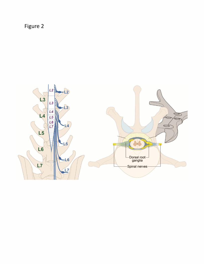
Figure 2

Figure 3
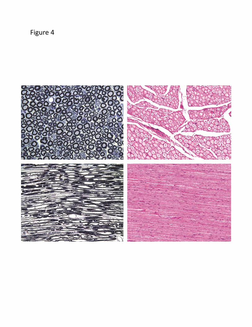
Figure 4

Figure 5
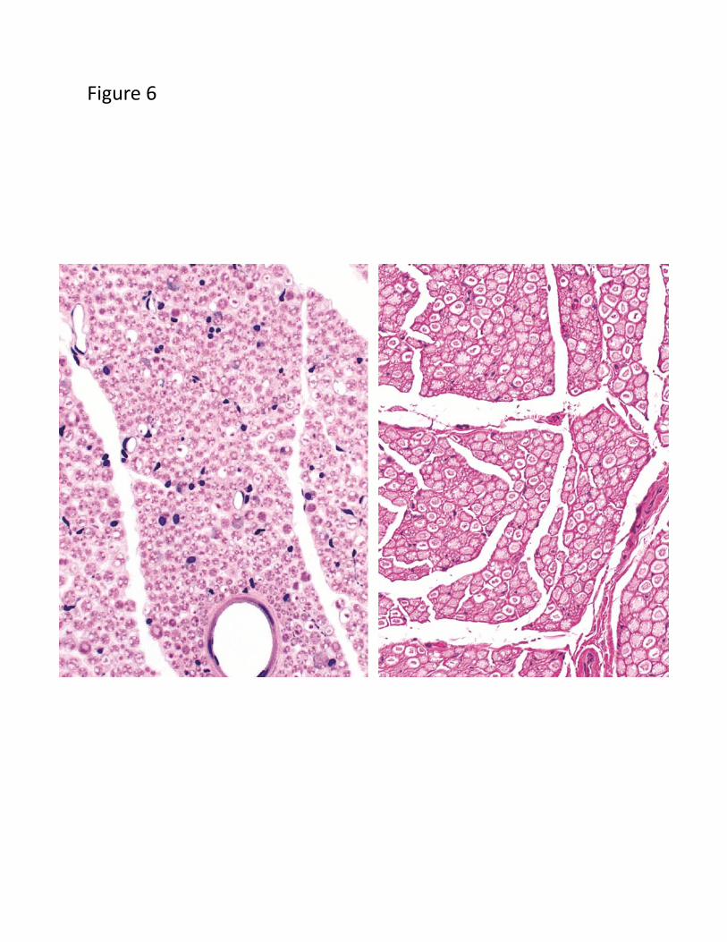
Figure 6

Figure 7