Stiffness Is Realized Form-Finding Model Shows How...
17
Form-Finding Model Shows How Cytoskeleton Network Stiffness Is Realized Jinghai Gong 1,2 , Daxu Zhang 2 , Yiider Tseng 3 , Baolong Li 2 , Denis Wirtz 4 , Benjamin William Schafer 1* 1 Department of Civil Engineering, Johns Hopkins University, Baltimore, Maryland, United States of America, 2 Department of Civil Engineering, Shanghai Jiao Tong University, Shanghai, China, 3 Department of Chemical Engineering, University of Florida, Gainesville, Florida, United States of America, 4 Department of Chemical and Biomolecular Engineering, Department of Oncology, Johns Hopkins University, Baltimore, Maryland, United States of America Abstract In eukaryotic cells the actin-cytoskeletal network provides stiffness and the driving force that contributes to changes in cell shape and cell motility, but the elastic behavior of this network is not well understood. In this paper a two dimensional form-finding model is proposed to investigate the elasticity of the actin filament network. Utilizing an initially random array of actin filaments and actin-cross-linking proteins the form-finding model iterates until the random array is brought into a stable equilibrium configuration. With some care given to actin filament density and length, distance between host sites for cross-linkers, and overall domain size the resulting configurations from the form-finding model are found to be topologically similar to cytoskeletal networks in real cells. The resulting network may then be mechanically exercised to explore how the actin filaments deform and align under load and the sensitivity of the network’s stiffness to actin filament density, length, etc. Results of the model are consistent with the experimental literature, e.g. actin filaments tend to re-orient in the direction of stretching; and the filament relative density, filament length, and actin-cross-linking protein’s relative density, control the actin-network stiffness. The model provides a ready means of extension to more complicated domains and a three-dimensional form-finding model is under development as well as models studying the formation of actin bundles. Citation: Gong J, Zhang D, Tseng Y, Li B, Wirtz D, et al. (2013) Form-Finding Model Shows How Cytoskeleton Network Stiffness Is Realized. PLoS ONE 8(10): e77417. doi:10.1371/journal.pone.0077417 Editor: Laurent Kreplak, Dalhousie University, Canada Received January 3, 2013; Accepted September 10, 2013; Published October 17, 2013 Copyright: © 2013 Gong et al. This is an open-access article distributed under the terms of the Creative Commons Attribution License, which permits unrestricted use, distribution, and reproduction in any medium, provided the original author and source are credited. Funding: The authors would like to recognize the Institute for NanoBio Technology at the Johns Hopkins University for partial funding in the development of this work. The funders had no role in study design, data collection and analysis, decision to publish, or preparation of the manuscript. No additional external funding received for this study. Competing interests: The authors have declared that no competing interests exist. * E-mail: [email protected] Introduction Eukaryotic cells are the building blocks of higher organisms. The cytoskeleton forms the internal framework of eukaryotic cells and is responsible for a cell’s elasticity and thus plays an important role in cell shape and motility. The mechanical behavior of the cytoskeleton is determined by the interactions of three types of filaments: actin filaments, intermediate filaments, and microtubules [1]. Actin filaments provide cells with primary mechanical support and are engaged as a driving force in cell motility [2]. The stiffness of the cytoskeleton network governs passive and active mechanical performance of cells. Many biological functions are intimately associated to the mechanical response, and therefore the cell’s stiffness may also serve as a sensitive indicator for the development or health state of a cell [2]. In recent years, numerous efforts have been made to predict the elasticity of the cytoskeleton network by using computational models; including open-cell foam models [3], tensegrity models [4-7], cable network models [8], lattice-based models [9], coarse-grained models [10,11] and Brownian dynamics models [12,13]. The pioneering work of Satcher and Dewey [3] employed a unit cell model of the cytoskeleton, in which the cross-linked actin filament network is treated as a solid matrix with random open pores (an open-celled foam) [2]. The model provides a simplified representation of the complex topology of the cytoskeleton in an average sense, and depends primarily on the bending stiffness of the actin filaments, and is independent of pre-stress forces in the network. Calibration to experimental data is required. The model represents a fine starting place for analytical exploration, but is limited in its ability to capture details of actual cytoskeletal networks. Many researchers have employed tensegrity structures to describe the mechanical properties of a cell. Tensegrity is a structural principle based on the use of isolated compression elements inside a net of continuous tension elements with the aim of achieving a stable form in space [4]. Ingber and co- PLOS ONE | www.plosone.org 1 October 2013 | Volume 8 | Issue 10 | e77417
-
Upload
truongkien -
Category
Documents
-
view
222 -
download
1
Transcript of Stiffness Is Realized Form-Finding Model Shows How...

Form-Finding Model Shows How Cytoskeleton NetworkStiffness Is RealizedJinghai Gong1,2, Daxu Zhang2, Yiider Tseng3, Baolong Li2, Denis Wirtz4, Benjamin William Schafer1*
1 Department of Civil Engineering, Johns Hopkins University, Baltimore, Maryland, United States of America, 2 Department of Civil Engineering, Shanghai JiaoTong University, Shanghai, China, 3 Department of Chemical Engineering, University of Florida, Gainesville, Florida, United States of America, 4 Department ofChemical and Biomolecular Engineering, Department of Oncology, Johns Hopkins University, Baltimore, Maryland, United States of America
Abstract
In eukaryotic cells the actin-cytoskeletal network provides stiffness and the driving force that contributes to changesin cell shape and cell motility, but the elastic behavior of this network is not well understood. In this paper a twodimensional form-finding model is proposed to investigate the elasticity of the actin filament network. Utilizing aninitially random array of actin filaments and actin-cross-linking proteins the form-finding model iterates until therandom array is brought into a stable equilibrium configuration. With some care given to actin filament density andlength, distance between host sites for cross-linkers, and overall domain size the resulting configurations from theform-finding model are found to be topologically similar to cytoskeletal networks in real cells. The resulting networkmay then be mechanically exercised to explore how the actin filaments deform and align under load and thesensitivity of the network’s stiffness to actin filament density, length, etc. Results of the model are consistent with theexperimental literature, e.g. actin filaments tend to re-orient in the direction of stretching; and the filament relativedensity, filament length, and actin-cross-linking protein’s relative density, control the actin-network stiffness. Themodel provides a ready means of extension to more complicated domains and a three-dimensional form-findingmodel is under development as well as models studying the formation of actin bundles.
Citation: Gong J, Zhang D, Tseng Y, Li B, Wirtz D, et al. (2013) Form-Finding Model Shows How Cytoskeleton Network Stiffness Is Realized. PLoS ONE8(10): e77417. doi:10.1371/journal.pone.0077417
Editor: Laurent Kreplak, Dalhousie University, Canada
Received January 3, 2013; Accepted September 10, 2013; Published October 17, 2013
Copyright: © 2013 Gong et al. This is an open-access article distributed under the terms of the Creative Commons Attribution License, which permitsunrestricted use, distribution, and reproduction in any medium, provided the original author and source are credited.
Funding: The authors would like to recognize the Institute for NanoBio Technology at the Johns Hopkins University for partial funding in the developmentof this work. The funders had no role in study design, data collection and analysis, decision to publish, or preparation of the manuscript. No additionalexternal funding received for this study.
Competing interests: The authors have declared that no competing interests exist.
* E-mail: [email protected]
Introduction
Eukaryotic cells are the building blocks of higher organisms.The cytoskeleton forms the internal framework of eukaryoticcells and is responsible for a cell’s elasticity and thus plays animportant role in cell shape and motility. The mechanicalbehavior of the cytoskeleton is determined by the interactionsof three types of filaments: actin filaments, intermediatefilaments, and microtubules [1]. Actin filaments provide cellswith primary mechanical support and are engaged as a drivingforce in cell motility [2].
The stiffness of the cytoskeleton network governs passiveand active mechanical performance of cells. Many biologicalfunctions are intimately associated to the mechanical response,and therefore the cell’s stiffness may also serve as a sensitiveindicator for the development or health state of a cell [2]. Inrecent years, numerous efforts have been made to predict theelasticity of the cytoskeleton network by using computationalmodels; including open-cell foam models [3], tensegrity models
[4-7], cable network models [8], lattice-based models [9],coarse-grained models [10,11] and Brownian dynamics models[12,13].
The pioneering work of Satcher and Dewey [3] employed aunit cell model of the cytoskeleton, in which the cross-linkedactin filament network is treated as a solid matrix with randomopen pores (an open-celled foam) [2]. The model provides asimplified representation of the complex topology of thecytoskeleton in an average sense, and depends primarily onthe bending stiffness of the actin filaments, and is independentof pre-stress forces in the network. Calibration to experimentaldata is required. The model represents a fine starting place foranalytical exploration, but is limited in its ability to capturedetails of actual cytoskeletal networks.
Many researchers have employed tensegrity structures todescribe the mechanical properties of a cell. Tensegrity is astructural principle based on the use of isolated compressionelements inside a net of continuous tension elements with theaim of achieving a stable form in space [4]. Ingber and co-
PLOS ONE | www.plosone.org 1 October 2013 | Volume 8 | Issue 10 | e77417

workers [14] first introduced the tensegrity concept to explainhow cells and tissues are constructed and Stamenovic andCoughlin [8,15] have used a variety of tensegrity models fromsimple to complex to explore cytoskeletal networks. Stiffness oftensegrity models are dependent on the level of internal pre-stress, and opposite to open cell foam models, engage theaxial stiffness of actin filaments and are independent of thebending stiffness of the actin filaments. Inclusion of pre-stressis important as experiments have established that cell stiffnessis correlated with the level of pre-stress in both living [16-18]and reconstituted [19,20] actin networks. However, internal pre-stress in the network is a difficult quantity to measure, variesgreatly across experiments [1] and results in significantuncertainty in the application of the model. In addition, whiletensegrity does provide a stable three-dimensional cableconfiguration it is a very specific class of a much broader set ofthree-dimensional structures and little topologic evidence existsfor its exact use in the cytoskeleton. Instead, much like theopen-cell foam models it must be recognized as an intriguingstarting point, particularly for analytical exploration, but islimited in its ability to capture many details of actualcytoskeletal networks.
A feature of all of the aforementioned models is that theyhave regular geometries and topologies. However, the topologyof real cytoskeleton networks is highly diverse and complex.Randomness is a fundamental nature of cytoskeleton networksand should not be ignored. Attempts to introduce thisrandomness into cytoskeletal network models have beencompleted in tensegrity models [6] and through Monte Carlosimulation [11]. Another feature of developed computationalmodels is that they are coarse-grained; that is, the truecytoskeleton system has far more filaments than the structuralcomponents in these models. Bausch and Kroy [21] proposeda multi-scale modeling approach to infer mechanical propertiesof filaments at the atomic scale and translate these into coarse-grained models of large filaments or filament networks [11].Brownian dynamics models developed by Kim et al. capturesbroad features of the cytoskeleton network and are well-suitedto describe the mechanical response in vitro; however theshear modulus (G') calculated by such models is small andefforts are still needed for accurate in vivo simulations.
In this work, our objective is to demonstrate the possibility ofdesigning more complex and topologically relevant actin-network models using an equilibrium form-finding method. Thepaper addresses the development of the form-finding model,the components of which are generated by using a fullystochastic approach. The model is used to predict experimentalobservations of filament re-orientation, and study effects of thefilament relative density, the filament length, and the relativedensity of filament cross-linkers on elastic modulus of thefilament network.
Methods and Models
Existing computational models for actin-networks aretypically coarse-grained, drastically simplify and regularize theactual network topology, and require artificial externalcalibration. The objective of the form-finding model developed
herein is to provide a fine scale model that is topologicallyconsistent with actual actin-networks, does not require externalcalibration, and provides a means to directly explore the role ofactin-network components, e.g. the influence of the averagelength of actin filaments on the elastic modulus of the network.
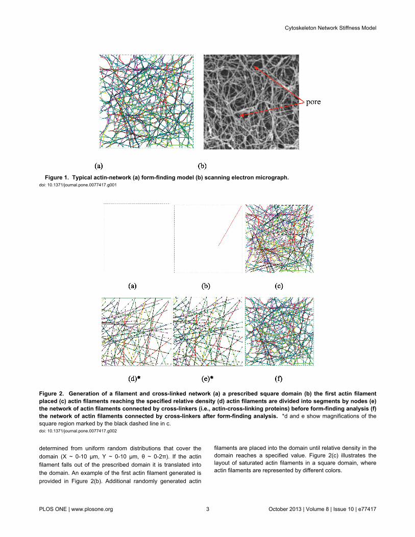
To achieve this modeling objective, the equilibrium form-finding concept of structural/architectural engineering has beenemployed to find the natural form of cytoskeleton networks.Form-finding investigates how inter-connected networksdeform into stable flexible structures. In structural/architecturalengineering, form-finding may be used to find stableconfigurations of members for complex roofs and buildingstructures. In nature, form-finding may be employed to discoverhow seemingly dis-organized structures are organized undermechanical stimulus. Form-finding analysis can be used todisclose the organizational principles of patterning in nature. Inthe mechanical sense, form-finding is the process of finding astable equilibrium configuration for an arbitrarily defined initialsystem within a set of boundary conditions for a particularmaterial or inter-connected components. Based on form-findingprinciples, models of actin-networks have been developed inthis paper. Figure 1 shows a comparison between the currenttwo-dimensional form-finding model and a scanning electronmicrograph (SEM) of a typical actin-network. It can be seenthat the form-finding model has the ability to provide atopologically realistic representation of the geometry of a realcytoskeleton.
Model constructionInitial conditions. Before creating a form-finding model, it is
essential to determine a domain. In this case the domaindefines the extent of the initial cytoskeleton network. As shownin Figure 2(a), a square domain has been chosen as theperiodic representative element. Key length scales in thedomain include the actin filament length and the domain sidelength. Obviously, the domain dimension has to be greater thanthe maximum actin filament length, but it is desirable to limitdomain size in order to minimize the computational cost since alarge number of stochastic simulations are necessary to studythe relation between the network’s stiffness and the propertiesof its constituents. Based on these considerations and typicalfilament length’s reported in the literature (e.g., [22]) anaverage actin filament length of 5 μm is selected along with asquare domain of 10 μm × 10 μm.
Model parameters. The form-finding and actin-networkstretching analyses are carried out with large deformationnonlinear finite element analysis. Beam and cable elements areused to model the actin filaments and their cross-linkers,respectively. Geometric and material properties for the networkare based on available experimental measurements and arelisted in Table 1.
Actin filament generation. To generate the geometricalmodel, actin filaments are placed sequentially within aprescribed two-dimensional space, as shown in Figure 2(a).For each actin filament, length is sampled from a truncated(negative values are thrown out) Gaussian distribution withmean and standard deviation as reported in Table 1.Centroidalcoordinates and angle of orientation of the filament are
Cytoskeleton Network Stiffness Model
PLOS ONE | www.plosone.org 2 October 2013 | Volume 8 | Issue 10 | e77417
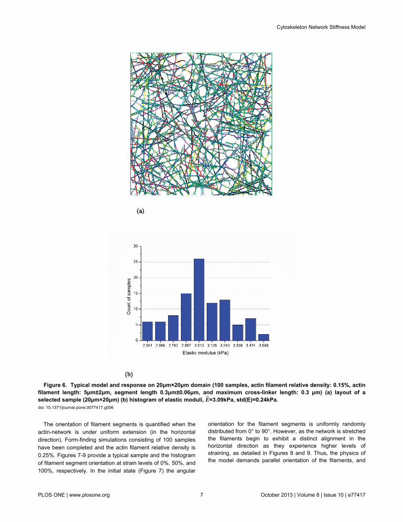
determined from uniform random distributions that cover thedomain (X ~ 0-10 μm, Y ~ 0-10 μm, θ ~ 0-2π). If the actinfilament falls out of the prescribed domain it is translated intothe domain. An example of the first actin filament generated isprovided in Figure 2(b). Additional randomly generated actin
filaments are placed into the domain until relative density in thedomain reaches a specified value. Figure 2(c) illustrates thelayout of saturated actin filaments in a square domain, whereactin filaments are represented by different colors.
Figure 1. Typical actin-network (a) form-finding model (b) scanning electron micrograph. doi: 10.1371/journal.pone.0077417.g001
Figure 2. Generation of a filament and cross-linked network (a) a prescribed square domain (b) the first actin filamentplaced (c) actin filaments reaching the specified relative density (d) actin filaments are divided into segments by nodes (e)the network of actin filaments connected by cross-linkers (i.e., actin-cross-linking proteins) before form-finding analysis (f)the network of actin filaments connected by cross-linkers after form-finding analysis. *d and e show magnifications of thesquare region marked by the black dashed line in c.doi: 10.1371/journal.pone.0077417.g002
Cytoskeleton Network Stiffness Model
PLOS ONE | www.plosone.org 3 October 2013 | Volume 8 | Issue 10 | e77417

Cross-linker generation. The modeled cytoskeletalnetwork consists of an actin-network (of beams) cross-linkedby proteins modeled as cables. Either α-actinin or filamin cross-linkers may be appropriate [23-26] here the model parameters(Table 1) are essentially aligned with filamin properties, but thestiffness of α-actinin may in the long term make it a moreappropriate choice. At this stage, the goal is to demonstrate themodel potential, but further detailed work on the cross-inkerparameters is needed. Cable elements are used as cross-linkers to connect the actin filaments. The cross-linkers aregenerated in two steps. First, actin filaments are divided intosegments to present their periodic binding sites to the cross-linkers as shown in Figure 2(d). Hence, cross-linkers may onlyattach at the binding sites (segment ends). The binding sitesare selected such that they match expected pore size (seeFigure 1(b)). Second, cross-linkers are created by connectingany two binding sites where the distance apart is less than themaximum cross-linker length. The resulting two-dimensionalnetwork model is provided in Figure 2(e) and is thus preparedfor the form-finding step.
Form-finding. After a model such as that shown in Figure2(e) is generated, a form-finding analysis is carried out tocompute the final equilibrium shape of the actin-network. Asmall tensile pre-stress force (~3 pN or 5% of the tensile yieldstrength of the cross-linkers, Table 1) is applied to the cross-linkers to mimic the force state of an actin-cross-linking proteinafter it establishes a link between two actin filaments; and thena nonlinear finite element form-finding analysis is performed tocompute the self-equilibrium configuration. During the form-finding analysis, the pre-stress force in the cross-linkers is keptconstant. The small constant pre-stress force in the cross-linkers is only to facilitate the form-finding analysis. The pre-stress magnitude can be calibrated by comparing the predictedfilament fluctuations with experimental observations.Preliminary studies of the cross-linker pre-stress magnitudeindicate the expected stiffness varies by about 5% betweenminimal pre-stress and a pre-stress equal to the tensile yieldstrength of the cross-linkers. Figure 2(f) provides typical form-finding results for a cytoskeleton network, including actinfilaments connected by actin-cross-linking proteins. The
Table 1. Dimensions and material properties of actinfilaments and cross-linkers.
Parameters DataElastic modulus of actin filaments 1.4 GPa [30]Diameter of actin filaments 7 nm [30]Length of actin filaments 5 ± 2 μm [17]Length of actin filament segments 1 0.3 ± 0.06 μmRelative density of actin filaments 2 0.15% ~ 0.3% [1]Yield tensile force of actin filaments 0.25 nN [31]Maximum length of cross-linkers 0.3 μm [19]Yield tensile force of cross-linkers 60 pN [18]1 The maximum length of cross-linkers is used to achieve an accurate prediction.2 Relative density is the amount of actin filaments per unit volume of filamentnetwork.doi: 10.1371/journal.pone.0077417.t001 resulting networks share a strong resemblance with the known
topology of actin filament networks such as shown in Figure1(b) or the EM images of [27].
Effective modulus determination. After form-finding thedeveloped representative volume element (e.g. Figure 2(f)) canbe exercised to determine effective properties of thecytoskeleton under mechanical stimuli. The simplest of which isthe effective elastic (Young’s) modulus. Elastic modulus isdetermined by the results from a simple extension of themodel. One side of the domain is fixed in longitudinaltranslation while the far side is displaced longitudinally (xdirection) a finite amount. Assuming plane stress conditions(Figure 3) for any subdomain in the model the effective elastic(Young’s) modulus is calculated by Hooke's law asE= σx
2−σy2 / σxεx−σyεy . For the simplest case where the
subdomain studied is the entire model the effective engineeringstrain (εx) is the finite stretch divided by the domain length (10μm). The effective engineering stress (σx) is the force requiredto create the finite stretch divided by the original cross-sectional area of the domain (10 μm x 1 μm) and σy=0.
Modeling sensitivity to sample and domain sizeTo obtain an optimum balance between computational cost
and accuracy, modeling sensitivity to sample and domain sizeis assessed before completing further analysis.
Modeling sensitivity to sample size. Elastic modulus ofnetworks with an actin filament relative density of 0.2% wasdetermined from sample sizes of 100 and 1000 realizations ofthe 10 μm × 10 μm domain. A typical sample is provided inFigure 4(a) and histograms of the developed elastic moduli areprovided for 100 samples in Figure 4(b) and 1000 samples inFigure 4(c). The larger number of samples provides a smootherapproximation of the resulting probability distribution function(PDF) and better explores the extremes of the distribution, butthe means, 4.38 kPa and 4.41 kPa, are very close. Since forthe studies here mean elastic moduli is the main concern, thesmaller sample size is deemed adequate.
Modeling sensitivity to domain size. Elastic modulus ofnetworks, with a relative density of 0.15%, was studied fordomains of 10 μm × 10 μm and 20 μm × 20 μm. Typicalnetwork topology and histograms from the 100 samples of
Figure 3. Schematic diagram of Hooke’s law for planestress state. doi: 10.1371/journal.pone.0077417.g003
Cytoskeleton Network Stiffness Model
PLOS ONE | www.plosone.org 4 October 2013 | Volume 8 | Issue 10 | e77417

Figure 4. Typical model and impact of number of simulations (actin filament relative density: 0.2%, size: 10μm×10μm,Actin filament length: 5μm±2μm, segment length 0.3μm±0.06μm, and maximum cross-linker length: 0.3μm) (a) layout of aselected sample (b) histogram of elastic moduli for 100 samples: E=4.38kPa, std(E)=0.47kPa (c) histogram of elasticmoduli for 1000 samples: E=4.41kPa, std(E)=0.52kPa. doi: 10.1371/journal.pone.0077417.g004
Cytoskeleton Network Stiffness Model
PLOS ONE | www.plosone.org 5 October 2013 | Volume 8 | Issue 10 | e77417

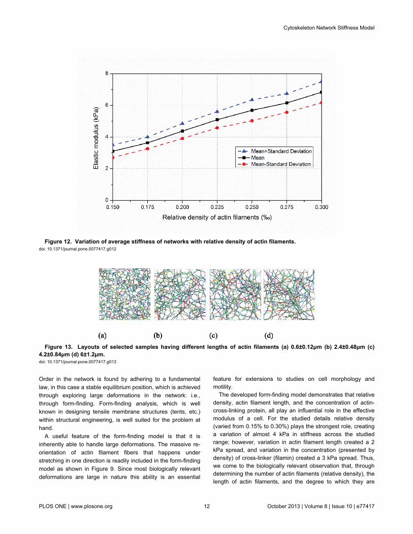
predicted elastic moduli for the two domain sizes are providedin Figure 5 and 6. Although the distributions of predicted elasticmoduli in Figures 5(b) and 6(b) are modestly different, theirmean values, 3.11 kPa and 3.09 kPa, are close and the 10 μm× 10 μm is deemed adequate. Note, here the fidelity of theform-finding model can be judged – the predictions are in thesame order of elastic moduli as measured by reliableexperimental techniques [1].
Results
Filaments orient under extensionActin filaments re-orient when a cell is under mechanical
stimulation. The exact nature of this re-orientation is complexand depends on the time-scales of the stimulation, the stiffnessof the substrate employed in the stretching and a host ofbiological factors. At short time scales as the filament networkresponds to elongation filaments may align parallel to thestretch [28]; for other cases alignment may be perpendicular tothe direction of stretch [29] unless specific proteins areknocked down[30]. The current form-finding analysis has beenused to explore this phenomenon.
Figure 5. Typical model and response on 10μm×10μm domain (100 samples, actin filament relative density: 0.15%, actinfilament length: 5μm±2μm, segment length 0.3μm±0.06μm, and maximum cross-linker length: 0.3 μm) (a) layout of aselected sample (10μm×10μm) (b) histogram of elastic moduli, E=3.11kPa, std(E)=0.40kPa. doi: 10.1371/journal.pone.0077417.g005
Cytoskeleton Network Stiffness Model
PLOS ONE | www.plosone.org 6 October 2013 | Volume 8 | Issue 10 | e77417
The orientation of filament segments is quantified when theactin-network is under uniform extension (in the horizontaldirection). Form-finding simulations consisting of 100 sampleshave been completed and the actin filament relative density is0.25%. Figures 7-9 provide a typical sample and the histogramof filament segment orientation at strain levels of 0%, 50%, and100%, respectively. In the initial state (Figure 7) the angular
orientation for the filament segments is uniformly randomlydistributed from 0° to 90°. However, as the network is stretchedthe filaments begin to exhibit a distinct alignment in thehorizontal direction as they experience higher levels ofstraining, as detailed in Figures 8 and 9. Thus, the physics ofthe model demands parallel orientation of the filaments, and
Figure 6. Typical model and response on 20μm×20μm domain (100 samples, actin filament relative density: 0.15%, actinfilament length: 5μm±2μm, segment length 0.3μm±0.06μm, and maximum cross-linker length: 0.3 μm) (a) layout of aselected sample (20μm×20μm) (b) histogram of elastic moduli, E=3.09kPa, std(E)=0.24kPa. doi: 10.1371/journal.pone.0077417.g006
Cytoskeleton Network Stiffness Model
PLOS ONE | www.plosone.org 7 October 2013 | Volume 8 | Issue 10 | e77417

the model will have to have additional feedback included toexhibit the type of behavior observed in [29].
Filament and filamin control network stiffnessThe relative densities of actin filaments and actin-cross-
linking proteins dramatically alter the stiffness of actin networks[31-36]. Moreover, actin filament length affects the rheology ofthese networks and therefore further “tunes” their elasticity [22].The developed form-finding model enables us to directlyinvestigate the effects of relative density of the filaments orcross-linkers, and filament length on the network stiffness.
Relative density of filaments positively correlated withcytoskeletal stiffness. We used seven sets of samples (100each), across filament relative densities of 0.15%, 0.175%,0.20%, 0.225%, 0.25%, 0.275% and 0.30%. The topology of
selected samples are provided in Figure 10 and show howrelative density influences pore size and inter-connectednessof the developed network. At each relative density 100 samplesare completed and the effective elastic modulus predicted.Histograms of the mean and standard deviation of elasticmodulus for each filament are provided in Figure 11. Relativedensity plays a powerful role in determining network stiffness,e.g. the modulus is 3.11 kPa and 6.83 kPa at 0.15% and 0.30%relative density, respectively. The variation of mean networkstiffness (modulus) with filament density is provided in Figure12, where an approximately linear relation can be observed.
Filament length positively correlated with cytoskeletalstiffness. Changing average filament length at a fixed relativedensity provides another means to alter the network topology(Figure 13) and resulting stiffness. A set of ten studies (100
Figure 7. Initial layout and orientation of filament segments (a) initial layout of a selected sample (b) histogram of anglesbetween filament segments and horizontal axis at the initial state. doi: 10.1371/journal.pone.0077417.g007
Cytoskeleton Network Stiffness Model
PLOS ONE | www.plosone.org 8 October 2013 | Volume 8 | Issue 10 | e77417

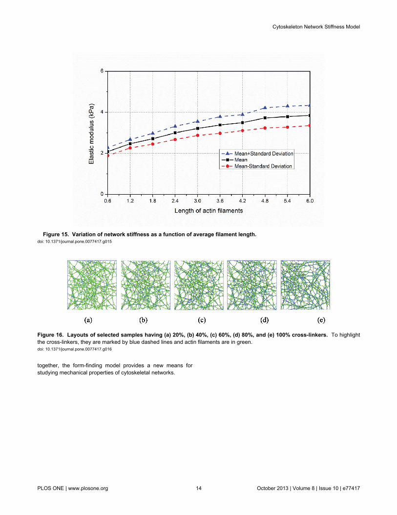
samples in each study) is completed where the average andstandard deviation of the filament length is varied - averagefilament length is varied from 0.6 μm to 6 μm in increments of0.6 μm and the standard deviation is perfectly correlated to thelength increasing from 0.12 μm up to 1.2 μm in increments of0.12 μm. At the shortest filament length (Figure 13(a))individual filaments typically only have one or two cross-linkedconnections to other filaments. At the longest filament length(Figure 13(d)) individual filaments have many, often more than10, cross-linked connections to other filaments. This inter-connectivity (even at the same overall relative density) isrewarded with higher average effective modulus (E): 3.85 kPafor the longest average filament length (Figure 13(d) and 14(d))versus only 2.08 kPa for the shortest average filament length(Figure 13(a) and 14(a)).
Sample histograms for the resulting effective modulus areprovided in Figure 14 and summary statistics across the fullstudy in Figure 15. Note, at the longest filament length(6.0±1.2μm,Figure 13(d) and 14(d)) a larger number of highstiffness outliers result –suggesting that we are near the limitfor our domain size and boundary effects are beginning toinfluence the statistics. Thus, longer filament lengths are notrecommended for study without increasing the domain size.Taken in total, Figure 15 shows that actin filament length varieseffective modulus by as much as nearly 2 kPa, similar to thetotal variation for studied relative density (Figure 12).
Relative density of cross-linkers positively correlatedwith cytoskeletal stiffness. In the developed form-findingmodel the cross-linking protein connects the actin-networktogether. In the baseline model the actin filaments have hostsites, which are 0.3 ± 0.06 μm apart (these are the segment
Figure 8. Deformed shape and orientation at 50% tensile strain (a) layout of a selected sample at 50% extension (b)histogram of angles between filament segments and horizontal axis. doi: 10.1371/journal.pone.0077417.g008
Cytoskeleton Network Stiffness Model
PLOS ONE | www.plosone.org 9 October 2013 | Volume 8 | Issue 10 | e77417

Figure 9. Deformed shape and orientation at 100% tensile strain (a) layout of a selected sample at 100% extension (b)histogram of angles between filament segments and horizontal axis. doi: 10.1371/journal.pone.0077417.g009
Figure 10. Layouts of selected samples with different actin filament relative densities. (a) 0.15% (b) 0.20% (c) 0.25% (d)0.30%.doi: 10.1371/journal.pone.0077417.g010
Cytoskeleton Network Stiffness Model
PLOS ONE | www.plosone.org 10 October 2013 | Volume 8 | Issue 10 | e77417

ends, described in Section 2.1.4) and any two host sites thatare less than 0.3 μm are connected by a cross-linker. Here,instead of connecting all host sites that are less than 0.3 μmonly a percentage are randomly connected, varying from 20%up to 100% (baseline). For a given actin filament topology (asingle network realization) the resulting cross-linking isdepicted in Figure 16. Histograms of the effective modulusacross 100 samples are provided for 20% to 80% cross-linkingdensity in Figure 17 and the average ± a standard deviation aredepicted for all results in Figure 18. Effective modulusincreases by 3 kPa from 20% up to 100% cross-linking.
Form-finding model provides basis for a more balancedapproach
The open-cell foam model and the tensegrity model ofcytoskeletal network stiffness are important idealizations, butthey suffer fundamentally in that they assume that all thedeformations are either bending (open-cell foam) or axial(tensegrity). In fact, no reason exists that this should be thecase, and the form-finding model demonstrates how bothmodes of deformation play a role in the solution.
For each actin filament, under deformation, we maydetermine the strain energy due to bending (SEbending) and the
strain energy due to axial deformation (SEaxial). The total strainenergy (SEtotal) is the summation of these two. For our baselinemodel (0.15% relative density, further details in Table 1) ahistogram of the fraction of axial strain energy (SEaxial/SEtotal) isprovided in Figure 19. As indicated in Figure 19, most of theactin filaments have very low axial strain energy and hencehigh bending strain energy. Hence the actin-network may beconsidered to be dominated by bending deformations andbehave in a beam-like fashion. Nonetheless, a large number ofactin filaments are dominated by axial (truss-like) deformations– and many more still are in between. The specifics of theseresults are sensitive to relative density, cross-linker density,mode of deformation, etc. Here, the form-finding model showsthat both modes of deformation are necessary for realisticmodeling of cytoskeletal network stiffness.
Discussion
The developed form-finding model provides a means togenerate realistic cytoskeletal network topologies with majorbiological features incorporated into the model. Instead ofbeginning from a set, ordered, topology as in existing modelsthe form-finding model begins with a random array of materials.
Figure 11. Impact of filament relative density on stiffness (a) filament relative density of 0.15%, E=3.11kPa,std(E)=0.40kPa; (b) filament relative density of 0.20%, E = 4.38kPa, std(E) = 0.47kPa; (c) filament relative density of 0.25%,E = 5.69kPa, std(E) = 0.66kPa; (d) filament relative density of 0.30%, E = 6.83kPa, std(E) = 0.67kPa. Eand std(E) denotesample mean and standard deviation of elastic modulus E, respectively. doi: 10.1371/journal.pone.0077417.g011
Cytoskeleton Network Stiffness Model
PLOS ONE | www.plosone.org 11 October 2013 | Volume 8 | Issue 10 | e77417
Order in the network is found by adhering to a fundamentallaw, in this case a stable equilibrium position, which is achievedthrough exploring large deformations in the network: i.e.,through form-finding. Form-finding analysis, which is wellknown in designing tensile membrane structures (tents, etc.)within structural engineering, is well suited for the problem athand.
A useful feature of the form-finding model is that it isinherently able to handle large deformations. The massive re-orientation of actin filament fibers that happens understretching in one direction is readily included in the form-findingmodel as shown in Figure 9. Since most biologically relevantdeformations are large in nature this ability is an essential
feature for extensions to studies on cell morphology andmotility.
The developed form-finding model demonstrates that relativedensity, actin filament length, and the concentration of actin-cross-linking protein, all play an influential role in the effectivemodulus of a cell. For the studied details relative density(varied from 0.15% to 0.30%) plays the strongest role, creatinga variation of almost 4 kPa in stiffness across the studiedrange; however, variation in actin filament length created a 2kPa spread, and variation in the concentration (presented bydensity) of cross-linker (filamin) created a 3 kPa spread. Thus,we come to the biologically relevant observation that, throughdetermining the number of actin filaments (relative density), thelength of actin filaments, and the degree to which they are
Figure 12. Variation of average stiffness of networks with relative density of actin filaments. doi: 10.1371/journal.pone.0077417.g012
Figure 13. Layouts of selected samples having different lengths of actin filaments (a) 0.6±0.12μm (b) 2.4±0.48μm (c)4.2±0.84μm (d) 6±1.2μm. doi: 10.1371/journal.pone.0077417.g013
Cytoskeleton Network Stiffness Model
PLOS ONE | www.plosone.org 12 October 2013 | Volume 8 | Issue 10 | e77417

cross-linked, the cell has fine-level control over the developedstiffness. Further, we note that these features are all capturedin the form-finding model.
Given the many length and time scales inherent in cellularresponse multi-scale models are a natural long-term directionfor model development. The form-finding model developedherein provides relevant features to cross scales. For example,at finer scales, actin-cross-linking protein binding sites on theactin filament are included in the model and detailed models ofthe cross-linking process could be included. At coarser scales,the overall effective modulus of a given realization ofcytoskeletal network can readily be included in a continuumscale model.
Significant future work remains to advance the form-findingmodel. The most important of which is the move from two-dimensions to three-dimensions. As the SEM of Figure 1(b)shows the network has depth and the filaments are clearlyoriented in space, not just a plane. Initial work in this directionhas been completed and it may be stated that stableequilibrium configurations in three-dimensions, using the sametechniques for building up the form-finding model, exist. Modelcomplexity and computational size quickly expand presentingcertain challenges to large stochastic simulations, but it is feltby the authors that this direction has the most promise and isan active area of current research. Additional work utilizing the
form-finding model to directly address cytoskeletalviscoelasticity is also needed. The form-finding model providesthe framework for this study, but detailed time-dependentparameters of the filaments and cross-linkers is required.
Conclusions
A stochastic form-finding model has been developed to studythe stiffness of the cytoskeleton network. The developed two-dimensional model includes actin filaments and cross-linkingproteins and is realized on a 10 μm × 10 μm domain. For agiven random initial actin filament topology and density ofcross-linkers a form-finding analysis is performed to generate astable configuration for the network. The resulting network istopologically similar to real actin-networks. Through simulationof simple stretching the performance of the network isassessed. Both bending and axial deformations occur withinthe actin filaments. Consistent with actual cell response it isalso shown how actin filaments align and re-orient under largestretching. The predicted effective modulus under stretching isconsistent with experimental observations. The form-findingmodel also shows how effective modulus is sensitive to relativedensity, actin filament length, and cross-linker density. Work toextend the model to three dimensions is underway. Taken
Figure 14. Impact of average actin filament length (L) on stiffness (filament relative density of 0.2%) (a) actin filamentlength L = 0.6±0.12μm, E = 2.08kPa, std(E) = 0.19kPa; (b) L = 2.4±0.48μm, E = 3.00kPa, std(E) = 0.32kPa; (c) L = 4.2±0.84μm,E = 3.50kPa, std(E) = 0.39kPa; (d) L = 6.0±1.2μm, E = 3.85kPa, std(E) = 0.49kPa. doi: 10.1371/journal.pone.0077417.g014
Cytoskeleton Network Stiffness Model
PLOS ONE | www.plosone.org 13 October 2013 | Volume 8 | Issue 10 | e77417
together, the form-finding model provides a new means forstudying mechanical properties of cytoskeletal networks.
Figure 15. Variation of network stiffness as a function of average filament length. doi: 10.1371/journal.pone.0077417.g015
Figure 16. Layouts of selected samples having (a) 20%, (b) 40%, (c) 60%, (d) 80%, and (e) 100% cross-linkers. To highlightthe cross-linkers, they are marked by blue dashed lines and actin filaments are in green.doi: 10.1371/journal.pone.0077417.g016
Cytoskeleton Network Stiffness Model
PLOS ONE | www.plosone.org 14 October 2013 | Volume 8 | Issue 10 | e77417

Figure 17. Impact of cross-linker density (f) on stiffness (filament relative density of 0.2): (a) cross-linker density f = 20%,E = 1.35 kPa, std(E) = 0.27kPa; (b) f = 40%, E = 2.39kPa, std(E) = 0.35kPa; (c) f = 60%, E = 3.15 kPa, std(E) = 0.42kPa; (d) f =80%, E = 3.77kPa, std(E) = 0.46kPa. doi: 10.1371/journal.pone.0077417.g017
Cytoskeleton Network Stiffness Model
PLOS ONE | www.plosone.org 15 October 2013 | Volume 8 | Issue 10 | e77417

Figure 18. Variation of network stiffness with percentage of cross-linkers. doi: 10.1371/journal.pone.0077417.g018
Figure 19. Histogram of actin filament energy fraction that is axial strain energy (10μm×10μm, actin filament relativedensity 0.15%, actin filament length 5μm±2μm, segment length 0.3±0.06μm, and maximum cross-linker length: 0.3μm). doi: 10.1371/journal.pone.0077417.g019
Cytoskeleton Network Stiffness Model
PLOS ONE | www.plosone.org 16 October 2013 | Volume 8 | Issue 10 | e77417

Author Contributions
Conceived and designed the experiments: JG DW BWS.Performed the experiments: JG DZ. Analyzed the data: JG DZ
YT DW BWS. Contributed reagents/materials/analysis tools:JG DZ BL BWS. Wrote the manuscript: JG DZ BL BWS.Critical assessment of biological relevance andrecommendations for modifications: YT.
References
1. Ethier CR, Simmons CA (2007) Introductory biomechanics: from cellsto organisms. Cambridge Univ Pr.
2. Pollard TD, Cooper JA (2009) Actin, a central player in cell shape andmovement. Science 326: 1208–1212. doi:10.1126/science.1175862.PubMed: 19965462.
3. Satcher RL Jr, Dewey CF Jr (1996) Theoretical estimates ofmechanical properties of the endothelial cell cytoskeleton. Biophys J71: 109-118. doi:10.1016/S0006-3495(96)79206-8. PubMed: 8804594.
4. Ingber DE (1993) Cellular tensegrity: defining new rules of biologicaldesign that govern the cytoskeleton. J Cell Sci 104: 613-613. PubMed:8314865.
5. Cañadas P, Laurent VM, Oddou C, Isabey D, Wendling S (2002) Acellular tensegrity model to analyse the structural viscoelasticity of thecytoskeleton. J Theor Biol 218: 155-173. doi:10.1006/jtbi.2002.3064.PubMed: 12381289.
6. Baudriller H, Maurin B, Cañadas P, Montcourrier P, Parmeggiani A etal. (2006) Form-finding of complex tensegrity structures: application tocell cytoskeleton modelling. Comp R Mec 334: 662-668. doi:10.1016/j.crme.2006.08.004.
7. Luo Y, Xu X, Lele T, Kumar S, Ingber DE (2008) A multi-modulartensegrity model of an actin stress fiber. J Biomech 41: 2379-2387. doi:10.1016/j.jbiomech.2008.05.026. PubMed: 18632107.
8. Coughlin MF, Stamenović D (2003) A prestressed cable network modelof the adherent cell cytoskeleton. Biophys J 84: 1328-1336. doi:10.1016/S0006-3495(03)74948-0. PubMed: 12547813.
9. Broedersz CP, Mao X, Lubensky TC, MacKintosh FC (2011) Criticalityand isostaticity in fibre networks. Nat Phys. 7: 983-988. doi:10.1038/nphys2127.
10. Chu JW, Voth GA (2006) Coarse-grained modeling of the actin filamentderived from atomistic-scale simulations. Biophys J 90: 1572-1582. doi:10.1529/biophysj.105.073924. PubMed: 16361345.
11. Kang J, Steward RL, Kim YT, Schwartz RS, LeDuc PR et al. (2011)Response of an actin filament network model under cyclic stretchingthrough a coarse grained Monte Carlo approach. J Theor Biol.274(1):109-119. doi:10.1016/j.jtbi.2011.01.011. PubMed: 21241710.
12. Kim T, Hwang W, Kamm R (2009) Computational analysis of a cross-linked actin-like network. Exp Mech 49: 91-104. doi:10.1007/s11340-007-9091-3.
13. Kim T, Hwang W, Lee H, Kamm RD (2009) Computational analysis ofviscoelastic properties of crosslinked actin networks. PLOS ComputBiol 5: e1000439. PubMed: 19609348.
14. Ingber DE, Madri JA, Jamieson JD (1981) Role of basal lamina inneoplastic disorganization of tissue architecture. Proc Natl Acad SciUSA 78: 3901–3905. doi:10.1073/pnas.78.6.3901. PubMed: 7022458.
15. Stamenović D, Coughlin MF (2000) A quantitative model of cellularelasticity based on tensegrity. J Biomech Eng 122: 39–43. doi:10.1115/1.429631. PubMed: 10790828.
16. Wang N, Tolić-Nørrelykke IM, Chen J, Mijailovich SM, Butler JP et al.(2002) Cell prestress. I. Stiffness and prestress are closely associatedin adherent contractile cells. Am J Physiol-Cell Physiol 282: C606-C616. doi:10.1152/ajpcell.00269.2001. PubMed: 11832346.
17. Fernández P, Pullarkat PA, Ott A (2006) A master relation defines thenonlinear viscoelasticity of single fibroblasts. Biophys J 90: 3796-3805.doi:10.1529/biophysj.105.072215. PubMed: 16461394.
18. Lam RH, Weng S, Lu W, Fu J (2012) Live-cell subcellularmeasurement of cell stiffness using a microengineered stretchablemicropost array membrane. Integr Biol 4: 1289-1298. doi:10.1039/c2ib20134h. PubMed: 22935822.
19. Gardel ML, Nakamura F, Hartwig JH, Crocker JC, Stossel TP et al.(2006) Prestressed F-actin networks cross-linked by hinged filamins
replicate mechanical properties of cells. Proc Natl Acad Sci U S A 103:1762-1767. doi:10.1073/pnas.0504777103. PubMed: 16446458.
20. Mizuno D, Tardin C, Schmidt CF, MacKintosh FC (2007)Nonequilibrium mechanics of active cytoskeletal networks. Science315: 370-373. doi:10.1126/science.1134404. PubMed: 17234946.
21. Bausch A, Kroy K (2006) A bottom-up approach to cell mechanics. NatPhys 2: 231-238. doi:10.1038/nphys260.
22. Kasza KE, Broedersz CP, Koenderink GH, Lin YC, Messner W et al.(2010) Actin filament length tunes elasticity of flexibly cross-linked actinnetworks. Biophys J 99: 1091-1100. doi:10.1016/j.bpj.2010.06.025.PubMed: 20712992.
23. Ferrer JM, Lee H, Chen J, Pelz B, Nakamura F et al. (2008) Measuringmolecular rupture forces between single actin filaments and actin-binding proteins. Proc Natl Acad Sci USA 105: 9221–9226. doi:10.1073/pnas.0706124105. PubMed: 18591676.
24. Furuike S, Ito T, Yamazaki M (2001) Mechanical unfolding of singlefilamin A (ABP-280) molecules detected by atomic force microscopy.FEBS Lett 498: 72-75. doi:10.1016/S0014-5793(01)02497-8. PubMed:11389901.
25. Sjöblom B, Salmazo A, Djinović-Carugo K (2008) a-Actinin structureand regulation. Cell Mol Life Sci 65: 2688-2701. doi:10.1007/s00018-008-8080-8. PubMed: 18488141.
26. Yamazaki M, Furuike S, Ito T (2002) Mechanical response of singlefilamin A (ABP-280) molecules and its role in the actin cytoskeleton. JMuscle Res Cell Motil 23: 525-534. doi:10.1023/A:1023418725001.PubMed: 12785102.
27. Wolosewick JJ, Porter KR (1979) Microtrabecular lattice of thecytoplasmic ground substance. Artifact or reality. J Cell Biol 82:114-139. doi:10.1083/jcb.82.1.114. PubMed: 479294.
28. Walcott S, Sun SX (2010) A mechanical model of actin stress fiberformation and substrate elasticity sensing in adherent cells. Proc NatlAcad Sci USA 107: 7757-7762. doi:10.1073/pnas.0912739107.PubMed: 20385838.
29. Kaunas R, Nguyen P, Usami S, Chien S (2005) Cooperative effects ofRho and mechanical stretch on stress fiber organization. Proc NatlAcad Sci U S A 102: 15895-15900. doi:10.1073/pnas.0506041102.PubMed: 16247009.
30. Niediek V, Born S, Hampe N, Kirchgeßner N, Merkel R et al. (2011)Cyclic stretch induces reorientation of cells in a Src family kinase-andp130Cas-dependent manner. Eur J Cell Biol.91(2): 118-128. PubMed:22178114.
31. Janmey PA, Hvidt S, Lamb J, Stossel TP (1990) Resemblance of actin-binding protein/actin gels to covalently crosslinked networks. Nature345: 89 - 92. doi:10.1038/345089a0. PubMed: 2158633.
32. Ruddies R, Goldmann WH, Isenberg G, Sackmann E (1993) Theviscoelasticity of entangled actin networks: the influence of defects andmodulation by talin and vinculin. Eur Biophys J 22: 309-321. doi:10.1007/BF00213554. PubMed: 8112218.
33. Wachsstock DH, Schwarz WH, Pollard TD (1994) Cross-linkerdynamics determine the mechanical properties of actin gels. Biophys J66: 801-809. doi:10.1016/S0006-3495(94)80856-2. PubMed: 8011912.
34. Tempel M, Isenberg G, Sackmann E (1996) Temperature-induced sol-gel transition and microgel formation in α-actinin cross-linked actinnetworks: a rheological study. Phys Rev E 54: 1802. doi:10.1103/PhysRevE.54.1802.
35. Xu J, Wirtz D, Pollard TD (1998) Dynamic cross-linking by α-actinindetermines the mechanical properties of actin filament networks. J BiolChem 273: 9570–9576. doi:10.1074/jbc.273.16.9570. PubMed:9545287.
36. Gardel ML, Shin JH, MacKintosh FC, Mahadevan L, Matsudaira P et al.(2004) Elastic behavior of cross-linked and bundled actin networks.Science 304: 1301-1305. doi:10.1126/science.1095087. PubMed:15166374.
Cytoskeleton Network Stiffness Model
PLOS ONE | www.plosone.org 17 October 2013 | Volume 8 | Issue 10 | e77417