STEMI after Dobutamine Stress Echocardiography in...
5
Case Report STEMI after Dobutamine Stress Echocardiography in Hyperthyroid State Mahmood Mubasher , Ashfaq Patel, Mohamed Magdi , and Tahir Hamid Hamad Medical Corporation, Qatar Correspondence should be addressed to Mahmood Mubasher; [email protected] Received 8 December 2018; Accepted 14 March 2019; Published 2 April 2019 Academic Editor: Antonio de Padua Mansur Copyright © 2019 Mahmood Mubasher et al. This is an open access article distributed under the Creative Commons Attribution License, which permits unrestricted use, distribution, and reproduction in any medium, provided the original work is properly cited. Uncontrolled hyperthyroidism has been associated with significant changes in cardiovascular hemodynamics. We report a case of a 39-year-old male who has been recently diagnosed with severe hyperthyroidism. He was undergoing dobutamine stress echocardiography (DSE) for evaluation of symptoms suggestive of stable angina. The exam was complicated by ST-segment elevation myocardial infarction- (STEMI-) required coronary angiography that showed mild coronary artery disease. 1. Introduction It is known that hyperthyroidism alters the hemodynamics of the cardiovascular system through increasing the oxygen demand [1]. Dobutamine stress echocardiography (DSE) is an effective and safe test to detect coronary artery disease (CAD) with low risk of adverse events [2–4]. STEMI was reported in patients undergoing DSE in about 0.1% of cases; most of them had underlying significant coronary arteries [5, 6]. We detail a case of a 39-year-old male who recently had severe hyperthyroidism. He developed chest pain, sig- nificant ST elevation, and raised cardiac enzymes following DSE for angina-type chest pain. Coronary angiography showed CAD with mild luminal stenosis. 2. Case A 39-year-old Asian male, with a recent diagnosis of severe hyperthyroidism and family history of coronary artery disease but no other cardiovascular risk factors, presented with intermittent angina-type chest pain of 8-month dura- tion. He also reported heat intolerance, recurrent palpitation, sweating, watery diarrhea, and weight loss of 10 kg over a 3-month duration. Methimazole and propranolol were initiated two days before the actual presentation. On exami- nation, he had normal heart sounds, no gallop, murmur, or rub tachycardia but with regular and synchronous heart rate, and no clinical signs of heart failure. Neck exam revealed diffuse mild thyroid enlargement and general exam showed fine hand tremors. Electrocardiography on presentation showed biphasic T waves in V1 and V2 (Figure 1). Three sets of cardiac enzymes were negative; TSH was <0.005 mIU/L (0.45-4.5) and T4 was 48.3 pmol/L (9-20) reference and units. Transthoracic echocardiography was normal with good left ventricular function (ejection fraction (EF): 70%) and no regional wall motion abnormalities. As the patient did not tolerate exercise treadmill stress test, he under- went a dobutamine stress echo. Dobutamine was infused at 3-minute intervals, starting with 10 μg/kg and increasing to 20 μg/kg, 30 μg/kg, and 40 μg/kg in addition to 0.5 mg atropine IV until a target heart rate of 153 bpm accounting for 95% of his maximal predicted heart rate. The patient developed severe chest pain and systemic hypotension with a blood pressure of 80/50 mmHg. ST elevation in the antero- lateral leads, new RBBB, and short runs of nonsustained ventricular tachycardia (VT) on continuous ECG monitoring were noted (Figure 2). Echocardiography showed new regional wall motion abnormalities in the form of akinesia of the apical, mid anteroseptal, and mid anterior walls and hypokinesia of the mid anterolateral and mid posterolateral walls with a drop of EF to 30%. The patient was transferred to the intensive Hindawi Case Reports in Cardiology Volume 2019, Article ID 7434071, 4 pages https://doi.org/10.1155/2019/7434071
Transcript of STEMI after Dobutamine Stress Echocardiography in...

Case ReportSTEMI after Dobutamine Stress Echocardiography inHyperthyroid State
Mahmood Mubasher , Ashfaq Patel, Mohamed Magdi , and Tahir Hamid
Hamad Medical Corporation, Qatar
Correspondence should be addressed to Mahmood Mubasher; [email protected]
Received 8 December 2018; Accepted 14 March 2019; Published 2 April 2019
Academic Editor: Antonio de Padua Mansur
Copyright © 2019 Mahmood Mubasher et al. This is an open access article distributed under the Creative Commons AttributionLicense, which permits unrestricted use, distribution, and reproduction in any medium, provided the original work isproperly cited.
Uncontrolled hyperthyroidism has been associated with significant changes in cardiovascular hemodynamics. We report a case of a39-year-old male who has been recently diagnosed with severe hyperthyroidism. He was undergoing dobutamine stressechocardiography (DSE) for evaluation of symptoms suggestive of stable angina. The exam was complicated by ST-segmentelevation myocardial infarction- (STEMI-) required coronary angiography that showed mild coronary artery disease.
1. Introduction
It is known that hyperthyroidism alters the hemodynamics ofthe cardiovascular system through increasing the oxygendemand [1]. Dobutamine stress echocardiography (DSE) isan effective and safe test to detect coronary artery disease(CAD) with low risk of adverse events [2–4]. STEMI wasreported in patients undergoing DSE in about 0.1% of cases;most of them had underlying significant coronary arteries[5, 6]. We detail a case of a 39-year-old male who recentlyhad severe hyperthyroidism. He developed chest pain, sig-nificant ST elevation, and raised cardiac enzymes followingDSE for angina-type chest pain. Coronary angiographyshowed CAD with mild luminal stenosis.
2. Case
A 39-year-old Asian male, with a recent diagnosis of severehyperthyroidism and family history of coronary arterydisease but no other cardiovascular risk factors, presentedwith intermittent angina-type chest pain of 8-month dura-tion. He also reported heat intolerance, recurrent palpitation,sweating, watery diarrhea, and weight loss of 10 kg overa 3-month duration. Methimazole and propranolol wereinitiated two days before the actual presentation. On exami-nation, he had normal heart sounds, no gallop, murmur, or
rub tachycardia but with regular and synchronous heart rate,and no clinical signs of heart failure. Neck exam revealeddiffuse mild thyroid enlargement and general exam showedfine hand tremors. Electrocardiography on presentationshowed biphasic T waves in V1 and V2 (Figure 1). Three setsof cardiac enzymes were negative; TSH was <0.005 mIU/L(0.45-4.5) and T4 was 48.3 pmol/L (9-20) reference andunits. Transthoracic echocardiography was normal withgood left ventricular function (ejection fraction (EF): 70%)and no regional wall motion abnormalities. As the patientdid not tolerate exercise treadmill stress test, he under-went a dobutamine stress echo. Dobutamine was infusedat 3-minute intervals, starting with 10 μg/kg and increasingto 20 μg/kg, 30 μg/kg, and 40 μg/kg in addition to 0.5 mgatropine IV until a target heart rate of 153 bpm accountingfor 95% of his maximal predicted heart rate. The patientdeveloped severe chest pain and systemic hypotension witha blood pressure of 80/50 mmHg. ST elevation in the antero-lateral leads, new RBBB, and short runs of nonsustainedventricular tachycardia (VT) on continuous ECGmonitoringwere noted (Figure 2).
Echocardiography showed new regional wall motionabnormalities in the form of akinesia of the apical, midanteroseptal, and mid anterior walls and hypokinesia of themid anterolateral and mid posterolateral walls with a dropof EF to 30%. The patient was transferred to the intensive
HindawiCase Reports in CardiologyVolume 2019, Article ID 7434071, 4 pageshttps://doi.org/10.1155/2019/7434071

care unit, and resuscitation with IV fluids led to improve-ment in his blood pressure and propranolol was uptitratedtill heart rate became controlled. Chest pain and ST elevationnormalized spontaneously resolved. Cardiac troponin Treached a peak of 1900 ng/L but serial troponins showed pro-gressive improvement. Global longitudinal strain measures
(Figure 3) were abnormal in the left anterior descending(LAD) territory (Figure 4). Dual antiplatelets, heparin infu-sion along with propranolol and statin, started and kept onIV hydrocortisone 100 mg three times daily for 3 days todecrease the possibility of thyroid storm with coronaryangiography (CAG). CAG was done 3 days later and showed
Figure 1
Figure 2
2 Case Reports in Cardiology
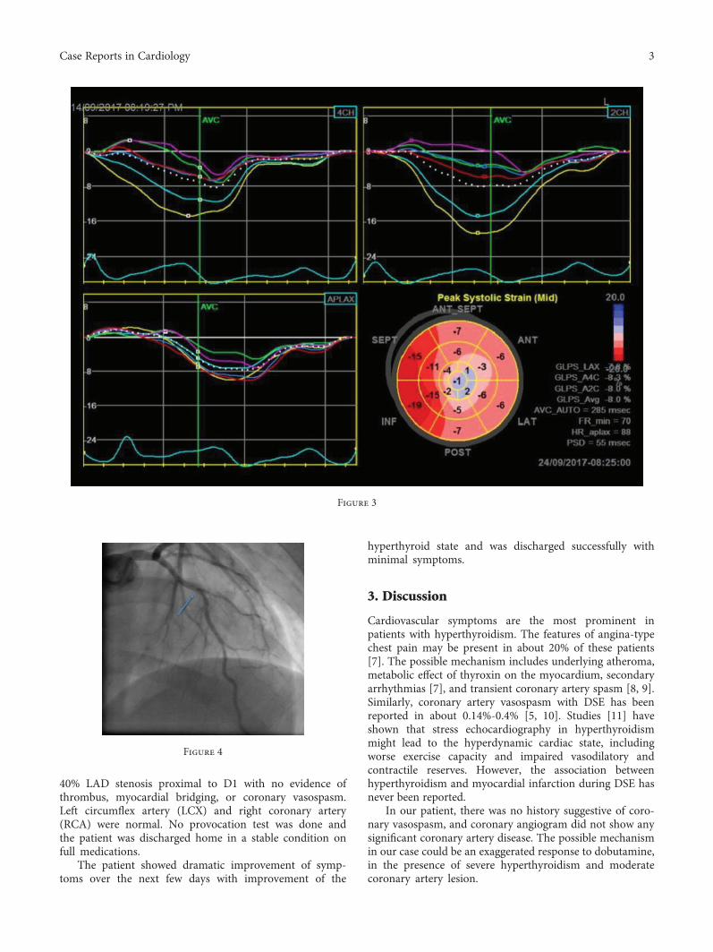
40% LAD stenosis proximal to D1 with no evidence ofthrombus, myocardial bridging, or coronary vasospasm.Left circumflex artery (LCX) and right coronary artery(RCA) were normal. No provocation test was done andthe patient was discharged home in a stable condition onfull medications.
The patient showed dramatic improvement of symp-toms over the next few days with improvement of the
hyperthyroid state and was discharged successfully withminimal symptoms.
3. Discussion
Cardiovascular symptoms are the most prominent inpatients with hyperthyroidism. The features of angina-typechest pain may be present in about 20% of these patients[7]. The possible mechanism includes underlying atheroma,metabolic effect of thyroxin on the myocardium, secondaryarrhythmias [7], and transient coronary artery spasm [8, 9].Similarly, coronary artery vasospasm with DSE has beenreported in about 0.14%-0.4% [5, 10]. Studies [11] haveshown that stress echocardiography in hyperthyroidismmight lead to the hyperdynamic cardiac state, includingworse exercise capacity and impaired vasodilatory andcontractile reserves. However, the association betweenhyperthyroidism and myocardial infarction during DSE hasnever been reported.
In our patient, there was no history suggestive of coro-nary vasospasm, and coronary angiogram did not show anysignificant coronary artery disease. The possible mechanismin our case could be an exaggerated response to dobutamine,in the presence of severe hyperthyroidism and moderatecoronary artery lesion.
Figure 3
Figure 4
3Case Reports in Cardiology

Similarly, studies [10, 12, 13] have shown STEMI dueto oxygen supply-demand mismatch, vasospasm, coronaryartery dissection, or coronary artery plaque rupture withsubsequent thrombus formation.
Β-blockade may leave α-adrenergic vasoconstrictionunopposed.Whether administration of propranolol enhancescoronary spasm is not well established. Alvarez et al. [14]reported coronary vasospasm after administration of pro-pranolol; however, other studies did not support this theory[15]. Other studies found no advantage of propranolol overdiltiazem in preventing coronary spasms [16]. Keeping inmind those previous studies done on euthyroid patients,it may be not applicable to our patient which showeddramatic improvement of symptoms after increasing thepropranolol doses.
In our case, we did not see any coronary artery dissectionor plaque rupture. It is difficult to rule out a plaque rupturewith superimposed thrombus formation, but the transientnature of ST elevation will go against this possibility.Similarly, the coronary angiogram procedure was delayedin order to control his hyperthyroid state firstly, andtherefore, the thrombus if present might have been resolvedduring this time.
4. Conclusion
We believe our patient had transient ST-elevation myocar-dial infarction, secondary to DSE in a poorly controlledhyperthyroid state and moderated coronary artery disease.We would suggest treating hyperthyroidism prior to request-ing noninvasive stress test and avoiding invasive coronaryangiogram due to thyroid storm risk, based on the clinicalpicture. Our case also suggests that propranolol may bebeneficial in controlling cardiac symptoms and possiblyvasospasms in patients with hyperthyroidism.
Conflicts of Interest
The authors declare that there is no conflict of interestregarding the publication of this paper.
References
[1] I. Klein and K. Ojamaa, “Thyroid hormone and the cardiovas-cular system,” The New England Journal of Medicine, vol. 344,no. 7, pp. 501–509, 2001.
[2] M. L. Geleijnse, P. M. Fioretti, and J. R. T. C. Roelandt,“Methodology, feasibility, safety and diagnostic accuracy ofdobutamine stress echocardiography,” Journal of the AmericanCollege of Cardiology, vol. 30, no. 3, pp. 595–606, 1997.
[3] F. Lattanzi, E. Picano, E. Adamo, and A. Varga, “Dobutaminestress echocardiography: safety in diagnosing coronary arterydisease,” Drug Safety, vol. 22, no. 4, pp. 251–262, 2000.
[4] B. M. Gordon, V. Mohan, A. T. Chapekis et al., “An analysis ofthe safety of performing dobutamine stress echocardiographyin an ambulatory setting,” Journal of the American Society ofEchocardiography, vol. 8, no. 1, pp. 15–20, 1995.
[5] N. Lamisse, A. Cohen, C. Chauvel et al., “Dobutamine stressechocardiography; a monocentric experience on consecutive
patients. Effect of age,” Archives des maladies du coeur et desvaisseaux, vol. 90, no. 11, pp. 1455–1461, 1997.
[6] M. Fineschi, V. Zaca, M. Padeletti, T. Gori, and P. Aitiani,“Transmural myocardial ischaemia complicating recoveryafter dobutamine-atropine stress echocardiography in patientswith non-significant coronary artery disease: insights frominvasive assessment of coronary physiology,” European Jour-nal of Echocardiography, vol. 12, no. 7, article E34, 2011.
[7] L. Resnekov and R. E. Falicov, “Thyrotoxicosis and lactateproducing angina pectoris with normal coronary arteries,”Heart, vol. 39, no. 10, pp. 1051–1057, 1977.
[8] J. A. Jaber, S. Haque, H. Noor, B. Ibrahim, and J. al Suwaidi,“Thyrotoxicosis and coronary artery spasm: case report andreview of the literature,” Angiology, vol. 61, no. 8, pp. 807–812, 2010.
[9] J. H. Kim, “A case of myocardial ischemia and myocardialinjury caused by coronary vasospasm associated with hyper-thyroidism,” Korean Circulation Journal, vol. 30, no. 3,pp. 369–372, 2000.
[10] N. Masencal, I. El Hajjaji, R. El Mahnoud, F. Digne, andO. Dubourg, “Prevalence of coronary artery spasm duringdobutamine stress echocardiography,” The American Journalof Cardiology, vol. 109, no. 6, pp. 800–804, 2012.
[11] G. J. Kahaly, S. Wagner, J. Nieswandt, S. Mohr-Kahaly, andT. J. Ryan, “Stress echocardiography in hyperthyroidism,”The Journal of Clinical Endocrinology & Metabolism, vol. 84,no. 7, pp. 2308–2313, 1999.
[12] A. Elhendy, W. Ginete, S. Shurmur, and T. R. Porter, “Acutemyocardial infarction after a negative dobutamine stressechocardiogram,” European Journal of Echocardiography,vol. 5, no. 6, pp. 469–471, 2004.
[13] I. Karabinos, A. Papadopoulos, S. Koulouris, A. Kranidis,S. Korovesis, and D. Katritsis, “Spontaneous coronary arterydissection during a dobutamine stress echocardiography,”Echocardiography, vol. 23, no. 3, pp. 232–234, 2006.
[14] L. Alvarez, J. Zamorano, L. Mataix, C. Almeria, R. Moreno,and J. L. Rodrigo, “Coronary spasm after administration ofpropranolol during dobutamine stress echocardiography,”Revista Española de Cardiología, vol. 55, no. 7, pp. 778–781, 2002.
[15] N. De Cesare, S. Cozzi, A. Apostolo et al., Coronary ArteryDisease, Lippincott-Raven Publishers, 1994.
[16] P. Y. Tilmant, J. M. Lablanche, F. A. Thieuleux, B. A. Dupuis,and M. E. Bertrand, “Detrimental effect of propranolol inpatients with coronary arterial spasm countered by combina-tion with diltiazem,” The American Journal of Cardiology,vol. 52, no. 3, pp. 230–233, 1983.
4 Case Reports in Cardiology

Stem Cells International
Hindawiwww.hindawi.com Volume 2018
Hindawiwww.hindawi.com Volume 2018
MEDIATORSINFLAMMATION
of
EndocrinologyInternational Journal of
Hindawiwww.hindawi.com Volume 2018
Hindawiwww.hindawi.com Volume 2018
Disease Markers
Hindawiwww.hindawi.com Volume 2018
BioMed Research International
OncologyJournal of
Hindawiwww.hindawi.com Volume 2013
Hindawiwww.hindawi.com Volume 2018
Oxidative Medicine and Cellular Longevity
Hindawiwww.hindawi.com Volume 2018
PPAR Research
Hindawi Publishing Corporation http://www.hindawi.com Volume 2013Hindawiwww.hindawi.com
The Scientific World Journal
Volume 2018
Immunology ResearchHindawiwww.hindawi.com Volume 2018
Journal of
ObesityJournal of
Hindawiwww.hindawi.com Volume 2018
Hindawiwww.hindawi.com Volume 2018
Computational and Mathematical Methods in Medicine
Hindawiwww.hindawi.com Volume 2018
Behavioural Neurology
OphthalmologyJournal of
Hindawiwww.hindawi.com Volume 2018
Diabetes ResearchJournal of
Hindawiwww.hindawi.com Volume 2018
Hindawiwww.hindawi.com Volume 2018
Research and TreatmentAIDS
Hindawiwww.hindawi.com Volume 2018
Gastroenterology Research and Practice
Hindawiwww.hindawi.com Volume 2018
Parkinson’s Disease
Evidence-Based Complementary andAlternative Medicine
Volume 2018Hindawiwww.hindawi.com
Submit your manuscripts atwww.hindawi.com