
Stem cells therapy for peripheral arterial occlusive disease
41
Stem Cells Therapy for Peripheral Arterial Occlusive Disease Dong-Ik Kim, MD. , Ph.D ([email protected]) Division of Vascular Surgery, Samsung Medical Center Sungkyunkwan University School of Medicine, Seoul, Korea
-
Upload
uvcd -
Category
Presentations & Public Speaking
-
view
57 -
download
1
Transcript of Stem cells therapy for peripheral arterial occlusive disease

Stem Cells Therapy for Peripheral Arterial Occlusive Disease
Dong-Ik Kim, MD. , Ph.D ([email protected])Division of Vascular Surgery, Samsung Medical Center
Sungkyunkwan University School of Medicine, Seoul, Korea

Prospect of Stem Cell Therapy
• Will the stem cell be effective for the
treatment of peripheral arterial occlusive
disease in the future?
Yes, it will be.

Evidences of Stem Cell Effect
• Animal Study
• Clinical Study

Animal Study (2002 - )
• Whole bone marrow stem cell
• Bone marrow derived mononuclear cell
• Bone marrow derived MSC
• Cord blood derived MNC
• Cord blood derived MSC
• Adipose tissue derived MSC

vWF HNA Merge
Angiogenesis : Differentiated from injected stem cell

Angiogenesis - angiogram
Control Treat

Dog Ischemic Limb Model- Mongrel Dog : male, 20-25 ㎏ - Femoral Arteries : occluded with ameroid constrictor

Mouse limb ischemia model

Animal Model
• Methods of stem cell injection : intramuscular injection
• Harvest : 8 weeks & 6 month after stem cell injection
• Assessment for angiogenic effect
1) Angiography
2) Immunohistochemical staining

0.8
1
1.2
1.4
1.6
1.8
2
2.2
2.4
capillary rato
HCB-MNC
Control 4x10^6 4x10^7 4x10^8
*
**
**
Human cord blood mononuclear cell transplantation : capillary ratios

Human cord blood mononuclear cell transplantation: Angiogenic factor - Western blot

(A) (B) (C)
(D) (E) (F)
Human cord blood mononuclear cell transplantation : Angiogenic factor - Tunnel assay & immunohistochemical stain

*
*
*
*
* **
*
#
#
#
#
Human cord blood mononuclear cell transplantation : Angiogenic gene expression - Real Time-PCR
Conclusions from animal study
• Angiogenesis comes from injected stem cells
• Paracrine effect of injected stem cells
1) secretion of cytokines : PDGF, VEGF etc.

Evidences of Stem Cell Effect
• Animal Study
• Clinical Study

Lists of Clinical Trials
• Autologous whole bone marrow stem cell implantation : Tibia bone fenestration technique (2004- 2006 )
• Autologous whole bone marrow stem cell implantation :
aspiration from iliac bone (2007- 2009)
• Homologous HCB-MNC Injection (2008-2010)
• Homologous HCB-MSC transplantation (2011- 2012)

T F
Autologous whole bone marrow stem cell implantation (2004):
Tibia bone fenestration technique

Demographics
• Total 27 patients ( 34 limbs)
• 26 male (33 limbs) / 1 female (1 limb)
• Mean age : 37.6 ± 6.9 years
• Mean FU periods : 19.1 ± 3.5 month (12.4 - 25month)

Angiographic findings
* (A, C, E) : pre-operative status (B, D, F) : post-operative status
* B : +1 D : +2 F : +3 angiogenic status
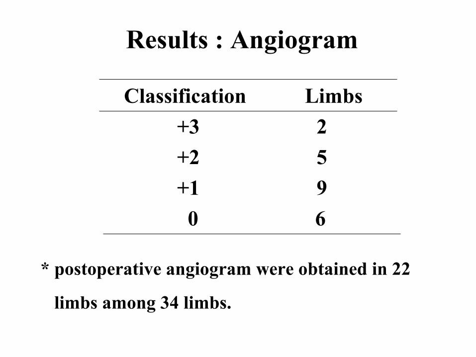
Results : Angiogram
Classification Limbs
+3 2
+2 5
+1 9
0 6
* postoperative angiogram were obtained in 22
limbs among 34 limbs.

Pre stem cell therapy
Poststem cell therapy
Rates of clinical improvement : 79.7 %


Clinical Trials
• Autologous whole bone marrow stem cell implantation : Tibia bone fenestration technique (2004- 2006 )
• Autologous whole bone marrow stem cell implantation :
aspiration from iliac bone (2007- 2009)
• Homologous HCB-MNC Injection (2008-2010)
• Homologous HCB-MSC transplantation (2011- 2012)

VİDEO

July 2004 - June 2009
90 ischemic limbs with 67 patients 1. Mean age: 39.8 ± 7.9 years 2. Mean follow-up period: 29.3 ± 18.1 months
Symptoms 1. Intermittent claudication : 44 limbs 2. Critical limb ischemia : 46 limbs
Demographics

Angiographic outcomes (n=37)1. recanalization of the run-off vessels: 3 limbs (8.1%)2. development of new collateral vessels (angiogenesis) : 16 limbs (43.2%)3. increased diameter and/or length of preexisting collateral vessels (arteriogenesis) : 15 limbs (40.5%)
Rates of angiographic improvement : 43.2 %
Rates of clinical improvement : 55.6 %
Results

Number of injected cells
Total MNCs (/ml) CD34+ (/ml)
Mean No. P Mean No. P
PB
Day 0 2.71×10⁶ ± 1.13×10⁶ 0.247 2.23×10⁵ ± 2.57×10⁵ 0.620
Day 1 3.19×10⁶ ± 1.77×10⁶ 0.688 3.34×10⁵ ± 3.43×10⁵ 0.509
Day 2 2.87×10⁶ ± 1.68×10⁶ 0.472 3.59×10⁵ ± 4.61×10⁵ 0.906
BM Day 3 4.46×10⁶ ± 1.02×10⁷ 0.090 1.62×10⁵ ± 2.53×10⁵ 0.167
CD133+ (/ml) CD34+CD133+ (/ml)
Mean No. P Mean No. P
PB
Day 0 1.22×10⁵ ± 1.61×10⁵ 0.943 6.19×10⁴ ± 1.63×10⁵ 0.143
Day 1 2.13×10⁵ ± 2.42×10⁵ 0.464 8.38×10⁴ ± 9.19×10⁴ 0.150
Day 2 2.04×10⁵ ± 2.05×10⁵ 0.706 6.84×10⁴ ± 6.93×10⁴ 0.345
BM Day 3 1.16×10⁵ ± 1.35×10⁵ 0.142 4.73×10⁴ ± 6.74×10⁴ 0.225


Clinical Trials
• Autologous whole bone marrow stem cell implantation : Tibia bone fenestration technique (2004- 2006 )
• Autologous whole bone marrow stem cell implantation :
aspiration from iliac bone (2007- 2009)
• Homologous HCB-MNC Injection (2008-2010)
• Homologous HCB-MSC transplantation (2011- 2012)

• 7 ischemic limbs with 7 patients• Number of injected stem cells : 4×10⁸
• Safety : No adverse effect • Rates of clinical improvement : 57%• Rates of angiographic improvement : 71.4%
Homologous HCB-MNC Injection


Clinical Trials
• Autologous whole bone marrow stem cell implantation : Tibia bone fenestration technique (2004- 2006 )
• Autologous whole bone marrow stem cell implantation :
aspiration from iliac bone (2007- 2009)
• Homologous HCB-MNC Injection (2008-2010)
• Homologous HCB-MSC transplantation (2011- 2012)

• Title : The Safety of Human Cord Blood-derived Mesenchymal Stem Cells Therapy in Patients with PAOD : Phase I Clinical Study• KFDA approval : 2011. 5 • IRB approval from SMC : 2011. 5 • Target disease : peripheral arterial occlusive disease• Inclusion criteria : Rutherford’s classification IIb, III, IV• Source of Stem Cells : Human Cord Blood-derived Mesenchymal Stem Cells • Injected cell number : 1 X 10⁷ • Injection site : calf muscle & vicinity of tibioperoneal artery
HCB –MSC injection (Phase I Study)

• Primary endpoint : Safety evaluation (1,3,6 month)1) death 2) cardiovascular event3) anaphylactic shock or allergic reaction4) acute or chronic graft-versus host disease5) procedure-related complications
• Secondary endpoint1) wound healing rate2) change of segmental limb pressure3) change of the scale for gauging changes in clinical status
End Point of Phase I study

NoAGE
pre postGauging change
Angio TotalRutherford category
Sx VAS ABIRutherford category
Sx VAS ABI
1 31 5 Ulcer 7 ATA 0.31PTA 0.46
2 Healed 0 ATA 0.44PTA 0.47 +1 +2
2 46 3 Claudication 7 ATA 0
PTA 0.682 improve 1 ATA 0
PTA 0.8 +2
3∫ 72 3 Claudication 7 ATA 0.24
PTA 0.472 improve 3 ATA 0.46
PTA 0.52 +1 +2
4 56 4 Claudication 7
ATA 0.26PTA 0.3
3 improve 5ATA 0.19 PTA 0.73 +2
5 56 5 Ulcer 8 ATA 0.32PTA 0.36
4 Healed 2 ATA 0.19PTA 0.33 +1
6* 48 5 Ulcer 10 ATA 0PTA 1.02
1 Healed 1 ATA 1.18PTA 0.94 +2
7 77 6 Ulcer 6ATA 0.57PTA
06 improve 2
ATA 0.56PTA
00 +1
8 48 3 Claudication 8 ATA 0.4
PTA 0.613 improve 5 ATA 0.25
PTA 0.44 0
9∬ 44 5 Ulcer 5 ATA 0.55PTA 0.68
5 improve 2 ATA 0.59PTA 0.68 0
10* 47 5 Ulcer 5 ATA 1.08PTA 0.68
1 Healed 0 ATA 1.09PTA 0.74 +1
*: Toe amputation, ∫: Cr elevation, ∬: urticaria
Results : HCB –MSC injection

• Ministry of Health and Welfare• Approve the autologous bone marrow stem cell therapy for PAOD
as a New Health Technology (2013.2)• Autologous Bone Marrow Stem Cell Transplantation became a
common treatment modality to the patient for PAOD in Korea.
Stem Cell therapy for PAOD in Korea

Conclusions from clinical trials
• Stem cell therapy is safe and effective for the treatment for the peripheral arterial occlusive disease.

What’s your choice?
International Journal of Stem Cells (http://www.ijstemcell.com)

Acknowledgment

Thank you


![ARTERIAL PERIPHERAL VASCULAR DISEASES.ppt [Read-Only]ocw.usu.ac.id/course/download/1110000113-cardiovascular-system/… · arterial peripheral vascular diseases acute arterial occlusion](https://static.fdocuments.us/doc/165x107/604e83caf1418f71db611c5a/arterial-peripheral-vascular-read-onlyocwusuacidcoursedownload1110000113-cardiovascular-system.jpg)


