Stem cell-based reporter assays for the identification of...
69
UNIVERSIDADE DE LISBOA Faculdade de Ciências Departamento de Biologia Animal Stem cell-based reporter assays for the identification of carcinogenic properties of (non)-genotoxic chemicals Cátia Sofia Alão Gonçalves DISSERTAÇÃO MESTRADO EM BIOLOGIA HUMANA E AMBIENTE 2013
Transcript of Stem cell-based reporter assays for the identification of...

UNIVERSIDADE DE LISBOA Faculdade de Ciências
Departamento de Biologia Animal
Stem cell-based reporter assays for the identification of carcinogenic properties of
(non)-genotoxic chemicals
Cátia Sofia Alão Gonçalves
DISSERTAÇÃO
MESTRADO EM BIOLOGIA HUMANA E AMBIENTE
2013
UNIVERSIDADE DE LISBOA Faculdade de Ciências
Departamento de Biologia Animal
Stem cell-based reporter assays for the identification of carcinogenic properties of
(non)-genotoxic chemicals
Cátia Sofia Alão Gonçalves
Dissertação orientada por: Dr. Harry Vrieling (Toxicogenetic/LUMC)
Professora Dra. Deodália Dias (DBA/FCUL)
DISSERTAÇÃO
MESTRADO EM BIOLOGIA HUMANA E AMBIENTE
2013

i
Acknowledgements
First of all I would like to express my deep and sincere gratitude to Harry
Vrieling, for having welcomed me in your workgroup and give me the opportunity to
develop my dissertation. I also have to thank Giel Hendriks for the useful discussions
and uplifting spirits that allowed me to develop my dissertation and allowed me to
develop, not only the professional level, but also the personal one.
A special thanks to the entire lab, in particular to Brunno for helping me with the
BAC recombennering and the great talks, to Fabienne for the tips on how to culture the
mES cell lines and for her constant assistance in the Lab and to Binni for helping me
understand a little better the Dutch culture. I wish to thank all of my colleagues at the
Department of Toxicogenetics, at the Leiden University Medical Centre, for all the
support.
I thank Professor Deodália Dias for all the support provided under this thesis and
also in my training.
I want to thank to all the friends that I made during my time in Leiden, without
them, probably this dissertation would not be completed. They made the time in Leiden,
one of the best experiences I've ever had.
And of course, to my friends, that even separated by 2.440 km, gave me all the
support in the world, they always believed that I had the strength to go further.
The last but not least, to my family who always believed in me and gave me that
moral and financial support to pursue my dreams.
I would like to thank all those that believe in me
To all,
Obrigada, Thank you, Gracias, Dank je wel, Merci, Grazie.

ii
The project which allowed the realization of this dissertation was financially
supported by the Dutch Technology Foundation STW, on the Toxicogenetics
department in the group of Dr. Harry Vrieling with the supervising of Giel Hendriks.
Tables, figures and bibliographic references in this thesis are in accordance with
the standards of the journal Mutation Research/Fundamental and Molecular
Mechanisms of Mutagenesis.
Nothing in Biology Makes Sense
Except in the Light of Evolution”
Theodosius Dobzhansk, 1973.

iii
Resumo
Nos dias que correm, existem a geração contínua de novos produtos químicos, quer seja para o desenvolvimento de drogas, quer para aplicação na indústria alimentar, cosmética ou mesmo na agricultura. Portanto, antes que um composto possa ser colocado à venda no mercado, é necessário realizar vários testes, para que se possa excluir aquelas propriedades nocivas para a saúde humana, incluindo o potencial risco de cancro. No entanto devido ao crescente número de novos produtos químicos, existe a forte necessidade de encontrar ensaios in vitro que sejam rápidos em fornecer os resultados, baratos, relativamente fáceis de aplicar e ainda que apresentem uma taxa alta de sensibilidade de fiabilidade para avaliação do risco de cancro para o Homem.
O DNA é uma molécula complexa, que necessita de alguma integridade, para tal esta tem de ser protegida dos agentes ambientais, tais como a luz UV, produtos químicos provenientes de alimentos, medicamentos, ou mesmo gerada espontaneamente durante o metabolismo do DNA. Por esta razão, os organismos foram capazes de desenvolver vários mecanismos de defesa celular, incluindo as vias de reparação do DNA, os “checkpoint” do ciclo celular ou a indução da apoptose, de modo a proteger contra os efeitos tóxicos, mutagénicos de um possível exposição.
As propriedades carcinogénicas dos químicos são frequentemente associados com o seu potencial genotóxico, ou seja, a capacidade de provocar lesões no DNA. Como resultado, estes químicos podem modificar a estrutura do DNA, levar à formação de ligações cruzadas, ductos, quebras simples e duplas da cadeia de DNA, bloquear a progressão do garfo de replicação ou os mecanismos de transcrição de um gene. A interferência da replicação do DNA ou da transcrição de um genes pode levar a mutações e rearranjos cromossómicos e, por vezes, pode levar a um evento oncogénico.
Muitos dos químicos carcinogénicos são capazes de levar a lesões do DNA e são “genotóxicos” no seu modo de acção. Existem, no entanto, um grupo de compostos carcinogénicos que induzem cancro na forma não-genotóxicos, ou seja, sem levar a

iv
alterações do DNA, quer seja a nível do número de cromossoma quer seja a nível da estrutura.
O modo pelo qual os químicos carcinogénicos não-genotóxicos exercem a sua acção pode incluir os supressores imunológicos, proliferados de peroxissomas, agonistas dos receptores de hormonas, inibidores da fosfatase e os metalóides.
No entanto, ao contrário dos compostos carcinogéneos genotóxicos, o mecanismo pelo qual os carcinogénicos não-genotóxicos levam à carcinogénese ainda é desconhecido. Como tal, actualmente não existem ensaios in vitro disponíveis para estabelecer as propriedades potencialmente carcinogéneas dos compostos cancerígenos não-genotóxicos. Usualmente, para detectar agentes cancerígenos não-genotóxicos recorresse a estratégias que utilizam ensaios baseados em parâmetros genotóxicos. O teste de Ames é um teste bacteriano que utiliza a mutagenicidade como parâmetros genotóxicos, mas pode-se também recorrer a genotoxicidade em células de mamíferos. No entanto, estes compostos não são detectados por estes ensaios, e acabam por passar despercebido. Logo, o ensaio mais utilizado para este caso de compostos químicos é recorrer a bioensaios para detectar a carcinogenicidade, usando roedores.
Logo, face ao aumento do número de composto e com vista à diminuição de bioensaios, realizámos este projecto que tinha como objectivo descrever a geração de um novo repórter baseado na utilização de GFP que permitia detectar as possíveis propriedades de compostos cancerígenos não-genotóxicos, que era específico para a resposta a proteínas mal enoveladas (UPR, do inglês: Unfolded Protein Response)
Para que se pudesse desenvolver estes repórteres baseados em GFP, realizámos um “genome-wide transcription profiling” utilizando mES que foram previamente expostas a diversos agentes cancerígenos não-genotóxicos.
Após análise dos “genome-wide transcription profiling”, foi possível seleccionar quatro genes biomarcadores, Armet, Derl3, Dnajc3 e Ddit3, que eram induzidos após exposição a supressores imunológicos e a metalóides. Recorrendo ao qRT-PCR confirmámos esta indução, e verificámos que os genes biomarcadores Armet, Derl3 e Dnajc3 eram induzidos após exposição com o supressor imunológico cisclosporina A, e o gene biomarcador Ddit3 era induzido após exposição ao metalóide arsenito de sódio.
Para que os genes biomarcadores que seleccionámos pudessem ser utilizados como repórteres baseados em GFP, recorremos à técnica de recombinação de cromossoma artificial bacteriano (BAC, do inglês: bacterial artificial chromosome). Esta técnica consiste em gerar linhas celulares que expressam uma proteína de fusão de GFP e, portanto, é possível determinar facilmente indução do gene pela quantidade de

v
fluorescência emitida pela proteína de fusão de GFP modificada após o tratamento com um determinado composto.
Uma vez que os genes biomarcadores, Armet-GFP, Derl3-GFP, Dnajc3-GFP e Ddit3-GFP, se encontraram funcionais, estes foram expostos a cancerígenos não-genotóxicos, que incluía a: cisclosporina A; o arsenito de sódio; e a tunicamicina, e a carcinogéneos genotóxicos, nomeadamente a cisplatina, a doxorrubicina e a peróxido de hidrogénio. Após análise da citometria de fluxo, verificámos que os nossos genes biomarcadores não eram induzidos por químicos carcinogéneos genotóxicos, mas que por sua vez eram fortemente induzidos quer por a cisclosporina A e arsenito de sódio, mas também por tunicamicina, que nos levou a afirmar que estes biomarcadores eram específicos para o UPR e que o supressor imunológico cisclosporina A e o ao metalóide arsenito de sódio estão envolvidos no UPR.
Uma vez desenvolvidos os repórteres GFP, o próximo passo era testar a especificidade e sensibilidade. Para tal, validámos, juntamente com os genes biomarcadores para os compostos cancerígenos não-genotóxicos, uns repórteres GFP desenvolvidos anteriormente para os compostos carcinogéneos genotóxicos. Os repórteres GFP foram expostos a 53 químicos propostos pela ECVAM, que inclui compostos carcinogéneos genotóxicos, cancerígenos não-genotóxicos e compostos não-carcinogénicos. Após análise dos dados, observámos que tínhamos desenvolvido repórteres GFP com diferentes sensibilidades, uns eram capazes de detectar compostos carcinogéneos genotóxicos que induziam lesões genotóxicas ou stress oxidativo, e outros que eram capazes de detectar compostos cancerígenos não-genotóxicos que detectavam compostos que induziam lesões nas proteínas.
Para que pudéssemos correlacionar a activação dos repórteres GFP com um fim biológico, decidimos recorrer a um ensaio in vitro vulgarmente utilizado para detectar genotoxicidade. Recorremos à utilização do ensaio do cometa, visto este ser um ensaio que é amplamente utilizado para avaliar lesões no DNA. No entanto, este ensaio ainda não tinha sido descrito para as mES. Começamos por optimizar o ensaio do cometa e concluímos que este não tinha a capacidade de detectar compostos cancerígenos não-genotóxicos, e para além de que mesmo tendo a capacidade de detectar compostos carcinogéneos genotóxicos, não eram informativo sobre o tipo de lesão que induzia, quer fosse lesões no DNA ou dano oxidativo.
Em suma, podemos concluir que ao usar a combinação de diferentes linhagens celulares de repórteres baseados em GFP, somos capazes de discriminar entre compostos que induzem principalmente lesões genotóxicas, stress oxidativo ou lesões nas proteínas

vi
Com este estudo fomos ainda capazes de demonstrar que o ensaio do cometa, um ensaio tradicionalmente utilizado in vitro para detectar propriedades genotóxicos de compostos, não tem a mesma sensibilidade e especificidade que os repórteres GFP que desenvolvemos, na medida em que o ensaio do cometa não permitiu detectar compostos carcinogéneos não-genotóxicos e não forneceu qualquer informação sobre o tipo de lesão causada pelos compostos carcinogéneos genotóxicos.
Com este estudo, podemos afirmar que desenvolvemos um ensaio in vitro, que é altamente sensível e específico, que pode fornecer informações sobre a toxicidade relativa de produtos químicos, e que ao mesmo tempo é rápido e tem elevada fiabilidade para avaliação do risco de cancro para o Homem e que permite de certa forma levar a redução de biosensaios.
Palavras-chave: Repórteres-GFP, carcinogéneos não-genotóxicos, genotoxicidade, UPR.
vii
Abstract
There is a continuous generation of new chemicals for drug development or applications in food industry, cosmetics and agriculture. Before compounds are allowed on the market, they first require testing to exclude hazardous properties on human health, including potential cancer risk. Due to the increasing number of new chemicals there is a strong demand for rapid, easy to use high-throughput in vitro assays for human cancer risk assessment. The induction of cancer is strongly associated with DNA damage and mutations upon exposure to genotoxic compounds. However, there are many non-genotoxic (NGTX) compounds that have been shown to be carcinogenic. The mechanisms by which these non-genotoxic carcinogens (NGTXC) induce cancer is often unclear. Here we describe the development of four different mES reporter cell lines for the detection of possible carcinogenic properties of non-genotoxic chemicals. The GFP-based reporter cell lines are based on the activation of biomarker genes that are predictive for exposure to specific NGTXC. By genome transcription profiling of mES and qRT-PCR we identified genes that were transcriptionally activated upon exposure to NGTXC. Four genes (Armet, Dnajc3, Derl3 and Ddit3) were induced by the immune suppressors CsA and Fk506 and by the metalloid NaAsO2. These genes were generated by BAC recombineering, were GFP-reporters genes were stably integrated in mES cells. These biomarker genes have previously been associated with the induction of the unfolded protein response (UPR), suggesting the UPR is a mode of action of CsA and NaAsO2. These GFP-reporters cell lines are specifically activated upon exposure to compounds that induce protein damage. A panel of different mES reporter cell lines will allow identification of the potential carcinogenic properties of non-genotoxic chemicals and in addition can detect protein damage.
Keywords: GFP-reporter, non-genotoxic carcinogens, genotoxicity, UPR
viii
Table of contents
Resumo ................................................................................................................ iii Abstract ............................................................................................................... vii Table of contents ................................................................................................ viii
List of Figures .................................................................................................. ix List of Tables .................................................................................................... x
Abbreviations ....................................................................................................... xi
Chapter 1. Introduction ................................................................................................. 1
1.1. Genotoxic carcinogens .............................................................................................................. 1
1.2. DNA repair systems ................................................................................................................... 2
1.2.1. DNA damage response (DDR) .......................................................... 3 1.3. Current Methods for (Geno) toxicity Testing ................................................................ 4
1.4. Non-genotoxic carcinogens .................................................................................................... 6
Objectives ............................................................................................................. 7
Chapter 2. Material and Methods ................................................................................. 9
Embryonic stem cell culture and treatments ......................................................... 9 Gene expression profiling ..................................................................................... 9 Generation of GFP reporter cell lines ................................................................. 10 Detection of GFP expression .............................................................................. 11 Validation of GFP reporter cell lines .................................................................. 11 Comet assay ........................................................................................................ 12
Chapter 3. Results ........................................................................................................ 14
3.1. Reporters for non-genotoxic carcinogens ...................................................................... 14
3.1.1. Identification of biomarkers genes .................................................. 14 3.1.2. Development and selection of GFP-reporter cell lines .................... 20 3.1.3. GFP-based reporters: sensitivity and specificity ............................. 24
3.2. Validation of the GFP-reporters for non-genotoxic carcinogens ........................ 27
3.3. Comet assay .................................................................................................................................. 34
Chapter 4. Discussion ................................................................................................... 38
References...................................................................................................................... 43
Supplementary data ..................................................................................................... xii
Supplementary Method 1: BAC recombineering ................................................................ xii
Supplementary Method 2: RNA analysis protocol ............................................................. xv
Supplementary Method 3: Comet assay protocol .............................................................. xvi
ix
List of Figures
Figure 1. DNA damage agents, repair process and consequences.. ................................ 2
Figure 2. Representation of a comet . ............................................................................ 13
Figure 3. Expression of possible biomarker genes for immune suppressor by qRT-PCR
................................................................................................................................ 17
Figure 4. Expression of biomarker genes for PPARα ligands by qRT-PCR. ................ 18
Figure 5. Expression of possible biomarker genes for metalloids by qRT-PCR ........... 19
Figure 6. Schematic representation of the RecE and RecT recombination enzymes and
recombination of GFP-ires-Neo cassette into the BAC]. ....................................... 20
Figure 7. Gel electrophoresis of PCR analysis of modified BAC-derived reporter genes.
................................................................................................................................ 21
Figure 8. Monoclonal mES cell line selection ............................................................... 22
Figure 9. Clone selection. ............................................................................................. 23
Figure 10. The mES reporter cell lines respond preferentially to compounds that induce
protein damage ....................................................................................................... 25
Figure 11. Check the sensitivity and specificity of the GFP-reporter ........................... 26
Figure 12. Validation for the reporters, tested with 53 compounds suggested by
ECVAM .................................................................................................................. 32
Figure 13. ToxPlot for Ddit3-GFP, Blvrb-GFP, Btg2-GFP and Rtkn-GFP .................. 33
Figure 14. Alkaline comet assay .................................................................................... 35
Figure 15. Neutral comet assay. .................................................................................... 36

x
List of Tables
Table 1. Chemicals and concentrations used for exposure of wt mES cell line ............ 15
Table 2. Biomarker genes that were selected by genome wide transcription profiling of
mES ........................................................................................................................ 16
Table 3. Various GFP reporters and the biological effects ............................................ 28
Table 4. Tested compounds for the validation of the GFP-reporters. ............................ 29
xi
Abbreviations
CDC25 Cell division cycle 25 cDNA Complementary deoxyribonucleic acid Chk1 Checkpoint kinase 1 Chk2 Checkpoint kinase 2 DMEM Dulbecco's Modified Eagle Medium DMSO Dimethyl sulfoxide DNA Deoxyribonucleic acid EDTA Ethylenediaminetetraacetic acid FACS Fluorescence-activated cell sorting GADD45α Growth arrest and DNA damage inducible alpha GFP Green fluorescent protein G418 Geneticin Hprt Hypoxanthine Phosphoribosyltransferase 1 mES Mouse embryonic stem cells MRE11 Meiotic recombination 11 NaOH Sodium hydroxide NBS1 Nijmegen Breakage Syndrome 1 NF-kB Nuclear Factor Kappa Beta Nrf2 nuclear factor (erythroid-derived 2)-like 2 PBS Phosphate-buffered saline PCR Polymerase chain reaction PKC Protein kinase C qRT-PCR Quantitative reverse transcriptase PCR RNA RiboNucleic Acid ROS Reactive oxygen species siRNA Small interfering RNA SD Standard deviation TBE Tris/Borate/EDTA TE Tris-EDTA UV Ultraviolet YWHAD Protein Kinase C Inhibitor Protein-1

1
Chapter 1
Introduction
Every year, industry developed compounds for a broad range of applications. However, these compounds can have harmful properties on human health, since they can interact with different cellular structures. Compound exposure can induce mutation, affect genome stability, result in ROS formation, contribute to aging processes and can potentiate genetic diseases and cancer risk [1,2] (Fig. 1).
According to estimates of the International Agency for Research on Cancer (IARC), cancer is a leading cause of death and was responsible, in 2008, for 7.6 million deaths worldwide [3]. Therefore, to determine the future risk of cancer it is important to assess the hazard of compounds, as well as developing new techniques to assess the risk.
1.1. Genotoxic carcinogens
DNA is a complex organic molecule responsible for the genetic information of cell bodies. Thus, for the information to be successfully transmitted from generation to generation, DNA integrity must be maintained [4]. To maintain genomic integrity, DNA must be protected from damage induced by environmental agents, such as UV light, chemicals from food, drugs, or generated spontaneously during DNA metabolism [5]. For this reason, organisms have evolved various cellular defence mechanisms, including DNA repair pathways, cell cycle checkpoints or induction of apoptosis, in order to protect against the toxic, mutagenic, and possible oncogenic consequences of exposure [6] (Fig. 1).
The carcinogenic properties of compounds are often associated with their genotoxic potential, i.e., the ability to cause DNA damage. As a result, these compounds can modify the DNA structure, lead to the formation of cross-links, bulky DNA adducts, single- and double strand breaks (SSBs and DSBs) and block progression of the DNA

Chapter 1. Introduction
2
replication or gene transcription machineries. Interference with DNA replication or gene transcription can lead to mutations and chromosomal rearrangements and sometimes can result in an oncogenic event [7].
Fig. 1. DNA damage agents, repair process and consequences. a) DNA damaging agents (top); DNA lesions induced by these agents (middle); DNA repairs systems responsible for the removal of the lesions (bottom). b) DNA damage effects on cell-cycle progression, with the arrest in the G1, S, G2 and M phases (top); and on DNA metabolism (middle); Long-term consequences of DNA injury (bottom) (adapted from Hoeijmakers [5]).
1.2. DNA repair systems
In order for the DSB repair mechanisms to be successfully carried out, eukaryotic cells have developed a cellular mechanisms that regulate the recruitment of DNA repair factors to sites of DNA damage, activate those factors, and coordinate the choice of the pathways to employ for efficient DNA repair [8].
A variety of damage can occur in the DNA, thus, to respond to DNA damage, repair mechanisms specific for many types of lesion have evolved directly to a specific type of damage. Bulky distortions in the DNA helix are detected and repaired by nucleotide excision repair (NER) [9,10], while base excision repair (BER) repairs smaller lesions such as deamination of a base through excision of the damaged base [11]. Mispaired DNA bases are replaced with correct bases by mismatch repair (MMR) [5,12].
Chapter 1. Introduction
3
SSBs are repaired by single-strand break repair (SSBR), whereas DSBs are processed either by homologous recombination (HR) or non-homologous end joining (NHEJ). HR always takes place during replication as it uses the information of the sister chromatid as a template for repair, to restores the genomic sequence of the broken DNA ends. While NHEJ does not make use of a sister chromatid, it merely joints and ligates the broken ends independently of the previous DNA sequence. Thus, NHEJ can take place at any time point in the cell-cycle and promotes the potentially inaccurate relegation of DSBs [13].
However, the cell is not always able to repair the damage and therefore the cell might try to bypass the damage by different mechanisms and continue replication. During replication the cell might bypass the damage via two routes either by damage avoidance or by translesion synthesis (TLS) [2,6]. Damage avoidance uses the sister chromatid as template for further replication and thus is error free. Whereas translation synthesis uses a different polymerase than can more easily read over the damage. However, this polymerase has greatly reduced template specificity, therefore this polymerase has a high error rate and often incorporates incorrect bases [6]. A high error rate can ultimately lead to incorporation of mutations into the genome and can therefore be a cause of carcinogen.
Each pathway consists of numerous proteins forming a cascade in order to repair the damage as accurate as possible. The activation of cell cycle checkpoints stops the proliferating cell in its cell cycle progression in order to give time to the DNA damage repair machinery to repair the lesion [14,15]. However, when the repair process fails, the cell cycle can be blocked permanently. Therefore, the most efficient mechanism to protect cells against the genotoxicity of a compound is apoptosis. By apoptosis cells that express high levels of DNA damage are removed from the exposed tissue, preventing mutagenesis of the cells [6]. Thereby, all the mechanisms contribute to prevent incorrect genetic information from being passed onto the progeny
1.2.1. DNA damage response (DDR)
The DNA damage response is a kinase-dependent signalling pathways, composed by the atagexia telangiectasia mutated (ATM) and atagexia telangiectasia and Rad3-related (ATR), that have the ability to sense DNA damage and transduce this information to the cell to influence cellular response to DNA damage [1,16].
Both kinases promote cell cycle checkpoint activation and DNA repair systems in response to genotoxic agents. ATM is recruited to DNA double-strand breaks (DSBs)
Chapter 1. Introduction
4
by its regulator the MRN (MRE11, Rad50-NBS1) and is required for efficient DSB repair [7,17]. The ATR kinase with its regulator ATRIP (ATR-interacting protein) senses single-strand DNA (ssDNA) and is activated in response to stalling of DNA replication and is recruited to stalled replication forks. Activation of ATM results in phosphorylation of the Chk2, whereas ATR activates the Chk1. Both Chk1 and Chk2 inhibit CDC25 phosphatase and the p53 tumor suppressor, thereby blocking activation of the cyclin-dependent kinases, inhibiting cell cycle progression, and activating the induction of apoptosis [17,18].
1.3. Current Methods for (Geno) toxicity Testing
Due to the fast development of new compounds and prior to their introduction on the market, it is necessary to develop an assay that is able to assess the potential genotoxic or other deleterious properties of newly developed compounds. It’s crucial for industries, that the assays are fast, cheap and predictive for animals and humans health risk, as well as the environment. High sensitivity and specificity, together with few false-positives or negatives are the features for a good predictive assay [19]. Furthermore, an assay should also provide information about the mode of toxicity of a compound and be able to predict adverse health effects.
There are various validated in vitro and in vivo assay to test the potential cancer hazard of a compound, as well as to detect biological endpoints of exposure. The assays have the ability to measures the DNA damage, mutation or chromosomal aberrations [20].
One of the classic tests for genotoxicity is the Ames test. The Ames test is a bacterial assay that is able to assess the mutagenic potential of a compound by means of the reversion of mutations in a modified Salmonella Typhimurium [7]. The bacterium has a mutation in the histidine gene, making it dependant on a histidine for growth. A possible mutagen compound is considered a mutagen when it is able to mutate the bacterium in a way that it loses its dependence on histidine [20]. However, the Ames test has a relative low sensitivity and therefore often fails in identifying genotoxic properties [21]. Other tests for bacterial mutagenicity, is the SOS/umu-test. This test makes use of the SOS response, in which the cell cycle is arrested and DNA repair is induced. This response is activated upon exposure to genotoxic compound [21].
To establish genotoxicity in eukaryotes, several yeast-based assays have been developed. The DELL assay is based on the revision of mutation in a modified Saccharomyces cerevisiae strains containing a mutation in the his3 gene. The

Chapter 1. Introduction
5
GreenScreen GS assay contains a GFP coupled to the Rad54 gene promoter. This assay monitors the activation of the DNA damage response, by the activation of Rad54 gene expression, when induced upon exposure to various DNA damaging agent [21].
Nevertheless, to assess the risk to human is crucial to use genotoxicity tests based on mammalian cells, however has proven to be much more challenging. The comet assay (single cell gel electrophoresis) detects DNA SSB and DSB, alkaline labile sites and excision repair processes in individual cells. The Comet assay detects DNA breaks via a protocol of cell lysation and electrophoresis under low voltage allowing migration of damaged DNA, if DNA damage has occurred the electrophoresed DNA fragments migrate towards the anode, and appear as a diffuse tail behind the nucleus, producing a shape resembling a comet [22].
The Mouse lymphoma TK assay (MLA) is able to detect mutagenic and clastogenic events at the thymidine kinase (tk) locus of L5178Y mouse lymphoma tk (+/-) cells. The assay makes use of the acquirement of resistance to trifluorothymidine and uses the number and size of cultured clones as read out [23].
The GreenScreen HC assay and the ToxTracker assay are newly developed assays that make use of cellular signalling pathways. The principle of these two tests is based on the activation of biomarker genes involved in the pathways activated upon DNA damage1. The GreenScreen HC makes use of human lymphoblastoid TK6 cells and is based on activation of the GADD45α gene, which is a known target of the p53 tumor suppressor. However, also other signalling pathways, Nrf2 and Nf-kB, transcription factor that play a role in inflammation, which have been associated with Gadd54α induction [7,24].
The ToxTracker assay makes use of transcriptional changes after cell treatment. The assay uses a panel of three GFP-based mES reporter cell lines whose genes are induced after treatment with genotoxic carcinogen compounds. The biomarker genes are the Bscl2 (Bernardelli-Seip congenital lipotrophy 2), the Srxn1 (sulfiredoxin 1) and the Btg2 (B-cell translocation gene 2) [1]. Bscl2-GFP is specifically induced upon exposure to direct DNA damaging agents, the GFP reporter gene is activated in response to DNA replication inhibition and depends on ATR-Chk1 signalling from stalled replication forks [1,7]. The Srxn1-GFP reporter gene is strongly activated upon cellular oxidative stress and directly controlled by the Nrf2 antioxidant pathway [1,7]. The Btg2-GFP reporter is a p53-depend and responsive to DNA-damaging agents and pro-oxidant [7,21]. Together these different cell lines are able to discriminate between compounds that primarily induce genotoxic or oxidative stress [7].

Chapter 1. Introduction
6
1.4. Non-genotoxic carcinogens
Most chemical carcinogens are able to induce DNA damage and are ‘genotoxic’ in their carcinogenic mode of action. There is, however, a group of carcinogens that induce cancer in a non-genotoxic manner, without altering DNA, chromosome number or structure.
Among the processes that non-genotoxic carcinogens have been shown to include immune suppressors, peroxisome proliferators, hormone receptor agonists, phosphatase inhibitors and metalloids [7,25]. However, unlike genotoxic carcinogens which share a unifying characteristic that is genotoxicity, non-genotoxic carcinogens can have diverse modes of action which are, for the most part, tissue and species specific [25], and it´s unclear the exact mechanism by which exposure of these compounds contributes to carcinogenesis [19]. Albeit, the monitoring of non-genotoxic carcinogens is difficult and there are no general markers suitable for all compound classes [19].
Non-genotoxic carcinogens are known to interact with the function of peroxisome proliferators. Peroxisome proliferators are normally involved in mitochondrial and peroxisomal fatty-acid uptake and beta-oxidation. The binding of peroxisome proliferator receptors (PPARα) by non-genotoxic compounds can result in inhibition of apoptosis and in cell proliferation [26]. Cell proliferation is one of the characteristics of carcinogenesis and can also be dependent on the binding of estrogenic substances to intracellular estrogen receptors [27]. Have been previously described that some non-genotoxic carcinogens are estrogen agonists, and also have been implied in the activation of PKC via a target site or receptor [28].
Some non-genotoxic carcinogens have been proven to be immune suppressors [29]. Immune suppression is also known to play a role in carcinogenesis. The immune system attack harmful cells, however when the immune suppressor are administered the immune reactions are suppressed [29], making occurrence and growth of tumors more likely. Nevertheless, in most cases a link between the mode of action of a compound and its carcinogenic effects has not been established [7].
In contrast to genotoxic carcinogens, there are currently no validated in vitro assays available to establish potentially carcinogenic properties for the non-genotoxic compounds [27]. Usually, to detect non-genotoxic carcinogens the used strategy are based on genotoxic endpoints including the Ames mutagenicity test in bacteria, genotoxicity in mammalian cells and germ cell mutagenicity tests, nevertheless non-genotoxic carcinogens are negative in these tests and thus go undetected [25]. Therefore, the traditional assay to detect carcinogenicity is to use rodent 2-year cancer
Chapter 1. Introduction
7
bioassay [30], however there is a new European policy which try to reduce the number of cancer bioassays [25].
It´s crucial to develop an assay that is fast, cheap and allows to reduce the number of animals that are used in the bioassays. However, the lack of assays is due to the diversity of effects elicited by different non-genotoxic carcinogens on the cells physiology. This diversity of effects makes the development of assays to assess carcinogenic effects highly challenging.
A way to solve the problem of lack of assays is to select biomarkers for the difference modes of action the non-genotoxic carcinogens and to assess the gene expression of a cell after treatment with different groups of chemicals [31]. Thus, gene expression profiling might be able to distinct genes specific for a certain response pathway activated after treatment. When a gene is selective for a certain class of compounds it can be used as a biomarker gene for distinction between classes of compounds, like genotoxic carcinogens and non-genotoxic carcinogens [31].
Objectives
The aim of this project was to describe the generation of a novel GFP-based reporter for the detection of possible carcinogenic properties of non-genotoxic compounds, that were specific for the unfolded protein response (UPR).
A systematic genome-wide transcription profiling in mES cells was previously performed on cells exposed to different groups of non-genotoxic carcinogens. Based on the genome-wide transcription profiling, we selected a set of putative biomarker genes for exposure to different classes of NGTXC. We selected three biomarker genes that were specifically induced upon exposure to different immune suppressors and one biomarker genes upon exposure to metalloids. The GFP-based reporter cell lines were generated with BAC recombinational techniques. This technique generates cell lines expressing a GFP fusion protein and therefore functions as a high-throughput method for the examination of the proteins function and induction inside the cell [31]. The GFP biomarker genes were preferentially activated upon exposure to compounds that induce the UPR.
Besides, we validated GFP-based reporters that were developed for NGTXC together with GFP reporters that were developed for detection of carcinogenic properties of genotoxic carcinogens compounds. In order to correlate the GFP-reporter

Chapter 1. Introduction
8
activation with a biological endpoint we used an in vitro assay to establish reporter activation. We use the comet assay, since is an assay that is widely used to measure DNA damage [22]. We optimized the comet assay for the mES, since it is not yet described for this cells type.
9
Chapter 2
Material and Methods
Embryonic stem cell culture and treatments
mES cells were cultured as described previously [1]. C57/Bl6 B4418 wild-type mES cells were cultured in Knockout DMEM (Gibco) which contained 10% fetal bovine serum, 2mM GlutaMAX (Gibco), 1mM sodium pyruvate (Gibco), 100μM β-mercaptoethanol (Gibco), and leukemia inhibitory factor (LIF). mES cells were culture on irradiated primary mouse embryonic fibroblasts as feeders according to the described protocol [21].
mES cells were seeded 24hrs prior to compounds exposure on gelatin-coated 96-well plates in the absence of feeder cells in buffalo rat liver cell (BRL)-conditioned ES cell medium, as has previously been described1. The mES cells were treated with five increasing concentrations per compound that were prepared in DMSO or PBS and added directly to the culture medium.
For treatment that did not required metabolic activation, the cells were continuously exposed for 24hrs. 24hr after cells were washed with PBS, trypsinized and PBS+2% serum was added before FACS analysis.
Gene expression profiling
Induction of the genes selected during the previously performed genome wide transcription profiling was validated using qRT-PCR. Cells were on gelatin-coated 6-well plates, as described [1].
Detailed protocol for the RNA isolation and cDNA synthesis can be found in supplementary method 2. Total RNA was isolated after 8 or 16 h treatment using TRIzol (Invitrogen) and the total RNA concentration was determined by
Chapter 2. Material and Methods
10
spectrophotometer NanoDrop (Isogen ND-1000). cDNA was synthesized using oligo (dT) 12–18 primers and SuperScript Reverse Transcriptase III (Invitrogen) according to the manufacturer’s protocol. Gene expression was determined using specific primers with SYBR Green Supermix (BIO RAD) on a Real Time System C1000 Touch Thermal Cycler (BIO RAD). Expression was normalized using expression of the YWHAD and Hprt genes.
Generation of GFP reporter cell lines
Selection of biomarker genes that were used to create GFP-based reporters was based on the genome wide transcription profiling that has been described [21]. The genes: Bhlhb2, Grasp, Ddit3, Atf3 and Krt8 were selected to generate the GFP reporters. The GFP reporters were generated by BAC recombineering as described by Poser et al. [32] and Hendriks et al. [1]. See supplementary method 1 for a detailed protocol for BAC recombineering.
The bacterial artificial chromosome (BAC) containing the biomarker gene were selected using mouse BAC finder and purchased from BACPAC. We order the BAC RP24-228C19 for the biomarker gene Bhlhb2, BAC PR24-312P21 for Grasp, BAC RP24-175A16 for Ddit3, BAC RP24-318C6 for Atf3 and the BAC RP24-152H23 for Krt8.
The biomarker gene were modified with a C-terminal GFP green fluorescent marker [32] using the Quick & Easy BAC modification Kit (Gene Bridges).
BAC strains were transformed with the pRedE/T plasmid followed by Tet selection. PCR fragments encoding a GFP-ires-Neo reporter cassette were generated using primers that contain an additional 50 nucleotides sequence homologous to the 3’ sequence of the biomarker gene on the BAC, and were transformed into the BAC strain, as has been described by Hendriks et al. [1]. BAC with the PCR fragment were grown on kanamycin plates, the clones were analysed for proper integration of the GFP cassette and the modified BAC were isolated with the Nucleobond PC100 DNA isolation kit (Macherey-Nagel).
The BACs were analysed for proper integration by PCR and confirmed by sequence analysis. 6*105 mES cells were seeded on gelatin-coated culture dishes 24h prior to transfection, in BRL-conditioned ES cell medium in the absence of feeder cells. The mES cells were transfected using Lipofectamine 2000 (Invitrogen), as described by Poser et al. [32]. The next day the cells were washed with PBS and BRL-conditioned

Chapter 2. Material and Methods
11
ES cell medium was added. After 24h G418 was added to the medium, to select for Neomycin resistance. Two weeks after transfection, monoclonal cell lines were picked and grown under constant selection of G418. Monoclonal mES cell lines were selected based on GFP expression after exposure to either clofibrate or sodium arsenite according to the non-genotoxic compound that were selected. GFP expression was determined by flow cytometry
Detection of GFP expression
To test the GFP reporter expression induced by a compound, the cells were cultured on gelation-coated 96-wells plates, and 24h after were treated with five different concentrations. The compounds were prepared in DMSO or PBS and diluted in BRL-condition ES cell medium.
GPF reporter expression was determined by flow cytometry (Guava easyCyte HT, EMD Millipore) as the mean fluorescence intensity of 5000 intact cells. Cell viability was also assessed by FACS.
Validation of GFP reporter cell lines
The GFP-based reporters cell lines were validated with 53 genotoxic and non-genotoxic compounds, belonging to a list of chemicals suggested for validation in vitro genotoxicty test assay by the European Centre for Validation of Alternative Methods (ECVAM) [33] (table 4).
Compound concentrations were based on cytotoxicity, which had been previously described1. 10mM was the highest concentration that was used [30], when the compound did not affect the cell viability. Induction of GFP-based reporters was assessed after 24h exposure by the flow cytometer (Guava easyCyte HT, EMD Millipore).
We considered an activation of a reporter cell line positive when the exposure to a compound resulted in > 1.5-fold induction of GFP expression that is at least five times greater than the SD in background fluorescence in mock-exposed cells, described by Hendriks et al. [1]. Activation of a reporter cells line is based on the mean of at least three independent experiments, treated with five different concentrations compounds.

Chapter 2. Material and Methods
12
Comet assay
The comet assay was performed under alkaline and neutral conditions according to Olive & Banáth [34], with some minor modifications, that are described in detail in supplemental method 3.
The mES cells were seeded as described previously, with different compounds on gelatin-coated 6-well plate in BRL-conditioned medium, for 15min, 30min, 1, 2, 4, 6 and 8hrs according to the administered compound (supplemental table 1).
Cells suspensions (3,5*105 cells) were chilled on ice and centrifuged at 1000rmp for 5min. After the supernatant was removed, the cells were re-suspended in a solution of 0.8% of low melting point agarose (LMA) (NuSieve GTG) dissolved in PBS. 100µL of cell suspension was spread over a microscope slide, previously coated with 1.5% agarose (Roche) and immediately covered with a cover glass to make a microgel on the slide. The slides were kept at low temperature for at least 10min to allow the agarose to solidify. The cover glass was removed and the slides were immersed in cold Lysis Solution (Trevigen, Sanbio). The slides were 1h on cold lysis solution in the dark. Slides were removed from lysis solution, and then, for alkaline condition, the slides were submerged for 30min in unwinding solution (10M NaOH and 0.5M EDTA, pH ~13). The slides were placed on a horizontal electrophoresis unit, and the DNA was allowed to unwind for 30 min in freshly prepared alkaline electrophoresis buffer. Electrophoresis was conducted at 27 V and 300 mA. All technical steps were conducted at low temperature (4C) to avoid additional DNA damage. After electrophoresis, the slides were gently removed from the electrophoresis unit, and were placed three times in neutralization buffer containing 0.4M Tris at pH 7.5, 5min and dipped in 70% alcohol for 30min. The slides were air dried and stored at room temperature until analysis.
For neutral condition, after the lysis step, cells were washed twice with neutral solution (1xTBE), 5min each. Slides were placed on electrophoresis unit, and the DNA was allowed to migrate for 20 min in the buffer, at low temperature. After electrophoresis, the slides were washed two times with bi-distilled water during 5min each, and rinsed with 70% alcohol for 30min. The slides were air dried and stored at room temperature until analysis.
Slides were stained with SYBR Gold nucleic acid gel stain (Molecular Probes-Invitrogen), TE buffer and β-mercaptoethanol, over 30min. After 30min, the slides were washed with TE and covered with a cover glass. They were allowed to dry and then analysed.

Chapter 2. Material and Methods
13
For scoring the DNA damage, the samples were examined under a 20x objective on a fluorescence microscope using FITC filters (490nm excitation/520nm emission) (Zeiss AXIO, Germany). The images of the comets were analysed using the CometScore software (TriTek, AutoComet.com). The parameter used to measure DNA damage in the cells was the tail moment. The tail moment is the product of ‘tail length’ and ‘%DNA in tail / 100’ (Fig. 2 ).
At least fifty randomly selected cells per slide were scored for the cell culture experiments. The mean of the tail moment for each experiment was calculated. DNA damage was considered positive when exposure to a compound resulted in an increase of more than 0.0912 in tail moment, which is five times higher than the SD in the untreated cells.
Fig. 2. Representation of a comet. Comet of 30min 500µM of hydrogen peroxide.
Tail Head
14
Chapter 3
Results
3.1. Reporters for non-genotoxic carcinogens
3.1.1. Identification of biomarkers genes
In order to identify biomarker genes for non-genotoxic carcinogens, mES were exposed to 23 non-genotoxic (NGTX) carcinogen and non-genotoxic noncarcinogen (table 1) for 8h, and a genome wide transcription profiling of mES cells was performed. Table 1 shows which compounds and concentration were used to select biomarker genes.

Chapter 3. Results
15
Table 1 Chemicals and concentrations used for exposure of wt mES cell line. The table shows two different classes of compounds subdivided into different groups and their biological relevance, abbreviation and the concentrations that were tested with a genome wide transcription profiling and the concentration tested with the qRT-PCR.
After data normalization and statistical analysis of the genome wide transcription profiling, genes that showed specific increase in expression after treatment with non-genotoxic carcinogens were ranked based on the fold change in gene expression (data not shown). The biomarker genes that showed the highest induction belonged to immune suppressor, PPARα and metalloids, and are grouped by classes in table 2.
Class Biological relevance Treatment Abbreviation Conc. (µM) Array
Conc. (µM) qRT-PCR
NGTX carcinogens Immune suppressor Cyclosporin A CsA 10 50 Tacrolimus Fk506 10 25 PPARα ligand Wyeth-14,643 WY 250 250 Clofibrate CLO 100 400 CAR ligand Phenobarbital PB 3000
1,4-bis[2-(3,5-dichloro pyridyloxy)]benzene
TCPOBOP 5
Organochloride pesticides β-Hexachlo rocyclohexane β-HCH 50
Heptachlor Epoxide Hept. Epox 5
Ahr ligand 2,3,7,8-Tetrachloro dibenzo-p-dioxin TCDD 0.1
Aroclor 1254 Aroclor 1254 25
PKC activator Phorbol 12-myristate 13 acetate PMA 1
Phorbol 12,13-dibutyrate PDBU 1
Ozone depletion Carbon Tetrachloride CT 3 1,1,1-trichloethane TCE 3 PP2A inhibitor Okadaic Acid OA 4*10-3 Calyculin A CLA 0.5*10-3 Metalloid Sodium Arsenite NaAsO2 2 5 Lead Acetate PbAc 10 100 Erα activator Diethylstibestrol DES 2.5
NGTX noncarcinogens PPARα ligand Diisodecyl phthalate DIDP 2000 Tributyltinoxide TBTO 0.25 Sugar Alcohol D-mannitol Man 2000 Ahr/Erα/SXR/PXR activator Bisphenol A BPA 80

Chapter 3. Results
16
Table 2 Biomarker genes that were selected by genome wide transcription profiling of mES. The biomarker genes were selected after treatment with different classes of non-genotoxic carcinogens, to select the biomarker for the immune suppressor cells were exposed to CsA and Fk506, to PPARα ligand mES were exposed to WY and CLO and to metalloids mES were exposed to NaAsO2 and PbAc.
To confirm the induction of biomarker genes that were identified by genome wide transcription profiling we analysed the expression of the 22 genes by qRT-PCR. For this, wild-type mES cells were exposed for 8 and 16h to CsA and Fk506 (immune suppressor), WY and CLO (PPARα ligands), NaAsO2 and PbAc (metalloids) and to cisplatin, a genotoxic agent. After qRT-PCR analysis, only the biomarker genes that showed the highest induction were chosen.
For the different biomarker genes that were selected for the immune suppressor we selected Armet, Derl3, Dnajc3 and H47 as biomarker genes, since were the ones that showed the higher fold change after treatment (Fig. 3). These four genes when exposed to CsA showed a higher induction, compared with exposure of Fk506, and none of the genes showed induction when exposed to the genotoxic agent. In contrast, genes Sdf211 and Pdia4 showed no change when exposed to the treatment.
Immune suppressor PPARα ligand Metalloid
Armet Kl11 Ddit3 Sdf211 Cib4 Gpr124 Dnajc3 Makp4 Grb10 H47 Hebp1 Atf3 Derl3 Cln5 Krt8 Pdia4 Bhlhb2 Slc40a1
Grasp Sympo2
Camk2n Ceacam1

Chapter 3. Results
17
Fig. 3. Expression of possible biomarker genes for immune suppressor by qRT-PCR. Genes were tested on their expression with a qRT-PCR after 8 and 16h exposure to CisPt 5µM, CsA 50µM and Fk506 25µM.
Of the various biomarkers genes that were selected for PPARα ligand, we selected Bhlhb2 and Grasp, because were the only two genes that showed the higher induction after treatment (Fig. 4).
The biomarker gene Bhlhb2 was selected based on gene induction after 8h treatment to CLO and did not show induction after exposure to cisplatin. Grasp was induced for both the peroxisome proliferators (WY and CLO), but only after 16h treatment. When exposed to cisplatin showed a slight induction, but was not significant. We exposed the mES to CsA, however these genes were not induced by this compound, thus indicating some specificity.

Chapter 3. Results
18
Fig. 4. Expression of biomarker genes for PPARα ligands by qRT-PCR. Genes were tested on their expression with a qRT-PCR after 8 and 16 hrs exposure to CisPt 5µM, CsA 50µM, WY 250µM and CLO 400µM.
Chapter 3. Results
19
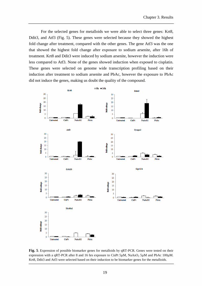
For the selected genes for metalloids we were able to select three genes: Krt8, Ddit3, and Atf3 (Fig. 5). These genes were selected because they showed the highest fold change after treatment, compared with the other genes. The gene Atf3 was the one that showed the highest fold change after exposure to sodium arsenite, after 16h of treatment. Krt8 and Ddit3 were induced by sodium arsenite, however the induction were less compared to Atf3. None of the genes showed induction when exposed to cisplatin. These genes were selected on genome wide transcription profiling based on their induction after treatment to sodium arsenite and PbAc, however the exposure to PbAc did not induce the genes, making us doubt the quality of the compound.
Fig. 5. Expression of possible biomarker genes for metalloids by qRT-PCR. Genes were tested on their expression with a qRT-PCR after 8 and 16 hrs exposure to CisPt 5µM, NaAsO2 5µM and PbAc 100µM. Krt8, Ddit3 and Atf3 were selected based on their induction to be biomarker genes for the metalloids.

Chapter 3. Results
20
Therefore, based on genome wide transcription profiling and on qRT-PCR we selected genes that were specifically activated after exposure to immune suppressor, PPARα ligand and metalloids. The biomarker genes that were selected seem to be specific for non-genotoxic carcinogens, since they did not show any induction when exposed to cisplatin, a genotoxic compounds.
3.1.2. Development and selection of GFP-reporter cell lines
Next we generate the GFP-based reporter for each gene. Therefore, we used the BAC recE/T recombineering to develop the GFP-reporters, as previously described [1].
The biomarker genes that were selected for the immune suppressor, Armet, Derl3, Dnajc3 and H47, were previously generated. However, it was not possible to generate a GFP reporter for the gene H47. We developed GFP-based reporter cell line for the biomarker genes Bhlhb2, Grasp, Krt8, Ddit3 and Atf3.
The GFP-based reporter cell line was constructed in order to easily determine gene induction by the amount fluorescence emitted by the modified GFP fusion protein after compound treatment. First, electrocompetent BAC strains were transformed with the pRed/ET plasmid, which contains the RecE and RecT recombination enzymes. We generated PCR fragments encoding a GFP-ires-neomycin/kanamycin reporter cassette, using primers that contain an additional 50 nucleotides sequence homologous to the 3’ sequence of the biomarker gene on the BAC (Fig. 6).
Fig. 6. Schematic representation of the RecE and RecT recombination enzymes and recombination of GFP-ires-Neo cassette into the BAC, adapted from Hendriks et al. [1].
Chapter 3. Results
21
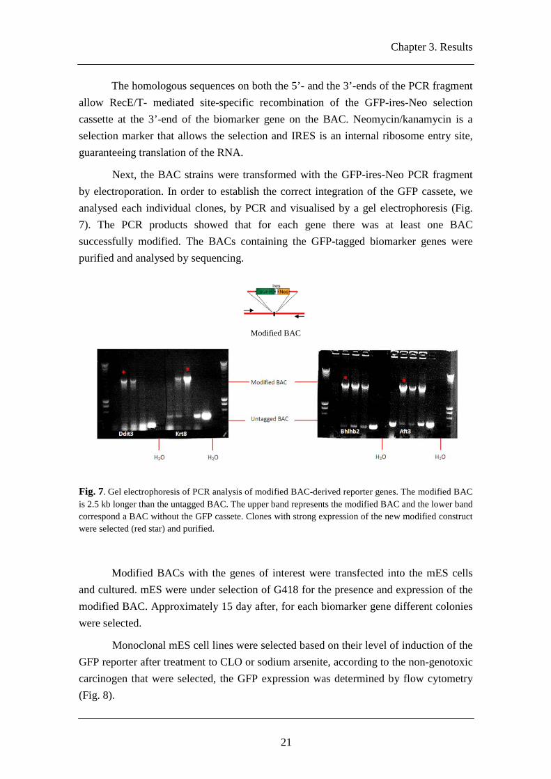
The homologous sequences on both the 5’- and the 3’-ends of the PCR fragment allow RecE/T- mediated site-specific recombination of the GFP-ires-Neo selection cassette at the 3’-end of the biomarker gene on the BAC. Neomycin/kanamycin is a selection marker that allows the selection and IRES is an internal ribosome entry site, guaranteeing translation of the RNA.
Next, the BAC strains were transformed with the GFP-ires-Neo PCR fragment by electroporation. In order to establish the correct integration of the GFP cassete, we analysed each individual clones, by PCR and visualised by a gel electrophoresis (Fig. 7). The PCR products showed that for each gene there was at least one BAC successfully modified. The BACs containing the GFP-tagged biomarker genes were purified and analysed by sequencing.
Fig. 7. Gel electrophoresis of PCR analysis of modified BAC-derived reporter genes. The modified BAC is 2.5 kb longer than the untagged BAC. The upper band represents the modified BAC and the lower band correspond a BAC without the GFP cassete. Clones with strong expression of the new modified construct were selected (red star) and purified.
Modified BACs with the genes of interest were transfected into the mES cells and cultured. mES were under selection of G418 for the presence and expression of the modified BAC. Approximately 15 day after, for each biomarker gene different colonies were selected.
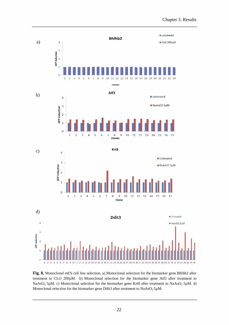
Monoclonal mES cell lines were selected based on their level of induction of the GFP reporter after treatment to CLO or sodium arsenite, according to the non-genotoxic carcinogen that were selected, the GFP expression was determined by flow cytometry (Fig. 8).
Modified BAC
Chapter 3. Results
22
Fig. 8. Monoclonal mES cell line selection. a) Monoclonal selection for the biomarker gene Bhlhb2 after treatment to CLO 200µM. b) Monoclonal selection for the biomarker gene Atf3 after treatment to NaAsO2 5µM. c) Monoclonal selection for the biomarker gene Krt8 after treatment to NaAsO2 5µM. d) Monoclonal selection for the biomarker gene Ddit3 after treatment to NaAsO2 5µM.
d)
c)
b)
a)

Chapter 3. Results
23
We are able to develop GFP reporters for all biomarker genes that we select, however for the biomarker gene Grasp we were not able to introduce the GFP-cassete into the BAC strain.
The GFP reporters Bhlhb2 and Atf3 were successfully generated, nevertheless after treatment and flow cytometry analysis there were no clones showing significant GFP expression after treatment exposure (Fig. 8a and 8b).
For Krt8- and Ddit3-GFP we were able to selected 6 clones each. For each clone the GFP induction was assessed by flow cytometry, and the amount of GFP was used to determine the clone which best represent the GFP-reporter (Fig. 9). Both cells cell lines were exposed to five increasing concentrations of sodium arsenite. All cells lines behave the same way.
Fig. 9. Clone selection. a) Clone selection for Krt8-GFP, when exposed to five increasing concentration of NaAsO2. However we were not able to select clones, since none of then showed an increase of GFP induction. b) Clone selection for Ddit3-GFP, when exposed to five increasing concentration of NaAsO2. We select clone 42, since was the one that showed the higher GFP induction. The cell survival was assessed by flow cytometry for each reporter, showing an increase of toxicity.
a)
b)

Chapter 3. Results
24
For the reporter Ddit3-GFP we select the clone 42, since was the one that showed the highest GFP induction after exposure (Fig. 9b). However, for the reporter Krt8-GFP we were not able to select one clone, because for all the clones the GFP induction after treatment was not significant (Fig. 9a). The cell survival was assessed, and with increasing concentrations there was an increase in toxicity, meaning that the clones for Krt8-GFP were not activated even though there was an apparent toxicity effect.
The biomarker genes Bhlhb2, Atf3 and Krt8 were selected based on their high expression by qRT-PCR (Fig. 4 and Fig. 5), although we were able to developed GFP-reporters for all the biomarker genes, the GFP expression was not relevant for these reporter. We can point the cause of the lack of induction to the stability of the GFP protein. If the protein is not stable and degrades, we could not measure the GFP expression.
3.1.3. GFP-based reporters: sensitivity and specificity
We were able to successfully develop a reporter for non-genotoxic carcinogen, which was selected after treatment to sodium arsenite, together with GFP reporters that were previously develop for immune suppressor, Armet-, Dnajc3- and Derl3-GFP we have a set of reporter. Therefore, the next step was to assess the sensitivity and specificity of the mES of Armet-, Dnajc3-, Derl3- and Ddit3-GFP.
It has previously been described that Armet is mediated by ERSE-II response element, Dnajc3 is mediated by ATF6, Derl3 is a target for IRE1/XBP1 and Ddit3 encodes for CHOP [35]. Thus, meaning that all the reporters are related with the unfolded protein response (UPR).
To verify sensitivity and specificity we start to expose the reporters to five increasing concentrations of sodium arsenite, cyclosporin A and WY, and then measure the GFP expression by flow cytometry (Fig. 10).

Chapter 3. Results
25
Fig. 10. The mES reporter cell lines respond preferentially to compounds that induce protein damage. GFP reporter cells were exposed to NaAsO2, CsA, Wyeth 14.643 and tunicamycin. 24h after exposure the GFP expression was determined by flow cytometry
It was previous been described that immune suppressor cyclosporin A and metalloid sodium arsenite are related with the UPR [36,37], and we confirm after exposure to these compounds, that these GFP-reporters were induced either by cyclosporin A and sodium arsenite (Fig. 10). The reporters that were developed for the immune suppressor were induced just when exposed to cyclosporin A. Ddit3-GFP was induced after treatment to sodium arsenite, cyclosporin A and Wyeth-14.643 (Fig. 10).
To confirm that the mode of action of these reporters was the UPR, we exposed the GFP-reporters to tunicamycin, which is a compound that induces the UPR [38]. After exposure, all the reporters were induced, although Ddit3-GFP showed the higher GFP expression. We test the GFP-reporters with genotoxic compounds that induce DNA damage and oxidative stressor to check if they are specific only of non-genotoxic carcinogens (Fig. 11).

Chapter 3. Results
26
Fig. 11. Check the sensitivity and specificity of the GFP-reporter. The GFP reporter cells were exposed 24h to DNA-damaging agents: Cisplatin; MMC; doxorubicin; and etoposide, and to oxidative stress–inducing agents: DEM, H2O2 and MMS. The GFP induction was assessed 24h after treatment by flow cytometry.

Chapter 3. Results
27
When the GFP-reporters cells lines developed for the UPR were exposed to genotoxic compounds, they did not show any induction, or the induction was not significant (Fig. 11), showing that the GFP-reporters that we developed are specific for non-genotoxic carcinogens and aren´t induced when exposed to genotoxic compounds. We assessed the cell survival for all the tested compounds, although the compounds did not induce the reporters, with increasing concentrations there was an increase in toxicity, and we observed that the lack of induction of GFP-reporter when exposed to genotoxic compounds was not due to the condition of the compound, but the lack of induction by compounds (supplemental figure 1).
Therefore, we were able to developed reporters that were induced by the UPR, and there are specific for non-genotoxic carcinogens, especial to CsA and NaAsO2.
3.2. Validation of the GFP-reporters for non-genotoxic carcinogens
Thus, the next step was to establish the sensitivity and specificity of the Armet-, Dnajc3-, Derl3- and Ddit3-GFP reporters, together with Sdc1, Eng, Rtkn, Btg2 and Blvrb-GFP reporters.
The Sdc1-, Eng-, Rtkn-, Btg2- and Blvrb-GFP are reporters that were previously developed to detect carcinogenic properties of genotoxic carcinogens compounds (table 3). They can distinguish between DNA damage, cellular stress and oxidative stress. The GFP-reporters Sdc1, Eng and Rtkn are reporters that are specifically induced when mES cells are exposed to genotoxic compounds. The Btg2-GFP is a p53-dependent, which is responsive to DNA-damaging agents and pro-oxidants [21]. And the Blvrb-GFP is induced after exposure to chemicals that result in increased levels of cellular oxidative stress.

Chapter 3. Results
28
Table 3 Various GFP reporters and the biological effects.
These nine GFP-reporters were exposed to a broad range of carcinogenic and noncarcinogenic compounds, as suggested by the ECVAM [33]. Those compounds are divided in three classes. The ECVAM Class 1 includes the compounds that are in vivo genotoxins carcinogens with a mutagenic mode of action, should score positive in an in vitro genotoxicity assay. The ECVAM class 2 includes non-genotoxic carcinogens or noncarcinogens with a nonmutagenic mode of action, should be negative in in vitro genotoxicity tests. And the ECVAM class 3 includes noncarcinogens with a nonmutagenic mode of action. Negative in in vivo genotoxicity studies, but nonetheless they have been reported positive in some in vitro genotoxicity tests.
In total, the GFP-reporters were exposed to 53 different compounds, mainly ECVAM-suggested genotoxins, pro-oxidants, and nongenotoxins (table 4 and Fig. 12).
Biological damage Biomarker gene
NGTXC UPR
Armet Dnajc3 Derl3 Ddit3
GTXC
DNA damage
Sdc1 Eng Rtkn
Cellular stress Btg2 Oxidative stress Blvrb

Chapter 3. Results
29
Table 4 Tested compounds for the validation of the GFP-reporters. 35 compounds have been proposed by the ECVAM [34] and 18 compounds are additional to the ECVAM.
Compound Highest conc. for mES cell exposure (µM)
ECVAM class 1
Cisplatin 10
Methyl methosulfate 500
Cadmium chloride 20
p-chloroaniline 1000
Sodium arsenite 5
Taxol 1
Etoposide 2
ECVAM class 2
n-butyl chloride 10000
Phenformin HCl 1000
(2-Chloroethyl) trimethy NH4Cl 10000
Dicyclohexyl thiourea 10000
Cyclohexanone 10000
Erythromycin stearate 2500
Fluometron 1000
D-Limonene 1000
Amitrole 10000
Tert-butanol 1000
Diethanolamine 10000
Hexachloroethane 1000
Methy carbamate 10000
Pyridine 10000
tris (2-ethylhexyl)phosphate 10000
2-chloroethanol 10000
ECVAM class 3
Anthranilic acid 10000
Resorcinol 1000
Sulfizoxazole 1000
Ethionamide 10000
Benzyl alcohol 10000
Sodium saccharin 10000
Nitrophenol 1000 2,3-dichlorophenol 1000 Eugenol 1000 Ethyl acrylate 1000 Isobutyraldehyde 1000 Propyl gallate 1000 Additional genotoxins Doxorubicin 0.5 Mitomycin C 1.5 Diethyl maleate 250 Hydrogen peroxide 1000 Copper sulfate 400 Potassium bromate 2500 4-Nitroquinelone-1-oxide 2 4 Hydroxy-2-nonenal 40 Cytarabine 50 Campthothecin 0.3 Hydroxyurea 1000 1-Nitropyrene 100 Non-genotoxic carcinogens Cyclosporin A 50 Tacrolimus 25 Wyeth-14643 500 Clofibrate 100 Lead Acetate 100 Tunicamycin 2

Chapter 3. Results
30
To study the GFP expression of the reporters, we develop a software, the ToxPolt that is able to create a heatmap that plots the GFP expression against 50% toxicity given by the compound. After compound exposure the ToxPlot cluster the GFP-reporters based on their specificity, being possible to discriminate between compounds that give DNA damage, oxidative stress and protein damage.
All ECVAM class 1 compounds scored positive for Sdc1-, Eng-, Rtkn-, Btg2- and Blvrb-GFP reporters cell lines, except p-chloroaniline. Although this compound is an in vivo carcinogen, it was also not identified as genotoxin in other in vitro genotoxicity assays [1]. The reporters for UPR scored negative for all the ECVAM class 1, indicating that these GFP-reporters are specific only for non-genotoxic carcinogens, therefore are not induced by these class of compounds (Fig. 12).
All tested ECVAM Class 2 compounds failed to induce the GFP reporters for the genotoxic carcinogens. However, the compounds n-butyl chloride, phenformin HCL, 2-chloroethy trimethyl NH4Cl, dicyclohexyl thiourea, erythromycin strearate and tris 2-ethylhexylphosphate induced the reporter Ddit3-GFP. The ECVAM Class 2 included non-genotoxic compounds, suggesting that the activation of Ddit3-GFP is associated with the toxic properties of these compounds, with the induction of protein damage, and fits with an assumed non-mutagenic mode of action (Fig. 12).
The compounds of ECVAM class 3 failed to induce all the GFP-reporters. However, the compounds sulfisoxazole, benzyl alcohol, eugenol, ethyl acrylate and propyl gallate induced the reporters for GTXC suggesting that these compounds contain in vitro genotoxic properties even though exposure to these compounds has not been found to cause cancer [1], while the compounds resorcinol, p-nitrophenol and dichlorophenol induced the reporters for the UPR, that fits with an assumed non-mutagenic mode of action (Fig. 12).
An additional set of 18 non-ECVAM suggested compounds were tested to identify the specificity of the GFP-reporters, and were all correctly identified as genotoxic, oxidative stress or protein damage. Nitropyrene was one of the additional compounds that induced both non-genotoxic carcinogens and genotoxic carcinogens GFP-based reporters. This compound induces DNA damage, give oxidative stress and protein damage, and is associated with carcinogenicity [38] (Fig. 12).
Together, these results show that the reporters for genotoxic carcinogens (Sdc1-, Eng-, Rtkn-, Btg2- and Blvrb-GFP) and for non-genotoxic carcinogens (Armet-, Dnajc3-, Derl3- and Ddit3-GFP) can provide a sensitive and selective assay to establish potential toxic activities of compounds, and provide insight in the primary toxic properties of compounds.

Chapter 3. Results
31
To show the capacity of the GFP-reporters, we selected just the reporters that gave the stronger GFP expression after treatment for each group. For the UPR we selected the reporter Ddit3-GFP, for DNA damage we selected Rtkn-GFP, and for cellular stress and oxidative stress we selected Btg2 and Blvrb-GFP, respectively. With these four GFP reporters we showed that they were able to distinguish between compounds that induce DNA damage, protein damage and give oxidative stress. We selected the compounds that were score positive for each group of reporters, and we create a heatmap where it´s visible that the reporter for UPR is strongly induce by cyclosporin A and tunicamycin. The Blvrb-GFP is strongly induced by MMS and DEM and Btg2 and Rtkn were both induced by cisplatin and mitomycin C (Fig. 13).

Chapter 3. Results
32
Fig. 12. Validation for the reporters, tested with 53 compounds suggested by ECVAM. The GFP-reporters cell lines were exposed to different compounds for 24h and the induction in total GFP was assessed by flow cytometry. The ToxPlot create the heatmap was made based on GFP expression at the 50% toxicity

Chapter 3. Results
33
Fig. 13. ToxPlot for Ddit3-GFP, Blvrb-GFP, Btg2-GFP and Rtkn-GFP. mES reporter cells respond preferentially to protein damage, pro-oxidants or genotoxic compounds, respectively. Selection of compounds that induce our reporters, the GFP expression was assessed 24h after treatment by flow cytometry. The heatmap plots the GFP induction at 50% toxicity.
Protein Oxidative Cellular DNA damage stress stress damage
Chapter 3. Results
34
We were able to develop and validate GFP-reporters cell lines for non-genotoxic carcinogens and genotoxic carcinogens. The GFP reporters Armet, Dnajc3, Derl3 and Ddit3 are induced by compounds that give protein damage, and did not show any induction when exposed to compounds that give DNA damage or pro-oxidants, although the expression of Ddit3-GFP was higher compared with the other reporters for the UPR. These reporters are specific for non-genotoxic carcinogens that induce protein damage. The reporters Sdc1-, Eng-, Rtkn-, Btg2- and Blvrb-GFP are specific for genotoxic carcinogens, and are induced either by DNA damage or oxidative stress.
3.3. Comet assay
In order to correlate the GFP-reporter activation with a biological endpoint we use an in vitro assay to establish that activation. We selected the comet assay, because under alkaline conditions (pH >13), the assay can detect single and double-stranded breaks, alkaline labile sites and excision repair processes in individual cells [22,39]. Following the single-cell electrophoresis, the lengths of the comets (DNA tails) depended on the treatment, concentration and exposure time, in which longer tails indicate a higher DNA damage.
Nevertheless, there are no available protocols for comet assay using the mES, therefore we had to optimize the assay, and once the protocol was working we could test the chemicals and establish the reporter activation.
To correlate the reporter activation with a biological endpoint, we had to expose the mES used in the comet assay with the same chemicals used for the validation of the GFP-reporters, thus we test 23 chemicals (Fig. 14) that belong to table 4. The compounds used in the comet assay were selected based on the induction of a reporter, or in cases that did not lead to reporter activation the chemical showed high levels of toxicity. For all the chemicals, we tested different concentrations and time points (supplemental table 1) and for each compound we plot the higher tail moment (Fig. 14).
We consider a negative result (no tailing) when the value for tail moment is lower than 0.0912 (five time higher that the SD in untreated cells), and a positive result when the tail moment is above that value.

Chapter 3. Results
35
Fig. 14. Alkaline comet assay. We test 23 compounds divided in 4 classes: the ECVAM class 1, 2 and 3 and additional compounds. For each compound we plotted only the higher tail moment (mean±SD of at least 50 cells).
The chemicals belonging to the ECVAM class 1 induced DNA breaks, which we can be correlated with the reporter’s activation, since they have a mutagenic mode of action [33]. Yet, cisplatin and taxol are both negative in the comet assay, and are chemicals that strongly induce the GFP-reporters for DNA damage. However it has been described that cisplatin reacts with DNA, leading to the formation of inter-strand and intra-strand cross-links and decrease the DNA migration, although induces the GFP-reporters, in comet assay lead to a reduction of the tail [40,41,42].
The chemical p-chloroaniline induced the GFP-reporter Ddit3, a reporter for protein damage (Fig. 12 and Fig. 13), however, in the comet assay this chemical was scored negative, leading us to believe that the comet assay cannot detect protein damage.
Chemicals from the ECVAM class 2 and 3 did not induced DNA damage that could be detected by the comet assay. These classes of chemicals include non-genotoxic carcinogens and noncarcinogens [33] that cannot be measurable by the comet assay. However, using the combination of GFP-reporters we were able to detect DNA damage caused by these chemicals (Fig. 13).
We tested six additional compounds (Fig. 14), and were selected because they induce our GFP-reporter. These chemicals were scored positive in the comet assay, however mitomycin C, tunicamycin and cyclosporin A failed to induce a comet tail.

Chapter 3. Results
36
Mitomycin C is well known cross-linking agents and induces DNA breaks [9,43], however reduces DNA migration, that is why is scored negative in the comet assay [40,41,42].
Tunicamycin and cyclosporin A are both non-genotoxic carcinogens that lead to the activation of the reporter Ddit3-GFP, specific for protein damage, however these chemicals couldn´t induce tail in the comet assay, causing us to question the sensitivity of the assay. Therefore, we can state that the GFP-reporters are much more sensitive and specific to test the genotoxic properties of a compound.
We performed the comet assay under neutral conditions (Fig. 15), and although the neutral and the alkaline comet assays detect both single- and double-strand breaks and it is impossible to distinguish between them, the results in the alkaline comet have proved to be much better.
On the neutral comet the negative control has a big background that makes it difficult to analyze and compare between chemicals, however we reach the same conclusions.
Fig. 15. Neutral comet assay. We test 19 compounds divided in 4 classes: the ECVAM class 1, 2 and 3 and additional compounds. Only the higher tail moment for each compound was plotted (mean±SD of at least 50 cells).
The ECVAM class 1 induces DNA breaks, except cisplatin and taxol. ECVAM class 2 and 3, in the neutral comet induced a comet tail, but in some cases are not

Chapter 3. Results
37
relevant. And in the additional compound, mitomycin A, tunicamycin and cyclosporin A fail to induce a comet tail.
Therefore, using the combination of different GFP-reporters we can predict the genotoxic properties of compounds. Using the traditional assay, like comet assay, to measure the genotoxic properties, we cannot distinguish between chemicals that give DNA damage, oxidative stressor or protein damage.
Is much wiser to use an assay that can detect possible carcinogenic properties of non-genotoxic compounds, once traditional assays are not able to do it, so using the reporters that we developed for the UPR we can detect these properties, that otherwise was not possible to do using an in vitro assay.

38
Chapter 4
Discussion
The aim of our research was to develop an in vitro assay, a GPF-based report assay, for the identification of possible carcinogenic properties of non-genotoxic compounds, and build up hypotheses about its molecular mode of action. Here, we described a set of genes, Armet, Dnajc3, Derl3, and Ddit3, which consists of different GFP fluorescence mES reporter cell lines that are preferentially responsive to protein damage.
Although have not yet been identified marker for non-genotoxic carcinogens, with this project we have selected biomarker genes for a subset of non-genotoxic carcinognes. These markers show an involvement in the unfolded protein response (UPR), when mES were exposed to certain non-genotoxic carcinogens, namely the immune suppressor’s cysclosporin A and tacrolimus, and metalloid sodium arsenite (Fig. 10). Although still much disputed the UPR has been associated with carcinogenesis [44]. Some research even shows that the UPR is required for carcinogenesis [45].
Cells are able to activate a series of complementary adaptive mechanisms to cope with the damaged and unfolded proteins, which is known as the unfolded protein response (UPR) (supplemental fig. 2). Unfolded protein signalling originates from the endoplasmic reticulum (ER) or cytoplasm [46]. ER stress triggers the UPR as a mechanism to limit cellular damage, to initiate repair of misfolded proteins, to dispose of irreparable proteins, and to initiate programmed cell death in the context of extreme ER stress.
UPR is a complex network of signalling events that target multiple cellular responses and is associated with the activation of three major stress sensors: inositol-requiring protein 1 (IRE1) (both α and β isoforms), transcription factor 6 (ATF6) (both
Chapter 4. Discussion
39
α and β isoforms) and protein kinase RNA-like ER kinase (PERK) [46,47,48]. In the unstressed ER, these sensor are associated with the chaperone, glucose regulated protein 78 kDa (GRP78/BiP) [46]. Upon unfolded/misfolded proteins accumulate in the ER, GRP78 is released from the sensor proteins, enabling activation of their three pathways [7,47]. PERK activation leads to phosphorylation of the α subunit of the eukaryotic translation initiator factor 2α (eIF2α) to attenuate general protein synthesis. The phosphorylated eIF2α allows the translation of ATF4 mRNA, which encodes a transcription factor controlling the transcription of genes involved in autophagy, apoptosis, amino acid metabolism and antioxidant responses [48]. Upon dissociation of GRP78, IRE1α dimerization, followed by autotransphosphorylation, triggers its RNase activity, which processes the mRNA encoding spliced X-box binding protein 1 (XBP1u) to produce an active transcription factor, spliced XBP1 (XBP1s). The XBP1s controls the transcription of genes encoding proteins involved in protein folding, ER-associated degradation (ERAD), protein quality control and phospholipid synthesis [47,48]. ATF6 is localized at the ER in unstressed cells, although, after dissociation of GRP78, ATF6 is transported to the Golgi apparatus through interaction with the coat protein II (COPII) complex, where is cleaved by site 1 and site 2 proteases (S1P and S2P), releasing its cytosolic domain fragment (ATF6f) [47,48]. ATF6f controls the upregulation of genes encoding ERAD components and also XBP1 [48].
The promoter of most of the UPR-target genes contain the ERSE sequence leading to their activation [49]. The protein chaperones play an important role in the UPR. They are involved in the holding and folding of proteins [50], they bind to unfolded sequences and show preference for hydrophobic parts of the protein. They do this to prevent premature associations with other proteins, during heat shock when proteins tend to unfold, and to stabilize unstable proteins [51]. The chaperones that are activated in the UPR are members of the heat shock proteins. The heat shock proteins have been identified as factors that play a role in one or more of the steps leading to carcinogenesis [51]. The binding of certain heat shock proteins is required for the stability of proteins involved in pathways leading to carcinogenesis, such as the factors of extracellular-regulated-signal-kinase (ERK) pathway and the pro-survival PI3K pathway and heat shock proteins also help by evading fast programmed cell death [51]. Certain heat shock proteins also facilitate the emergence of polymorphisms and mutations by stabilizing mutated proteins [9, 15].
As mentioned previously Armet, Danjc3, Derl3 and Ddit3 are involved in the UPR. Armet (arginine-rich mutated in early stage tumors) it has been proven to be ER-stress responsive gene [52]. Armet contains intra-molecular disulfide bonds and is a
Chapter 4. Discussion
40
soluble protein resident in the ER (27-aster). Armet induction requires either ATF6 or XBP1 [52]. The UPR of Armet is mediated by ERSE-II response element, suggesting that Armet is involved in protein-quality control [53, 54]. There are studies showing that the P58IPK, a protein homologs to Dnajc3 (Interferon-induced, double-stranded RNA-activated protein kinase inhibitor) is implicated in the UPR [55]. The induction of P58IPK has been mediated by ATF6 and contributes to the down-regulation of the PERK/eIF2α/ATF4 response pathway [56,57]. Derl3 (Der1-like domain family, member 3) is up-regulated by UPR, and plays as role in the ERAD for misfolded glycoproteins and that it is a target for IRE1/XBP1[58]. Ddit3 (DNA-Damage-Inducible Transcript 3) is induced by UPR, and encodes for the C/EBP homologous protein (CHOP) [46]. CHOP was the first identified protein that mediates ER stress-induced apoptosis [48, 59].
By, flow cytometer we showed that Armet, Dnajc3 and Derl3 were preferentially induced by immune suppressors cyclosporin A and FK506, and Ddit3 by the metalloid sodium arsenite (Fig. 10). However, when we exposed the GFP-reporters to DNA damage or oxidative stressor the GFP expression was either weak or not significant (Fig. 11).
Cyclosporin A and Fk506 are immunosuppressive agents used to avoid transplant rejection and to treat autoimmune diseases. The mode of action of their immune suppression is based on their interference with T-cell activation and T-cell growth by inhibition of transcription of early lymphokines [29]. Du et al. [29] report that administration of cyclosporin A in a reporter mouse induced the UPR in vivo. The GFP-based reporter Armet, Dnajc3 and Derl3, showed to be induced by cyclosporin A (Fig. 10), therefore, our results indicate that both cyclosporin A and FK506 lead to the activation of the UPR. The mode of action of metalloids, it’s still unclear. However, sodium arsenite is able to activate the promoters of a great number of genes that are involved in differentiation, proliferation and apoptosis [60], and can induces protein misfolding, as well as protein aggregation [48].
Thus, using the reporter Ddit3-GFP we settle that sodium arsenite activate the UPR, however Ddit3 was also induced by cyclosporin A. This chemical induces CHOP [29] confirm what we saw on Fig.10, that Ddit3 was induced either by cyclosporin A and sodium arsenite. On the other hand, when mES GFP reporter were exposed to tunicamycin, a compound which is commonly used as positive control in UPR, since it interferes with the glycoprotein synthesis causing ER-stress [46], they showed GFP induction (Fig. 10). Together with the data from the expose to nitropyrene (Fig. 12), that gives protein damage and is associated with carcinogenicity [37], and induces the four GFP-reports. Therefore taken together, these suggest that the reporters that we develop

Chapter 4. Discussion
41
are biomarker genes are good candidates for non-genotoxic carcinogens, involved in the UPR, and are good biomarker genes to detect protein damage. However, the GFP expression of Ddit3-GFP when exposed to all compounds was comparatively higher than Armet, Dnajc3 and Derl3, thus we affirm that Ddit-GFP is the best candidate to detect protein damage. Although many players of the UPR, such as the protein chaperones have previously been associated with carcinogenesis [1,7,11], this makes the correlation between UPR activation and carcinogenesis an obvious one.
All of the already developed cell lines showed induction of GFP after treatment, from which can be concluded that the fusion protein is synthesized in all cell lines. However, the amount of GFP induction of mRNA is not in accordance with the amount of fusion protein synthesized, causing the GFP induction to be just a part of the total mRNA induction. This difference could be caused by a number of effects, which all need to be investigated before the further validation of the cell lines continues. Therefore, before the reason for this difference is investigated it is essential to first determine whether the activation of the reporter is representative for the endogenous gene activation by means of qRT-PCR and with a Western blot on protein level. When both endogenous and fusion gene activation are similar the difference in mRNA and protein level can be further investigated. The first possibility for the difference may merely be time, as mRNA is isolated after 8 and 16h and the GFP induction is assessed after 24h. However, protein synthesis takes longer than mRNA transcription and therefore the optimum of protein synthesis may lie respectively later or earlier than 24 h. Thus cells should be treated for different time periods to look for the optimal GFP induction. It could also be that the cell produces more mRNA than is actually necessary for protein synthesis. But, this is thought of as highly unlikely as under selection pressure a cell looks for the optimal use of energy and production of extra mRNA would be a waste of energy. Another possibility to explain the difference in mRNA and protein level is that the fusion protein is not stable or the protein is usually fast degraded after synthesis. It is therefore essential to assess the kinetics of the fusion protein. Kinetics can be assessed by bleaching of the GFP reporter and then determine how quickly new proteins are synthesized. One could also block all protein degradation in order to see whether the GFP-induction rises. However one cannot specifically block the degradation of the fusion protein and thus other cell mechanisms might influence the outcome.
It is also advisable to assess the location of the fusion protein under the microscope to assess the exact location of the proteins in the cell and whether this is in
Chapter 4. Discussion
42
accordance with the endogenous protein. In conclusion much has to still be investigated before one can continue with the development of these cell lines.
Even though the development of a validated GFP based reporter cell lines for the identification of possible carcinogenic responses of NGTX carcinogens is not finished it is important to discuss the representativeness of the mES cell lines for the induction of cancer in humans. Even though these reporters are based on mES cells, they are still be very representative for the human model, because the strategy to deal with ER-stress by the UPR is well conserved from yeast to humans [10]. Therefore, it adds to the probability that these mES reporter cell line when finished have the same predictive value as human reporter cells lines and would therefore be very applicable for the identification of chemicals that have a possible carcinogenic effect in humans. Although it is likely that this in vitro assay is representative for in vivo risk assessment this does has to be validated. Stem cells are untransformed non-cancerous cell lines that are proficient in the main DNA damage signaling and cell-cycle control systems and are genetically stable, on the contrary most commonly used cell lines in in vitro genotoxicity testing are either derived from cancers or other malignancies, or are transformed cells with disrupted cell-cycle control.
Thus, by using combination of different GFP-reporter cell lines, we were able to discriminate between compounds that primarily induce genotoxic, oxidative stress or protein damage. With this study we demonstrate one example of a traditional in vitro assay to detect genotoxic properties of compounds, the comet assay, that doesn´t have the same sensitive, and specific as the GFP-reporters, in which we were not able to detect a comet tail when cell were exposed to chemicals of the ECVAM 2 and 3, therefore with this study we developed a new in vitro assay that has an assay that is highly sensitive, and specific, and can provide insights in the relative toxicity potential of chemicals, is fast and allows us to reduce the number of mice used in bioassays.
43
References
[1] G. Hendriks, M. Atallah, B. Morolli, F. Calléja, N. Ras-Verloop, I. Huijskens, et al., The ToxTracker assay: novel GFP reporter systems that provide mechanistic insight into the genotoxic properties of chemicals., Toxicological Sciences : an Official Journal of the Society of Toxicology. 125 (2012) 285–98.
[2] J.H.J. Hoeijmakers, DNA damage, aging, and cancer., The New England Journal of Medicine. 361 (2009) 1475–85.
[3] J. Ferlay, H.R. Shin, F. Bray, D. Forman, C. Mather, D.M. Parkin. GLOBOCAN 2008 v1.2. Cancer Incidence and Mortality Worldwide: IARC CancerBase 2010. Acess date: 04-07-2013; No. 10 [Available from: http://globocan.iarc.fr].
[4] S. Lagerwerf, M.G. Vrouwe, R.M. Overmeer, M.I. Fousteri, L.H.F. Mullenders, DNA damage response and transcription., DNA Repair. 10 (2011) 743–50.
[5] J.H.J. Hoeijmakers, Genome maintenance mechanisms for preventing cancer, Nature. 411 (2001) 366–374.
[6] E.C. Friedberg, DNA damage and repair, Nature. 421 (2003) 436–440
[7] G. Hendriks, B. van de Water, W. Schoonen, H. Vrieling, Cellular-signaling pathways unveil the carcinogenic potential of chemicals., Journal of Applied Toxicology : JAT. 33 (2013) 399–409.
[8] J.N. Kavanagh, K.M. Redmond, G. Schettino, K.M. Prise, DNA Double Strand Break Repair: A Radiation Perspective., Antioxidants & Redox Signaling. 18 (2013).
[9] I. Kamileri, I. Karakasilioti, G. a Garinis, Nucleotide excision repair: new tricks with old bricks., Trends in Genetics : TIG. 28 (2012) 566–73.
[10] A.R. Lehmann, DNA polymerases and repair synthesis in NER in human cells., DNA Repair. 10 (2011) 730–3.
[11] G.L. Dianov, U. Hübscher, Mammalian base excision repair: the forgotten archangel., Nucleic Acids Research. 41 (2013) 3483–90.
[12] J.M. Furgason, E.M. Bahassi, Targeting DNA repair mechanisms in cancer., Pharmacology & Therapeutics. 137 (2013) 298–308.
[13] S.E. Polo, S.P. Jackson, Dynamics of DNA damage response proteins at DNA breaks: a focus on protein modifications., Genes & Development. 25 (2011) 409–33.
[14] J.H. Houtgraaf, J. Versmissen, W.J. van der Giessen, A concise review of DNA damage checkpoints and repair in mammalian cells., Cardiovascular

44
Revascularization Medicine : Including Molecular Interventions. 7 (2006) 165–72.
[15] M.B. Kastan, J. Bartek, Cell-cycle checkpoints and cancer., Nature. 432 (2004) 316–23
[16] D. Woods, J. Turchi, Chemotherapy induced DNA damage response: Convergence of drugs and pathways, Cancer Biology & Therapy. 14 (2013) 1–11.
[17] J. Smith, L.M. Tho, N. Xu, D.A. Gillespie, The ATM-Chk2 and ATR-Chk1 pathways in DNA damage signaling and cancer., Advances in Cancer Research. 108 (2010) 73-112.
[18] A. Ciccia, S.J. Elledge, The DNA damage response: making it safe to play with knives., Molecular Cell. 40 (2010) 179–204.
[19] W.P. Watson, A. Mutti, Role of biomarkers in monitoring exposures to chemicals: present position, future prospects., Biomarkers : Biochemical Indicators of Exposure, Response, and Susceptibility to Chemicals. 9 (2004) 211–42.
[20] D. Kirkland, L. Reeve, D. Gatehouse, P. Vanparys, A core in vitro genotoxicity battery comprising the Ames test plus the in vitro micronucleus test is sufficient to detect rodent carcinogens and in vivo genotoxins., Mutation Research. 721 (2011) 27–73.
[21] G. Hendriks, M. Atallah, M. Raamsman, B. Morolli, H. van der Putten, H. Jaadar, et al., Sensitive DsRed fluorescence-based reporter cell systems for genotoxicity and oxidative stress assessment., Mutation Research. 709-710 (2011) 49–59
[22] R.D. Bowden, M.R. Buckwalter, J.F. McBride, D. a Johnson, B.K. Murray, K.L. O’Neill, Tail profile: a more accurate system for analyzing DNA damage using the Comet assay, Mutation Research/Genetic Toxicology and Environmental Mutagenesis. 537 (2003) 1–9.
[23] M. Lloyd, D. Kidd, The mouse lymphoma assay., Methods in Molecular Biology. 817 (2012) 35-54.
[24] R.M. Walmsley, M. Tate, The GADD45a-GFP GreenScreen HC assay., Methods in Molecular Biology. 817 (2012) 231-50.
[25] L.G. Hernández, H. van Steeg, M. Luijten, J. van Benthem, Mechanisms of non-genotoxic carcinogens and importance of a weight of evidence approach., Mutation Research. 682 (2009) 94–109.
[26] R.A. Roberts, S. Chevalier, S.C. Hasmall, N.H. James, S.C. Cosulich, PPARa and the regulation of cell division and apoptosis, Toxicology. 181 - 182 (2002) 167–170.
[27] R.D. Combes, The use of structure-activity relationships and markers of cell toxicity to detect non-genotoxic carcinogens., Toxicology in Vitro : an International Journal Published in Association with BIBRA. 14 (2000) 387–99.

45
[28] G. Krauter, C.V. Von der Lieth, R. Schmidt, E. Hecker, Structure/activity relationships of polyfunctional diterpenes of the tigliane type. A pharmacophore model for protein-kinase-C activators based on structure/activity studies and molecular modeling of the tumor promoters 12-O-tetradecanoylphorbol 13-acetate and 3-O-tetradecanoylingenol., European Journal of Biochemistry. 242 (1996) 417-27.
[29] S. Du, N. Hiramatsu, K. Hayakawa, A. Kasai, M. Okamura, T. Huang, et al., Suppression of NF-kappaB by cyclosporin a and tacrolimus (FK506) via induction of the C/EBP family: implication for unfolded protein response., Journal of Immunology (Baltimore, Md. : 1950). 182 (2009) 7201–11.
[30] R.M. Walmsley, N. Billinton, How accurate is in vitro prediction of carcinogenicity?, British Journal of Pharmacology. 162 (2011) 1250–8.
[31] J.H.M. van Delft, E. van Agen, S.G.J. van Breda, M.H. Herwijnen, Y.C.M. Staal, J.C.S. Kleinjans, Discrimination of genotoxic from non-genotoxic carcinogens by gene expression profiling., Carcinogenesis. 25 (2004) 1265–76.
[32] I. Poser, M. Sarov, J.R.A. Hutchins, J. Hériché, A. Pozniakovsky, D. Weigl, et al., BAC TransgeneOmics: a high-throughput method for exploration of protein function in mammals, 5 (2010) 409–415.
[33] D. Kirkland, P. Kasper, L. Müller, R. Corvi, G. Speit, Recommended lists of genotoxic and non-genotoxic chemicals for assessment of the performance of new or improved genotoxicity tests: a follow-up to an ECVAM workshop., Mutation Research. 653 (2008) 99–108.
[34] P.L. Olive, J.P. Banáth, R.E. Durand, J.P. Banath, Heterogeneity in Radiation-Induced DNA Damage and Repair in Tumor and Normal Cells Measured Using the “Comet” Assay, Radiation Research. 122 (1990) 86.
[35] Gene [Internet] [cited 2013 Jul 17] Available from: http://www.ncbi.nlm.nih.gov/gene/. 2013. Ref Type: Online Source
[36] A.M. Bolt, F. Zhao, S. Pacheco, W.T. Klimecki, Arsenite-induced autophagy is associated with proteotoxicity in human lymphoblastoid cells., Toxicology and Applied Pharmacology. 264 (2012) 255–61.
[37] S. Oyadomari, M. Mori, Roles of CHOP/GADD153 in endoplasmic reticulum stress., Cell Death and Differentiation. 11 (2004) 381–9.
[38] H. Andersson, E. Piras, J. Demma, B. Hellman, E. Brittebo, Low levels of the air pollutant 1-nitropyrene induce DNA damage, increased levels of reactive oxygen species and endoplasmic reticulum stress in human endothelial cells., Toxicology. 262 (2009) 57–64.
[39] B. Azevedo, F., Marques, F., Fork, H., Oliveira, R. and Johansson, Measuring oxidative DNA damage and DNA repair using the yeast comet assay, Yeast. 28 (2011) 55–61.
[40] L.M. Mendonça, G.C. dos Santos, R.A. dos Santos, C.S. Takahashi, M.D.L.P. Bianchi, L.M.G. Antunes, Evaluation of curcumin and cisplatin-induced DNA damage in PC12 cells by the alkaline comet assay., Human & Experimental Toxicology. 29 (2010) 635–43.

46
[41] I.U. De Silva, P.J. McHugh, P.H. Clingen, J. a Hartley, Defects in interstrand cross-link uncoupling do not account for the extreme sensitivity of ERCC1 and XPF cells to cisplatin., Nucleic Acids Research. 30 (2002) 3848–56
[42] F. Nesslany, N. Zennouche, S. Simar-Meintières, I. Talahari, E.-N. Nkili-Mboui, D. Marzin, In vivo Comet assay on isolated kidney cells to distinguish genotoxic carcinogens from epigenetic carcinogens or cytotoxic compounds., Mutation Research. 630 (2007) 28–41.
[43] T. Andoh, R. Ishida, Catalytic inhibitors of DNA topoisomerase II., Biochimica et Biophysica Acta. 1400 (1998) 155–71.
[44] Y. Ma, L.M. Hendershot, The role of the unfolded protein response in tumour development: friend or foe?, Nature Reviews Cancer. 4 (2004) 966-77
[45] L. Romero-Ramirez, H. Cao, D. Nelson, E. Hammond, A.H. Lee, H. Yoshida, et al., XBP1 is essential for survival under hypoxic conditions and is required for tumor growth. Cancer Research. 64 (2004) 5943-7.
[46] S. Oyadomari, C. Yun, E. a Fisher, N. Kreglinger, G. Kreibich, M. Oyadomari, et al., Cotranslocational degradation protects the stressed endoplasmic reticulum from protein overload., Cell. 126 (2006) 727–39.
[47] C. Hetz, The unfolded protein response: controlling cell fate decisions under ER stress and beyond., Nature Reviews. Molecular Cell Biology. 13 (2012) 89–102.
[48] A.M. Bolt, F. Zhao, S. Pacheco, W.T. Klimecki, Arsenite-induced autophagy is associated with proteotoxicity in human lymphoblastoid cells., Toxicology and Applied Pharmacology. 264 (2012) 255–61.
[49] K. Kokame, H. Kato, T. Miyata, Identification of ERSE-II, a new cis-acting element responsible for the ATF6-dependent mammalian unfolded protein response., The Journal of Biological Chemistry. 23 (2001) 9199-205.
[50] H. Wegele, L. Muller, J. Buchner, Hsp70 and Hsp90--a relay team for protein folding., Reviews of Physiology, Biochemistry and Pharmacology. 151 (2004) 1-44.
[51] S.K. Calderwood, M.A. Khaleque, D.B. Sawyer, D.R. Ciocca, Heat shock proteins in cancer: chaperones of tumorigenesis., Trends in Biochemical Sciences. 31 (2006) 164-72.
[52] A. Apostolou, Y. Shen, Y. Liang, J. Luo, S. Fang, Armet, a UPR-upregulated protein, inhibits cell proliferation and ER stress-induced cell death., Experimental Cell Research. 314 (2008) 2454–67.
[53] N. Mizobuchi, J. Hoseki, H. Kubota, S. Toyokuni, J. Nozaki, M. Naitoh, et al., ARMET is a soluble ER protein induced by the unfolded protein response via ERSE-II element., Cell Structure and Function. 32 (2007) 41–50.
[54] A. Lee, N.N. Iwakoshi, L.H. Glimcher, XBP-1 Regulates a Subset of Endoplasmic Reticulum Resident Chaperone Genes in the Unfolded Protein Response, Molecular and Cellular Biology. 23 (2003) 7448–7459.

47
[55] S. Oyadomari, C. Yun, E. a Fisher, N. Kreglinger, G. Kreibich, M. Oyadomari, et al., Cotranslocational degradation protects the stressed endoplasmic reticulum from protein overload., Cell. 126 (2006) 727–39.
[56] B. Li, D. Li, G.-G. Li, H.-W. Wang, A.-X. Yu, P58(IPK) inhibition of endoplasmic reticulum stress in human retinal capillary endothelial cells in vitro., Molecular Vision. 14 (2008) 1122–8.
[57] R. van Huizen, J.L. Martindale, M. Gorospe, N.J. Holbrook, P58IPK, a novel endoplasmic reticulum stress-inducible protein and potential negative regulator of eIF2alpha signaling., The Journal of Biological Chemistry. 278 (2003) 15558–64.
[58] Y. Oda, T. Okada, H. Yoshida, R.J. Kaufman, K. Nagata, K. Mori, Derlin-2 and Derlin-3 are regulated by the mammalian unfolded protein response and are required for ER-associated degradation., The Journal of Cell Biology. 172 (2006) 383–93.
[59] F. Holtrup, A. Bauer, K. Fellenberg, R. a Hilger, M. Wink, J.D. Hoheisel, Microarray analysis of nemorosone-induced cytotoxic effects on pancreatic cancer cells reveals activation of the unfolded protein response (UPR)., British Journal of Pharmacology. 162 (2011) 1045–59.
[60] D. Beyersmann, Effects of carcinogenic metals on gene expression, Toxicology Letters 127 (2002) 63-8.

xii
Supplementary data
Supplementary Method 1: BAC recombineering
Antibiotics:
Chloramphenicol (Cam) working concentration is 12.5 µg/ml Tetracycline (Tet) working concentration is 3 µg /ml Kanamycin (Kan) working concentration is 25 µg/ml
Gene construct
PCR mix:
Plasmid GFP-IRES-Neo 1 µL Primer forward (10pmol/µL) 1.5 µL Primer reverse (10pmol/µL) 1.5 µL DNTPs (10mM stock) 1.5 µL 5x KAPA HiFi fidelity buffer 10 µL KaPa HiFi polymerase 1 µL H2O 33.5 µL Total 50 µL
PCR program:
980C 5min 980C 30s 550C 30s 680C 2,50min 680C 10min
Introducing pRedE/T vector into the BAC:
1. Inoculate 4 mL LC + Cam with one colony of the BAC O/N shaking at
370C 2. Add 400 µL of O/N BAC culture to 40 mL LC + Cam, mix well and
divide equally into 4 (50ml) falcon tubes (10mL each), incubate for 2 to 3 hours shaking at 370C till OD600=~0,4.
3. Always keep the cells cold on ice, from now on. 4. Collect cell together and centrifuge for 10 min at 40C (4000rpm) 5. Remove supernatant and wash pellet with 40 mL of cold 10% glycerol.
20x

xiii
6. Centrifuge for 10 min at 40C (4000rpm) 7. Remove supernatant, wash cell for a second time with 40 mL of cold
10% glycerol 8. Centrifuge for 10 min at 40C (4000rpm) 9. Remove as much as possible from the supernatant 10. Add 100 µL of 10% glycerol to the cells, resuspend the electro-
competent cells and add 50 µL to an eppendorf 11. Add 1 µL pRedE/T plasmid (20ng/µL) to the eppendrof with cells, mix
by tapping, and leave on ice. 12. Electroporate cells: place cells in cold cuvette (1mm gap) and apply pulse
( bacteria program 1.8kV, 200 Ω, 25uF) 13. Move cells quickly from cuvette with a Pasteur pipette and place into a
tube with 1mL LC. 14. Incubate shaking at 300C for ½ to 2 hours. 15. Plate 100 µL on LC + Cam + tet on 1 plate and the rest on another plate. 16. Incubate at 300C O/N in the dark.
Introducing the IRES-Neo into the BAC:
17. Inoculate 4mL LC+ Cam + Tet with 10 colony of the BAC + pRedE/T
O/N shaking at 300C in the dark 18. Second day, add 400 µL of O/N BAC+ pRedE/T culture to 40mL LC +
Cam+ Tet mix well and divide equally into 4 (50ml) falcon tubes (10mL each), incubate for 2 to 3 hours shaking at 300C till OD600=~0,2-0,3.
19. Collect cells together, add up to 0,2% of (10%) L-arabinose (800 µL for 40mL), mix well and divide equally into 4 falcon tubes (10ml each), incubate for 45min shaking at 370C in the dark till OD600=~0,4.
20. Collect cell together and centrifuge for 10min at 40C (4000rpm) 21. Remove supernatant and wash pellet with 40mL of cold 10% glycerol. 22. Centrifuge for 10min at 40C (4000rpm) 23. Remove supernatant, wash cell for a second time with 40ml of cold 10%
glycerol 24. Centrifuge for 10min at 40C (4000rpm) 25. Remove as much as possible from the supernatant 26. Add 100 µL of 10% glycerol, and add 50uL to an eppendorf 27. Add to 300ng of the gene construct. 28. Electroporate cells: place cells in cold cuvette (1mm gap) and apply pulse
( bacteria program 1.8kV, 200 Ω, 25uF) 29. Move cells quickly from cuvette with a Pasteur pipette and place into a
tube with 1mL LC. 30. Incubate shaking at 370C for ½ to 2 hours. 31. Plate 100 µL on LC + Cam + tet + Kan on 1 plate and the rest on another
plate. 32. Grow O/N at 370C
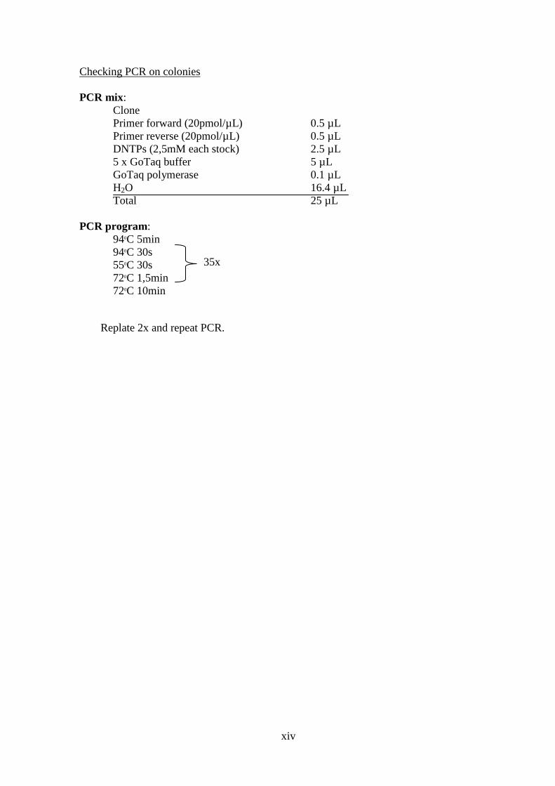
xiv
Checking PCR on colonies PCR mix:
Clone Primer forward (20pmol/µL) 0.5 µL Primer reverse (20pmol/µL) 0.5 µL DNTPs (2,5mM each stock) 2.5 µL 5 x GoTaq buffer 5 µL GoTaq polymerase 0.1 µL H2O 16.4 µL Total 25 µL
PCR program:
940C 5min 940C 30s 550C 30s 720C 1,5min 720C 10min
Replate 2x and repeat PCR.
35x

xv
Supplementary Method 2: RNA analysis protocol
RNA isolation
1. Culture cells on gelatine with conditioned medium until well is full 2. Remove medium and wash 1x PBS 3. Add appropriate amount of TRIzol (Invitrogen) for the used culture well and
incubate at room temperature for 5min 4. Add chloroform (40 µL per 100 µL TRIzol), shake for 15sec and incubate for 2-
3minutes at room temperature 5. Centrifuge for 15min 12000 rpm at 40C 6. Move water phase to new eppendorf, add ½ a volume of isopropanol and
incubate for 10min at room temperature. 7. Centrifuge for 10min 1200 rpm at 40C 8. Remove supernatant and wash pellet 1x with 100uL 75% Ethanol. 9. Centrifuge 10min 1200 rpm 40C 10. Resuspend pellet in 20 µL mQ and determine RNA concentration with
NanoDrop
cDNA synthesis
11. Use 1 µg of RNA and add mQ to a total volume of 9,5 µL 12. Add 0,5 µL oligo dt primer (12-18) and 4 µL dntps (2,5mM each) 13. Incubate for 5min at 650C and for 5min on ice 14. Add 4 µL 5x first strand buffer, 1 µL 0,1M dtt, 0,5 µL RNAzin and 0,5 µL
SuperScript reverse transcriptase III (Invitrogen) 15. Incubate for 1 hour at 500C and for 10min at 650C 16. GoTaq PCR 17. PCR Mix:
GoTaq buffer 5µL dntps (2.5mM/each) 2,5 µL Sus 3 primer (Aprt forward) 0,5 µL Sus 4 primer (Aprt reverse) 0,5 µL GoTaq polymerase 0,1 µL H2O 15,4 µL cDNA 1 µL Total 25 µL
18. PCR program: 940C 5min 940C 30s 550C 30s 720C 1,5min 720C 10min
19. Load PCR product on 1% agarose TAE gel. Aprt will give a product of ± 400bp
35x
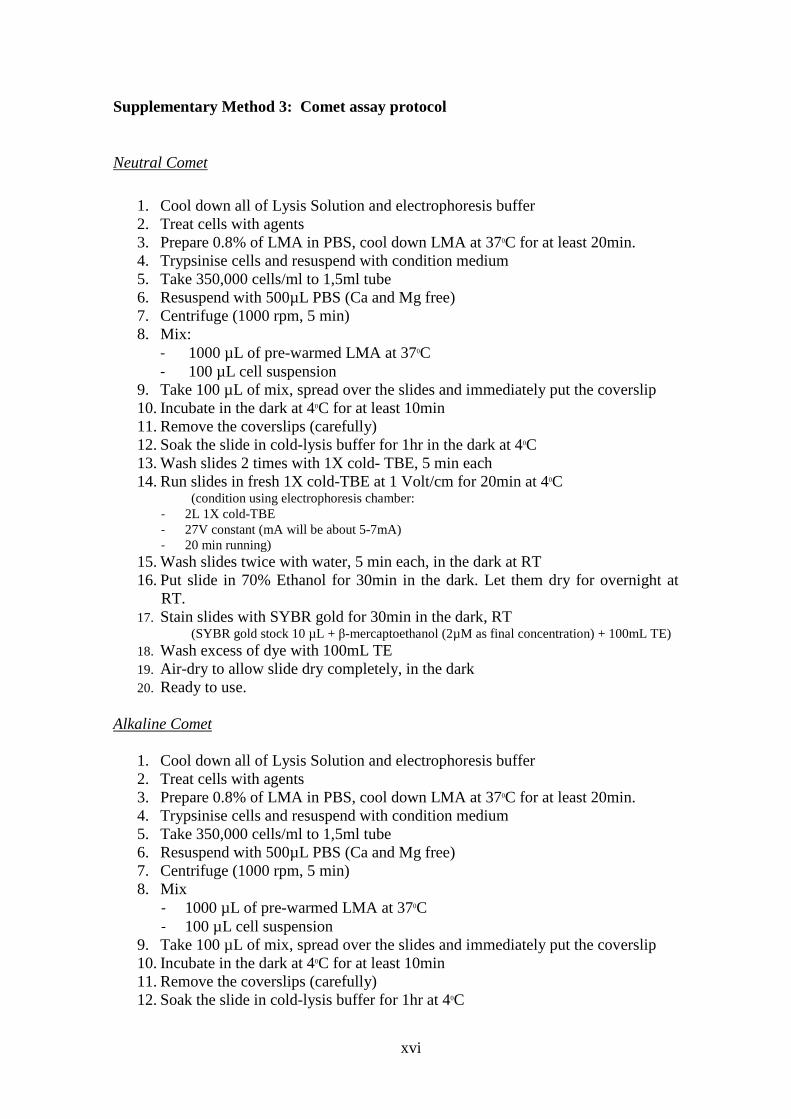
xvi
Supplementary Method 3: Comet assay protocol
Neutral Comet
1. Cool down all of Lysis Solution and electrophoresis buffer 2. Treat cells with agents 3. Prepare 0.8% of LMA in PBS, cool down LMA at 370C for at least 20min. 4. Trypsinise cells and resuspend with condition medium 5. Take 350,000 cells/ml to 1,5ml tube 6. Resuspend with 500µL PBS (Ca and Mg free) 7. Centrifuge (1000 rpm, 5 min) 8. Mix:
- 1000 µL of pre-warmed LMA at 370C - 100 µL cell suspension
9. Take 100 µL of mix, spread over the slides and immediately put the coverslip 10. Incubate in the dark at 40C for at least 10min 11. Remove the coverslips (carefully) 12. Soak the slide in cold-lysis buffer for 1hr in the dark at 40C 13. Wash slides 2 times with 1X cold- TBE, 5 min each 14. Run slides in fresh 1X cold-TBE at 1 Volt/cm for 20min at 40C
(condition using electrophoresis chamber: - 2L 1X cold-TBE - 27V constant (mA will be about 5-7mA) - 20 min running)
15. Wash slides twice with water, 5 min each, in the dark at RT 16. Put slide in 70% Ethanol for 30min in the dark. Let them dry for overnight at
RT. 17. Stain slides with SYBR gold for 30min in the dark, RT
(SYBR gold stock 10 µL + β-mercaptoethanol (2µM as final concentration) + 100mL TE) 18. Wash excess of dye with 100mL TE 19. Air-dry to allow slide dry completely, in the dark 20. Ready to use.
Alkaline Comet
1. Cool down all of Lysis Solution and electrophoresis buffer 2. Treat cells with agents 3. Prepare 0.8% of LMA in PBS, cool down LMA at 370C for at least 20min. 4. Trypsinise cells and resuspend with condition medium 5. Take 350,000 cells/ml to 1,5ml tube 6. Resuspend with 500µL PBS (Ca and Mg free) 7. Centrifuge (1000 rpm, 5 min) 8. Mix
- 1000 µL of pre-warmed LMA at 370C - 100 µL cell suspension
9. Take 100 µL of mix, spread over the slides and immediately put the coverslip 10. Incubate in the dark at 40C for at least 10min 11. Remove the coverslips (carefully) 12. Soak the slide in cold-lysis buffer for 1hr at 40C

xvii
13. Dip slides in unwinding solution for 30min at 40C, in the dark 14. Run slides in fresh unwinding solution at 1 Volt/cm for 30min at 40C
(condition using electrophoresis chamber: - 2L alkaline solution - 27 V constant (mA will be about 330-350mA) - 20 min running)
15. Wash slides 3x with 0.4M Tris pH7,5, 5 min each, in the dark at RT 16. Put slide in 70% Ethanol for 30min in the dark. Let them dry for overnight at
RT. 17. Stain slides with SYBR gold for 30min in the dark, RT
(SYBR gold stock 10 µL + β-mercaptoethanol (2µM as final concentration) + 100mL TE) 18. Wash excess of dye with 100mL TE 19. Air-dry to allow slide dry completely, in the dark. 20. Ready to use.
**Positive control: mES B4418 exposed to 30min of 500 µM H2O2.

xviii
Supplemental fig. 1. Cell survival for all the tested compounds. The toxicity curve is based on the average for the four GFP-reporters. The cell survival was assessed after 24-h treatment by flow cytometry.

xix
Supplemental fig. 2. The UPR sensors. PERK, IRE1α, and ATF6 control mRNA translation and transcriptional induction of UPR-regulated genes. Upon accumulation of unfolded protein, PERK is activated by homodimerization and autophosphorylation to phosphorylate eIF2α, thus reducing the rate of mRNA translation and the biosynthetic protein-folding load on the ER. eIF2α phosphorylation increases translation of Atf4, a transcription factor that activates expression of genes encoding protein chaperones, ERAD machinery, enzymes that reduce oxidative stress. Dimerization of IRE1α induces cleavage of Xbp1 mRNA. Then, Xbp1 mRNA is translated to produce XBP1s. ATF6 is transport to the Golgi where it’s cleaved to release a transcriptionally active fragment. Cleaved ATF6 acts in concert with XBP1s to induce expression of genes encoding protein chaperones and ERAD machinery.
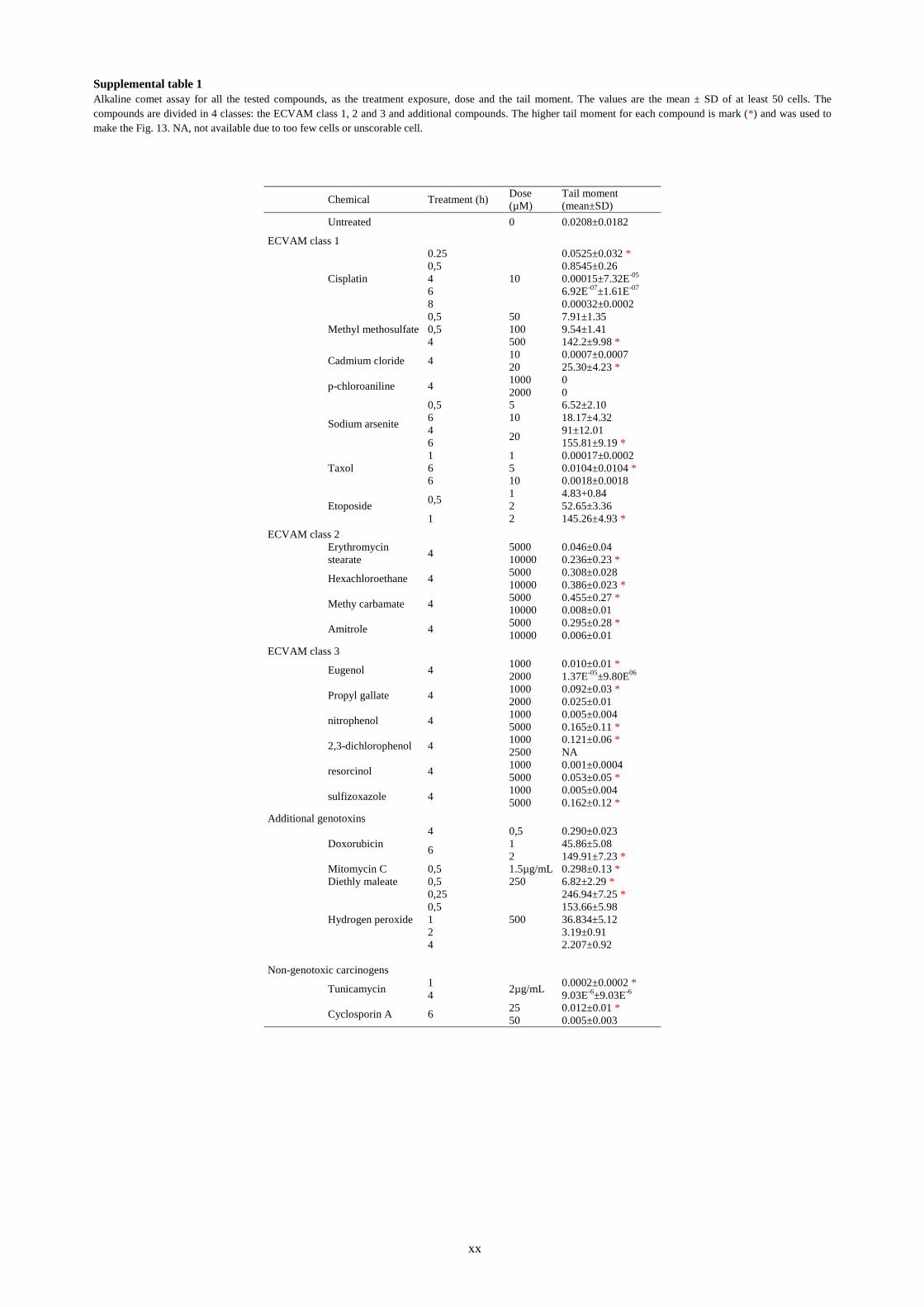
xx
Supplemental table 1 Alkaline comet assay for all the tested compounds, as the treatment exposure, dose and the tail moment. The values are the mean ± SD of at least 50 cells. The compounds are divided in 4 classes: the ECVAM class 1, 2 and 3 and additional compounds. The higher tail moment for each compound is mark (*) and was used to make the Fig. 13. NA, not available due to too few cells or unscorable cell.
Chemical Treatment (h) Dose (µM)
Tail moment (mean±SD)
Untreated 0 0.0208±0.0182 ECVAM class 1
Cisplatin
0.25
10
0.0525±0.032 *
0,5 0.8545±0.26
4 0.00015±7.32E-05
6 6.92E-07±1.61E-07
8 0.00032±0.0002
Methyl methosulfate 0,5 50 7.91±1.35
0,5 100 9.54±1.41
4 500 142.2±9.98 *
Cadmium cloride 4 10 0.0007±0.0007
20 25.30±4.23 *
p-chloroaniline 4 1000 0
2000 0
Sodium arsenite
0,5 5 6.52±2.10
6 10 18.17±4.32
4 20 91±12.01
6 155.81±9.19 *
Taxol 1 1 0.00017±0.0002
6 5 0.0104±0.0104 *
6 10 0.0018±0.0018
Etoposide 0,5 1 4.83+0.84
2 52.65±3.36
1 2 145.26±4.93 * ECVAM class 2 Erythromycin
stearate 4 5000 0.046±0.04
10000 0.236±0.23 *
Hexachloroethane 4 5000 0.308±0.028
10000 0.386±0.023 *
Methy carbamate 4 5000 0.455±0.27 *
10000 0.008±0.01
Amitrole 4 5000 0.295±0.28 *
10000 0.006±0.01 ECVAM class 3 Eugenol 4 1000 0.010±0.01 *
2000 1.37E-05±9.80E06
Propyl gallate 4 1000 0.092±0.03 *
2000 0.025±0.01
nitrophenol 4 1000 0.005±0.004
5000 0.165±0.11 *
2,3-dichlorophenol 4 1000 0.121±0.06 *
2500 NA
resorcinol 4 1000 0.001±0.0004
5000 0.053±0.05 *
sulfizoxazole 4 1000 0.005±0.004
5000 0.162±0.12 * Additional genotoxins Doxorubicin
4 0,5 0.290±0.023
6 1 45.86±5.08
2 149.91±7.23 *
Mitomycin C 0,5 1.5µg/mL 0.298±0.13 *
Diethly maleate 0,5 250 6.82±2.29 *
Hydrogen peroxide
0,25
500
246.94±7.25 * 0,5 153.66±5.98 1 36.834±5.12 2 3.19±0.91 4 2.207±0.92 Non-genotoxic carcinogens Tunicamycin 1 2µg/mL 0.0002±0.0002 * 4 9.03E-6±9.03E-6 Cyclosporin A 6 25 0.012±0.01 * 50 0.005±0.003