Statistical Estimation: Least Squares, Maximum Likelihood and
67
CS5540: Computational Techniques for Analyzing Clinical Data Lecture 7: Statistical Estimation: Least Squares, Maximum Likelihood and Maximum A Posteriori Estimators Ashish Raj, PhD Image Data Evaluation and Analytics Laboratory (IDEAL) Department of Radiology Weill Cornell Medical College New York
Transcript of Statistical Estimation: Least Squares, Maximum Likelihood and

CS5540: Computational Techniques for Analyzing Clinical Data
Lecture 7:
Statistical Estimation: Least
Squares, Maximum Likelihood and Maximum A Posteriori Estimators
Ashish Raj, PhD
Image Data Evaluation and Analytics Laboratory (IDEAL)
Department of Radiology
Weill Cornell Medical College
New York

IDEA Lab, Radiology, Cornell
2
Outline
� Part I: Recap of Wavelet Transforms
� Part II : Least Squares Estimation
� Part III: Maximum Likelihood Estimation
� Part IV: Maximum A Posteriori Estimation : Next week
Note: you will not be tested on specific examples shown here, only on general principles

IDEA Lab, Radiology, Cornell
Basis functions in WT
3
Haar
Mexican Hat
Daubechies
(orthonormal)
IDEA Lab, Radiology, Cornell
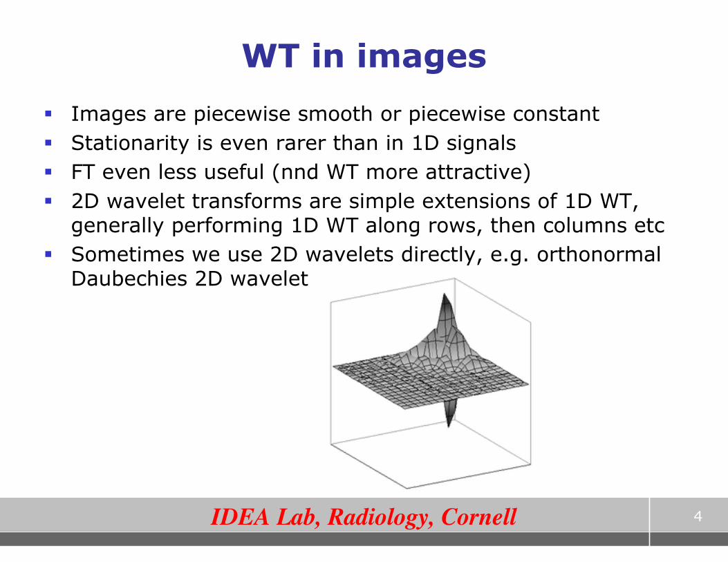
WT in images
� Images are piecewise smooth or piecewise constant
� Stationarity is even rarer than in 1D signals
� FT even less useful (nnd WT more attractive)
� 2D wavelet transforms are simple extensions of 1D WT, generally performing 1D WT along rows, then columns etc
� Sometimes we use 2D wavelets directly, e.g. orthonormal Daubechies 2D wavelet
4

IDEA Lab, Radiology, Cornell
WT on images
5
time
scale
2D generalization of scale-
time decomposition
Successive application of dot product with wavelet of increasing width.
Forms a natural pyramid structure. At each scale:
H = dot product of image rows with wavelet
V = dot product of image columns with wavelet
H-V = dot product of image rows then columns with wavelet
Scale 0
Scale 1
Scale 2
H
V H-V

IDEA Lab, Radiology, Cornell
Wavelet Applications
� Many, many applications!
� Audio, image and video compression
� New JPEG standard includes wavelet compression
� FBI’s fingerprints database saved as wavelet-compressed
� Signal denoising, interpolation, image zooming, texture analysis, time-scale feature extraction
6
� In our context, WT will be used primarily as a feature extraction tool
� Remember, WT is just a change of basis, in order to extract useful information which might otherwise not be easily seen

IDEA Lab, Radiology, Cornell
WT in MATLAB
� MATLAB has an extensive wavelet toolbox
� Type help wavelet in MATLAB command window
� Look at their wavelet demo
� Play with Haar, Mexican hat and Daubechies wavelets
7

IDEA Lab, Radiology, Cornell
Project Ideas
8
� Idea 1: use WT to extract features from ECG data
– use these features for classification
� Idea 2: use 2D WT to extract spatio-temporal features from 3D+time MRI data
– to detect tumors / classify benign vs malignant tumors
� Idea 3: use 2D WT to denoise a given image

IDEA Lab, Radiology, Cornell
Idea 3: Voxel labeling from contrast-enhanced MRI
� Can segment according to time profile of 3D+time
contrast enhanced MR data of liver / mammography
Typical plot of time-resolved
MR signal of various tissue
classes
Temporal models used to
extract features
Instead of such a simple temporal model, wavelet decomposition could provide spatio-temporal features that you can use for clustering

IDEA Lab, Radiology, Cornell
Liver tumour quantification from DCE-MRI

IDEA Lab, Radiology, Cornell
Further Reading on Wavelets
11

Part II : Least Squares Estimation and Examples

IDEA Lab, Radiology, Cornell
13
A simple Least Squares problem – Line fitting
� Goal: To find the “best-fit” line representing a bunch of points
� Here: yi are observations at location xi,
� Intercept and slope of line are the unknown model parameters to be estimated
� Which model parameters best fit the observed points?
This can be written in matrix notation, as
θθθθLS = arg min ||y – Hθθθθ||2
What are H and θθθθ?
),(minarg),(Best
)(where,))((),(
intint
int
2
int
myEmy
mxyxhxhymyE i
i
ii
=
+=−=∑
IDEA Lab, Radiology, Cornell
14
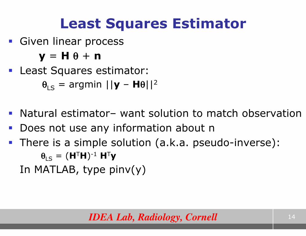
Least Squares Estimator
� Given linear process
y = H θθθθ + n
� Least Squares estimator:
θθθθLS = argmin ||y – Hθθθθ||2
� Natural estimator– want solution to match observation
� Does not use any information about n
� There is a simple solution (a.k.a. pseudo-inverse):
θθθθLS = (HTH)-1 HTy
In MATLAB, type pinv(y)

IDEA Lab, Radiology, Cornell
15
Example - estimating T2 decay constant in repeated spin echo MR data
IDEA Lab, Radiology, Cornell
16
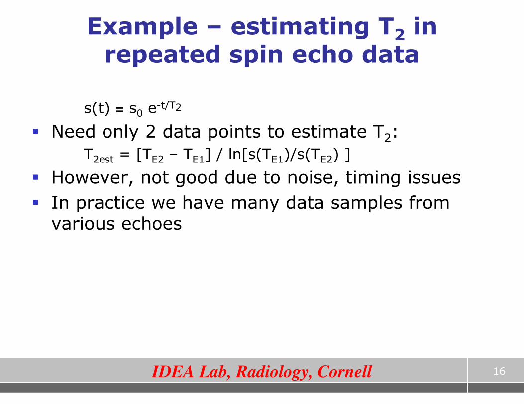
Example – estimating T2 in repeated spin echo data
s(t) = s0 e-t/T2
� Need only 2 data points to estimate T2:
T2est = [TE2 – TE1] / ln[s(TE1)/s(TE2) ]
� However, not good due to noise, timing issues
� In practice we have many data samples from various echoes

IDEA Lab, Radiology, Cornell
17
Example – estimating T2
θ LS = (HTH)-1HTy
T2 = 1/rLS
H ln(s(t1))
ln(s(t2))
M
ln(s(tn))
1 -t1
1 -t2
M
1 -tn
= a
r
θ
y
Least Squares estimate:

IDEA Lab, Radiology, Cornell
18
Estimation example - Denoising
� Suppose we have a noisy MR image y, and wish to obtain the noiseless image x, where
y = x + n
� Can we use LSE to find x?
� Try: H = I, θ = x in the linear model
� LS estimator simply gives x = y!
� we need a more powerful model
� Suppose the image x can be approximated by a polynomial, i.e. a mixture of 1st p powers of r:
x = Σi=0p ai r
i

IDEA Lab, Radiology, Cornell
19
Example – denoising
θ LS = (HTH)-1HTy
x = Σi=0p ai r
i
H
θ
y
Least Squares estimate:
y1
y2
M
yn
1 r11 L r1
p
1 r21 L r2
p
M
1 rn1 L rn
p
=
a0
a1
M
ap
n1
n2
M
nn
+

Part III : Maximum Likelihood Estimation and Examples

IDEA Lab, Radiology, Cornell
21
Estimation Theory
� Consider a linear process
y = H θθθθ + n
y = observed data
θθθθ = set of model parameters
n = additive noise
� Then Estimation is the problem of finding the statistically optimal θθθθ, given y, H and knowledge of noise properties
� Medicine is full of estimation problems

IDEA Lab, Radiology, Cornell
22
Different approaches to estimation
� Minimum variance unbiased estimators
� Least Squares
� Maximum-likelihood
� Maximum entropy
� Maximum a posteriori
has no
statistical
basis
uses
knowledge of
noise PDF
uses prior
information
about θ

IDEA Lab, Radiology, Cornell
23
Probability vs. Statistics
� Probability: Mathematical models of uncertainty predict outcomes
– This is the heart of probability
– Models, and their consequences
• What is the probability of a model generating some particular data as an outcome?
� Statistics: Given an outcome, analyze different models
– Did this model generate the data?
– From among different models (or parameters), which one generated the data?

IDEA Lab, Radiology, Cornell
24
Maximul Likelihood Estimator for Line Fitting Problem
� What if we know something about the noise? i.e. Pr(n)…
� If noise not uniform across samples, LS might be incorrect
This can be written in matrix notation, as
θθθθML = arg min ||W(y – Hθθθθ)||2
What is W?
noise σ = 0.1
noise σ = 10
ML estimate
),(minarg),(Best
)(where,))(
(),(
intint
int
2
int
myEmy
mxyxhxhy
myEi i
ii
=
+=−
=∑σ

IDEA Lab, Radiology, Cornell
25
Definition of likelihood
� Likelihood is a probability model of the uncertainty in output given a known input
� The likelihood of a hypothesis is the probability that it would have resulted in the data you saw
– Think of the data as fixed, and try to chose among the possible PDF’s
– Often, a parameterized family of PDF’s
• ML parameter estimation

IDEA Lab, Radiology, Cornell
26
Gaussian Noise Models
� In linear model we discussed, likelihood comes from noise statistics
� Simple idea: want to incorporate knowledge of noise statistics
� If uniform white Gaussian noise:
� If non-uniform white Gaussian noise:
−=
−=
∑∏ 2
2
2
2
2
||
exp1
2
||exp
1)Pr(
σσi
i
i
i
n
Z
n
Zn
−=∑
2
2
2
||
exp1
)Pr(i
i
in
Z σn

IDEA Lab, Radiology, Cornell
27
Maximum Likelihood Estimator - Theory
� n = y-Hθ, Pr(n) = exp(- ||n||2/2σ2)
� Therefore Pr(y for known θ) = Pr(n)
� Simple idea: want to maximize Pr(y|θ) - called the likelihood function
� Example 1: show that for uniform independent Gaussian noise
θML = arg min ||y-Hθ||2
� Example 2: For non-uniform Gaussian noise
θML = arg min ||W(y-Hθ)||2

IDEA Lab, Radiology, Cornell
28
Maximum Likelihood Estimator
� But if noise is jointly Gaussian with cov. matrix C
� Recall C , E(nnT). Then
Pr(n) = e-½ nT C-1 n
L(y|θ) = ½ (y-Hθ)T C-1 (y-Hθ)
θML = argmin ½ (y-Hθ)TC-1(y-Hθ)
� This also has a closed form solution
θML = (HTC-1H)-1 HTC-1y
� If n is not Gaussian at all, ML estimators become complicated and non-linear
� Fortunately, in MR noise is usually Gaussian

IDEA Lab, Radiology, Cornell
MLE
� Bottomline:
� Use noise properties to write Pr(y|θ)
� Whichever θ maximize above, is the MLE
29

IDEA Lab, Radiology, Cornell
30
Example – Estimating main frequency of ECG signal
� Model: y(ti) = a sin(f ti) + ni
� What is the MLE of a, f ?
� Pr(y | θ ) = exp(-Σi (y(ti) - a sin(f ti) )2 / 2 σ2)

IDEA Lab, Radiology, Cornell
31
Maximum Likelihood Detection
� ML is quite a general, powerful idea
� Same ideas can be used for classification and detection of features hidden in data
� Example 1: Deciding whether a voxel is artery or vein
� There are 3 hypotheses at each voxel:
� Voxel is artery, or voxel is vein, or voxel is parenchyma

IDEA Lab, Radiology, Cornell
32
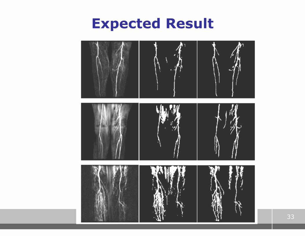
Example: MRA segmentation
� artery/vein may have similar intensity at given time point
� but different time profiles
� wish to segment according to time profile, not single intensity
IDEA Lab, Radiology, Cornell
33
Expected Result

IDEA Lab, Radiology, Cornell
Example: MRA segmentation
� First: need a time model of all segments
� Lets use ML principles to see which voxel belongs to which model
� Artery:
� Vein:
� Parench:
)|( ath θ
)|( vth θ
iaii nthy += )|( θ
ivii nthy += )|( θ
ipii nthy += )|( θ

IDEA Lab, Radiology, Cornell
−−=
2
2
2
))|((exp)|Pr(
σ
θθ aii
ai
thyy
−−=
2
2
2
))|((exp)|Pr(
σ
θθ vii
vi
thyy
−−=
2
2
2
))|((exp)|Pr(
σ
θθ pii
pi
thyy
Maximum Likelihood Classification
iaii nthy += )|( θ
ivii nthy += )|( θ
ipii nthy += )|( θ
Artery:
Vein:
Paren:
So at each voxel, the best model is one that maximizes:
Or equivalently, minimizes:
∏=i
ii yy )|Pr()|Pr( θθ
∑ −i
ii thy2))|(( θ
−
−=∑
2
2
2
))|((
expσ
θi
ii thy

IDEA Lab, Radiology, Cornell
� Data: Tumor model Rabbit DCE-MR data
� Paramegnetic contrast agent , pathology gold standard
� Extract temporal features from DCE-MRI
� Use these features for accurate detection and quantification of tumour
Liver tumour quantification from Dynamic Contrast Enhanced MRI

IDEA Lab, Radiology, Cornell
Liver Tumour Temporal models
37
Typical plot of time-resolved
MR signal of various tissue
classes
Temporal models used to
extract features
)|( θth

IDEA Lab, Radiology, Cornell
Liver tumour quantification from DCE-MRI

IDEA Lab, Radiology, Cornell
39
Slides by Andrew Moore (CMU): available on
course webpage
Paper by Jae Myung, “Tutorial on Maximum
Likelihood”: available on course webpage
http://www.cs.cmu.edu/~aberger/maxent.html
ML Tutorials
Max Entropy Tutorial

Part IV : Maximum A Posteriori (Bayesian) Estimation and Examples

IDEA Lab, Radiology, Cornell
41
Failure modes of ML
� Likelihood isn’t the only criterion for selecting a model or parameter
– Though it’s obviously an important one
� ML can be overly sensitive to noise
� Bizarre models may have high likelihood
– Consider a speedometer reading 55 MPH
– Likelihood of “true speed = 55”: 10%
– Likelihood of “speedometer stuck”: 100%
� ML likes “fairy tales”
– In practice, exclude such hypotheses
� There must be a principled solution…
– We need additional information to unwedge ML from bad solutions

IDEA Lab, Radiology, Cornell
If PDF of θ (or x) is also known…
y = H x + n, n is Gaussian (1)
ML
� If we know both Likelihood AND some prior knowledge about the unknown x
� Then can exploit this knowledge
� How? Suppose PDF of x is known
MAP
y = H x + n, n, x are Gaussian (2)

IDEA Lab, Radiology, Cornell
43
MAP for Line fitting problem
� If model estimated by ML and Prior info do not agree…
� MAP is a compromise between the two
LS estimate
Most probable
prior model
MAP estimate

IDEA Lab, Radiology, Cornell
44
Maximum a Posteriori Estimate
� Prior knowledge about random variables is generally expressed in the form of a PDF Pr(x)
� Once the likelihood L(x) and prior are known, we have complete statistical knowledge
� MAP (aka Bayesian) estimates are optimal
Bayes Theorem:
Pr(x|y) = Pr(y|x) . Pr(x)
Pr(y)
likelihood
prior posterior

IDEA Lab, Radiology, Cornell
45
Maximum a Posteriori (Bayesian) Estimate
� Consider the class of linear systems y = Hx + n
� Bayesian methods maximize the posterior probability:
Pr(x|y) � Pr(y|x) . Pr(x) � Pr(y|x) (likelihood function) = exp(- ||y-Hx||2)
� Pr(x) (prior PDF) = exp(-G(x))
� Non-Bayesian: maximize only likelihood
xest = arg min ||y-Hx||2
� Bayesian: xest = arg min ||y-Hx||2 + G(x) ,
where G(x) is obtained from the prior distribution of x

IDEA Lab, Radiology, Cornell
46
Example Bayesian Estimation
� Example: Gaussian prior centered at zero: Pr(x) = exp{- ½ xT Rx
-1 x}
� Bayesian methods maximize the posterior probability:
Pr(x|y) � Pr(y|x) . Pr(x) � Pr(y|x) (likelihood function) = exp(- ||y-Hx||2)
� ML estimate: maximize only likelihood
xest = arg min ||y-Hx||2
� MAP estimate:
xest = arg min ||y-Hx||2 + λ xT Rx-1 x
Makes Hx close to y Tries to make x a sample from
a Gaussian centered at 0

IDEA Lab, Radiology, Cornell
47
MAP Example: Multi-variate FLASH
� Acquire 6-10 accelerated FLASH data sets at different flip angles or TR’s
� Generate T1 maps by fitting to:
( )( )
( )1*
2
1
1 expexp sin
1 cos exp
TR TS TE T
TR Tα
α
− −= −
− −
• Not enough info in a single voxel
• Noise causes incorrect estimates
• Error in flip angle varies spatially!

IDEA Lab, Radiology, Cornell
48
Spatially Coherent T1, ρρρρ estimation
� First, stack parameters from all voxels in one big vector x
� Stack all observed flip angle images in y
� Then we can write y = M (x) + n
� Recall M is the (nonlinear) functional obtained from
( )( )
( )1*
2
1
1 expexp sin
1 cos exp
TR TS TE T
TR Tα
α
− −= −
− −
� Solve for x by non-linear least square fitting, PLUS spatial prior:
xest = arg minx || y - M (x) ||2 + µ2||Dx||2
� Minimize via MATLAB’s lsqnonlin function
E(x)
Makes M(x) close to y Makes x smooth

IDEA Lab, Radiology, Cornell
49
Multi-Flip Results – combined ρρρρ, T1 in pseudocolour

IDEA Lab, Radiology, Cornell
50
Multi-Flip Results – combined ρρρρ, T1 in pseudocolour

IDEA Lab, Radiology, Cornell
51
MAP example 2: Spatial Priors For Dynamic Imaging
Frames are tightly distributed around mean
After subtracting mean, images are close to Gaussian
time
frame Nf
frame 2
frame 1
Prior: -mean is µx
-local std.dev. varies as a(i,j)
mean
mean µx(i,j)
variance
envelope a(i,j)

IDEA Lab, Radiology, Cornell
52
Spatial Priors for MR images
� Stochastic MR image model:
x(i,j) = µx (i,j) + a(i,j) . (h ** p)(i,j) (1) ** denotes 2D convolution
µx (i,j) is mean image for class
p(i,j) is a unit variance i.i.d. stochastic process
a(i,j) is an envelope function h(i,j) simulates correlation properties of image x
x = ACp + µ (2)
where A = diag(a) , and C is the Toeplitz matrix generated by h
� Can model many important stationary and non-stationary cases
stationary process
r(τ1, τ2) = (h ** h)(τ1, τ2)

IDEA Lab, Radiology, Cornell
53
MAP estimate for Imaging Model (3)
� The Wiener estimate
xMAP - µ x = HRx (HRxHH + Rn)
-1 (y- µ y) (3)
Rx, Rn = covariance matrices of x and n
Stationarity � Rx has Toeplitz structure � fast processing
xMAP - µ x = HACCHAH ( HACCHAHHH + σn2 I )-1 (y- µ y) (4)
� Direct inversion prohibitive; so use CG iterative method
� (4) better than (3) since A and C are O(N log N) operations, enabling much faster processing

IDEA Lab, Radiology, Cornell
54
How to obtain estimates of A, C ?
� Need a training set of full-resolution images xk, k = 1,…,K
� Parallel imaging doesnt provide un-aliased full-res images
� Approaches: 1. Use previous full-res scans
- time consuming, need frequent updating 2. Use SENSE-reconstructed images for training set
- very bad noise amplification issues for high speedups
3. Directly estimate parameters from available parallel data
- Aliasing may cause inaccuracies

IDEA Lab, Radiology, Cornell
55
MAP for Dynamic Parallel Imaging
time
frame Nf
frame 2
frame 1
� Nf images available for parameter estimation!
� All images well-registered
� Tightly distributed around pixel mean
� Parameters can be estimated from aliased data
frame 1 frame 2 frame Nf
� Nf /R full-res images kx
ky

IDEA Lab, Radiology, Cornell
56
MAP-SENSE Preliminary Results
Unaccelerated 5x faster: MAP-SENSE
� Scans acceleraty 5x
� The angiogram was computed by: avg(post-contrast) – avg(pre-contrast)
5x faster: SENSE

Part V : Time Series Analysis
Finding statistically optimal models for time series data

IDEA Lab, Radiology, Cornell
Time Series Analysis
� Lots of clinical data are time series
– ECG, MR contrast enhanced tumor data,
– ER caseload, cancer patient survival curves,…
� How to extract temporal features from these data?
– Use Fourier, Wavelet Transforms, fit piecewise linear models
– Good, but… features are arbitrary, no optimality properties
� Now: better models for time series with memory
– Autoregressive (AR) model
– Moving Average (MA) model
– Both AR + MA 58

IDEA Lab, Radiology, Cornell
Autoregressive models
� When current signal depends on past signal
– Data has “memory”
� Quite natural for biological and physiological data
– Because real systems cannot change arbitrarily
– Current state depends on previous state
� Model:
59
How to estimate phi1?
AR parameters Noise or input process

IDEA Lab, Radiology, Cornell
Estimating AR parameters
� Example: For
� If noise is independent Gaussian, then show that ML estimate is
60

IDEA Lab, Radiology, Cornell
Estimating AR parameters
� Can you do this for any p?
http://www-stat.wharton.upenn.edu/~steele/Courses/956/Resource/YWSourceFiles/YW-Eshel.pdf
61

IDEA Lab, Radiology, Cornell
Moving Average Process
� MA process model:
� Autoregressive moving average (ARMA) model:
� Combines both AR and MA processes
62
Noise or input process
MA parameters
Model order
IDEA Lab, Radiology, Cornell
Estimating MA and ARMA
parameters � MA parameters = from Fourier Transforms
� ARMA: more complicated, but most languages have library routines that do this
� E.g. in MATLAB: ar(), arma(), etc
� What is the benefit of estimating AR and ARMA parameters?
– Might provide better temporal features than FT, WT or arbitrary temporal features
� Example: could apply this to mammography or liver tumor data
63

IDEA Lab, Radiology, Cornell
Examples
� Maybe these are better modeled as ARMA???
64
Artery/vein in MR
angiography
Liver Tumour
time profiles

IDEA Lab, Radiology, Cornell
65
References
� Simon Kay. Statistical Signal Processing. Part I: Estimation Theory. Prentice Hall 2002
� Simon Kay. Statistical Signal Processing. Part II: Detection Theory. Prentice Hall 2002
� Haacke et al. Fundamentals of MRI.
� Zhi-Pei Liang and Paul Lauterbur. Principles of MRI – A Signal Processing Perspective.
Info on part IV:
� Ashish Raj. Improvements in MRI Using Information Redundancy. PhD thesis, Cornell University, May 2005.
� Website: http://www.cs.cornell.edu/~rdz/SENSE.htm

IDEA Lab, Radiology, Cornell
66
Next lecture (Friday)
� Non-parametric density estimation
– Histograms + various fitting methods
– Nearest neighbor
– Parzen estimation
Next lecture (Wednesday)
� Maximum A Posteriori Estimators
� Several examples

CS5540: Computational Techniques for Analyzing Clinical Data
Lecture 7:
Statistical Estimation: Least
Squares, Maximum Likelihood and Maximum A Posteriori Estimators
Ashish Raj, PhD
Image Data Evaluation and Analytics Laboratory (IDEAL)
Department of Radiology
Weill Cornell Medical College
New York