Staphylococcus (Staphylococcus aureus, Staphylococcus epidermidis, and Staphylococcus saprophyticus)

Staphylococcus aureus clumping factor A is aforce-sensitive molecular switch thatactivates bacterial adhesionPhilippe Herman-Bausiera,1, Cristina Labatea,1, Aisling M. Towellb, Sylvie Derclayea, Joan A. Geogheganb,2,and Yves F. Dufrênea,c,2
aInstitute of Life Sciences, Université Catholique de Louvain, B-1348 Louvain-la-Neuve, Belgium; bDepartment of Microbiology, Moyne Institute ofPreventive Medicine, School of Genetics and Microbiology, Trinity College Dublin, Dublin 2, Ireland; and cWalloon Excellence in Life Sciences andBiotechnology, B-1300 Wavre, Belgium
Edited by Richard P. Novick, New York University School of Medicine, New York, NY, and approved April 13, 2018 (received for review October 17, 2017)
Clumping factor A (ClfA), a cell-wall–anchored protein from Staph-ylococcus aureus, is a virulence factor in various infections andfacilitates the colonization of protein-coated biomaterials. ClfApromotes bacterial adhesion to the blood plasma protein fibrino-gen (Fg) via molecular forces that have not been studied so far. Aunique, yet poorly understood, feature of ClfA is its ability to favoradhesion to Fg at high shear stress. Unraveling the strength anddynamics of the ClfA–Fg interaction would help us better under-stand how S. aureus colonizes implanted devices and withstandsphysiological shear stress. By means of single-molecule experi-ments, we show that ClfA behaves as a force-sensitive molecularswitch that potentiates staphylococcal adhesion under mechanicalstress. The bond between ClfA and immobilized Fg is weak(∼0.1 nN) at low tensile force, but is dramatically enhanced(∼1.5 nN) by mechanical tension, as observed with catch bonds.Strong bonds, but not weak ones, are inhibited by a peptide mim-icking the C-terminal segment of the Fg γ-chain. These resultspoint to a model whereby ClfA interacts with Fg via two distinctbinding sites, the adhesive function of which is regulated by me-chanical tension. This force-activated mechanism is of biologicalsignificance because it explains at the molecular level the abilityof ClfA to promote bacterial attachment under high physiologicalshear stress.
Staphylococcus aureus | ClfA | fibrinogen | shear stress |atomic force microscopy
The bacterial pathogen Staphylococcus aureus binds to hostextracellular matrix proteins using a variety of cell-wall–
anchored proteins (1). Among these, the fibrinogen (Fg)-bindingmicrobial surface component recognizing the adhesive matrixmolecule (MSCRAMM) protein clumping factor A (ClfA) is animportant virulence factor of S. aureus involved in various in-fections (2–4). In addition, ClfA promotes bacterial attachmentto plasma protein-coated biomaterials allowing the bacteria tocolonize and form a biofilm (5). Vaccination with recombinantClfA is protective against infection, and ClfA has been includedas an antigen in a number of multivalent S. aureus vaccinescurrently in clinical trials (2, 6, 7). ClfA has an N-terminal Aregion composed of three separately folded subdomains: N1, N2,and N3. N2 and N3 form the minimum ligand-binding region andbind the carboxy-terminus of the γ-chain of Fg (Fig. 1A and refs.8–10) through a variation of the multistep “dock, lock, and latch”(DLL) mechanism (11) first described for the binding of theStaphylococcus epidermidis protein SdrG to Fg (12). The carboxy-terminus of the γ-chain of Fg docks in a ligand-binding trenchlocated between subdomains N2 and N3. The DLL mechanisminvolves dynamic conformational changes of the adhesin thatresult in a greatly stabilized adhesin-ligand complex. The overallaffinity of the interaction of ClfA with Fg is increased throughinteractions at a recently described second site that lies at the topof subdomain N3 outside of the DLL ligand-binding trench (13).
Antibiotic treatments have proven to be less and less effectiveover the years due to the emergence of multidrug-resistantstrains (14, 15). Therefore, there is great interest in developingalternative strategies to fight bacterial infections (15, 16). Anappealing approach is the use of antiadhesion compounds toblock cell adhesion and biofilm development (17). A well-knownexample is the use of cranberry juice to treat urinary tract in-fections by uropathogenic Escherichia coli bacteria (18). Thedevelopment of novel antiadhesion therapeutics targeting ClfArequires a detailed understanding of the ligand-binding mecha-nisms of this adhesin.An interesting trait of ClfA is its ability to favor adhesion to
blood proteins at high shear stress (19–21). Despite the bi-ological relevance of such stress-induced adhesion, the un-derlying molecular mechanism has not yet been elucidated.We hypothesized that the interaction of ClfA with Fg mightstrengthen through a force-sensitive mechanism. To test thisidea, we measured the strength of the bond between ClfA andimmobilized Fg at various tensile loads, using atomic force mi-croscopy (AFM) (Fig. 1C) (22–25). We found that, while theClfA–Fg bond is weak at low applied force, the bond strength isdramatically increased at high force. These results providecompelling evidence that ClfA functions as a force-sensitive
Significance
The Staphylococcus aureus surface protein clumping factor A(ClfA) binds to the blood plasma protein fibrinogen (Fg) viamolecular interactions that are poorly understood. Here, weunravel the forces guiding the interaction between ClfA andimmobilized Fg, showing that it is dramatically enhanced bytensile loading. Our findings favor a model whereby ClfA in-teracts with Fg via two distinct binding sites, the adhesivefunction of which is tightly regulated by mechanical force.Reminiscent of a catch bond mechanism, this force-enhancedadhesion explains the ability of ClfA to promote S. aureuscolonization of host tissues and biomedical devices underphysical stress.
Author contributions: P.H.-B., C.L., A.M.T., S.D., J.A.G., and Y.F.D. designed research;P.H.-B., C.L., A.M.T., and S.D. performed research; P.H.-B., C.L., A.M.T., S.D., J.A.G., andY.F.D. analyzed data; P.H.-B., C.L., A.M.T., S.D., J.A.G., and Y.F.D. wrote the paper.
The authors declare no conflict of interest.
This article is a PNAS Direct Submission.
This open access article is distributed under Creative Commons Attribution-NonCommercial-NoDerivatives License 4.0 (CC BY-NC-ND).1P.H.-B. and C.L. contributed equally to this work.2To whom correspondence may be addressed. Email: [email protected] or [email protected].
This article contains supporting information online at www.pnas.org/lookup/suppl/doi:10.1073/pnas.1718104115/-/DCSupplemental.
Published online May 7, 2018.
5564–5569 | PNAS | May 22, 2018 | vol. 115 | no. 21 www.pnas.org/cgi/doi/10.1073/pnas.1718104115
Dow
nloa
ded
by g
uest
on
June
6, 2
020

molecular switch that regulates the strength of adhesion of S. aureusto protein-conditioned biomaterials, thus emphasizing the role thatphysical forces play in activating the function of bacterial adhesins.This study holds promise for the design of antibacterial agents withthe capacity to inhibit S. aureus adhesion at high shear rate.
ResultsS. aureus Strongly Binds to Immobilized Fibrinogen. To investigateClfA–Fg interactions in living bacteria, we used S. aureusSH1000 clfA clfB fnbA fnbB [hereafter called S. aureus ClfA(−)
cells; ref. 26] and the same strain transformed with a plasmidexpressing the entire clfA gene [ClfA(+) cells]. Fg was immobi-lized on solid substrates using N-hydroxysuccinimide (NHS)surface chemistry. Optical microscopy images confirmed thatClfA(+) cells adhered in large amounts to Fg-coated substrates,while no adhesion was seen with ClfA(−) cells (Fig. 1B). This indi-cates that ClfA is well-expressed and represents the only Fg-bindingprotein found at the cell surface of this strain. By means of single-cell force spectroscopy (SCFS; Fig. 1C, Left) (22, 27–29), we ana-lyzed the ClfA–Fg-binding forces at the whole-cell level. Singlebacteria were attached onto colloidal cantilevers, and the forcesbetween the cell probes and Fg substrates were measured. Fig. 2Ashows the maximum adhesion forces and rupture lengths obtainedfor five representative S. aureus ClfA(+) cells (for more cells, see SIAppendix, Fig. S1A). Most force curves feature adhesion forcepeaks ranging from ∼1,000 to ∼10,000 pN with rupture lengths of∼250–300 nm. The characteristics of the curves did not substantially
change when recording consecutive force curves on different spotsof the substrate, meaning that force measurements did not alter thecell-surface properties. Adhesion forces were spread across awide range, suggesting that a variable number of molecularbonds were probed. Most adhesive forces were larger than theforces measured for other staphylococcal adhesins (30, 31),suggesting that bacterial adhesion to Fg is very strong. As theselarge forces were abolished in ClfA(−) cells (Fig. 2B and SIAppendix, Fig. S1B), we conclude that they mostly reflect spe-cific ClfA–Fg interactions. Most bonds ruptured at ∼250–300 nm, which is consistent with the length of fully unfoldedadhesins. Assuming that the processed mature ClfA proteincomprises 860 residues, that each amino acid contributes0.36 nm to the contour length of the polypeptide chain, and thatthe ClfA folded length is ∼25 nm, we expect that the fully ex-tended ClfA protein should be ∼285 nm long.
How Strong Is the ClfA–Fibrinogen Interaction? To quantify thestrength of single ClfA-Fg bonds we used single-molecule forcespectroscopy (SMFS; Fig. 1C, Right) (29, 32) with Fg-modifiedtips. Fg molecules were attached to the tips at low densityusing a PEG-benzaldehyde linker. In Fig. 3A we present theadhesion force maps, maximum adhesion forces, and rupturelengths obtained between three S. aureus ClfA(+) cells andAFM tips functionalized with PEG chemistry (for more cells, seeSI Appendix, Fig. S2A). Strong adhesion peaks were detected withmean forces of 1,999 ± 267 pN (mean and SD of n = 401 adhesivecurves), 1,909 ± 155 pN (n = 88), and 2,093 ± 68 pN (n = 107) forcell #1, cell #2, and cell #3, respectively. Similar adhesion forceswere observed when the tips were modified via the NHSchemistry (Fig. 3B), indicating that the detected forces werenot dependent on the grafting protocol. Adhesion was medi-ated by ClfA as it was abrogated in ClfA(−) cells (Fig. 3C andSI Appendix, Fig. S2B). Force maps revealed that ClfA wasexpressed at rather high density and heterogeneously distrib-uted on the cell surface.We believe that the ∼2-nN forces are associated with single
DLL-like interactions for the following reasons. First, for all cellsinvestigated, adhesion forces featured distributions that werenarrow and centered near ∼2 nN, which strongly supports theidea that single bonds were probed. When multiple bonds breaksimultaneously, a wide force range reflecting multiples of theweakest unit force should be observed, which was not the case.Second, forces with very similar sharp distributions wereobtained using two different tip chemistries (PEG vs. NHS) thatare known to favor single-molecule detection. Third, strongforces are in the range of the strength measured for the DLLinteraction between the structurally related SdrG protein and Fg,both on living bacteria (29) and on purified adhesins (33).Fourth, we generated an S. aureus strain (called ClfAPY)expressing ClfA carrying amino acid substitutions within theN2N3-binding trench (P336S and Y338A) that prevents ligandbinding by the DLL mechanism. We found that these substitu-tions almost completely abolished Fg binding to ClfA (SI Ap-pendix, Fig. S3), thus demonstrating that strong ClfA bindingforces are due to a DLL-like interaction.To study ClfA in the absence of other staphylococcal cell-wall
components, we also examined cells from a Lactococcus lactisstrain expressing ClfA [L. lactis ClfA(+) cells] (34). As illustratedin Fig. 4A (for more cells, see SI Appendix, Fig. S4), this strainfeatured the same behavior as the S. aureus ClfA(+) strain, withstrong forces of 2,070 ± 93 pN (mean and SD on n = 508 ad-hesive curves), 1,848 ± 119 pN (n = 425), and 1,891 ± 225 pN(n = 333) for cell #1, cell #2, and cell #3, respectively. L. lactisClfA(+) cells showed a higher adhesion frequency and some-times large force values >2 nN. Whole-cell dot immunoblots(SI Appendix, Fig. S5) suggest that this may be due to a higher
Fig. 1. Studying the ClfA–Fg interaction. (A) The N-terminal signal sequence(S) is followed by subdomains N1, N2, and N3 comprising the ligand-binding Aregion. At the junction between N2 and N3 is found a binding trench in whichthe ligand (red) inserts and is locked in place. A flexible serine-aspartate repeatregion links region A to the C-terminal Wall (W) spanning region and thesorting sequence. The LPXTG motif allows anchoring of the protein to cell-wallpeptidoglycan by sortase A. (B) Optical microscopy images of S. aureus bacteriaexpressing or not full-length ClfA [ClfA(+) and ClfA(−) cells] following incubationwith Fg-conditioned substrates. (C) Force nanoscopy of the ClfA–Fg interaction:(Left) SCFS; (Right) SMFS. For clarity, the N1 domain of ClfA is not shown.
Herman-Bausier et al. PNAS | May 22, 2018 | vol. 115 | no. 21 | 5565
MICRO
BIOLO
GY
Dow
nloa
ded
by g
uest
on
June
6, 2
020

expression of the adhesin at the cell surface of L. lactis ClfA(+)
compared with S. aureus ClfA(+).One may argue that our experiments may not mimic in vivo
conditions as Fg was attached to the tip. So we cannot extend ourfindings on immobilized Fg to conditions where the bacteria mayinteract with the soluble form of Fg. However, immobilized Fg isbiologically relevant as it is associated with blood clots and,importantly, deposited on biomedical device surfaces. Given thatthe concentration of free Ca2+ in blood is equivalent to the IC50
for Ca2+ to inhibit Fg binding (35), 50% of ClfA molecules willnot have soluble Fg bound and will be free to engage withimmobilized Fg in a clot or on a surface.
The ClfA–Fibrinogen Interaction Is Dramatically Enhanced by TensileLoading. During colonization of host tissues and biomedical de-vices, S. aureus is subjected to physical forces, such as fluid flowand cell-surface contacts (36). An interesting but poorly un-derstood feature of ClfA is its ability to promote S. aureus ad-hesion under shear stresses (19, 37). We postulated that the ClfAbinding strength might be enhanced by mechanical force. To testthis, the strength of the ClfA–Fg interaction (F) was measuredwhile varying the rate at which force increases (loading rate, LR;the effective LR was estimated from the force vs. time curves) (38).Fig. 4B shows that the dynamic force spectroscopy data obtainedon L. lactis ClfA(+) cells (data pooled from 4,452 adhesive curves
Fig. 2. Forces guiding the adhesion of single bacteria to Fg substrates. (A) Maximum adhesion force (Left) and rupture length (Right) histograms withrepresentative retraction force profiles (Insets) obtained by recording force-distance curves in PBS between five S. aureus ClfA(+) cells from different culturesand Fg substrates. (B) Force data obtained in the same conditions for a S. aureus ClfA(−) cell. Fluorescence images of the bacterial probes stained with theBacLight viability kit (Insets) confirmed that the cell membrane is intact even after 30 min of measurements.
Fig. 3. Binding strength of single ClfA–Fg bonds in living S. aureus bacteria. (A, Left) Maximum adhesion force histograms with force maps (Insets). (Scalebars: 100 nm.) (Right) Rupture length histograms with representative retraction force profiles (Insets) obtained by recording force-distance curves in PBSbetween three different S. aureus ClfA(+) cells and AFM tips functionalized with Fg using PEG chemistry. (B) Data obtained in the same conditions betweentwo different S. aureus ClfA(+) cells and AFM tips functionalized with Fg using NHS chemistry. (C) Force data between a S. aureus ClfA(−) cell and a AFM tipfunctionalized with Fg using the PEG chemistry.
5566 | www.pnas.org/cgi/doi/10.1073/pnas.1718104115 Herman-Bausier et al.
Dow
nloa
ded
by g
uest
on
June
6, 2
020
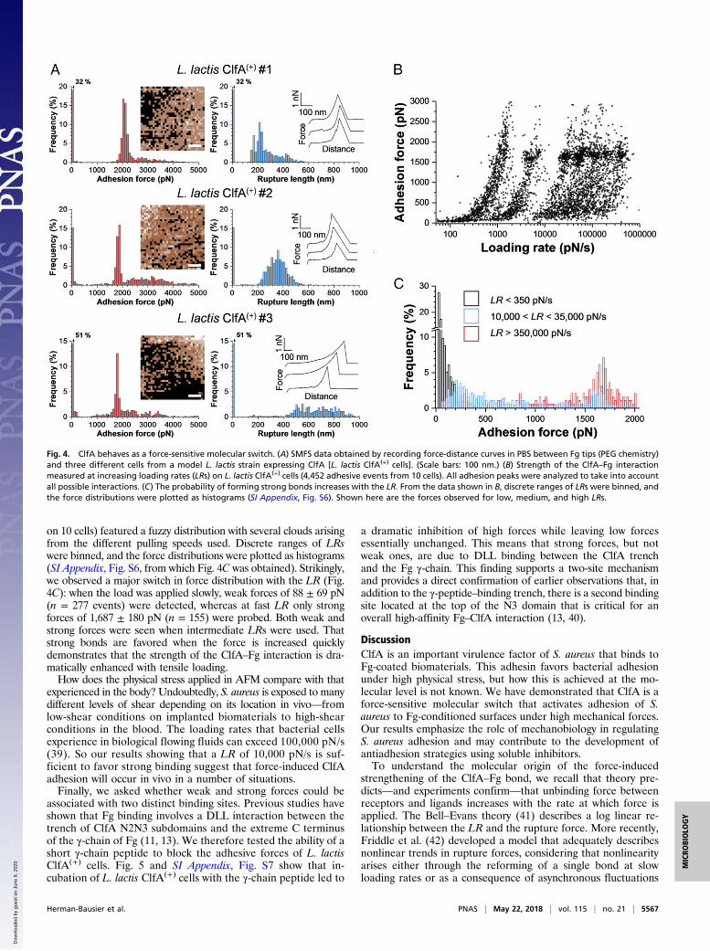
on 10 cells) featured a fuzzy distribution with several clouds arisingfrom the different pulling speeds used. Discrete ranges of LRswere binned, and the force distributions were plotted as histograms(SI Appendix, Fig. S6, from which Fig. 4C was obtained). Strikingly,we observed a major switch in force distribution with the LR (Fig.4C): when the load was applied slowly, weak forces of 88 ± 69 pN(n = 277 events) were detected, whereas at fast LR only strongforces of 1,687 ± 180 pN (n = 155) were probed. Both weak andstrong forces were seen when intermediate LRs were used. Thatstrong bonds are favored when the force is increased quicklydemonstrates that the strength of the ClfA–Fg interaction is dra-matically enhanced with tensile loading.How does the physical stress applied in AFM compare with that
experienced in the body? Undoubtedly, S. aureus is exposed to manydifferent levels of shear depending on its location in vivo—fromlow-shear conditions on implanted biomaterials to high-shearconditions in the blood. The loading rates that bacterial cellsexperience in biological flowing fluids can exceed 100,000 pN/s(39). So our results showing that a LR of 10,000 pN/s is suf-ficient to favor strong binding suggest that force-induced ClfAadhesion will occur in vivo in a number of situations.Finally, we asked whether weak and strong forces could be
associated with two distinct binding sites. Previous studies haveshown that Fg binding involves a DLL interaction between thetrench of ClfA N2N3 subdomains and the extreme C terminusof the γ-chain of Fg (11, 13). We therefore tested the ability of ashort γ-chain peptide to block the adhesive forces of L. lactisClfA(+) cells. Fig. 5 and SI Appendix, Fig. S7 show that in-cubation of L. lactis ClfA(+) cells with the γ-chain peptide led to
a dramatic inhibition of high forces while leaving low forcesessentially unchanged. This means that strong forces, but notweak ones, are due to DLL binding between the ClfA trenchand the Fg γ-chain. This finding supports a two-site mechanismand provides a direct confirmation of earlier observations that, inaddition to the γ-peptide–binding trench, there is a second bindingsite located at the top of the N3 domain that is critical for anoverall high-affinity Fg–ClfA interaction (13, 40).
DiscussionClfA is an important virulence factor of S. aureus that binds toFg-coated biomaterials. This adhesin favors bacterial adhesionunder high physical stress, but how this is achieved at the mo-lecular level is not known. We have demonstrated that ClfA is aforce-sensitive molecular switch that activates adhesion of S.aureus to Fg-conditioned surfaces under high mechanical forces.Our results emphasize the role of mechanobiology in regulatingS. aureus adhesion and may contribute to the development ofantiadhesion strategies using soluble inhibitors.To understand the molecular origin of the force-induced
strengthening of the ClfA–Fg bond, we recall that theory pre-dicts—and experiments confirm—that unbinding force betweenreceptors and ligands increases with the rate at which force isapplied. The Bell–Evans theory (41) describes a log linear re-lationship between the LR and the rupture force. More recently,Friddle et al. (42) developed a model that adequately describesnonlinear trends in rupture forces, considering that nonlinearityarises either through the reforming of a single bond at slowloading rates or as a consequence of asynchronous fluctuations
Fig. 4. ClfA behaves as a force-sensitive molecular switch. (A) SMFS data obtained by recording force-distance curves in PBS between Fg tips (PEG chemistry)and three different cells from a model L. lactis strain expressing ClfA [L. lactis ClfA(+) cells]. (Scale bars: 100 nm.) (B) Strength of the ClfA–Fg interactionmeasured at increasing loading rates (LRs) on L. lactis ClfA(+) cells (4,452 adhesive events from 10 cells). All adhesion peaks were analyzed to take into accountall possible interactions. (C) The probability of forming strong bonds increases with the LR. From the data shown in B, discrete ranges of LRs were binned, andthe force distributions were plotted as histograms (SI Appendix, Fig. S6). Shown here are the forces observed for low, medium, and high LRs.
Herman-Bausier et al. PNAS | May 22, 2018 | vol. 115 | no. 21 | 5567
MICRO
BIOLO
GY
Dow
nloa
ded
by g
uest
on
June
6, 2
020

of several independent interactions. So current models and dataall show continuous increases in rupture force with the LR. Bycontrast, the ClfA–Fg bond features an unusual switch in forcedistribution; that is, weak bonds (∼100 pN) dominate at low LRwhile strong bonds (∼1,500 pN) are favored at high LR. Becausethe strengths of weak and strong bonds differ by an order ofmagnitude and intermediate forces were rarely observed, webelieve that strong bonds do not result from the simultaneousrupture of multiple weak bonds.We propose that the unusual force-dependent strengthening of
the ClfA–Fg bond involves a force-induced conformational changein the adhesin, from a weak- to a strong-binding state. That theγ-chain peptide of Fg inhibits high forces but not low forces favors atwo binding site model, where the activity of the two binding sites istightly regulated by tensile force (Fig. 6). Under low tensile force, Fgbinds to the top of the ClfA N3 domain via weak bonds. Under highmechanical tension, extension and conformational changes inthe ClfA molecule trigger the ultrastrong DLL interaction bythe N2N3 subdomains. Such a mechanism may help us toidentify soluble ligands capable of inhibiting bacterial adhe-sion under high flow conditions.The force-dependent ClfA–Fg interaction is reminiscent of a
catch-bond behavior, that is, a specific bond that is reinforced bymechanical stress (43). A well-documented example is the E. coliFimH adhesin that binds mannose residues on epithelial cells(39). The FimH–mannose bond is weak and relatively short livedat low flow, whereas this bond is strengthened at high flow. Thisis explained by an allosteric model in which tensile mechanicalforce induces an allosteric switch from a low- to a high-affinityconformation of the adhesin (43, 44). Perhaps the ClfA–Fg in-teraction involves such an allosterically controlled mechanism,whereby stretching of the subdomains would suppress allostericinterplay and trigger strong DLL binding.The strength of the ClfA bond at high tensile load is in the
range of that of covalent bonds, despite a moderate affinity value(8–10). This discrepancy suggests that the unbinding pathway of
the adhesin may change when mechanical force is applied (45). So,when studying the mechanisms of bacterial adhesion under phys-iological shear, force measurements performed at nonequilibriummight be more relevant than equilibrium assays. That the ClfA–Fgcomplex resists very high forces is counterintuitive as rupture ofthe polypeptide backbones is expected to occur first. A possibleexplanation is that the complex may direct force along pathwaysnonparallel to the pulling direction, as shown for the mechanicallystable multidomain cellulosome protein complex (45).The high binding strength provides a molecular framework to
explain how ClfA promotes S. aureus adhesion on blood protein-coated surfaces under high shear stress conditions (19, 37). Fgbinding to ClfA expressed on the surface of S. aureus or L. lactisfacilitates platelet capture and thrombus formation under highshear conditions but not when low shear rates are applied (19,37). ClfA binding to Fg under shear conditions creates a bridgebetween the bacterium and integrin receptors expressed by en-dothelial cells (46). In addition, ClfA is involved in the shear-dependent adhesion of S. aureus to von Willebrand factor,thereby allowing the bacteria to resist shear forces of flowingblood (20, 21). It is possible that S. aureus has evolved force-dependent adhesion mechanisms such as the one unraveled hereto help the bacteria resist physical stress during host coloniza-tion, whereas weak adhesion forces at low shear stress wouldfavor cell detachment and thus the colonization of new sites.
MethodsS. aureus ClfA(−) is a S. aureus SH1000 clfA clfB fnbA fnbB strain defective inboth clumping factors A and B and fibronectin-binding proteins A and B (26)whereas S. aureus ClfA(+) is SH1000 clfA clfB fnbA fnbB transformed with theplasmid pALC2073::clfA (47). To study the effect of amino acid substitutionswithin the N2N3-binding trench, we used S. aureus ClfAWT, which is SH1000clfA clfB fnbA fnbB carrying the plasmid pCF77 expressing ClfA from its ownpromoter (48), and S. aureus ClfAPY, which is the same strain with P336S andY338A substitutions in the N2 subdomain of ClfA (49). Growth conditionsand AFM methods are described in SI Appendix.
ACKNOWLEDGMENTS. We thank David Alsteens and Timothy Foster forfruitful discussion. Work at the Université Catholique de Louvain was sup-ported by the European Research Council under the European Union’s Ho-rizon 2020 Research and Innovation Programme (Grant 693630); theWalloon Excellence in Life Sciences and Biotechnology (Grant WELBIO-CR-2015A-05); the National Fund for Scientific Research (FNRS); and the Re-search Department of the Communauté Française de Belgique (ConcertedResearch Action). Y.F.D. is Research Director at the FNRS.
Fig. 5. Adhesion to Fg involves two ClfA-binding sites. (A and B) Adhesionforces obtained by recording force-distance curves in PBS between threedifferent L. lactis ClfA(+) cells and Fg tips in the absence (A) or presence (B) ofthe C-terminal segment of the Fg γ-chain (0.2 mg·mL−1). All adhesion peakswere analyzed to take into account all possible interactions.
Fig. 6. Proposed model for the force-activated adhesion of ClfA. (Left)Under low mechanical force, Fg weakly binds to the top of the ClfAN3 domain. (Right) Under high force, extension and conformational changesof the ClfA N2N3 subdomains enable the γ-chain of Fg to dock in the ligand-binding trench and form strong DLL-like interactions.
5568 | www.pnas.org/cgi/doi/10.1073/pnas.1718104115 Herman-Bausier et al.
Dow
nloa
ded
by g
uest
on
June
6, 2
020

1. Foster TJ, Geoghegan JA, Ganesh VK, Höök M (2014) Adhesion, invasion and evasion:The many functions of the surface proteins of Staphylococcus aureus. Nat RevMicrobiol 12:49–62.
2. Josefsson E, Hartford O, O’Brien L, Patti JM, Foster T (2001) Protection against ex-perimental Staphylococcus aureus arthritis by vaccination with clumping factor A, anovel virulence determinant. J Infect Dis 184:1572–1580.
3. McAdow M, et al. (2011) Preventing Staphylococcus aureus sepsis through the in-hibition of its agglutination in blood. PLoS Pathog 7:e1002307.
4. Moreillon P, et al. (1995) Role of Staphylococcus aureus coagulase and clumpingfactor in pathogenesis of experimental endocarditis. Infect Immun 63:4738–4743.
5. Vaudaux PE, et al. (1995) Use of adhesion-defective mutants of Staphylococcus aureusto define the role of specific plasma proteins in promoting bacterial adhesion tocanine arteriovenous shunts. Infect Immun 63:585–590.
6. Schaffer AC, Lee JC (2009) Staphylococcal vaccines and immunotherapies. Infect DisClin North Am 23:153–171.
7. Proctor RA (2015) Recent developments for Staphylococcus aureus vaccines: Clinicaland basic science challenges. Eur Cell Mater 30:315–326.
8. McDevitt D, Francois P, Vaudaux P, Foster TJ (1994) Molecular characterization of theclumping factor (fibrinogen receptor) of Staphylococcus aureus. Mol Microbiol 11:237–248.
9. McDevitt D, Francois P, Vaudaux P, Foster TJ (1995) Identification of the ligand-binding domain of the surface-located fibrinogen receptor (clumping factor) ofStaphylococcus aureus. Mol Microbiol 16:895–907.
10. McDevitt D, et al. (1997) Characterization of the interaction between the Staphylo-coccus aureus clumping factor (ClfA) and fibrinogen. Eur J Biochem 247:416–424.
11. Ganesh VK, et al. (2008) A structural model of the Staphylococcus aureus ClfA-fibrinogen interaction opens new avenues for the design of anti-staphylococcaltherapeutics. PLoS Pathog 4:e1000226.
12. Ponnuraj K, et al. (2003) A “dock, lock, and latch” structural model for a staphylo-coccal adhesin binding to fibrinogen. Cell 115:217–228.
13. Ganesh VK, et al. (2016) Lessons from the crystal structure of the S. aureus surfaceprotein clumping factor A in complex with Tefibazumab, an inhibiting monoclonalantibody. EBioMedicine 13:328–338.
14. Foster TJ (2017) Antibiotic resistance in Staphylococcus aureus. Current status andfuture prospects. FEMS Microbiol Rev 41:430–449.
15. Bassetti M, et al. (2017) Antimicrobial resistance in the next 30 years, humankind,bugs and drugs: A visionary approach. Intensive Care Med 43:1464–1475.
16. Czaplewski L, et al. (2016) Alternatives to antibiotics: A pipeline portfolio review.Lancet Infect Dis 16:239–251.
17. Geoghegan JA, Foster TJ, Speziale P, Dufrêne YF (2017) Live-cell nanoscopy in anti-adhesion therapy. Trends Microbiol 25:512–514.
18. Flores-Mireles AL, Walker JN, Caparon M, Hultgren SJ (2015) Urinary tract infections:Epidemiology, mechanisms of infection and treatment options. Nat Rev Microbiol 13:269–284.
19. Kerrigan SW, et al. (2008) Molecular basis for Staphylococcus aureus-mediatedplatelet aggregate formation under arterial shear in vitro. Arterioscler Thromb VascBiol 28:335–340.
20. Claes J, et al. (2014) Adhesion of Staphylococcus aureus to the vessel wall under flowis mediated by von Willebrand factor-binding protein. Blood 124:1669–1676.
21. Claes J, et al. (2017) Clumping factor A, von Willebrand factor-binding protein andvon Willebrand factor anchor Staphylococcus aureus to the vessel wall. J ThrombHaemost 15:1009–1019.
22. Helenius J, Heisenberg CP, Gaub HE, Müller DJ (2008) Single-cell force spectroscopy.J Cell Sci 121:1785–1791.
23. Xiao J, Dufrêne YF (2016) Optical and force nanoscopy in microbiology. Nat Microbiol1:16186.
24. Dufrêne YF, et al. (2017) Imaging modes of atomic force microscopy for application inmolecular and cell biology. Nat Nanotechnol 12:295–307.
25. Dufrêne YF (2017) Microbial nanoscopy: Breakthroughs, challenges, and opportuni-ties. ACS Nano 11:19–22.
26. O’Neill E, et al. (2008) A novel Staphylococcus aureus biofilm phenotype mediated bythe fibronectin-binding proteins, FnBPA and FnBPB. J Bacteriol 190:3835–3850.
27. Beaussart A, et al. (2013) Single-cell force spectroscopy of probiotic bacteria. Biophys J104:1886–1892.
28. Beaussart A, et al. (2014) Quantifying the forces guiding microbial cell adhesion usingsingle-cell force spectroscopy. Nat Protoc 9:1049–1055.
29. Herman P, et al. (2014) The binding force of the staphylococcal adhesin SdrG is re-markably strong. Mol Microbiol 93:356–368.
30. Herman-Bausier P, et al. (2016) Mechanical strength and inhibition of the Staphylo-coccus aureus collagen-binding protein Cna. MBio 7:e01529-16.
31. Feuillie C, et al. (2017) Molecular interactions and inhibition of the staphylococcalbiofilm-forming protein SdrC. Proc Natl Acad Sci USA 114:3738–3743.
32. Hinterdorfer P, Dufrêne YF (2006) Detection and localization of single molecularrecognition events using atomic force microscopy. Nat Methods 3:347–355.
33. Milles LF, Schulten K, Gaub HE, Bernardi RC (2018) Molecular mechanism of extrememechanostability in a pathogen adhesin. Science 359:1527–1533.
34. O’Brien L, et al. (2002) Multiple mechanisms for the activation of human plateletaggregation by Staphylococcus aureus: Roles for the clumping factors ClfA and ClfB,the serine-aspartate repeat protein SdrE and protein A. Mol Microbiol 44:1033–1044.
35. O’Connell DP, et al. (1998) The fibrinogen-binding MSCRAMM (clumping factor) ofStaphylococcus aureus has a Ca2+-dependent inhibitory site. J Biol Chem 273:6821–6829.
36. Otto M (2014) Physical stress and bacterial colonization. FEMS Microbiol Rev 38:1250–1270.
37. Kerrigan SW, Loughmann A, Meade G, Foster TJ, Cox D (2006) Staphylococcus aureusclumping factor mediates rapid thrombus formation under high shear. Blood 108:1816.
38. Alsteens D, et al. (2015) Imaging G protein-coupled receptors while quantifying theirligand-binding free-energy landscape. Nat Methods 12:845–851.
39. Yakovenko O, et al. (2008) FimH forms catch bonds that are enhanced by mechanicalforce due to allosteric regulation. J Biol Chem 283:11596–11605.
40. Geoghegan JA, et al. (2010) Molecular characterization of the interaction of staph-ylococcal microbial surface components recognizing adhesive matrix molecules(MSCRAMM) ClfA and Fbl with fibrinogen. J Biol Chem 285:6208–6216.
41. Merkel R, Nassoy P, Leung A, Ritchie K, Evans E (1999) Energy landscapes of receptor-ligand bonds explored with dynamic force spectroscopy. Nature 397:50–53.
42. Friddle RW, Noy A, De Yoreo JJ (2012) Interpreting the widespread nonlinear forcespectra of intermolecular bonds. Proc Natl Acad Sci USA 109:13573–13578.
43. Sokurenko EV, Vogel V, Thomas WE (2008) Catch-bond mechanism of force-enhancedadhesion: Counterintuitive, elusive, but ... widespread? Cell Host Microbe 4:314–323.
44. Sauer MM, et al. (2016) Catch-bond mechanism of the bacterial adhesin FimH. NatCommun 7:10738.
45. Schoeler C, et al. (2015) Mapping mechanical force propagation through bio-molecular complexes. Nano Lett 15:7370–7376.
46. McDonnell CJ, et al. (2016) Inhibition of major integrin αV β3 reduces Staphylococcusaureus attachment to sheared human endothelial cells. J Thromb Haemost 14:2536–2547.
47. McCormack N, Foster TJ, Geoghegan JA (2014) A short sequence within subdomainN1 of region A of the Staphylococcus aureusMSCRAMM clumping factor A is requiredfor export and surface display. Microbiology 160:659–670.
48. Hartford OM, Wann ER, Höök M, Foster TJ (2001) Identification of residues in theStaphylococcus aureus fibrinogen-binding MSCRAMM clumping factor A (ClfA) thatare important for ligand binding. J Biol Chem 276:2466–2473.
49. Loughman A, et al. (2005) Roles for fibrinogen, immunoglobulin and complementin platelet activation promoted by Staphylococcus aureus clumping factor A.Mol Microbiol 57:804–818.
Herman-Bausier et al. PNAS | May 22, 2018 | vol. 115 | no. 21 | 5569
MICRO
BIOLO
GY
Dow
nloa
ded
by g
uest
on
June
6, 2
020