Staphylococcal Toxic Shock Syndrome
22
Staphylococcal Toxic Shock Syndrome: Mechanisms and Management Silversides J, Lappin E, Ferguson AJ Curr Infect Dis Rep (Published online 19 th June 2010) DOI:10.1007/s119080100119y Please note: This is the author’s accepted manuscript for this article. There may have been minor changes in formatting prior to publication of the final publisher’s PDF version. The publisher’s PDF of this article is available at springerlink.com by clicking here.
-
Upload
meducationdotnet -
Category
Documents
-
view
241 -
download
1
Transcript of Staphylococcal Toxic Shock Syndrome
Staphylococcal Toxic Shock Syndrome: Mechanisms and Management
Silversides J, Lappin E, Ferguson AJ
Curr Infect Dis Rep (Published online 19th June 2010)
DOI:10.1007/s11908-‐010-‐0119-‐y
Please note:
This is the author’s accepted manuscript for this article. There may have been minor changes in formatting prior to publication of the final publisher’s PDF version. The publisher’s PDF of this
article is available at springerlink.com by clicking here.
Staphylococcal Toxic Shock Syndrome: Mechanisms and Management
Dr Jonathan A Silversides BSc (Hons) MB BCh BAO (Hons) FRCA
Specialty Registrar in Anaesthesia and Intensive Care Medicine
Regional Intensive Care Unit, Royal Victoria Hospital, Grosvenor Road, Belfast BT12 6BA, United Kingdom
Phone: +44 7718 196319
Fax: +44 2838 868565
Email: [email protected]
Dr Emma Lappin MB BCh BAO FCARCSI MRCP(UK)
Specialist Registrar in Anaesthesia
Belfast City Hospital, Lisburn Road, Belfast BT9 7AB, United Kingdom
Phone: +44 2890 329241
Fax: +44 2838 868565
Email: [email protected]
Dr Andrew J Ferguson* MEd FRCA FCARCSI DIBICM FCCP
Consultant in Intensive Care Medicine and Anaesthesia
Craigavon Area Hospital, 68 Lurgan Road, Portadown, United Kingdom BT63 5AL
Phone: +44 2838 612307
Fax: +44 2838 868565
Email: [email protected]
*Corresponding author
Word Count = 4007 including abstract
Keywords: Staphylococcus aureus; Superantigen; Toxic shock syndrome; Septic shock; Infection; Gram-‐positive; Immunoglobulin; Clindamycin; Linezolid; Daptomycin; Tigecycline; Toll-‐like receptor;
T-‐cell receptor; Cytokine; Systemic inflammatory response syndrome; Early goal directed therapy; Nuclear factor kappa B; Tumour necrosis factor alpha; Interleukin 10; Immunomodulation; Toxic
shock syndrome toxin 1; Methicillin resistant Staphylococcus aureus (MRSA); Pathogen associated molecular patterns (PAMP); Polymorphism
Abstract
Staphylococcal toxic shock syndrome is a rare complication of Staphylococcus aureus infection in
which bacterial toxins act as superantigens, activating very large numbers of T cells and generating
an overwhelming immune-‐mediated cytokine avalanche which manifests clinically as fever, rash,
shock and rapidly progressive multiple organ failure, often in young, previously healthy patients.
The syndrome can occur with any site of S. aureus infection, and so clinicians of all medical
specialties should have a firm grasp of the presentation and management. In this article we review
the literature on the pathophysiology, clinical features, and treatment of this serious condition with
emphasis on recent insights into pathophysiology and on information of relevance to the practicing
clinician.
Introduction
Staphylococcal toxic shock syndrome (TSS) is a rare complication of infection with Staphylococcus
aureus, specifically toxin-‐producing strains. While the precipitating infection may appear to be
minor, toxins, the most commonly implicated of which is toxic shock syndrome toxin-‐1 (TSST-‐1), act
as superantigens, generating a disproportionately exuberant immune response and cytokine
avalanche. This brings about a rapidly progressive clinical syndrome of multiple organ dysfunction
virtually indistinguishable from septic shock and associated with a significant mortality.
It is critical that all clinicians appreciate the pathophysiology and management of this potentially life-‐
threatening condition, given the multiple clinical presentations of staphylococcal infection and the
rise in prevalence of gram positive infections including both hospital and community acquired
methicillin resistant Staphylococcus aureus (MRSA) infection.
TSS should be considered in the differential diagnosis of any patient with severe systemic
inflammatory response syndrome of unclear aetiology, but particularly in the situation of an
overwhelming systemic response to a relatively minor source of gram-‐positive infection.
Epidemiology
Toxic shock syndrome was first described in 1978 [1] and there were subsequent reports during the
1980s in previously healthy young women in association with the introduction of highly absorbent
tampons. Following identification of these tampons as a risk factor for TSS, and their subsequent
removal from the market, the incidence declined steadily in the United States between 1980 and
1996 from a peak of 6-‐12 cases per 100,000 inhabitants per year [2]. Changes to the case definition,
and a reliance on physicians to report the disease, have made accurate incidence figures difficult to
obtain, with between 71 and 101 cases notified to the CDC per year in the United States in the last 5
years [3]. In one active surveillance area in Minneapolis-‐St Paul, the incidence was reported to have
increased from 0.9 to 3.4 cases per 100,000 in the period from 2000 to 2003 [4], however more
recent figures from the same surveillance programme suggest an incidence of 2.1 per 100,000.
Colonisation of upper respiratory tract, skin, and genital tract mucosa with S. aureus is common
even in healthy individuals, with persistent nasal carriage in up to 27% of the population [5] and
vaginal colonisation in just under 10% [6]. Overall only a small proportion, under 10%, of S. aureus
isolates carry tst, the gene encoding TSST-‐1 [7] and the prevalence of vaginal carriage of a toxigenic
strain of S. aureus is in the order of 1-‐3% of the adult female population [8]. The events that
culminate in the shift of S. aureus from colonisation to infection are unclear. TSS may develop from
staphylococcal infections in any site, although in many cases no focal source of infection is identified.
MRSA strains are an increasingly common problem, in the community as well as the hospital
population, and geographic spread of TSST-‐1-‐producing MRSA strains has been reported in Europe
and Japan [9,10], although the status of these strains in the United States is unclear. While the
existence of TSST-‐1-‐producing MRSA strains is of concern, there is conflicting evidence as to a
possible association between methicillin resistance and superantigen production [7, 11-‐13].
Due to non-‐specific clinical features and lack of widely available rapid diagnostic tools it is likely that
many cases of staphylococcal TSS go undiagnosed or are coded as septic shock, and available figures
may well underestimate the true incidence.
Pathophysiology
S. aureus produces a range of protein exotoxins which are key to understanding the pathogenesis of
TSS. These bacterial toxins include the staphylococcal enterotoxins (SEs), TSST-‐1 and the
staphylococcal enterotoxin-‐like toxins (SEIs) (so-‐called as their emetic potential remains unproven)
[14**]. All are virulence factors acting as superantigens to trigger excessive and non-‐conventional T-‐
cell activation with potentially catastrophic over-‐amplification of the inflammatory cytokine cascade.
The term “superantigen” was first used in the late 1980s to describe the mechanism behind the
powerful T-‐cell-‐stimulating properties of streptococcal enterotoxin B [15].
Superantigens bypass normal mechanisms regulating antigen presentation and processing, in which
peptide fragments are presented to the T cell via a specific peptide-‐binding groove of the major
histocompatibility complex (MHC) type II molecule on the antigen presenting cell (APC). This
conventional process allows T cell responses only when both the class II molecule and specific
antigen fragment are recognised. Superantigens directly stimulate T cells by binding as unprocessed,
intact proteins directly to the T cell receptor (TCR) and MHC class II molecule in combination and at
locations remote from the conventional peptide binding area [16]. This cross-‐linking mechanism
involves the variable portion of the TCR β chain and can induce a clonal expansion of T cells
possessing the corresponding TCR Vβ pattern. Many superantigens are thought to interact with
selected TCR Vβ regions, and identification of this characteristic Vβ pattern or signature may be
diagnostically useful. However, a recent French study has shown that although each Vβ signature
analysed was stimulated by at least one staphylococcal superantigen, there was considerable
overlap and redundancy in superantigen induced Vβ populations with some, but not all,
superantigens having characteristic Vβ patterns [17*]. The list of superantigens with unique
signatures included TSST-‐1, SEA, SEG, SEH, SEIJ, SEIK, SEIL, SEIN, SEIM SEIO, SEIQ, SER, SEIU, and
SEIV. The mitogenic potential of a particular superantigen appears to correlate directly with the
binding affinity between the TCR and the superantigen [18]. Superantigens are capable of
stimulating over 20% of host T cells, far in excess of that caused by conventional antigen
presentation, and with intense potency (femtogram concentrations of superantigen are all that is
required in vitro) [14**].
T cell activation by superantigens leads to a massive, uncoordinated release of proinflammatory
cytokines responsible for the clinical picture of toxic shock syndrome. Experimentally, cytokine
release is biphasic, with an initial rise in interleukin-‐2 (IL-‐2), tumour necrosis factor-‐α (TNF α) and IL-‐
6 followed by a more gradual increase in IL-‐12 and interferon-‐γ (IFN γ) [19]. Cytokine activation
seems to be linked to induction of the transcription factor nuclear factor kappa-‐B (NFκB), which
plays a key role in the expansion of the inflammatory response [20]. In vitro it has been
demonstrated that it is the early cytokine burst that is responsible for lethality and is mediated via
TNF-‐α, rather than the underlying Th1 response [19].
Recently it has been shown that superantigens have the ability to up-‐regulate monocytic toll-‐like
receptor 2 (TLR2) expression through MHC class II signalling [21]. TLR2 is one of many recognition
receptors involved in the detection of gram-‐positive organism components (so-‐called pathogen
associated molecular patterns – PAMP) such as lipoteichoic acid, and the production of a subsequent
immune response [22*]. Although enhanced TLR2 expression has been demonstrated clinically in
patients with Group A streptococcal TSS (but not staphylococcal TSS) there does not seem to be a
linear relationship between expression and TLR2 signalling, especially in critical illness. Toll-‐like
receptor signalling is considered pro-‐inflammatory as their activation co-‐ordinates both the innate
and adaptive immune responses. However, it seems counterproductive to the survival and growth of
an invading organism to induce such a marked inflammatory reaction that either the organism or the
host, or both, will be killed. It has recently been hypothesised that staphylococcal cell wall
peptidoglycans that bind TLR2 can actually downregulate superantigen induced T cell activation via
IL-‐10 (generated by antigen-‐presenting cells) and cause apoptosis of monocytes and macrophages
[23*]. The authors, interestingly, suggest that S. aureus may use TLR2 signalling to dampen the
exotoxin-‐induced host immune response, , and so enhance its chances of survival. In addition to
benefitting the organism, this immunomodulation reduces the risk of TSS in the host, and may
explain in part why TSS is not more common in patients with staphylococcal infection. It is likely that
the exact mechanisms underlying TLR2 mediated immunomodulation differ depending on the S.
aureus strain and organism load (perhaps immunomodulation is more likely with low organism
loads), the tissue site, and the responding immune cells [24**]
Not all S. aureus isolates will produce superantigens; 50-‐80% of S. aureus isolates are positive for at
least one superantigen gene [23*]. Toxin encoding genes are often contained within mobile genetic
elements such as prophages, plasmids and pathogenicity islands. These are not uniformly distributed
between isolates, and horizontal transfer can occur between strains leading to genetic diversification
[14**]. A worrying study from Japan looking at over 250 S. aureus samples from hospital inpatients
has shown that MRSA isolates harboured more superantigenic toxin genes than the methicillin-‐
sensitive S. aureus (MSSA) isolates.
The most clearly apparent superantigen-‐disease relationship is between menstrual TSS and
staphylococcal TSST-‐1. This toxin has been implicated in over 95% of cases, presumed to be due to
the toxin’s ability to traverse mucosal barriers. Staphylococcal cytolysin α-‐toxin induces a strong pro-‐
inflammatory response in vaginal mucosal cells, promoting release of IL-‐6, IL-‐1β and TNF-‐α, and
disrupting the mucosal surface to enhance penetration of TSST-‐1 [25]. Although the incidence of
menstrual TSS is in decline, TSST-‐1 has also been associated with non-‐menstrual TSS in around 50%
of cases, the remainder being primarily due to the enterotoxin SEB and less often SEC, SEG and SEI.
Host factors are also critical in disease development. Deficient host immunity remains a major factor
in the development of menstrual TSS with one early study demonstrating that only 9.5% of patients
with menstrual TSS had developed antibodies to TSST-‐1 in acute phase sera in the first week of
illness, and the subsequent rate of sero-‐conversion remained low [26]. This failure to acquire
immunity may result from a lack of Th2 response and also the ability of TSST-‐1 to induce T-‐cell
dependant apoptosis of B cells. The host genetic profile may also alter disease trajectory with
evidence suggesting that HLA haplotype can also impact clinical susceptibility to the toxic effects of
individual superantigens. Most staphylococcal enterotoxins preferentially bind HLA-‐DR rather than
HLA-‐DQ, and it has recently been observed that SEA binding to HLA-‐DR4 and HLA-‐DR15 is markedly
greater than to HLA-‐DR11, suggesting haplotype-‐specific binding variation. In contrast, differences of
SEB binding to various HLA-‐DR molecules were small [27]. The role of HLA class II polymorphisms
may well have a greater significance in the progression of streptococcal TSS than staphylococcal TSS.
Polymorphism within genes encoding inflammatory or coagulation cascade products may also
translate into altered disease expression in response to exposure to superantigenic material.
Clinical Features, Investigations and Diagnosis
TSS is multisystem disease that usually presents with rapid onset of fever, hypotension and
progressive multi-‐organ failure over the course of several hours, often without a very obvious septic
focus. While the largest proportion of TSS is menstrual related, other reported sources of toxigenic
S. aureus include surgical wounds, soft tissue infections including infected burns, postpartum
infections, intrauterine devices, nasal packs and pneumonia. Post-‐operative TSS most commonly
occurs on the second postoperative day and may be associated with a benign-‐looking wound [28].
Carriage of TSST-‐1 producing S. aureus strains has recently been identified in a significant proportion
of patients with chronic rhinosinusitis [29], and a recent review of 76 cases of paediatric TSS found
evidence of acute rhinosinusitis without other sources of infection in 17 (21%), suggesting that this
may be a common and under-‐recognised source of toxigenic S. aureus [30].
A prodromal influenza-‐like illness, consisting of fever, chills, myalgia and often gastrointestinal
disturbance including nausea, vomiting and diarrhoea is frequently present for 1-‐2 days before
medical assistance is sought.
At the time of presentation, patients are often profoundly unwell with high fever, tachycardia,
vasodilatation, tachypnoea, incipient or actual hypotension, dizziness, confusion or decreased level
of consciousness. A widespread macular erythrodermic rash may be present, although this is not
invariable and may be transient and limited in extent. Desquamation of palmar and plantar surfaces
may occur, although this is usually not diagnostically useful at presentation since it often occurs 1-‐2
weeks after disease onset.
Progression to multiple organ failure is usual over the course of 6-‐12 hours, with fluid-‐unresponsive
hypotension due to vasodilatation and massive capillary leak, acute kidney injury, disseminated
intravascular coagulation (DIC), acute respiratory distress syndrome (ARDS) and hepatic dysfunction
developing in the course of the illness in a manner indistinguishable from septic shock. A consensus
definition of TSS is given in Table 1 [31].
In addition to the clinical features of TSS, evidence of a precipitating staphylococcal infection may
provide a useful diagnostic clue as well as opportunity for therapeutic intervention to reduce
bacterial and toxin load.
Making a clinical diagnosis of TSS is often difficult, particularly in patients with co-‐morbidities and in
the postoperative setting. A high index of suspicion is vital if the diagnosis is to be made early, and
TSS must be considered particularly in young female patients during menstruation, in the post-‐
partum period, in patients with nasal packs in situ following nasal surgery, or manifestations of
sinusitis, and in patients who develop features of systemic inflammatory response syndrome (SIRS)
out of proportion to a minor skin or soft tissue infection. Vaginal examination should be carried out
to exclude infection, foreign body, or tampon.
The differential diagnosis is wide, comprising the many and varied causes of gram positive and gram
negative shock, particularly where the characteristic rash is absent or difficult to detect, for example
in non-‐Caucasian patients. Differential diagnoses (in addition to conventional septic shock) include
streptococcal toxic shock, meningococcal septicaemia, scarlet fever, Rocky Mountain spotted fever
(in at-‐risk areas), and leptospirosis.
Investigations are used to exclude alternative diagnoses, to identify and track progression of organ
dysfunction, and to provide supportive evidence for a diagnosis of TSS.
Haematological investigations will commonly reveal a neutrophilic leucocytosis and evidence of DIC
(elevated prothrombin and activated partial thromboplastin times and decreased platelet count). A
transient leucopenia has occasionally been observed, which has been attributed to neutrophil
sequestration in lymph nodes and spleen [32]. Biochemical analysis will demonstrate multiorgan
injury and may show increased urea and creatinine concentrations, elevated hepatic transaminases
and bilirubin, hypoalbuminaemia and abnormal electrolyte concentrations. Cultures and gram
staining of any likely sites of infection are mandatory, with vaginal swabs positive for Staphylococcus
aureus in over 90% of menstrual-‐related cases even in the absence of overt vaginal infection. In
contrast to streptococcal toxic shock syndrome, blood cultures may be positive for S. aureus in less
than 5% of cases. Chest X-‐ray findings are likely to be those of acute respiratory distress syndrome,
although a staphylococcal pneumonia or empyema may be the infective source. Other radiological
investigations (including CT and MRI) may be indicated to exclude alternative diagnoses or occult
infective foci.
While the diagnosis is usually made on the basis of compatible clinical features with or without
evidence of staphylococcal infection, correlative laboratory testing is available in some centres.
Polymerase chain reaction-‐based detection of staphylococcal superantigen genes [33] may provide
prompt support for the diagnosis. Anti-‐TSST-‐1 antibody assays may also provide supportive data,
with antibody deficiency serving as a marker of susceptibility [34]. Flow cytometric analysis of T cell
populations may be rapidly available and provide corroborative diagnostic information: it may be
possible to detect characteristic Vβ T-‐cell responses to staphylococcal superantigens (classically
transient T-‐cell depletion followed by massive expansion of a Vβ2-‐positive T cell subset for TSST-‐1)
and this can help to differentiate TSS from staphylococcal septic shock [35]. A diagnostic approach
utilising this test to complement clinical criteria has been shown to reduce the time taken for
diagnosis and anecdotal evidence supports its use [35,36]. If the local prevalence of individual
staphylococcal strains and their association with toxin production and antibiotic resistance is known,
identification of a staphylococcus with a particular resistance pattern can be used to infer toxin-‐
producing potential [37*].
Treatment
Treatment of staphylococcal toxic shock syndrome comprises supportive measures, targeted
antibiotic therapy, and adjunctive immunomodulatory therapy. In addition, a number of potentially
useful therapies are under development.
The majority of patients will require admission to an intensive care unit for invasive monitoring and
physiologic support, although resuscitative measures should not be delayed pending admission.
Principles for the initial resuscitation of a patient with staphylococcal toxic shock syndrome are
those applicable to any patient with septic shock, and key aspects are outlined in the guidelines of
the Surviving Sepsis Campaign [38**]. This incorporates the concept of ‘early goal-‐directed therapy’
based on a study of the protocol-‐guided management of septic shock patients in the emergency
department [39]. The approach is outlined in Figure 1 and includes basic measures such as
administration of supplemental oxygen therapy, and fluid resuscitation with isotonic crystalloids or
colloids targeted to a mean arterial pressure of 65 mmHg and urine output of 0.5 ml kg-‐1 hour-‐1,
which can be commenced on a general ward or the emergency department. More advanced
resuscitation targets include a central venous pressure (CVP) of greater than 8 mmHg and superior
vena caval oxygen saturation (ScvO2) greater than or equal to 70%, although normalisation of serum
lactate is an equally valid resuscitation endpoint [40*]. Failure to achieve a satisfactory mean
arterial pressure despite adequate fluid loading is an indication for vasopressor therapy, generally
with norepinephrine or dopamine. Many units prefer norepinephrine due to its side effect profile
[41]. Failure to achieve adequate oxygen delivery as evidenced by low ScvO2 or ongoing elevation of
lactate should lead to further fluid challenges, transfusion of packed red cells if the haematocrit is
less than 30%, or addition of a dobutamine infusion especially if significant ventricular dysfunction is
present.
Patients with TSS frequently require endotracheal intubation and mechanical ventilation to improve
oxygenation, particularly in the context of acute lung injury, and a lung-‐protective ventilatory
strategy (tidal volumes of 6 ml kg-‐1 predicted body weight, plateau pressure ≤ 30 cm H2O, use of
PEEP, 40° head up position, permissive hypercapnia if necessary) should be utilised. Other
supportive measures may include hydrocortisone (in doses < 300mg/day) and/or vasopressin (0.03
units/minute) for catecholamine-‐resistant shock, glycaemic control (goal glucose 150 mg/dl), blood
products, enteral (preferred) or parenteral nutrition, venous thrombosis and stress ulcer
prophylaxis, and renal replacement therapy.
Bacterial source control, whether removal of a tampon, debridement of an infected wound or
drainage of a focal collection, must be undertaken at an early stage. Appropriate antibiotic therapy
should be initiated within an hour of the diagnosis, with blood cultures taken prior to this: although
this has not been specifically studied in toxic shock syndrome, delay is strongly associated with
increased mortality in severe sepsis.
As therapy will often be commenced before the diagnosis of TSS is clear, initial antimicrobial regimes
must be sufficiently broad to cover all likely pathogens based on the available information.
Inadequate initial antimicrobial therapy worsens outcome in severe sepsis. There are many potential
regimens for cases where a diagnosis of TSS has been made. The β-‐lactam agents nafcillin,
cloxacillin, and flucloxacillin are widely used as therapy for methicillin-‐sensitive Staphylococcus
aureus strains (with or without an aminoglycoside). However, in vitro studies suggest that use of
these bactericidal drugs increases expression and release of toxins such as TSST-‐1. Vancomycin,
commonly used as a first-‐line agent for MRSA, has a similar mechanism of action to β-‐lactams,
although no specific effect on TSST-‐1 concentrations has been reported. In addition, vancomycin
resistance is on the increase in many areas. Clindamycin, a bacteriostatic lincosamide, has been
demonstrated to reduce TSST-‐1 production by up to 90% in vitro and is a useful agent to include
along with a bactericidal agent, at least initially. Clindamycin is unsuitable for monotherapy due to
high constitutive and inducible resistance rates, particularly among methicillin-‐resistant strains
[42,43]. In light of the recent data on TLR2 related immunomodulation by S. aureus, it has been
postulated that perhaps bacteriostatic agents such as clindamycin maintain the presence of TLR2-‐
stimulating bacterial cell wall components, and in so-‐doing indirectly lead to down-‐regulation of the
T-‐cell response [24**]. It is also useful to note that linezolid and tigecycline have been shown to
have inhibitory effects on toxin production [44,45] and may be useful alternatives, particularly in the
context of MRSA. There are of course several other agents with potent anti-‐staphylococcal activity,
either alone or in combination with another drug. Quinupristin/dalfopristin has been shown to be
particularly effective against intracellular S. aureus [46], and rifampicin and fusidic acid may have a
role as supplementary agents. Potential antimicrobial options are summarised in Table 2, although it
must be emphasised that there is no in vivo data to support any particular regime, and local
practices and resistance patterns should be taken into account. Similarly, no experimental data
exists to support an extended duration of therapy beyond that indicated for the source infection and
guided by clinical and laboratory response.
On the basis that patients lacking an effective antibody response to TSST-‐1 and other superantigens
are at increased risk for toxic shock syndrome, intravenous immunoglobulin has been used as
adjunctive therapy. Several case reports and one small randomised trial suggested clinical
improvement following its use in streptococcal toxic shock syndrome although large-‐scale trials are
lacking [47,48]. In vitro suppression of T-‐cell proliferation and cytokine release in response to
staphylococcal enterotoxin B has been demonstrated even in the absence of specific antibodies,
suggestive of an immunosuppressive effect beyond antibody-‐mediated toxin neutralisation [49].
Little data exists on the use of immunoglobulin in staphylococcal TSS, although immunoglobulin has
been shown to inhibit leucocyte proliferation in response to staphylococcal superantigens in vitro
[50]. Of note in this study, however, was the finding that the immunoglobulin dose required to
inhibit the response to staphylococcal superantigen activity was significantly higher than that
required to inhibit the response to streptococcal superantigens, and the concentration varied with
the immunoglobulin preparation used, presumably reflecting varying antibody activity among
donors. In summary, adjuvant therapy with human immunoglobulin may be of benefit and should
be considered in patients unresponsive to conventional therapy after several hours, although the
optimal dose and duration of therapy is unknown.
Activated protein C (Drotrecogin alfa) has been used successfully in staphylococcal toxic shock
syndrome, although criteria for its use in this setting are unclear. Current guidelines for septic shock
recommend consideration of activated protein C in patients without contraindications who are
considered to be at high risk of death, typically with multiple organ dysfunction and Acute
Physiology And Chronic Health Evaluation (APACHE) II scores greater than 25 in patients [38**].
Current areas of research into therapy for staphylococcal toxic shock syndrome include the
development of a neutralising monoclonal antibody to TSST-‐1 and other superantigens, the use of
TLR2 ligands to induce immunomodulation, and the use of fixed antibodies in high-‐affinity columns
to extract toxin from plasma.
Outcomes
TSS has a mortality rate of 4-‐22%. Mortality is significantly higher in non-‐menstrual than menstrual
cases, reflective of the wider age range, frequent delayed diagnosis, and increased co-‐morbidities in
this group. Although rare, recurrence of staphylococcal toxic shock syndrome has been reported in
both menstrual and non-‐menstrual cases.
Conclusions
Staphylococcal toxic shock syndrome is an uncommon but important condition resulting from an
overwhelming superantigen-‐mediated T-‐cell activation resulting in rapidly progressive shock and
multiple organ dysfunction, often in young and previously-‐healthy patients and usually requiring
intensive care. A high index of suspicion is critical to making the diagnosis as the clinical picture is
frequently indistinguishable from classical septic shock and sources of staphylococcal infection or
colonisation must be actively sought. Anti-‐staphylococcal treatment should include antimicrobials
which have been shown to reduce the rate of toxin release such as clindamycin, linezolid or
tigecycline, as well as an antistaphylococcal bactericidal agent such as nafcillin or vancomycin.
Human immunoglobulin and activated protein C may be considered as adjunctive therapy in the
most severely ill patients poorly responsive to conventional therapy. Despite the aggressive nature
of the disease, the likelihood of a good outcome can be improved with prompt recognition, targeted
resuscitation, aggressive antimicrobial therapy and organ support within an intensive care unit.
References
1. Todd J, Fishaut M, Kapral F, et al. Toxic-‐shock syndrome associated with phage-‐group-‐1 staphylococci. Lancet 1978, 2:1116-‐1118.
2. Hajjeh RA, Reingold A, Weil A, et al. Toxic Shock Syndrome in the United States: Surveillance
Update, 1979-‐1996. Emerg Infect Dis 1999, 5:807-‐810. 3. Centers for Disease Control and Prevention (CDC). Notifiable Diseases and Mortality Tables.
MMWR 2010, 59:398-‐411. 4. Schlievert PM, Tripp TJ, Peterson ML. Reemergence of Staphylococcal Toxic Shock Syndrome in
Minneapolis-‐St Paul, Minnesota, during the 2000-‐2003 Surveillance Period. J Clin Microbiol 2004, 42:2875-‐2876.
5. Wertheim HFL, Melles DC, Vos MC, et al. The role of nasal carriage in Staphylococcus aureus
infections. Lancet Infect Dis 2005, 5:751-‐762. 6. Guinan ME, Dan BB, Guidotti RJ, et al. Vaginal Colonization with Staphylococcus aureus in
Healthy Women. Ann Intern Med 1982, 96:944-‐947.
7. Schlebusch S, Schooneveldt JM, Huygens F, et al. Prevalence of Staphylococcus aureus strains in an Australian cohort, 1989–2003: evidence for the low prevalence of the toxic shock toxin and Panton–Valentine leukocidin genes. Eur J Clin Microbiol Infect Dis 2009, 28:1183–1189.
8. Parsonnet J, Hansmann MA, Delaney ML, et al. Prevalence of toxic shock syndrome toxin 1-‐
producing Staphylococcus aureus and the presence of antibodies to this superantigen in menstruating women. J Clin Microbiol 2005, 43:4628-‐4634.
9. Durand G, Bes, M, Meugnier H, et al. Detection of New Methicillin-‐Resistant Staphylococcus
aureus Clones Containing the Toxic Shock Syndrome Toxin 1 Gene Responsible for Hospital-‐ and Community-‐Acquired Infections in France. J Clin Microbiol 2006, 44:847-‐853.
10. Parsonnet J, Goering RV, Hansmann MA, et al. Prevalence of Toxic Shock syndrome Toxin 1 (TSST-‐1)-‐Producing Strains of Staphylococcus aureus and Antibody to TSST-‐1 among Healthy Japanese Women. J Clin Microbiol 2008, 46:2731–2738.
11. Souza RR, Coelho LR, Botelho AMN, et al. Biofilm formation and prevalence of lukF-‐pv, seb, sec and tst genes among hospital-‐ and community-‐acquired isolates of some international methicillin-‐resistant Staphylococcus aureus lineages. Clin Microbiol Infect 2009, 15:203–207.
12. Limbago B, Fosheim GE, Schoonover V, et al. Characterization of Methicillin-‐Resistant
Staphylococcus aureus Isolates Collected in 2005 and 2006 from Patients with Invasive Disease: a Population-‐Based Analysis. J Clin Microbiol 2009, 47:1344–1351.
13. Hu D, Omoe K, Inoue F, et al. Comparative prevalence of superantigenic toxin genes in
meticillin-‐resistant and methicillin susceptible Staphylococcus aureus isolates. J Med Microbiol 2008, 57:1106–1112.
14. **Fraser JD, Proft T. The bacterial superantigen and superantigen-‐like proteins. Immunol Rev
2008, 225:226-‐243.
A thorough and useful overview of current knowledge of superantigen structure, activity and the pathophysiology of superantigen-‐mediated disease.
15. White J, Herman A, Pullen AM, et al. The V-‐beta specific superantigen staphylococcal
enterotoxin B: stimulation of mature T cells and clonal deletion in neonatal mice. Cell 1989, 56:27-‐35.
16. Llewelyn M, Cohen J. Superantigens: microbial agents that corrupt immunity. The Lancet
Infectious Diseases 2002, 2:156-‐162.
17. *Thomas D, Dauwalder O, Brun V, et al. Staphylococcus aureus superantigens elicit redundant and extensive human Vβ patterns. Infection and Immunity 2009, 77:2043-‐2050.
This study goes into considerable depth in elucidating specific superantigen Vβ signatures although the authors found significant variation and overlap in Vβ T cell responses to staphylococcal superantigens.
18. McCormick JK, Yarwood JM, Schlievert PM. Toxic shock syndrome and bacterial superantigens:
an update. Annu Rev Microbiol 2001, 55:77-‐104.
19. Faulkner L, Cooper A, Fantino C, et al. The mechanism of superantigen mediated toxic shock: not a simple Th1 cytokine storm. J Immunol 2005, 175: 6870-‐6877.
20. Trede NS, Castigli E, Geha RS, et al. Microbial superantigens induce NF-‐kappa B in the human
monocytic cell line THP-‐1. J Immunol 1993, 150:5604-‐13.
21. Hopkins P, Pridmore AC, Ellmerich S, et al. Increased surface toll-‐like receptor 2 expression in superantigen shock. Crit Care Medicine 2008, 36:1267-‐1276.
22. *Iwasaki A, Medzhitov R. Regulation of adaptive immunity by the innate immune system.
Science 2010, 327: 291-‐295.
A very good review of the process of pathogen recognition by the host, including secreted, transmembrane, and cytosolic receptors for pathogen associated molecular patterns (PAMPs).
23. *Chau TA, McCully ML, Brintnell W, et al. Toll-‐like receptor 2 ligands on the staphylococcal cell
wall downregulate superantigen-‐induced T cell activation and prevent toxic shock syndrome. Nature Medicine 2009, 15:641-‐649.
This study offers a potential explanation for the low frequency of toxic shock syndrome despite widespread colonisation and infection, based on the ability of TLR2 activation to modulate inflammation.
24. **Mele T, Madrenas J. TLR2 signalling: at the crossroads of commensalism, invasive infections
and toxic shock syndrome by Staphylococcus aureus. Int J Biochem Cell Biol 2010, in press.
An excellent review highlighting the potential for TLR2 mediated immunomodulation and the impact of a dual pro-‐ and anti-‐inflammatory outcome from staphylococcal exposure on our approach to TSS.
25. Brosnahan A, Mantz MJ, Squier CA, et al. Cytolysins augment the superantigen penetration of stratified mucosa. J Immunol 2009, 182:2364-‐2373.
26. Stolz SJ, Davis JP, Vergeront JM, et al. Development of serum antibody to toxic shock toxin
among individuals with toxic shock syndrome in Wisconsin. J Infect Dis 1985, 151:883-‐889.
27. Llewelyn M, Sriskandan S, Peakman M, et al. HLA class II polymorphisms determine responses to bacterial superantigens. J Immunol 2004, 172:1719-‐1726.
28. Strausburgh LJ. Toxic shock syndrome: Are you recognizing its changing presentations?
Postgrad Med 1993, 94:107-‐108. 29. El-‐Fiky LM, Khamis N, Mostafa Bel D, Adly AM. Staphylococcal infection and toxin production in
chronic rhinosinusitis. Am J Rhinol Allergy 2009, 23:264-‐267. 30. Chan KH, Kraai TL, Richter GT, et al. Toxic Shock Syndrome and Rhinosinusitis in Children. Arch
Otolaryngol Head Neck Surg 2009, 135:538-‐542. 31. Wharton M, Chorba Tl, Vogt RL, et al. Case definitions for public health surveillance. MMWR
Recomm Rep 1990, 39:1-‐43. 32. Waclavicek M, Stich N, Rappan I, et al. Analysis of the early response to TSST-‐1 reveals Vbeta-‐
unrestricted extravasation, compartmentalization of the response, and unresponsiveness but not anergy to TSST-‐1. J Leukoc Biol 2009, 85:44-‐54.
33. Granger K, Rundell MS, Pingle MR, et al. Multiplex PCR-‐ligation detection reaction assay for
simultaneous detection of drug resistance and toxin genes from Staphylococcus aureus, Enterococcus faecalis, and Enterococcus faecium. J Clin Microbiol 2010, 48:277-‐80.
34. Javid Khojasteh V, Rogan MT, Edwards-‐Jones V, et al. Detection of antibodies to Staphylococcus
aureus Toxic Shock Syndrome Toxin-‐1 using a competitive agglutination inhibition assay. Lett Appl Microbiol 2003, 36:372-‐376.
35. Ferry T, Thomas D, Perpoint T, et al. Analysis of superantigenic toxin Vb T-‐cell signatures
produced during cases of staphylococcal toxic shock syndrome and septic shock. Clin Microbiol Infect 2008, 14: 546–554.
36. Ferry T, Thomas D, Bouchut J, et al. Early diagnosis of staphylococcal toxic shock syndrome by
detection of the tsst-‐1 v beta signature in peripheral blood of a 12-‐year-‐old boy. Ped Infect Dis J 2008, 27:274-‐277.
37. *Gbaguidi-‐Haore H,Thouverez M, Couetdic G, et al. Usefulness of antimicrobial resistance
pattern for detecting PVL-‐ or TSST-‐1-‐producing methicillin resistant Staphylococcus aureus in a French university hospital. J Med Microbiol 2009, 58:1337–1342.
This useful paper demonstrates the potential to determine the likelihood of toxin production by MRSA based on local antimicrobial resistance pattern, once a reference study on toxin production by local isolates has been carried out.
38. **Dellinger RP, Levy MM, Carlet JM, et al. Surviving Sepsis Campaign: International guidelines
for management of severe sepsis and septic shock: 2008. Int Care Med 2008, 34:17-‐60.

An evidence-‐based, worldwide, consensus statement on current therapy for sepsis and septic shock, emphasising the importance of early intervention and attention to detail.
39. Rivers E, Nguyen B, Havstad S, et al. Early goal-‐directed therapy in the treatment of severe sepsis and septic shock. N Engl J Med 2001, 345:1368-‐1377.
40. *Jones AE, Shapiro NI, Trzeciak S, et al. Lactate clearance vs central venous oxygen saturation as
goals of early sepsis therapy: a randomized clinical trial. JAMA 2010, 303:739-‐746.
Randomised controlled trial of two resuscitation endpoints, demonstrating non-‐inferiority of the more readily measured lactate clearance as compared to central venous oxygen saturation.
41. DeBacker D, Biston P, Devriendt J, et al. Comparison of Dopamine and Norepinephrine in the Treatment of Shock. NEJM 2010, 362:779-‐789.
42. Gupta V, Datta P, Rani H, Chander J. Inducible clindamycin resistance in Staphylococcus aureus:
a study from North India. J Postgrad Med 2009, 55:176-‐9.
43. Zhanel GG, DeCorby M, Nichol KA, et al. Characterisation of methicillin-‐resistant Staphylococcus aureus, vancomycin-‐resistant enterococci and extended-‐spectrum beta-‐lactamase-‐producing Escherichia coli in intensive care units in Canada: Results of the Canadian National Intensive Care Unit (CAN-‐ICU) study (2005-‐2006). Can J Infect Dis Med Microbiol 2008, 19:243-‐249.
44. Stevens DL, Ma Y, Salmi DB, et al. Impact of antibiotics on expression of virulence-‐associated
exotoxin genes in methicillin-‐sensitive and methicillin-‐resistant Staphylococcus aureus. J Infect Dis 2007, 195:202-‐211.
45. Saliba R, Paasch L, El Solh A. Tigecycline attenuates staphylococcal superantigen-‐induced T-‐cell
proliferation and production of cytokines and chemokines. Immunopharmacol Immunotoxicol 2009, 31:583-‐588.
46. Baudoux P, Lemaire S, Denis O, et al. Activity of quinupristin/dalfopristin against extracellular and intracellular Staphylococcus aureus with various resistance phenotypes. J Antimicrob Chemother 2010, in press.
47. Schlievert PM. Use of intravenous immunoglobulin in the treatment of staphylococcal and streptococcal toxic shock syndromes and related illnesses. J Allergy Clin Immunol 2001, 108:S107-‐10.
48. Darenberg J, Ihendyane N, Sjolin J. Intravenous immunoglobulin G therapy in streptococcal toxic shock syndrome: a European randomized, double-‐blind, placebo-‐controlled trial. Clin Infect Dis 2003, 37:333-‐340.
49. Kato K, Sakamoto T, Ito K. Gamma-‐globulin inhibits superantigen-‐induced lymphocyte
proliferation and cytokine production. Allergol Int 2007, 56: 439–444.
50. Darenberg J, Soderquist B, Normark BH, et al. Differences in potency of intravenous polyspecific immunoglobulin G against streptococcal and staphylococcal superantigens: implications for therapy of toxic shock syndrome. Clin Inf Dis 2004, 38:836–842.
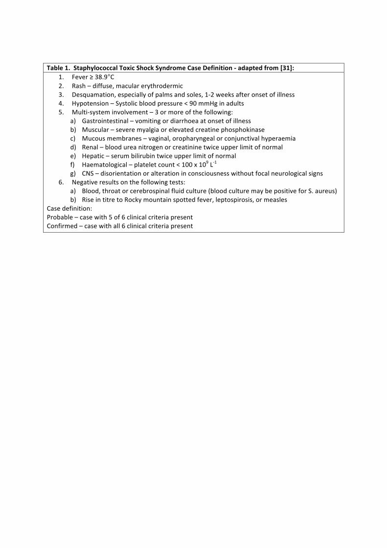
Table 1. Staphylococcal Toxic Shock Syndrome Case Definition -‐ adapted from [31]: 1. Fever ≥ 38.9°C 2. Rash – diffuse, macular erythrodermic 3. Desquamation, especially of palms and soles, 1-‐2 weeks after onset of illness 4. Hypotension – Systolic blood pressure < 90 mmHg in adults 5. Multi-‐system involvement – 3 or more of the following:
a) Gastrointestinal – vomiting or diarrhoea at onset of illness b) Muscular – severe myalgia or elevated creatine phosphokinase c) Mucous membranes – vaginal, oropharyngeal or conjunctival hyperaemia d) Renal – blood urea nitrogen or creatinine twice upper limit of normal e) Hepatic – serum bilirubin twice upper limit of normal f) Haematological – platelet count < 100 x 109 L-‐1 g) CNS – disorientation or alteration in consciousness without focal neurological signs
6. Negative results on the following tests: a) Blood, throat or cerebrospinal fluid culture (blood culture may be positive for S. aureus) b) Rise in titre to Rocky mountain spotted fever, leptospirosis, or measles
Case definition: Probable – case with 5 of 6 clinical criteria present Confirmed – case with all 6 clinical criteria present
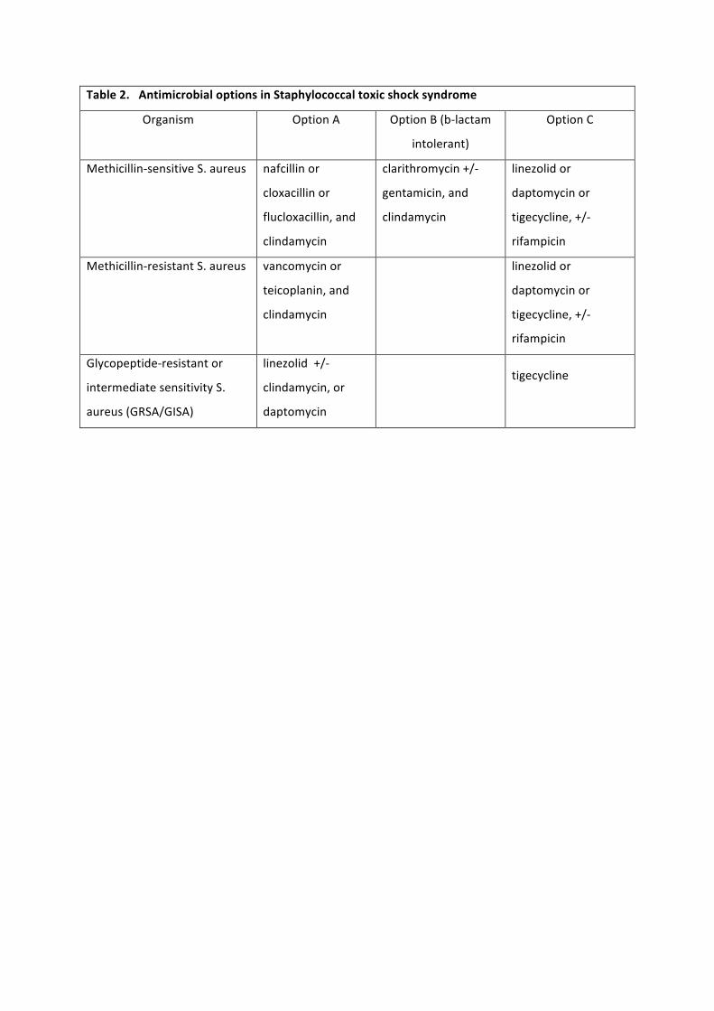
Table 2. Antimicrobial options in Staphylococcal toxic shock syndrome
Organism Option A Option B (b-‐lactam
intolerant)
Option C
Methicillin-‐sensitive S. aureus nafcillin or
cloxacillin or
flucloxacillin, and
clindamycin
clarithromycin +/-‐
gentamicin, and
clindamycin
linezolid or
daptomycin or
tigecycline, +/-‐
rifampicin
Methicillin-‐resistant S. aureus vancomycin or
teicoplanin, and
clindamycin
linezolid or
daptomycin or
tigecycline, +/-‐
rifampicin
Glycopeptide-‐resistant or
intermediate sensitivity S.
aureus (GRSA/GISA)
linezolid +/-‐
clindamycin, or
daptomycin
tigecycline
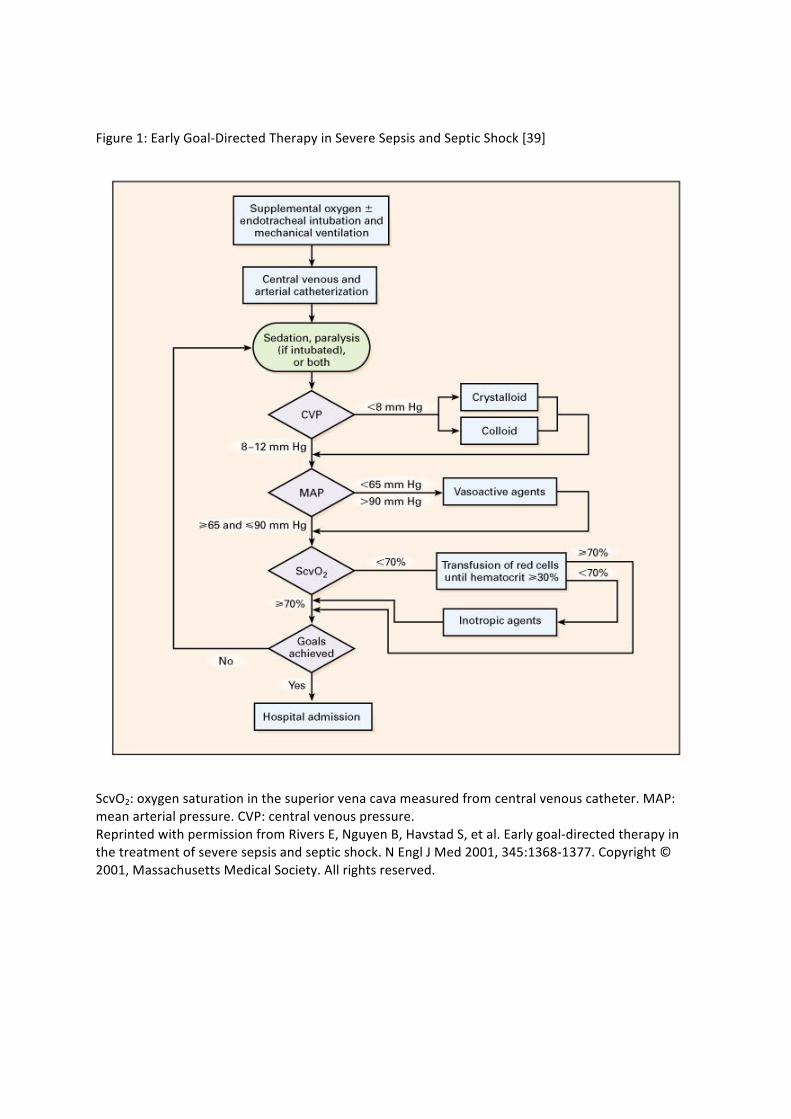
Figure 1: Early Goal-‐Directed Therapy in Severe Sepsis and Septic Shock [39]
ScvO2: oxygen saturation in the superior vena cava measured from central venous catheter. MAP: mean arterial pressure. CVP: central venous pressure. Reprinted with permission from Rivers E, Nguyen B, Havstad S, et al. Early goal-‐directed therapy in the treatment of severe sepsis and septic shock. N Engl J Med 2001, 345:1368-‐1377. Copyright © 2001, Massachusetts Medical Society. All rights reserved.
![Division of Public Health Services...Syndrome [Treponema pallidum] Tetanus [Clostridium tetani] Toxic-Shock Syndrome (TSS) [streptococcal or staphylococcal] Trichinosis [Trichinella](https://static.fdocuments.us/doc/165x107/600aad0a71e3742d46221fb0/division-of-public-health-services-syndrome-treponema-pallidum-tetanus-clostridium.jpg)