Staphylococcal Enterotoxin B-Induced MicroRNA-155 Targets ... · Staphylococcal Enterotoxin...
10
Staphylococcal Enterotoxin B-Induced MicroRNA-155 Targets SOCS1 To Promote Acute Inflammatory Lung Injury Roshni Rao, Prakash Nagarkatti, Mitzi Nagarkatti Department of Pathology, Microbiology, and Immunology, University of South Carolina School of Medicine, Columbia, South Carolina, USA Staphylococcal enterotoxin B (SEB) causes food poisoning in humans. It is considered a biological weapon, and inhalation can trigger lung injury and sometimes respiratory failure. Being a superantigen, SEB initiates an exaggerated inflammatory re- sponse. While the role of microRNAs (miRNAs) in immune cell activation is getting increasing recognition, their role in the reg- ulation of inflammatory disease induced by SEB has not been studied. In this investigation, we demonstrate that exposure to SEB by inhalation results in acute inflammatory lung injury accompanied by an altered miRNA expression profile in lung-infiltrating cells. Among the miRNAs that were significantly elevated, miR-155 was the most overexpressed. Interestingly, miR-155 / mice were protected from SEB-mediated inflammation and lung injury. Further studies revealed a functional link between SEB-in- duced miR-155 and proinflammatory cytokine gamma interferon (IFN-). Through the use of bioinformatics tools, suppressor of cytokine signaling 1 (SOCS1), a negative regulator of IFN-, was identified as a potential target of miR-155. While miR-155 / mice displayed increased expression of Socs1, the overexpression of miR-155 led to its suppression, thereby enhancing IFN- levels. Additionally, the inhibition of miR-155 resulted in restored Socs1expression. Together, our data demonstrate an impor- tant role for miR-155 in promoting SEB-mediated inflammation in the lungs through Socs1 suppression and suggest that miR- 155 may be an important target in preventing SEB-mediated inflammation and tissue injury. S taphylococcal enterotoxin B (SEB), a superantigen produced by Staphylococcus aureus, has deleterious effects in humans, such as food poisoning (1) and toxic shock (2). Because it can be easily aerosolized, SEB is classified as a category B agent by the Centers for Disease Control and Prevention (3). Upon inhalation exposure, SEB can trigger acute inflammatory lung injury charac- terized by immune cell infiltration, excessive cytokine production, tissue damage, and pulmonary edema (4, 5). Due to the distinct manner in which SEB binds to the non- polymorphic regions of major histocompatibility complex class II (MHC-II) on antigen-presenting cells and the specific V regions of the T-cell receptor (TCR), such as murine V8 (6), SEB exposure leads to the activation and proliferation of a large population (5 to 30%) of T lymphocytes (7). Activation of such a substantial number of T lymphocytes results in the ro- bust production of inflammatory cytokines, such as interleukin 2 (IL-2), tumor necrosis factor alpha (TNF-), and gamma interferon (IFN-)(8, 9). In most cases, IFN- is the main culprit in mediating the damaging and often lethal effects seen upon SEB exposure. For example, transgenic mice deficient in IFN- were protected from SEB-mediated toxic shock syn- drome (TSS) and subsequent mortality (10). Additionally, the neutralization of IFN-, after SEB exposure was shown to pre- vent lethal systemic inflammation (11), further suggesting the importance of SEB-mediated IFN- production. While the in- teraction between SEB and TCR, along with the subsequent T-cell proliferation and cytokine secretion, has been exten- sively studied (12–14), the role of microRNA (miRNA) in me- diating SEB-induced inflammation has not yet been elucidated. MicroRNAs are 21 to 23 nt long, single-stranded noncoding RNA molecules that can translationally repress or target mRNA for degradation, thereby acting as primary modulators of gene expression (15). Several studies have demonstrated a role for miRNA in modulating immune responses under various inflam- matory conditions (16). For example, while miR-125b is highly expressed in naive CD4 T cells, it becomes significantly down- regulated upon T-cell activation (17). Similarly, studies have dem- onstrated that overexpression of the (miR-17–92) cluster in T cells leads to lymphoproliferative disorders due to the repression of the proapoptotic molecule, BIM (18). Furthermore, mice deficient in miR-155 are resistant to developing experimental autoimmune encephalomyelitis (EAE), a mouse model of multiple sclerosis (19), while the overexpression of miR-155 exacerbates the symp- toms associated with the disease. Taken together, these studies strongly suggest that miRNAs play a major role in modulating immune cell activation, particularly T cells, as well as promoting proinflammatory responses. In the current study of SEB-induced acute inflammatory lung injury, we applied microarray analysis and quantitative real-time PCR (qRT-PCR) to establish important miRNAs that are dysregu- lated in response to SEB. Further, our data identified miR-155 as a major contributor to SEB-mediated lung inflammation. While it is known that SEB exposure leads to inflammation and the pro- duction of copious amounts of IFN-, we provide mechanistic insight through gain- and loss-of-function experiments into the role of miR-155 in this process. Our results may present an op- portunity to further therapeutically target miR-155 in the treat- ment of SEB-mediated acute inflammatory lung injury. Received 27 February 2014 Returned for modification 31 March 2014 Accepted 24 April 2014 Published ahead of print 28 April 2014 Editor: B. A. McCormick Address correspondence to Mitzi Nagarkatti, [email protected]. Copyright © 2014, American Society for Microbiology. All Rights Reserved. doi:10.1128/IAI.01666-14 July 2014 Volume 82 Number 7 Infection and Immunity p. 2971–2979 iai.asm.org 2971 on April 6, 2019 by guest http://iai.asm.org/ Downloaded from on April 6, 2019 by guest http://iai.asm.org/ Downloaded from on April 6, 2019 by guest http://iai.asm.org/ Downloaded from
Transcript of Staphylococcal Enterotoxin B-Induced MicroRNA-155 Targets ... · Staphylococcal Enterotoxin...
Staphylococcal Enterotoxin B-Induced MicroRNA-155 Targets SOCS1To Promote Acute Inflammatory Lung Injury
Roshni Rao, Prakash Nagarkatti, Mitzi Nagarkatti
Department of Pathology, Microbiology, and Immunology, University of South Carolina School of Medicine, Columbia, South Carolina, USA
Staphylococcal enterotoxin B (SEB) causes food poisoning in humans. It is considered a biological weapon, and inhalation cantrigger lung injury and sometimes respiratory failure. Being a superantigen, SEB initiates an exaggerated inflammatory re-sponse. While the role of microRNAs (miRNAs) in immune cell activation is getting increasing recognition, their role in the reg-ulation of inflammatory disease induced by SEB has not been studied. In this investigation, we demonstrate that exposure to SEBby inhalation results in acute inflammatory lung injury accompanied by an altered miRNA expression profile in lung-infiltratingcells. Among the miRNAs that were significantly elevated, miR-155 was the most overexpressed. Interestingly, miR-155�/� micewere protected from SEB-mediated inflammation and lung injury. Further studies revealed a functional link between SEB-in-duced miR-155 and proinflammatory cytokine gamma interferon (IFN-�). Through the use of bioinformatics tools, suppressorof cytokine signaling 1 (SOCS1), a negative regulator of IFN-�, was identified as a potential target of miR-155. While miR-155�/�
mice displayed increased expression of Socs1, the overexpression of miR-155 led to its suppression, thereby enhancing IFN-�levels. Additionally, the inhibition of miR-155 resulted in restored Socs1expression. Together, our data demonstrate an impor-tant role for miR-155 in promoting SEB-mediated inflammation in the lungs through Socs1 suppression and suggest that miR-155 may be an important target in preventing SEB-mediated inflammation and tissue injury.
Staphylococcal enterotoxin B (SEB), a superantigen producedby Staphylococcus aureus, has deleterious effects in humans,
such as food poisoning (1) and toxic shock (2). Because it can beeasily aerosolized, SEB is classified as a category B agent by theCenters for Disease Control and Prevention (3). Upon inhalationexposure, SEB can trigger acute inflammatory lung injury charac-terized by immune cell infiltration, excessive cytokine production,tissue damage, and pulmonary edema (4, 5).
Due to the distinct manner in which SEB binds to the non-polymorphic regions of major histocompatibility complexclass II (MHC-II) on antigen-presenting cells and the specificV� regions of the T-cell receptor (TCR), such as murine V�8(6), SEB exposure leads to the activation and proliferation of alarge population (5 to 30%) of T lymphocytes (7). Activation ofsuch a substantial number of T lymphocytes results in the ro-bust production of inflammatory cytokines, such as interleukin2 (IL-2), tumor necrosis factor alpha (TNF-�), and gammainterferon (IFN-�) (8, 9). In most cases, IFN-� is the mainculprit in mediating the damaging and often lethal effects seenupon SEB exposure. For example, transgenic mice deficient inIFN-� were protected from SEB-mediated toxic shock syn-drome (TSS) and subsequent mortality (10). Additionally, theneutralization of IFN-�, after SEB exposure was shown to pre-vent lethal systemic inflammation (11), further suggesting theimportance of SEB-mediated IFN-� production. While the in-teraction between SEB and TCR, along with the subsequentT-cell proliferation and cytokine secretion, has been exten-sively studied (12–14), the role of microRNA (miRNA) in me-diating SEB-induced inflammation has not yet been elucidated.
MicroRNAs are �21 to 23 nt long, single-stranded noncodingRNA molecules that can translationally repress or target mRNAfor degradation, thereby acting as primary modulators of geneexpression (15). Several studies have demonstrated a role formiRNA in modulating immune responses under various inflam-matory conditions (16). For example, while miR-125b is highly
expressed in naive CD4� T cells, it becomes significantly down-regulated upon T-cell activation (17). Similarly, studies have dem-onstrated that overexpression of the (miR-17–92) cluster in T cellsleads to lymphoproliferative disorders due to the repression of theproapoptotic molecule, BIM (18). Furthermore, mice deficient inmiR-155 are resistant to developing experimental autoimmuneencephalomyelitis (EAE), a mouse model of multiple sclerosis(19), while the overexpression of miR-155 exacerbates the symp-toms associated with the disease. Taken together, these studiesstrongly suggest that miRNAs play a major role in modulatingimmune cell activation, particularly T cells, as well as promotingproinflammatory responses.
In the current study of SEB-induced acute inflammatory lunginjury, we applied microarray analysis and quantitative real-timePCR (qRT-PCR) to establish important miRNAs that are dysregu-lated in response to SEB. Further, our data identified miR-155 as amajor contributor to SEB-mediated lung inflammation. While itis known that SEB exposure leads to inflammation and the pro-duction of copious amounts of IFN-�, we provide mechanisticinsight through gain- and loss-of-function experiments into therole of miR-155 in this process. Our results may present an op-portunity to further therapeutically target miR-155 in the treat-ment of SEB-mediated acute inflammatory lung injury.
Received 27 February 2014 Returned for modification 31 March 2014Accepted 24 April 2014
Published ahead of print 28 April 2014
Editor: B. A. McCormick
Address correspondence to Mitzi Nagarkatti, [email protected].
Copyright © 2014, American Society for Microbiology. All Rights Reserved.
doi:10.1128/IAI.01666-14
July 2014 Volume 82 Number 7 Infection and Immunity p. 2971–2979 iai.asm.org 2971
on April 6, 2019 by guest
http://iai.asm.org/
Dow
nloaded from
on April 6, 2019 by guest
http://iai.asm.org/
Dow
nloaded from
on April 6, 2019 by guest
http://iai.asm.org/
Dow
nloaded from
MATERIALS AND METHODSMice. Female C57BL/6 mice (6 to 8 weeks old) were purchased from theNational Cancer Institute (NCI). miR-155�/� (B6.Cg-Mir155 tm1.1 Rsky/J)mice were purchased from The Jackson Laboratory. All mice were housedunder pathogen-free conditions at the Animal Resource Facility (ARF),University of South Carolina (USC) School of Medicine. The use of ver-tebrate animals in the experiments performed was preapproved by theInstitutional Animal Care and Use Committee (IACUC) at USC. Thisstudy was carried out in strict accordance with the recommendations inthe Guide for the Care and Use of Laboratory Animals of the NationalResearch Council (20).
Induction of SEB-induced acute lung injury (ALI). SEB was obtainedfrom Toxin Technologies (Sarasota, FL). SEB dissolved in sterile phos-phate-buffered saline (PBS) (2 mg/ml) was administered by the intranasal(i.n.) route in a volume of 25 �l for a dose of 50 �g per mouse, as describedpreviously (4, 21, 22). Mice were euthanized 48 h after SEB exposure.
Lung histopathological analysis. At the time of euthanasia, lungswere obtained and fixed in 10% formalin. The tissue was then paraffinembedded and serial sections (5 �m) were made. The sections were sub-sequently deparaffinized by being dissolved with xylene, followed by re-hydration in several changes of alcohol (100%, 95%, and 90%). The slideswere then stained with hematoxylin and eosin (H&E) and evaluated witha Nikon E600 light microscopy system.
Antibodies. Fluorescein isothiocyanate-conjugated anti-CD8 (clone53.6.7) and phycoerythrin-conjugated anti-CD4 (clone GK1.5) antibod-ies (Abs) were purchased from BioLegend (San Diego, CA).
Preparation of lung-infiltrating cells and flow cytometry. Mice wereexposed to SEB as described above. Forty-eight hours after SEB exposure,lungs were harvested and homogenized using a Stomacher 80 Biomasterblender from Seward (Davie, FL) in 10 ml of sterile PBS. After beingwashed with sterile PBS, the cells were carefully layered on Ficoll His-topaque-1077 from Sigma-Aldrich (St. Louis, MO) and separated by den-sity gradient centrifugation at 500 g for 30 min at 24°C with the brakeoff. The mononuclear cell layer isolated was then enumerated using theTrypan blue exclusion method. To determine the subsets of immune cellsinfiltrating the lung, cells were stained with fluorescent-conjugated anti-bodies (anti-CD4, anti-CD8) and analyzed using the Beckman Coulter500 flow cytometer (Indianapolis, IN).
Recovery of bronchoalveolar lavage fluid (BALF) and cytokine de-tection. Forty-eight hours after SEB exposure, mice were euthanized, tra-cheae from vehicle- or SEB-treated mice were tied with a suture, and thelung was excised as an intact unit. With 1 ml sterile ice-cold PBS, thetrachea was lavaged to collect the BALF fluid. Cytokine analysis for IFN-�was carried out using BALF. All cytokines were measured using BioLeg-end (San Diego, CA) enzyme-linked immunosorbent assay (ELISA) MAXstandard kits.
Total RNA isolation. Total RNA (including small RNAs) was isolatedfrom lung-infiltrating mononuclear cells or in vitro from lymph nodes(LN) or splenocytes using the miRNeasy kit from Qiagen (Valencia, CA)by following the manufacturer’s instructions. The purity and concentra-tion of the RNA was confirmed spectrophotometrically, while the integ-rity of miRNA was further assessed using an Agilent 2100 Bioanalyzer(Agilent Tech, Palo Alto, CA).
miRNA expression profiling and analysis. To profile miRNA expres-sion in the lung, the Affymetrix GeneChip miRNA 1.0 array platform wasused. The array, which is composed of 609 mouse miRNA probes, makesuse of the FlashTag biotin HSR hybridization technique and was carriedout according to the manufacturer’s instructions (Affymetrix, SantaClara, CA). Fluorescent intensities obtained from hybridization were logtransformed and visualized in the form of a heatmap. Hierarchical clus-tering was carried out using Ward’s method, and similarity measurementwas calculated using half square Euclidean distance. miRNA expressionfold changes obtained from the microarray were then further analyzedusing the commercially available analysis tool Ingenuity Systems Ingenu-ity Pathway analysis (IPA), (Mountain View, CA, USA.) In brief, the data
set of 609 miRNAs was uploaded into IPA and only miRNAs that were3-fold or higher were considered for analysis. Core analysis was carriedout and a “top network” of miRNA and its associated molecules wasgenerated.
Analysis of miRNA target genes. IPA was also used to determine andcollate the highly predicted, moderately predicted, and experimentallyobserved mRNA target genes of those miRNAs that were highly (�3-fold)upregulated using IPA’s miRNA target filter tool. These targets were fur-ther sorted based on their role in cytokine signaling, cellular growth andproliferation, and cellular immune response. Additionally, to assign im-munological functions to our list of miRNA targets, gene ontology (GO)mapping of miRNA target genes was carried out using Cytoscape 3.0.1equipped with ClueGO and CluePedia applications.
qRT-PCR. Total RNA (miRNA and mRNA) was converted to cDNAusing the miScript cDNA synthesis kit (Qiagen) according to the manu-facturer’s instructions. For miRNA validation, the miScript SYBR greenPCR kit (Qiagen) was used, and fold change of miRNA was determined byusing the 2�CT method. Snord96a was used as the small RNA endoge-nous control. For mRNA validation, an SSO advanced SYBR green PCRkit from Bio-Rad (Hercules, CA) was used according to the manufactur-er’s instructions, and �-actin was used as the endogenous control. Thefollowing primers were used: �-actin forward (F) (5=-GGCTGTATTCCCCTCCAT G-3=) and reverse (R) (5=-CCAGTT GGTAACAATGCCATGT-3=); Socs1 F (5=-GGTTGTAGCAGCTTGTGTC-3=) and R (5=-AATGAAGCCAGAGACCCTC-3=); IFN-� F (5=-GCGTCATTGAATCACACCTG-3=) and R (5=-GAGCTCATTGAATGCTTGGC-3=).
Transfection with miR-155 mimic and inhibitors. Lymph nodes (ax-illary and inguinal) from naive C57BL/6 mice were harvested and culturedin 10 ml of complete medium at 37°C and 5% CO2. Complete mediumwas comprised of RPMI 1640 medium (Gibco Laboratories, Grand Island,NY) supplemented with 10% fetal bovine serum (FBS), 10 mM L-glu-tamine, 10 mM HEPES, 50 �M �-mercaptoethanol, and 100 �g/ml pen-icillin. Cells were seeded at 2 105 cells in 24-well plates and transfectedwith either 40 nM synthetic mmu-miR-155-3p miScript miRNA mimic(CUCCUACCUGUUAGCAUUAAC) or AllStar negative-control smallinterfering RNA (siRNA). For inhibition of miR-155, cells were activatedwith SEB (1 �g/ml) and treated with 100 nM anti-mmu-miR-155-3pmiScript miRNA inhibitor (CUCCUACCUGUUAGCAUUAAC) or miS-cript inhibitor negative control for 24 h using HiperFect transfection re-agent (Qiagen) according to the manufacturer’s instructions.
Luciferase assay. The following plasmids were purchased fromGeneCopoeia (Rockville, MD): 3= untranslated region (3=UTR), Socs1(MmiT028883), and control plasmid (CmiT000001-MT01). Chinesehamster ovary (CHO) cells were cotransfected with 100 ng of plasmid and100 nM miRIDIAN microRNA mmu-miR-155-5p mimic or miRIDIANmicroRNA mimic negative control using DharmaFECT DUO transfec-tion reagent by following the manufacturer’s instructions (Thermo Sci-entific, Pittsburgh, PA). Twenty-four hours following transfection, Lucif-erase activity was measured using LucPair miR-Duo Luciferase assay kitfrom GeneCopoeia.
Statistics. All statistical analyses were carried out using GraphPadPrism Software (San Diego, CA). In all experiments, the number of miceused was 4 or 5 per group, unless otherwise specified. Results are expressedas means � standard errors of the means (SEM). Student’s t test was usedto compare wild-type (WT) and miR-155�/� data, whereas multiplecomparisons were made using one-way analysis of variance (ANOVA),followed by post hoc analysis using Tukey’s method. A P value of �0.05was considered statistically significant. Individual experiments were per-formed in triplicate, and each experiment was performed independentlyat least three times to test reproducibility of results.
Microarray accession number. All microRNA microarray data weredeposited the in ArrayExpress database (www.ebi.ac.uk/arrayexpress) un-der accession number E-MTAB-2379.
Rao et al.
2972 iai.asm.org Infection and Immunity
on April 6, 2019 by guest
http://iai.asm.org/
Dow
nloaded from

RESULTSSEB exposure triggers inflammation in the lung. Previously, asingle dose of SEB (50 �g) by intranasal delivery was found toinduce cellular infiltration, increase cytokine production, andcause histopathological lesions and edema in C57BL/6 mice (4, 21,22), mimicking the symptoms of acute inflammatory lung injuryin humans (23). In this study, we first sought to investigate theinflammatory effect of SEB exposure in the lungs. Forty-eighthours after SEB exposure, H&E-stained sections of the lungs fromSEB-exposed mice showed massive infiltration of cells and signs ofedema as evidenced by fluid-filled bronchioles (Fig. 1A). BecauseSEB is a potent activator of T cells, we examined the effect of SEBon T-cell subsets within the lung. Immediately after euthanasia,the lungs were harvested and mononuclear cells were isolatedfrom the lungs by density gradient centrifugation to determine thephenotypic characteristics of the cells. SEB exposure not only ledto an overall increase in mononuclear cells but, specifically, anincrease in CD4� and CD8� T cells (Fig. 1B) was seen. BecauseSEB exposure triggers an increase in IFN-�, a major proinflam-matory cytokine previously reported to orchestrate the inflamma-tory cascade and cause tissue damage (10, 24, 25), we analyzed theconcentration of IFN-� in the bronchoalveolar lavage fluid(BALF) and found a high concentration (up to 3,000 pg/ml) ofIFN-� in the lungs of SEB-exposed mice (Fig. 1C). These datasuggested that SEB administration via the intranasal route triggersacute inflammation in the lungs.
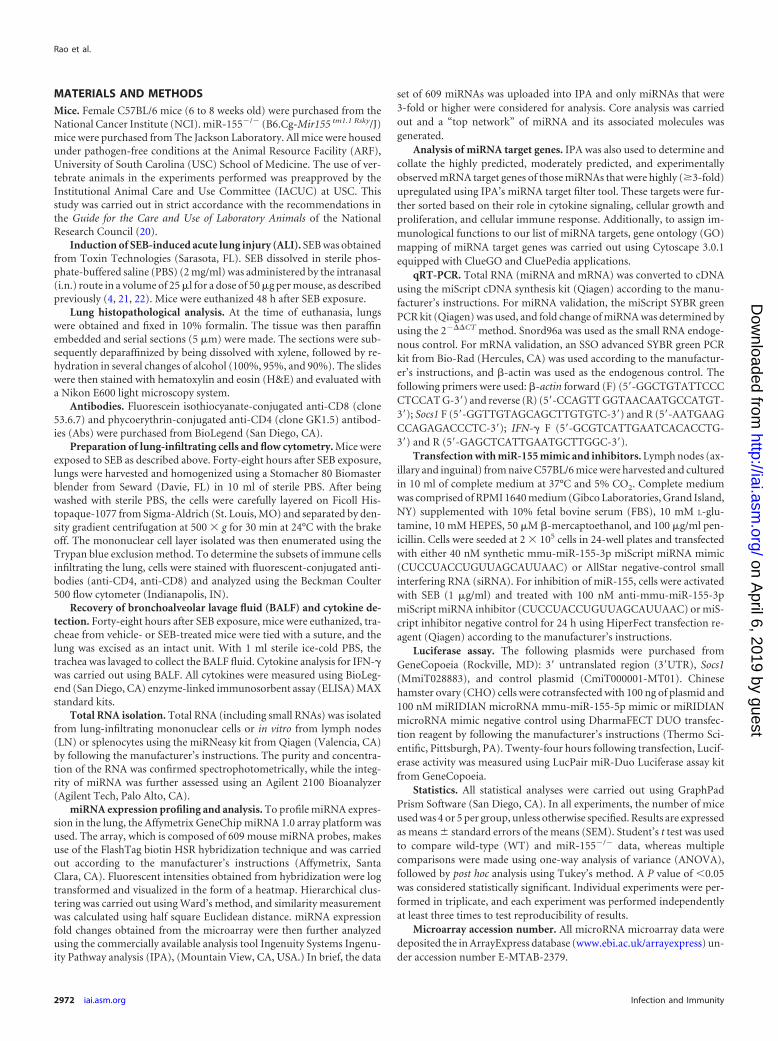
SEB exposure modulates miRNA expression in the lungs.The dysregulation of specific miRNAs in response to SEB expo-sure has not been elucidated. Because miRNAs play a critical rolein mediating inflammation, we examined the miRNA profile afterSEB exposure. Total miRNA was isolated from lung-infiltratingmononuclear cells, and the relative abundance of miRNA in SEB-exposed and vehicle-treated mice was determined using microar-ray miRNA analysis. A heatmap was generated based on hierar-chical clustering of miRNA, highlighting a stark differencebetween vehicle- and SEB-exposed mice (Fig. 2A). Further exam-ination of miRNA expression revealed that of the 609 miRNAsassessed, most remained unchanged, but a few showed significantup- or downregulation, as seen in the fold change distribution plot(Fig. 2B). Ingenuity pathway analysis (IPA) generated a “topnetwork” of miRNAs that was comprised of five upregulatedmiRNAs, including miR-155, miR-31, miR-182, miR-20b, andmiR-222 and their associated molecules (Fig. 2C). This networkwas characterized by IPA as responses involving inflammation,cellular development, cellular growth, and proliferation. To assignsignificant immunological functions to the genes in the aforemen-tioned top network, Cytoscape (ClueGO�CluePedia application)was employed. Gene ontology (GO) mapping revealed that thegenes associated with the miRNA in the top network were func-tionally relevant to T-cell activation (GO 0042110) and prolifera-tion (GO 0042098), IFN-� signaling (GO 0060333), and Toll-likereceptor signaling (GO 0002224) (Fig. 2D). Next, we validated the
FIG 1 SEB induces lung inflammation. (A) Representative H&E images (20) of cross sections of the lung from mice exposed to either vehicle or SEB. (B)Lung-infiltrating mononuclear cells obtained by density gradient centrifugation and total number of viable cells were counted using a hemocytometer. Cells werefurther stained with monoclonal antibody (MAb) to identify CD4� and CD8� cells and were analyzed on a flow cytometer. The percentage of the immune cellsubsets was multiplied by the total number of cells found in the lung and divided by 100 to yield the absolute cell numbers shown. (C) The concentration of IFN-�protein present in the BALF was determined using a standard ELISA kit. Data are means � SEM (n 5) and are representative of three independent experiments.Statistical significance compared to SEB-vehicle is indicated as follows: **, P � 0.01; ***, P � 0.001; ****, P � 0.0001.
SEB-Induced miR-155 Promotes Inflammatory ALI
July 2014 Volume 82 Number 7 iai.asm.org 2973
on April 6, 2019 by guest
http://iai.asm.org/
Dow
nloaded from
expression levels of these miRNAs in lung-infiltrating mononu-clear cells by qRT-PCR, which corroborated the expression pat-terns seen using the microarray (Fig. 2E). Among the miRNAs wevalidated, miR-155 was the most highly expressed (�8-fold) inthe lungs upon SEB exposure. Based on this, the role of miR-155 inthe development of SEB-induced ALI was further investigated.
miR-155 is important for SEB-mediated inflammation. Be-cause miR-155 was highly upregulated in response to SEB, wehypothesized that it might play a crucial role in facilitating theinflammation observed during the disease. To that end, WT andmiR-155�/� mice were exposed to SEB to determine the effects ondisease parameters. H&E-stained sections of the lung revealed thatWT mice exposed to SEB exhibited numerous layers of infiltrationinterspersed with edema. Interestingly, miR-155�/� mice exposedto SEB presented with almost normal lung architecture (Fig. 3A).Additionally, the miR-155-deficient mice expressed significantlydecreased total numbers of mononuclear cells within the lungupon SEB exposure compared to those of their WT counterparts.Upon closer examination of the mononuclear cell phenotypewithin the lungs, absolute numbers of CD4� and CD8� in themiR-155�/� mice were decreased compared to those of WT mice(Fig. 3B). Additionally, cytokine analysis of the bronchoalveolarlavage fluid (BALF) in the lungs of WT mice demonstrated highconcentrations of proinflammatory cytokine IFN-�. In contrast,IFN-� levels were significantly diminished in miR-155�/� mice
(Fig. 3C). Taken together, these results provided clear evidencethat miR-155�/� mice were protected from SEB-mediated ALI,suggesting that miR-155 plays a critical role in SEB-induced in-flammation.
miR-155 expression is critically linked to IFN-� production.Because SEB exposure leads to the release of copious amounts ofIFN-� and also results in increased miR-155 expression, we con-sidered if there was a positive correlation between IFN-� secretionand the expression of miR-155. To explore this possibility, we firstassessed the expression of IFN-� after transfection of LN T cellswith a synthetic miR-155 mimic. Interestingly, we found a sub-stantial increase in IFN-� levels not only in WT mice (Fig. 4A) butalso in miR-155-deficient mice that were transfected with themimic (Fig. 4B). Additionally, inhibition of miR-155 with a syn-thetic inhibitor conversely resulted in the diminished expressionof IFN-� (Fig. 4C), suggesting a crucial link between miR-155expression and that of IFN-� after SEB exposure.
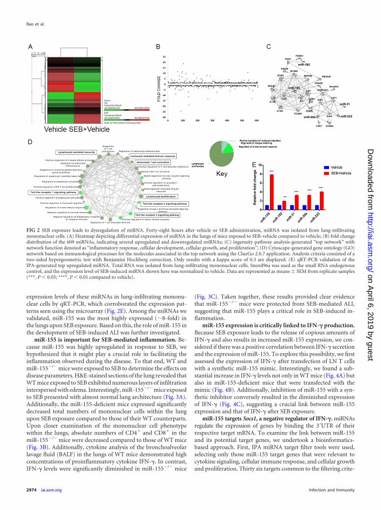
miR-155 targets Socs1, a negative regulator of IFN-�. miRNAsregulate the expression of genes by binding the 3=UTR of theirrespective target mRNA. To examine the link between miR-155and its potential target genes, we undertook a bioinformatics-based approach. First, IPA miRNA target filter tools were used,selecting only those miR-155 target genes that were relevant tocytokine signaling, cellular immune response, and cellular growthand proliferation. Thirty six targets common to the filtering crite-
FIG 2 SEB exposure leads to dysregulation of miRNA. Forty-eight hours after vehicle or SEB administration, miRNA was isolated from lung-infiltratingmononuclear cells. (A) Heatmap depicting differential expression of miRNA in the lungs of mice exposed to SEB-vehicle compared to vehicle; (B) fold changedistribution of the 609 miRNAs, indicating several upregulated and downregulated miRNAs; (C) ingenuity pathway analysis-generated “top network” withnetwork function denoted as “inflammatory response, cellular development, cellular growth, and proliferation”; (D) Cytoscape-generated gene ontology (GO)network based on immunological processes for the molecules associated in the top network using the ClueGo 2.0.7 application. Analysis criteria consisted of atwo-sided hypergeometric test with Benjamini Hochberg correction. Only results with a kappa score of 0.3 are displayed. (E) qRT-PCR validation of theIPA-generated top upregulated miRNA. Total RNA was isolated from lung-infiltrating mononuclear cells. Snord96a was used as the small RNA endogenouscontrol, and the expression level of SEB-induced miRNA shown here was normalized to vehicle. Data are represented as means � SEM from replicate samples(***, P � 0.05; ****, P � 0.01 compared to vehicle).
Rao et al.
2974 iai.asm.org Infection and Immunity
on April 6, 2019 by guest
http://iai.asm.org/
Dow
nloaded from

ria applied were selected (Fig. 5A). Next, an IPA-generated net-work was used to sort the targets based on those that were highlypredicted and experimentally observed (Fig. 5B). Conclusively, agene known as suppressor of cytokine signaling 1 (Socs1) was
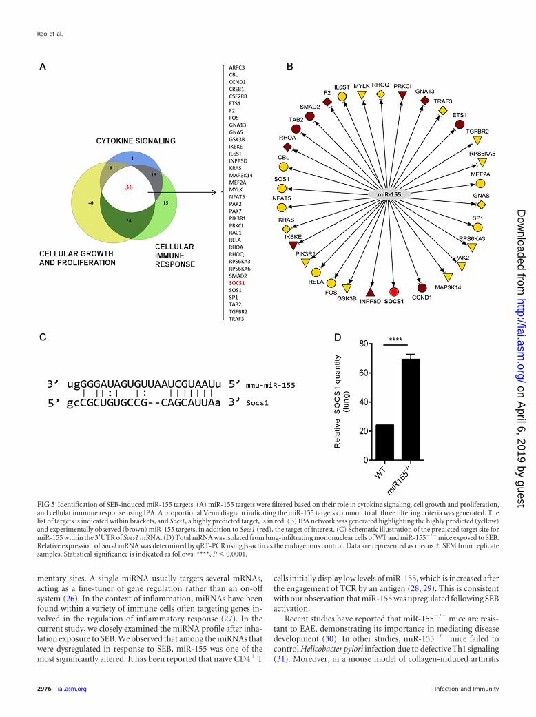
found to be a prominent miR-155 target due to miR-155’s abilityto bind to the 3=UTR of the Socs1 mRNA (Fig. 5C). Because Socs1is induced by IFN-� and acts as a negative regulator of IFN-�, therelationship between miR-155 and Socs1 in the context of IFN-�production was examined. miR-155�/� mice that were exposed toSEB had significantly increased expression of Socs1 mRNA inlung-infiltrating mononuclear cells compared to that of the WT(Fig. 5D). Accordingly, we hypothesized that miR-155 may targetSocs1 to promote IFN-�-mediated inflammation during SEB-in-duced inflammatory ALI. To confirm Socs1 as an miR-155 target,we first measured relative luciferase activity after cotransfection ofmiR-155 mimic and plasmid containing the 3= UTR of Socs1.Compared to the mimic control, miR-155 mimic led to a signifi-cant decrease in luciferase activity (Fig. 6A), validating Socs1 as atarget for miR-155. Next, the impact of miR-155 mimic on Socs1levels was explored. We found that both in WT (Fig. 6B) andmiR-155-deficient cells (Fig. 6C) that were transfected with miR-155 mimic, Socs1 levels remained suppressed. On the other hand,whereas SEB activation continued to lead to a repression in Socs1,miR-155 inhibition of SEB-activated cells resulted in its derepres-sion (Fig. 6D), confirming the role of miR-155 in suppressingSocs1 during SEB-mediated activation of immune cells.
DISCUSSION
With the discovery of miRNA, a novel and exciting mechanism ofgene regulation has arisen. miRNAs are small noncoding endog-enous RNA molecules that bind 3=UTR of genes carrying comple-
FIG 3 miR-155 plays a critical role in SEB-induced ALI. WT (C57BL/6) and miR-155�/� (B6.Cg-Mir155 tm1.1 Rsky/J) mice were exposed to SEB and euthanized48 h later. (A) Representative H&E images (20) of sections of lung indicating immune cell infiltration; (B) phenotypic characterization of cells infiltrating thelung was determined by staining of mononuclear cells with fluorescent-conjugated MAb against CD4 and CD8; (C) levels of IFN-� cytokine in the BALF wasdetermined by ELISA. Data are means � SEM (n 5) and are representative of two independent experiments. Statistical significance compared to WT isindicated as follows: **, P � 0.01.
FIG 4 IFN-� forms a critical link between SEB and subsequent miR-155 in-duction. (A) Lymph node (LN) T cells obtained from naive wild-type (WT)mice were transfected either with miR-155 mimic (Mimic) or mimic control(Control) for 24 h. IFN-� levels were determined by RT-PCR. (B) LN T cellsobtained from miR-155�/� mice were also transfected with 40 nM miR-155mimic or mimic control as indicated for 24 h, and IFN-� levels were assessed.(C) LN cells were activated with SEB (1 �g/ml) for 24 h. Cells were thentransfected with 100 nM miR-155 inhibitor (Inh) or inhibitor control (InhControl) for another 24 h. IFN-� levels were determined via RT-PCR. Data arerepresented as means � SEM from replicate samples. Statistical significance isindicated as follows: **, P � 0.01; ****, P � 0.0001.
SEB-Induced miR-155 Promotes Inflammatory ALI
July 2014 Volume 82 Number 7 iai.asm.org 2975
on April 6, 2019 by guest
http://iai.asm.org/
Dow
nloaded from
mentary sites. A single miRNA usually targets several mRNAs,acting as a fine-tuner of gene regulation rather than an on-offsystem (26). In the context of inflammation, miRNAs have beenfound within a variety of immune cells often targeting genes in-volved in the regulation of inflammatory response (27). In thecurrent study, we closely examined the miRNA profile after inha-lation exposure to SEB. We observed that among the miRNAs thatwere dysregulated in response to SEB, miR-155 was one of themost significantly altered. It has been reported that naive CD4� T
cells initially display low levels of miR-155, which is increased afterthe engagement of TCR by an antigen (28, 29). This is consistentwith our observation that miR-155 was upregulated following SEBactivation.
Recent studies have reported that miR-155�/� mice are resis-tant to EAE, demonstrating its importance in mediating diseasedevelopment (30). In other studies, miR-155�/� mice failed tocontrol Helicobacter pylori infection due to defective Th1 signaling(31). Moreover, in a mouse model of collagen-induced arthritis
FIG 5 Identification of SEB-induced miR-155 targets. (A) miR-155 targets were filtered based on their role in cytokine signaling, cell growth and proliferation,and cellular immune response using IPA. A proportional Venn diagram indicating the miR-155 targets common to all three filtering criteria was generated. Thelist of targets is indicated within brackets, and Socs1, a highly predicted target, is in red. (B) IPA network was generated highlighting the highly predicted (yellow)and experimentally observed (brown) miR-155 targets, in addition to Socs1 (red), the target of interest. (C) Schematic illustration of the predicted target site formiR-155 within the 3=UTR of Socs1 mRNA. (D) Total mRNA was isolated from lung-infiltrating mononuclear cells of WT and miR-155�/� mice exposed to SEB.Relative expression of Socs1 mRNA was determined by qRT-PCR using �-actin as the endogenous control. Data are represented as means � SEM from replicatesamples. Statistical significance is indicated as follows: ****, P � 0.0001.
Rao et al.
2976 iai.asm.org Infection and Immunity
on April 6, 2019 by guest
http://iai.asm.org/
Dow
nloaded from
(CIA), the deficiency of miR-155 led to a decrease in pathogenic Tcells. In humans, it has also been reported that soldiers undergo-ing a battlefield-like stress program demonstrated an increase inhsa-miR-155 levels in leukocytes exposed to SEB ex vivo (32),indicating that stress-related inflammation could also potentiallylead to the increase in miR-155. The current study further suggeststhat the acute inflammatory response in the lungs to a bacterialsuperantigen is also regulated by miR-155, inasmuch as SEB-ex-posed miR-155�/� mice having fewer infiltrating T cells than theirWT counterparts and almost normal lung histopathology.
Usually, an appropriate regulation of IFN-� is necessary formediating Th1 responses and blunting infection (33). SEB expo-sure, however, causes an excessive release of IFN-�. T cells ex-posed to IFN-� proliferate further, thus perpetuating a cycle ofinflammation (34, 35). Studies carried out with SEB activation ofsplenocytes have demonstrated that early cytokines released in-clude IL-2 and TNF-�, followed by the massive production ofIFN-� by T-helper cells (36). Additionally, in vitro SEB activationof rat splenocytes results in the release of IFN-� for up to 48 hpostactivation and promotes the proliferation of CD4� T cells,similar to that seen in mice and humans (37). We have noted inthe current model (data not shown) that the peak of acute inflam-matory lung injury occurs at 48 h after SEB exposure. In line withthe typical kinetics of cytokine release seen with SEB activation, wedid not detect early cytokines, TNF-� and IL-2, in the BALF at thistime point (data not shown). However, our data indicated sub-stantial release of SEB-induced IFN-� in WT mice, suggesting thatthis particular cytokine may significantly contribute to SEB-in-duced inflammation. Moreover, the correlation between de-creased IFN-� in the BALF of miR-155�/� mice exposed to SEBand lack of significant inflammation in the lungs is suggestive of amajor role for IFN-� in our model.
The relationship between miR-155 and IFN-� has been brieflyexplored in previous studies. For example, when miR-155 is over-expressed in human NK cells, the subsequent downregulation of atarget, SHIP-1, promotes IFN-� expression (38). During colla-gen-induced arthritis, miR-155�/� mice display significantlylower numbers of IFN-�-producing cells than WT mice (39).Likewise, our data demonstrated that while transfection with asynthetic miR-155 mimic leads to the induction of IFN-�, the
blockade of miR-155 diminishes IFN-� production. Our resultsclearly indicate that the SEB-mediated induction of IFN-� can beexplained, at least in part, by the induction of miR-155.
To uncover the relationship between miR-155 and IFN-� inresponse to SEB exposure, we employed bioinformatics tools andconducted an extensive literature search. These efforts suggestedsuppressor of cytokine signaling 1 (SOCS1) as a possible link be-tween miR-155 and IFN-�. SOCS1 belongs to a family of eightproteins (SOCS1 to SOCS7 and CIS) that regulate the productionof several cytokines (40). In particular, SOCS1 is induced byIFN-� for autoregulation of the IFN-� proinflammatory responseby inhibiting the JAK/STAT1 signaling pathway (41). Recent ex-periments in numerous cell types have revealed that miR-155 tar-gets SOCS1 (42–44). For example, macrophages infected with anRNA virus demonstrated enhanced type I interferon productiondue to miR-155 targeting of Socs1 (45). In the current study, wenoted that the miR-155�/� mice that are exposed to SEB showed
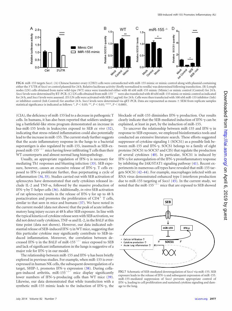
FIG 7 Schematic of SEB-mediated downregulation of Socs1 via miR-155. SEBexposure leads to the release of IFN-� and subsequent expression of miR-155.miR-155-mediated suppression of Socs1 prevents appropriate control ofIFN-�, leading to cell proliferation and sustained cytokine signaling and dam-age to the lung.
FIG 6 miR-155 targets Socs1. (A) Chinese hamster ovary (CHO) cells were cotransfected with miR-155 mimic or mimic control along with plasmid containingeither the 3=UTR of Socs1 or control plasmid for 24 h. Relative luciferase activity (firefly normalized to renilla) was determined following transfection. (B) Lymphnodes (LN) cells obtained from naive wild-type (WT) mice were transfected either with 40 nM miR-155 mimic (Mimic) or mimic control (Control) for 24 h.Socs1 levels were determined by RT-PCR. (C) LN cells obtained from miR-155�/� were also transfected with 40 nM miR-155 mimic or mimic control as indicatedfor 24 h, and Socs1 levels were assessed. (D) LN cells were activated with SEB (1 �g/ml) for 24 h. Cells were then transfected with 100 nM miR-155 inhibitor (Inh)or inhibitor control (Inh Control) for another 24 h. Socs1 levels were determined via qRT-PCR. Data are represented as means � SEM from replicate samplesstatistical significance is indicated as follows: *, P � 0.05; **, P � 0.01; ****, P � 0.0001.
SEB-Induced miR-155 Promotes Inflammatory ALI
July 2014 Volume 82 Number 7 iai.asm.org 2977
on April 6, 2019 by guest
http://iai.asm.org/
Dow
nloaded from

increased expression in Socs1 mRNA levels compared to those ofSEB-exposed WT mice. The expression of Socs1 correlated in-versely with IFN-� production in the BALF. In addition, our gain-and loss-of-function studies with miR-155 mimic and inhibitorclearly demonstrated that miR-155-mediated targeting of Socs1regulates IFN-� production.
The results of the present study highlight the role of miR-155 inSEB-induced acute inflammatory lung injury. Specifically, wedemonstrate that the high levels of IFN-� production associatedwith SEB exposure can be attributed to the miR-155-mediatedrepression of Socs1, a critical regulator of IFN-� (Fig. 7). Further-more, the importance of miR-155 is made particularly evident, asmiR-155-deficient mice were found to be protected from SEB-mediated inflammation and acute lung injury, thereby suggestingthat therapeutic targeting of miR-155 may be useful in the treat-ment of SEB-triggered acute inflammatory lung injury.
ACKNOWLEDGMENTS
This work was supported by NIH grants P01AT003961, R01AT006888,R01ES019313, R01MH094755, and P20RR032684 and VA merit awardBX001357.
REFERENCES1. Pinchuk IV, Beswick EJ, Reyes VE. 2010. Staphylococcal enterotoxins.
Toxins 2:2177–2197. http://dx.doi.org/10.3390/toxins2082177.2. Savransky V, Rostapshov V, Pinelis D, Polotsky Y, Korolev S, Komisar
J, Fegeding K. 2003. Murine lethal toxic shock caused by intranasal ad-ministration of staphylococcal enterotoxin B. Toxicol. Pathol. 31:373–378. http://dx.doi.org/10.1080/01926230309725.
3. Ulrich RG, Sidell S, Taylor TJ, Wilhelmsen CL, Franz DR. 2001.Textbook of military medicine: medical aspects of chemical and biologicalwarfare. Office of The Surgeon General at TMM Publications, BordenInstitute, Walter Reed Army Medical Center, Washington, DC.
4. Saeed AI, Rieder SA, Price RL, Barker J, Nagarkatti P, Nagarkatti M. 2012.Acute lung injury induced by staphylococcal enterotoxin B: disruption ofterminal vessels as a mechanism of induction of vascular leak. Microsc. Mi-croanal. 18:445–452. http://dx.doi.org/10.1017/S1431927612000190.
5. Neumann B, Engelhardt B, Wagner H, Holzmann B. 1997. Induction ofacute inflammatory lung injury by staphylococcal enterotoxin B. J. Immu-nol. 158:1862–1871.
6. Bavari S, Ulrich RG. 1995. Staphylococcal enterotoxin A and toxic shocksyndrome toxin compete with CD4 for human major histocompatibilitycomplex class II binding. Infect. Immun. 63:423– 429.
7. Kozono H, Parker D, White J, Marrack P, Kappler J. 1995. Multiplebinding sites for bacterial superantigens on soluble class II MHC mol-ecules. Immunity 3:187–196. http://dx.doi.org/10.1016/1074-7613(95)90088-8.
8. Miethke T, Duschek K, Wahl C, Heeg K, Wagner H. 1993. Pathogenesisof the toxic shock syndrome: T cell mediated lethal shock caused by thesuperantigen TSST-1. Eur. J. Immunol. 23:1494 –1500. http://dx.doi.org/10.1002/eji.1830230715.
9. Bette M, Schafer MK, van Rooijen N, Weihe E, Fleischer B. 1993.Distribution and kinetics of superantigen-induced cytokine gene expres-sion in mouse spleen. J. Exp. Med. 178:1531–1539. http://dx.doi.org/10.1084/jem.178.5.1531.
10. Tilahun AY, Holz M, Wu TT, David CS, Rajagopalan G. 2011. Inter-feron gamma-dependent intestinal pathology contributes to the lethalityin bacterial superantigen-induced toxic shock syndrome. PLoS One6:e16764. http://dx.doi.org/10.1371/journal.pone.0016764.
11. Florquin S, Amraoui Z, Goldman M. 1995. T cells made deficient ininterleukin-2 production by exposure to staphylococcal enterotoxin B invivo are primed for interferon-gamma and interleukin-10 secretion. Eur.J. Immunol. 25:1148 –1153. http://dx.doi.org/10.1002/eji.1830250503.
12. Fleischer B, Gerardy-Schahn R, Metzroth B, Carrel S, Gerlach D,Kohler W. 1991. An evolutionary conserved mechanism of T cell activa-tion by microbial toxins. Evidence for different affinities of T cell receptor-toxin interaction. J. Immunol. 146:11–17.
13. Herman A, Kappler JW, Marrack P, Pullen AM. 1991. Superantigens:
mechanism of T-cell stimulation and role in immune responses. Annu.Rev. Immunol. 9:745–772. http://dx.doi.org/10.1146/annurev.immunol.9.1.745.
14. Saeed AI, Rieder SA, Price RL, Barker J, Nagarkatti P, Nagarkatti M. 2012.AcutelunginjuryinducedbystaphylococcalenterotoxinB:disruptionofterminalvesselsasamechanismof inductionofvascular leak.Microsc.Microanal. 18:445–452. http://dx.doi.org/10.1017/S1431927612000190.
15. Cai Y, Yu X, Hu S, Yu J. 2009. A brief review on the mechanisms ofmiRNA regulation. Genomics Proteomics Bioinformatics 7:147–154.http://dx.doi.org/10.1016/S1672-0229(08)60044-3.
16. O’Connell RM, Rao DS, Baltimore D. 2012. microRNA regulation ofinflammatory responses. Annu. Rev. Immunol. 30:295–312. http://dx.doi.org/10.1146/annurev-immunol-020711-075013.
17. Rossi RL, Rossetti G, Wenandy L, Curti S, Ripamonti A, Bonnal RJ,Birolo RS, Moro M, Crosti MC, Gruarin P, Maglie S, Marabita F,Mascheroni D, Parente V, Comelli M, Trabucchi E, De Francesco R,Geginat J, Abrignani S, Pagani M. 2011. Distinct microRNA signatures inhuman lymphocyte subsets and enforcement of the naive state in CD4� Tcells by the microRNA miR-125b. Nat. Immunol. 12:796 – 803. http://dx.doi.org/10.1038/ni.2057.
18. Xiao C, Srinivasan L, Calado DP, Patterson HC, Zhang B, Wang J,Henderson JM, Kutok JL, Rajewsky K. 2008. Lymphoproliferative diseaseand autoimmunity in mice with increased miR-17–92 expression in lympho-cytes. Nat. Immunol. 9:405–414. http://dx.doi.org/10.1038/ni1575.
19. O’Connell RM, Kahn D, Gibson WS, Round JL, Scholz RL, ChaudhuriAA, Kahn ME, Rao DS, Baltimore D. 2010. MicroRNA-155 promotesautoimmune inflammation by enhancing inflammatory T cell develop-ment. Immunity 33:607– 619. http://dx.doi.org/10.1016/j.immuni.2010.09.009.
20. Committee for the Update of the Guide for the Care and Use ofLaboratory Animals. 2011. Guide for the care and use of laboratory ani-mals, 8th ed. The National Academies Press, Washington, DC.
21. Rieder SA, Nagarkatti P, Nagarkatti M. 2012. Multiple anti-inflammatory pathways triggered by resveratrol lead to amelioration ofstaphylococcal enterotoxin B-induced lung injury. Br. J. Pharmacol. 167:1244 –1258. http://dx.doi.org/10.1111/j.1476-5381.2012.02063.x.
22. Rieder SA, Nagarkatti P, Nagarkatti M. 2011. CD1d-independent acti-vation of invariant natural killer T cells by staphylococcal enterotoxin Bthrough major histocompatibility complex class II/T cell receptor inter-action results in acute lung injury. Infect. Immun. 79:3141–3148. http://dx.doi.org/10.1128/IAI.00177-11.
23. Wheeler AP, Bernard GR. 2007. Acute lung injury and the acute respira-tory distress syndrome: a clinical review. Lancet 369:1553–1564. http://dx.doi.org/10.1016/S0140-6736(07)60604-7.
24. Miyata A, Natsuaki M, Yamanishi K. 2008. Staphylococcal enterotoxin Benhances a flare-up reaction of murine contact hypersensitivity throughup-regulation of interferon-gamma. Exp. Dermatol. 17:843– 848. http://dx.doi.org/10.1111/j.1600-0625.2008.00714.x.
25. Plaza R, Rodriguez-Sanchez JL, Juarez C. 2007. Staphylococcal entero-toxin B in vivo modulates both gamma interferon receptor expression andligand-induced activation of signal transducer and activator of transcrip-tion 1 in T cells. Infect. Immun. 75:306 –313. http://dx.doi.org/10.1128/IAI.01220-06.
26. Sonkoly E, Pivarcsi A. 2009. microRNAs in inflammation. Int. Rev.Immunol. 28:535–561. http://dx.doi.org/10.3109/08830180903208303.
27. Lindsay MA. 2008. microRNAs and the immune response. Trends Im-munol. 29:343–351. http://dx.doi.org/10.1016/j.it.2008.04.004.
28. Thai TH, Calado DP, Casola S, Ansel KM, Xiao C, Xue Y, Murphy A,Frendewey D, Valenzuela D, Kutok JL, Schmidt-Supprian M, RajewskyN, Yancopoulos G, Rao A, Rajewsky K. 2007. Regulation of the germinalcenter response by microRNA-155. Science 316:604 – 608. http://dx.doi.org/10.1126/science.1141229.
29. Stahl HF, Fauti T, Ullrich N, Bopp T, Kubach J, Rust W, Labhart P,Alexiadis V, Becker C, Hafner M, Weith A, Lenter MC, Jonuleit H,Schmitt E, Mennerich D. 2009. miR-155 inhibition sensitizes CD4� Thcells for TREG mediated suppression. PLoS One 4:e7158. http://dx.doi.org/10.1371/journal.pone.0007158.
30. Murugaiyan G, Beynon V, Mittal A, Joller N, Weiner HL. 2011. Silenc-ing microRNA-155 ameliorates experimental autoimmune encephalomy-elitis. J. Immunol. 187:2213–2221. http://dx.doi.org/10.4049/jimmunol.1003952.
31. Oertli M, Engler DB, Kohler E, Koch M, Meyer TF, Muller A. 2011.MicroRNA-155 is essential for the T cell-mediated control of Helicobacter
Rao et al.
2978 iai.asm.org Infection and Immunity
on April 6, 2019 by guest
http://iai.asm.org/
Dow
nloaded from
pylori infection and for the induction of chronic gastritis and colitis. J.Immunol. 187:3578–3586. http://dx.doi.org/10.4049/jimmunol.1101772.
32. Muhie S, Hammamieh R, Cummings C, Yang D, Jett M. 2013. Tran-scriptome characterization of immune suppression from battlefield-likestress. Genes Immun. 14:19 –34. http://dx.doi.org/10.1038/gene.2012.49.
33. Schoenborn JR, Wilson CB. 2007. Regulation of interferon-gamma dur-ing innate and adaptive immune responses. Adv. Immunol. 96:41–101.http://dx.doi.org/10.1016/S0065-2776(07)96002-2.
34. Krakauer T. 2013. Update on staphylococcal superantigen-induced sig-naling pathways and therapeutic interventions. Toxins 5:1629 –1654. http://dx.doi.org/10.3390/toxins5091629.
35. Lee CL, Lee SH, Jay FT, Rozee KR. 1990. Immunobiological study ofinterferon-gamma-producing cells after staphylococcal enterotoxin Bstimulation. Immunology 70:94 –99.
36. Assenmacher M, Lohning M, Scheffold A, Manz RA, Schmitz J, Rad-bruch A. 1998. Sequential production of IL-2, IFN-gamma and IL-10 byindividual staphylococcal enterotoxin B-activated T helper lymphocytes.Eur. J. Immunol. 28:1534 –1543. http://dx.doi.org/10.1002/(SICI)1521-4141(199805)28:05�1534::AID-IMMU1534�3.0.CO;2-R.
37. Huang W, Koller LD. 1998. Superantigen activation and kinetics of cy-tokines in the Long-Evans rat. Immunology 95:331–338. http://dx.doi.org/10.1046/j.1365-2567.1998.00613.x.
38. Trotta R, Chen L, Ciarlariello D, Josyula S, Mao C, Costinean S, Yu L,Butchar JP, Tridandapani S, Croce CM, Caligiuri MA. 2012. miR-155regulates IFN-gamma production in natural killer cells. Blood 119:3478 –3485. http://dx.doi.org/10.1182/blood-2011-12-398099.
39. Kurowska-Stolarska M, Alivernini S, Ballantine LE, Asquith DL, MillarNL, Gilchrist DS, Reilly J, Ierna M, Fraser AR, Stolarski B, McSharry C,Hueber AJ, Baxter D, Hunter J, Gay S, Liew FY, McInnes IB. 2011.
MicroRNA-155 as a proinflammatory regulator in clinical and experi-mental arthritis. Proc. Natl. Acad. Sci. U. S. A. 108:11193–11198. http://dx.doi.org/10.1073/pnas.1019536108.
40. Krebs DL, Hilton DJ. 2001. SOCS proteins: negative regulators of cytokinesignaling. Stem Cells 19:378–387. http://dx.doi.org/10.1634/stemcells.19-5-378.
41. Tamiya T, Kashiwagi I, Takahashi R, Yasukawa H, Yoshimura A.2011. Suppressors of cytokine signaling (SOCS) proteins and JAK/STAT pathways: regulation of T-cell inflammation by SOCS1 andSOCS3. Arterioscler. Thromb. Vasc. Biol. 31:980 –985. http://dx.doi.org/10.1161/ATVBAHA.110.207464.
42. Cardoso AL, Guedes JR, Pereira de Almeida L, Pedroso de Lima MC.2012. miR-155 modulates microglia-mediated immune response bydown-regulating SOCS-1 and promoting cytokine and nitric oxide pro-duction. Immunology 135:73– 88. http://dx.doi.org/10.1111/j.1365-2567.2011.03514.x.
43. Zhang M, Zhang Q, Liu F, Yin L, Yu B, Wu J. 2011. MicroRNA-155 mayaffect allograft survival by regulating the expression of suppressor of cyto-kine signaling 1. Med. Hypotheses 77:682– 684. http://dx.doi.org/10.1016/j.mehy.2011.07.016.
44. Jiang S, Zhang HW, Lu MH, He XH, Li Y, Gu H, Liu MF, Wang ED.2010. MicroRNA-155 functions as an OncomiR in breast cancer by tar-geting the suppressor of cytokine signaling 1 gene. Cancer Res. 70:3119 –3127. http://dx.doi.org/10.1158/0008-5472.CAN-09-4250.
45. Wang P, Hou J, Lin L, Wang C, Liu X, Li D, Ma F, Wang Z, Cao X. 2010.Inducible microRNA-155 feedback promotes type I IFN signaling in antiviralinnate immunity by targeting suppressor of cytokine signaling 1. J. Immunol.185:6226–6233. http://dx.doi.org/10.4049/jimmunol.1000491.
SEB-Induced miR-155 Promotes Inflammatory ALI
July 2014 Volume 82 Number 7 iai.asm.org 2979
on April 6, 2019 by guest
http://iai.asm.org/
Dow
nloaded from
Correction for Rao et al., Staphylococcal Enterotoxin B-InducedMicroRNA-155 Targets SOCS1 To Promote Acute Inflammatory LungInjury
Roshni Rao, Sadiye Amcaoglu Rieder, Prakash Nagarkatti, Mitzi Nagarkatti
Department of Pathology, Microbiology, and Immunology, University of South Carolina School of Medicine, Columbia, South Carolina, USA
Volume 82, no. 7, p. 2971–2979, 2014. Page 2971: The byline should read as given above.
Copyright © 2014, American Society for Microbiology. All Rights Reserved.
doi:10.1128/IAI.02193-14
AUTHOR CORRECTION
3986 iai.asm.org Infection and Immunity p. 3986 September 2014 Volume 82 Number 9